usual and unusual contents of inguinal hernia sac: a spectrum of
TRANSCRIPT

Usual and unusual contents
of inguinal hernia sac: A
spectrum of radiologic
findings
Hemang Kotecha, DO
Eduardo Scortegagna, MD
Heeseop Shin, MD
Young Hwan Kim, MD

Objectives
Brief review of the radiologic anatomy of
the inguinal canal and inguinal hernias
Case-based discussion of imaging
findings, pitfalls, and differential diagnosis
of usual and unusual contents of the
inguinal canal

Anatomy of the Inguinal Canal
Lined by the aponeuroses of the abdominal oblique musculature
Runs from the deep (internal) inguinal ring to the superficial (external) inguinal ring
The deep inguinal ring is formed by an opening in the transversalis fascia
The superficial inguinal ring is formed by a gap in the external oblique aponeurosis
Coronal image following the injection of intravenous contrast through a peritoneal dialysis catheter reveals contrast entering the left inguinal canal via a patent processus vaginalis.

Normal Inguinal Canal Contents
Male Spermatic cord
Ductus (vas) deferens
Testicular artery
Testicular veins (pampiniform plexus)
Genital branch of the genitofemoral nerve
Ilioinguinal nerve
Female Round ligament
Ilioinguinal nerve to the labia majora
Axial and coronal images
demonstrate the course of
the calcified vas deferens
(arrows) bilaterally.

Inguinal Hernia
The prevalence of groin hernia in the US is
between 5-10%1,2
96% inguinal, 4% femoral
Usually a clinical diagnosis
Physical examination performed by surgeons
is 75% sensitive and 96% specific for inguinal
hernia3

Direct Inguinal Hernia
Hernia (arrow) protrudes through a weakness in the transversalis fascia
(Hesselbach’s triangle), lateral to rectus abdominis, medial to the
inferior epigastric vessels (circle), and above the inguinal ligament.
Inferior Epigastric Artery

Indirect Inguinal Hernia
The hernia protrudes through the deep inguinal ring, lateral to the inferior
epigastric vessels and anteromedial to the spermatic cord.
Inferior Epigastric Vessels
BOWEL Inferior Epigastric Vessels
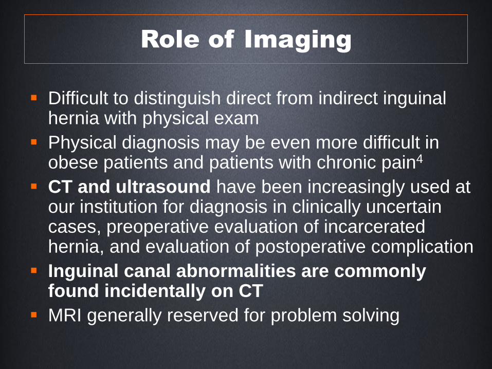
Role of Imaging
Difficult to distinguish direct from indirect inguinal hernia with physical exam
Physical diagnosis may be even more difficult in obese patients and patients with chronic pain4
CT and ultrasound have been increasingly used at our institution for diagnosis in clinically uncertain cases, preoperative evaluation of incarcerated hernia, and evaluation of postoperative complication
Inguinal canal abnormalities are commonly found incidentally on CT
MRI generally reserved for problem solving

Role of Imaging
Inguinal hernias may be clinically
occult in obese patients, even in
cases of large hernia.
3D reconstruction demonstrates an indirect
left inguinal hernia, which may be difficult to
palpate, deep to the subcutaneous adipose
tissue.
Inferior Epigastric Artery

ABNORMALITY
IN THE
INGUINAL
CANAL
Inguinal
Hernia and
Benign
Processes
Postsurgical
Changes
Neoplasm

ABNORMALITY
IN THE
INGUINAL
CANAL
Inguinal
Hernia and
Benign
Processes
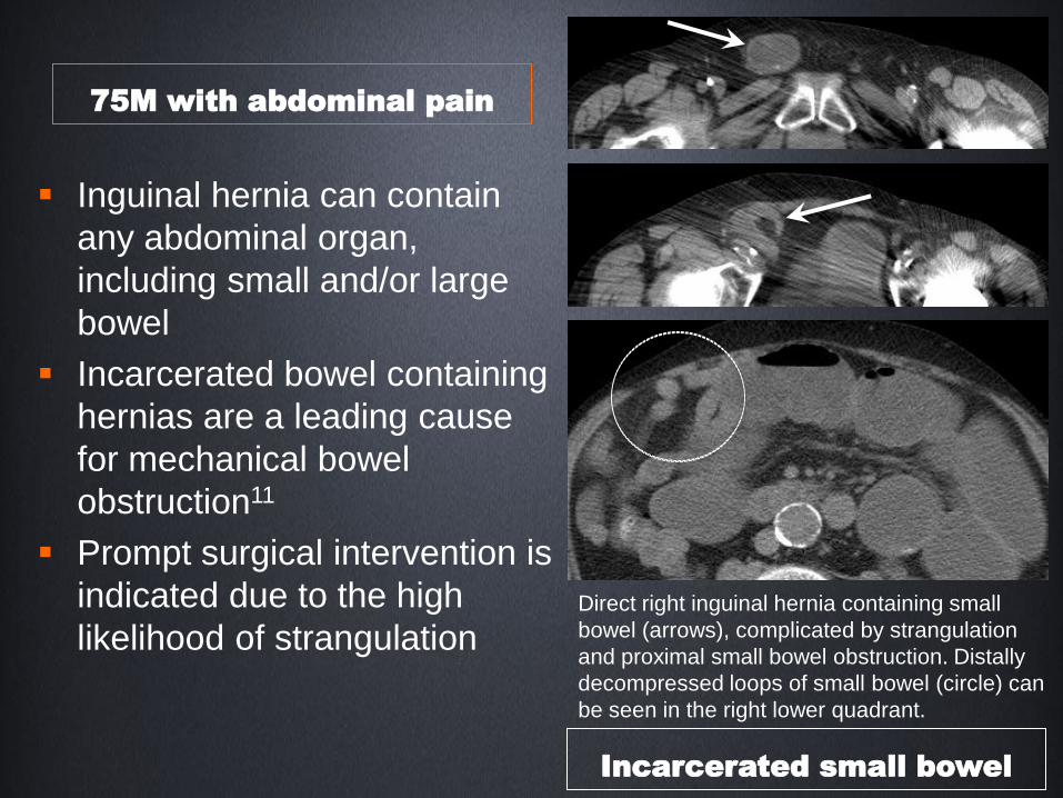
75M with abdominal pain
Direct right inguinal hernia containing small
bowel (arrows), complicated by strangulation
and proximal small bowel obstruction. Distally
decompressed loops of small bowel (circle) can
be seen in the right lower quadrant.
Inguinal hernia can contain
any abdominal organ,
including small and/or large
bowel
Incarcerated bowel containing
hernias are a leading cause
for mechanical bowel
obstruction11
Prompt surgical intervention is
indicated due to the high
likelihood of strangulation
Incarcerated small bowel

Scrotal ultrasound shows a complex tubular hypervascular mass extending from the inguinal canal to the left scrotum. Lack of peristalsis or sliding with valsalva argues against bowel hernia. Epididymitis was suspected.
35M with abdominal pain
Vasitis
Companion 1: Bowel Hernia
Companion 2: Epididymitis
Ultrasound shows a tubular structure that has peristalsis on real-time imaging, and slides with valsalva.
Ultrasound shows an enlarged, heterogeneous epididymis with markedly increased vascularity. Testicular vascularity is normal.

Vasitis
Inflammation of the vas deferens
Vasitis nodosa is a complication of vasectomy and may be asymptomatic
Acute pain and swelling in infectious vasitis5
Result of retrograde spread of pathogens from the urinary tract, prostate, or seminal vesicle6
Ultrasound used to evaluate for epididymitis, orchitis, and testicular torsion
CT is more definitive in differentiating vasitis from inguinal hernia5
Subsequent CT in the same patient shows
prominent prostate gland (not pictured), left
seminal vesicle (white arrow) and left vas
deferens (dashed arrow) with surrounding
stranding. Urine positive for N. gonorrhea.
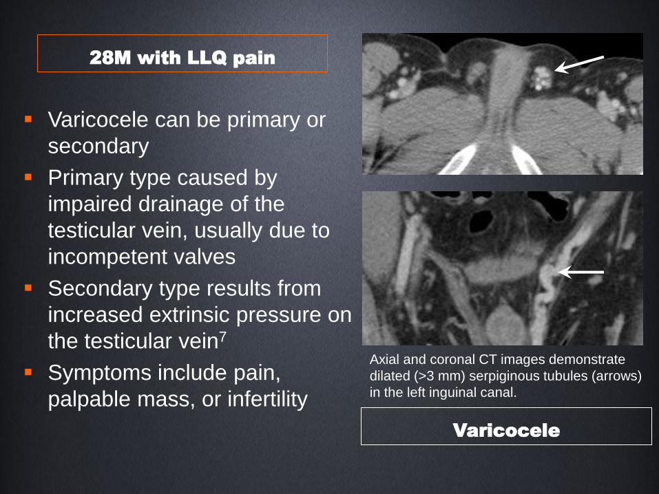
Axial and coronal CT images demonstrate
dilated (>3 mm) serpiginous tubules (arrows)
in the left inguinal canal.
Varicocele can be primary or
secondary
Primary type caused by
impaired drainage of the
testicular vein, usually due to
incompetent valves
Secondary type results from
increased extrinsic pressure on
the testicular vein7
Symptoms include pain,
palpable mass, or infertility
Varicocele
28M with LLQ pain
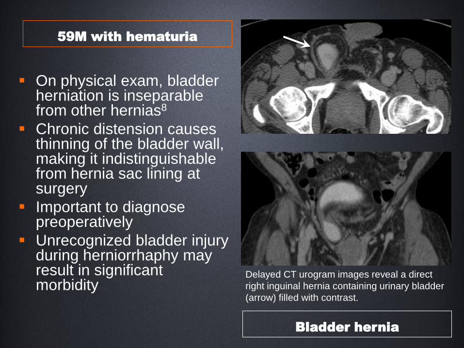
Delayed CT urogram images reveal a direct
right inguinal hernia containing urinary bladder
(arrow) filled with contrast.
On physical exam, bladder herniation is inseparable from other hernias8
Chronic distension causes thinning of the bladder wall, making it indistinguishable from hernia sac lining at surgery
Important to diagnose preoperatively
Unrecognized bladder injury during herniorrhaphy may result in significant morbidity
59M with hematuria
Bladder hernia
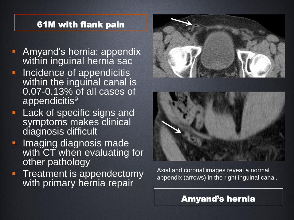
Axial and coronal images reveal a normal
appendix (arrows) in the right inguinal canal.
Amyand’s hernia: appendix within inguinal hernia sac
Incidence of appendicitis within the inguinal canal is 0.07-0.13% of all cases of appendicitis9
Lack of specific signs and symptoms makes clinical diagnosis difficult
Imaging diagnosis made with CT when evaluating for other pathology
Treatment is appendectomy with primary hernia repair
Amyand’s hernia
61M with flank pain

Axial image reveals an ovoid soft tissue density
(arrow) in the left inguinal canal.
The left ovary (arrow) and fallopian tube extend
into the inguinal canal on the coronal image.
Herniation of the ovary is rare (<3% of hernia in women)10
Generally seen in pediatric patients and often associated with congenital genitourinary tract anomalies
Complications include ovarian torsion, incarceration or salpingitis
At imaging, follow gonadal veins to identify each ovary to avoid misinterpretation
46F breast carcinoma staging
Ovary herniation

Axial CT image demonstrates a fat-containing
left inguinal hernia (arrow) with stranding of the
fat, suggesting incarceration.
Important to recognize that incarcerated fat containing hernias can be a cause of severe pain12
Unlike incarcerated hernias that contain bowel or other organs, this is not regarded as a surgical emergency
Differentiate fat herniation from inguinal canal lipoma, which does not connect to the extraperitoneal cavity
47F with LLQ pain
Incarcerated fat herniation

ABNORMALITY
IN THE
INGUINAL
CANAL
Postsurgical
Changes

Direct left inguinal hernia (solid arrow)
containing fat. The inferior epigastric
vessels (circle) are identified lateral to the
hernia sac. Prior hernia repair (dashed
arrow) is evident.
Mesh can be directly identified when radiopaque material used13, or indirectly by staples or suture calcifications
Recurrence rates following mesh repair vary based on technique, with a range of 5-10% at two years14
Most common long term complications include neuralgia, hematoma or seroma, orchitis and other testicular problems, and infection
46M with prior hernia repair
Recurrent inguinal hernia
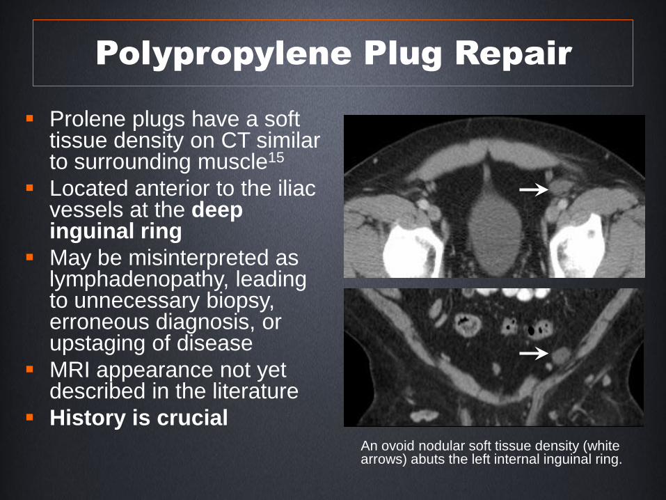
Polypropylene Plug Repair
Prolene plugs have a soft tissue density on CT similar to surrounding muscle15
Located anterior to the iliac vessels at the deep inguinal ring
May be misinterpreted as lymphadenopathy, leading to unnecessary biopsy, erroneous diagnosis, or upstaging of disease
MRI appearance not yet described in the literature
History is crucial An ovoid nodular soft tissue density (white arrows) abuts the left internal inguinal ring.

Axial and coronal CT images demonstrate a
soft tissue density (arrows) in the left inguinal
canal with surrounding stranding.
Axial MR images in the same patient show a
corresponding structure (arrowheads) with low
T1 and low T2 signal intensity. Findings
correspond to fibrous tissue surrounding a
migrated polypropylene plug.
T1WI
T2WI
45F with chronic groin pain
Migrated prolene plug

55F with RLQ pain 5d post
inguinal hernia repair
Axial and coronal images demonstrate air
and fluid within the right inguinal canal (white
arrows) suggesting abscess.
Most common immediate (<2 wks) postoperative complication is hematoma or seroma14
Wound infection is much less common
At imaging, gas within the collection suggests abscess
Important to correlate with systemic signs and symptoms of infection to avoid contaminating a sterile collection
Postoperative abscess

ABNORMALITY
IN THE
INGUINAL
CANAL
Neoplasm

Scrotal ultrasound reveals a large soft tissue
mass (M) containing amorphous hyperechoic
material posterior to the left testis (T). This was
initially diagnosed as an inguinal hernia
containing extraperitoneal fat.
M
T
Subsequent coronal CT image shows a large fat
attenuation mass (*) within the left inguinal
canal which does not communicate with the
intraperitoneal cavity. Polypropylene mesh plug
(arrow) is identified at the deep inguinal ring.
*
85M with groin bulge on exam
Liposarcoma

Inguinal Canal Liposarcoma
Liposarcoma makes up 7% of all paratesticular sarcomas, of which 12% occur in the inguinal canal16
Large fat attenuation mass on CT, which may have internal soft tissue nodules or septations
Most common type is well-differentiated, with no malignant potential
Treatment is surgical excision, with or without radiation and/or chemotherapy
Other primary neoplasms of the inguinal canal include other soft tissue sarcomas, testicular carcinomas, and Burkitt lymphoma17

75M restaging penile
squamous cell carcinoma
post conservative resection
Axial CT image reveals a soft tissue lesion in
the right inguinal canal, concerning for
metastatic disease. The lesion resolved
spontaneously on subsequent imaging and
was therefore determined to be postoperative
collection.
Penile cancer rare in the US
Nodal metastasis most important prognostic factor for 5-year survival18
Inguinal nodes most commonly involved
CT or MRI used for staging in patients with clinical adenopathy or obese patients
Treatment more aggressive if nodal metastases identified
Granulation tissue mimics
metastasis
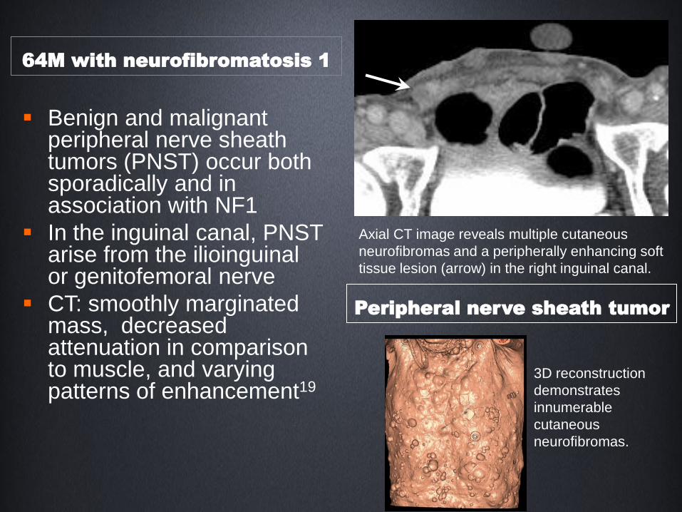
Benign and malignant peripheral nerve sheath tumors (PNST) occur both sporadically and in association with NF1
In the inguinal canal, PNST arise from the ilioinguinal or genitofemoral nerve
CT: smoothly marginated mass, decreased attenuation in comparison to muscle, and varying patterns of enhancement19
Axial CT image reveals multiple cutaneous
neurofibromas and a peripherally enhancing soft
tissue lesion (arrow) in the right inguinal canal.
3D reconstruction
demonstrates
innumerable
cutaneous
neurofibromas.
64M with neurofibromatosis 1
Peripheral nerve sheath tumor
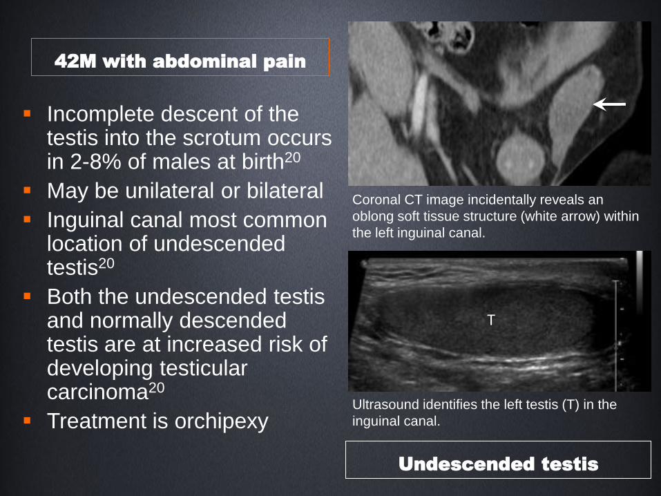
Coronal CT image incidentally reveals an
oblong soft tissue structure (white arrow) within
the left inguinal canal.
T
Ultrasound identifies the left testis (T) in the
inguinal canal.
Incomplete descent of the testis into the scrotum occurs in 2-8% of males at birth20
May be unilateral or bilateral
Inguinal canal most common location of undescended testis20
Both the undescended testis and normally descended testis are at increased risk of developing testicular carcinoma20
Treatment is orchipexy
42M with abdominal pain
Undescended testis
Summary
Most common abnormality in the inguinal canal is herniation of normal intra-abdominal organs
Inguinal hernia can be direct or indirect
Imaging performed in evaluation of uncertain cases, preoperative planning, and postoperative follow-up
Inguinal canal abnormalities are most often seen incidentally
Understanding of normal anatomy provides the foundation for creating a pertinent differential diagnosis

References
1. Dabbas N, Adams K, Pearson K, Royle G. Frequency of abdominal wall hernias: is
classical teaching out of date? JRSM Short Rep 2011; 2:5.
2. Rutkow IM, Robbins AW. Demographic, classificatory, and socioeconomic aspects of
hernia repair in the United States. Surg Clin North Am 1993; 73:413.
3. van den Berg JC, de Valois JC, Go PM, Rosenbusch G. Detection of groin hernia with
physical examination, ultrasound, and MRI compared with laparoscopic findings. Invest
Radiol 1999; 34:739.
4. Garvey JFW. Computed tomography scan diagnosis of occult groin hernia. Hernia 2012;
16:307.
5. Eddy K, Piercy GB, Eddy R. Vasitis: clinical and ultrasound confusion with inguinal hernia
clarified by computed tomography. Can Urol Assoc J. Aug 2011; 5(4): E74-E76.
6. Yang DM, Kim HC, Lee HL, Lim JW, Kim GY. Sonographic findings of acute vasitis. J
Ultrasound Med. 2010; 29: 1711-1715.
7. Kurklinsky AK, Rook TW. Nutcracker phenomenon and nutcracker syndrome. Mayo Clin
Proc. 2010;85(6):552-559.
8. Gomella LG, Spires SM, Burton JM, Ram MD, Flanigan The surgical implications of
herniation of the urinary bladder. RC.Arch Surg. 1985 Aug;120(8):964-7.
9. Ivashchuk G, et al. Amyand’s Hernia: A review. Medical Science Monitor, 2014; 20:140-
146.
10. Gurer A, Ozdogam M, Ozlem N, Yilidirim A, Kulacoglu H, Aydin R. Uncommon content in
groin hernia sac. Hernia: 2006; 10: 152-155.

References
11. Markogiannakis H, et al. Acute mechanical bowel obstruction: Clinical presentation,
etiology, management and outcome. World J Gastroenterol 2007 January 21; 13(3): 432-437.
12. Erickson KM et al. Abdominal hernias treatment and management. http://emedicine.medscape.com/article/189563-treatment. Accessed October 19, 2014.
13. Rakic S, Leblanc KA. The radiologic appearance of prosthetic materials used in hernia repair and a recommended classification. American Journal of Roentgenology. 2013;201: 1180-1183.
14. Neumayer L, Giobbie-Hurder A, Jonasson O, et al. Open mesh versus laparoscopic mesh repair of inguinal hernia. N Engl J Med. 2004;350:1819-27.
15. Cronin GC, Harisinghani MG, Catalano O, Blake M. Multitechnique imaging findings of prolene plug hernia repair. American Journal of Radiology. 2010;195:701-706.
16. Montgomery E, Buras R. Incidental liposarcomas identified during hernia repair operations. J Surg Oncol 1999;71:50–53.
17. Bhosale PR, Patnana M, Viswanathan C, Szklaruk J. The inguinal canal: anatomy and imaging features of common and uncommon masses. Radiographics 2008;28:819-835.
18. Delacroix SE Jr, Pettaway CA. Therapeutic strategies for advanced penile carcinoma. Curr Opin Palliat Care. 2010;4(4):285-92.
19. Ogose A, Hotta T, Morita T, et al. Diagnosis of peripheral nerve sheath tumors around the pelvis. Jpn J Clin Oncol. 2004;34(7)405-413.
20. Ritzen EM. Undescended testes: a consensus on management. European Journal of Endocrinology. 2008; 159 Suppl1:S87-90.