uti imaging algorithms revisited in the light of...
TRANSCRIPT

UTI imaging algorithms revisited in
the light of modern approaches -
when to image, whom, how?
Michael Riccabona Division of Pediatric Radiology
Department of Radiology
University Hospital LKH Graz, Austria
ESPR uroadiology task force ESUR pediatric work group

Introduction
• UTI common in children overall prevalence 2-8%
• Risk of recurrent UTI = 12- 30% in first year after initial UTI
particularly in first years of life

Introduction
• UTI common in children
• Risk of recurrent UTI = 12- 30%
• Complications & risks of upper UTI - acute complications = abscess …
- develop renal scars particularly in infancy
• Complications from renal scarring - hypertension
- proteinuria
- pregnancy-related complications
- end-stage renal failure
• UTI common in children, risk of recurrent UTI
• Complications & risks of upper UTI, renal scarring
• Symptoms in infancy often unspecific
proper urine sample essential - proper technique (sampling, culture, …)
- reliable results? additional laboratory data?
Introduction: UTI - diagnosis

• UTI common in children, risk of recurrent UTI
• Complications & risks of upper UTI, renal scarring
• Symptoms in infancy often unspecific
proper urine sample essential
classification - febrile = upper UTI / aPN
- lower UTI / cystitis & urethritis
- difference: renal involvement
NOTE: only renal involvement causes scaring
Introduction: UTI - diagnosis

Imaging in UTI
Aim of imaging in UTI
• to (early) identify risk factors & abnormalities
- that can be modified
• to decrease likelihood of recurrent (upper) UTI
• to reduce risk of renal scarring
.Saadeh SA, Mattoo TK. Managing urinary tract infections. Pediatr Nephrol. 2011

How to image: Top-down and/or Down-Up ?
Previously
• US
• IVU + DMSA - diagnose & localize UTI
• VCUG in all patients
- for detection of VUR + follow-up
BUT: Today's knowledge based on this approach
- though invasive and rigorous, still was helpful

• Congenital VUR often vanishes spontaneously
or at least diminishes
even higher grades
without sequalea
• Low grade VUR (I / II°) in itself without risk
not for UTI recurrence
not for renal damage
Today’s new knowledge on VUR

• Congenital VUR often vanishes spontaneously
• Low grade VUR (I / II°) in itself without risk
• High grade VUR has varying & unpredictable impact even in patients with UTI
often already fetal dysplasia (cRNP) – cannot be influenced
• Renal scarring can occur after UTI even without detectable VUR
BUT: limited VUR detection by VCUG
VUR in children with UTI without VUR on VCUG?
in many: VUR on ce-VUS or endoscopic techniques (PIC)
Today’s new knowledge on VUR

• Congenital VUR often vanishes spontaneously
• Low grade VUR (I / II°) in itself without risk
• High grade VUR has varying & unpredictable impact
• Renal scarring, UTI cause & course
many factors may impact UTI frequency & renal scarring
behavior / fashion / social aspects …
epidemiologic, kind & behavior of micro-organism
treatment onset, kind, response …
genetic preposition?
bladder function disturbance
Today’s new knowledge on VUR

• Congenital VUR often vanishes spontaneously
• Low grade VUR (I / II°) in itself without risk
• High grade VUR has varying & unpredictable impact
• Renal scarring, UTI reasons multi-factorial
• Treatment also changed less aggressive surgically, new endoscopic treatment options …
even AB-prophylaxis under discussion
lots of controversy, ongoing debate
Today’s new knowledge on VUR
Roussey-Kesler G, et al (2008) Antibiotic prophylaxis for the prevention of recurrent urinary tract infection in children with low grade vesicoureteral reflux: results from a prospective randomized study. J Urol
179:674–679, discussion 679
Montini G, et al. (2008) Prophylaxis after first febrile urinary tract infection in children? A multicenter, randomized, controlled, noninferiority trial. Pediatrics 122:1064–1071
Pennesi M, et al. (2008) Is antibiotic prophylaxis in children with vesicoureteral reflux effective in preventing pyelonephritis and renal scars? A randomized, controlled trial. Pediatrics 121:e1489
Craig JC, et al. (2009) Antibiotic prophylaxis and recurrent urinary tract infection in children. N Engl J Med 361:1748–1759
Brandstrom P, et al. (2010) The Swedish reflux trial in children: III. Urinary tract infection pattern. J Urol 184:286–29

• To discuss role of imaging in UTI reflecting new therapy concepts
based on new knowledge & insights into pathophysiology
address “bottom-up” versus “top-down” approach
• To describe relevant imaging techniques
• To give typical examples
• To propose imaging algorithm for diagnostic imaging
• To address how to deal with complications
Objective
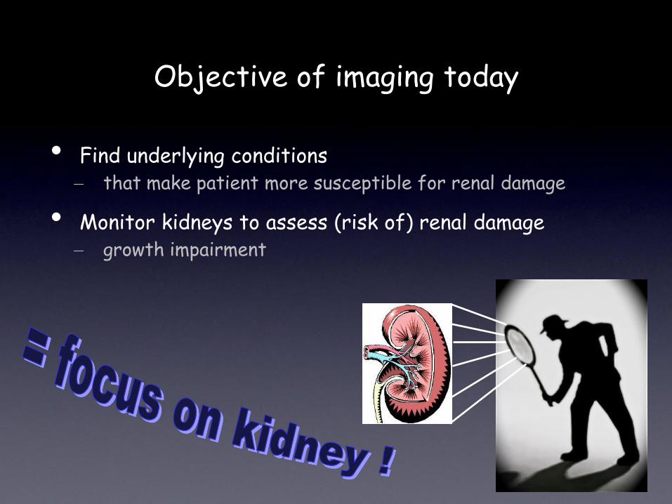
Objective of imaging today
• Find underlying conditions that make patient more susceptible for renal damage
• Monitor kidneys to assess (risk of) renal damage growth impairment

Imaging methods
•US
•VCUG / RNC, ce-VUS
•DMSA
•IVU
•MRI
•CT

Imaging methods
• US = accepted initial & universal imaging tool
HN, UTI, screening ...
• Requisites:
proper transducers
good hydration
full bladder
knowledge & experience
clinical data
post-void assessment
= need sufficient time

• US signs for VUR
gaping ostium (diagnostic)
VUR visualization by CDS (diagnostic)
ureteral / pelvic / caliceal dilatation
changing upper dilatation? (indirect signs)
Imaging methods - US

Imaging methods - US
• US signs for VUR
gaping ostium, VUR visualisation, dilatation
bladder wall thickening, trabeculation (unspecific)
lateralized or duplex ostium (indirect sign)
“urothelial sign” (indirect sign)

Imaging methods - US
• US signs for VUR
gaping ostium, VUR visualisation, dilatation
wall thickening, lateralized/duplex ostium
other (indirect) hints & signs (unspecific)
residual volume
renal scar, size difference, dysplasia

Imaging methods - US
• US signs for VUR
gaping ostium, VUR visualisation, dilatation
wall thickening, lateralized/duplex ostium
other (indirect) hints & signs (unspecific)
residual volume, renal scar, dysplasia
urethral & pelvic floor pathology
Imaging methods - US
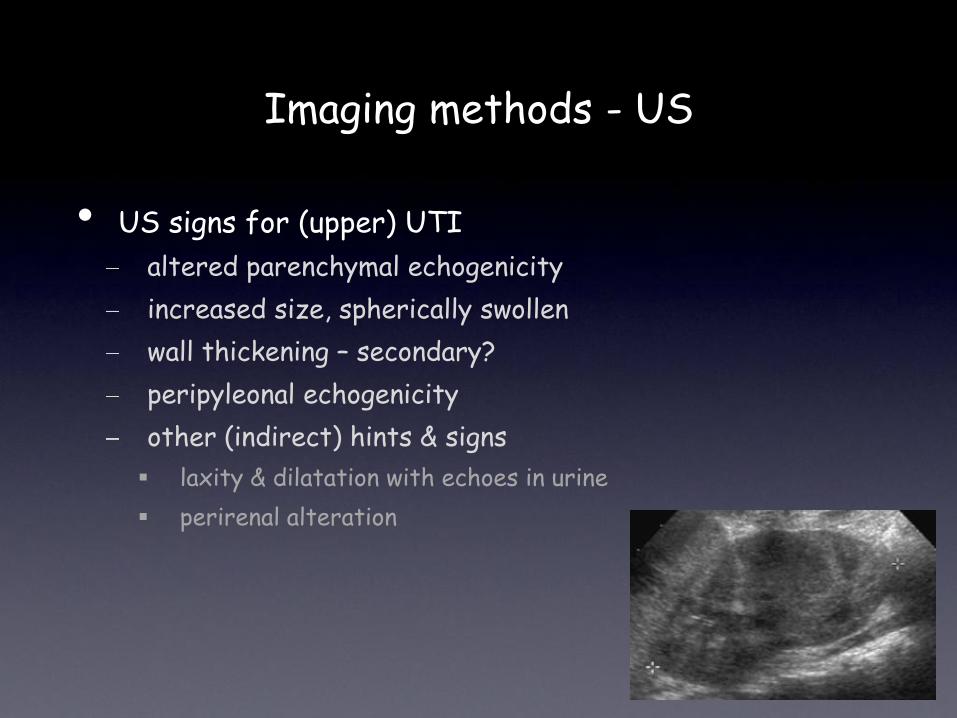
• US signs for (upper) UTI
altered parenchymal echogenicity
increased size, spherically swollen
wall thickening – secondary?
peripyleonal echogenicity
other (indirect) hints & signs
laxity & dilatation with echoes in urine
perirenal alteration

Imaging methods - US
• US in complications of UTI
pyohydronephrosis
necrosis / abscess
xanthogranulomatous pyelonephritis
stone formation, fungus ball
tuberculosis ….

Imaging methods - US
• US in complications of UTI
pyohydronephrosis
necrosis / abscess
xanthogranulomatous pyelonephritis
pseudotumnor / lobar nephroma

Imaging methods - US
• US in UTI – role of aCDS
segmental perfusion defects
necrosis / abscess
role of ce-US?

• US in UTI – role of aCDS
segmental perfusion defects necrosis/abscess, ce-US?
incidental alternate findings
infarction, retro-aortal left renal vein
tumor, stones …
Imaging methods - US

• Restrictions of basic US
no panoramic display
poor for definite VUR diagnosis
high observer variability
restrictions for
anatomy of urethra
ureteral anatomy
diverticula
(small) scars
bladder function assessment
additional imaging tools essential
Imaging methods - US Imaging methods - US
Imaging methods
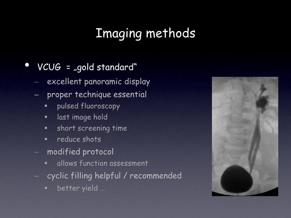
• VCUG = „gold standard“
excellent panoramic display
proper technique essential
pulsed fluoroscopy
last image hold
short screening time
reduce shots
modified protocol
allows function assessment
cyclic filling helpful / recommended
better yield …
Imaging methods - VCUG
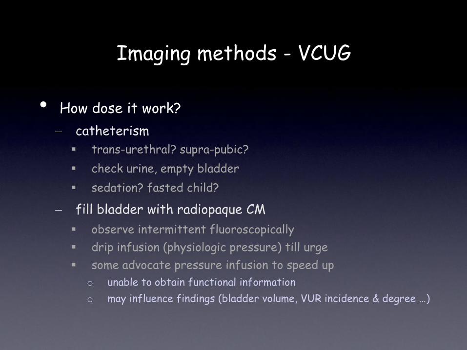
• How dose it work?
catheterism
trans-urethral? supra-pubic?
check urine, empty bladder
sedation? fasted child?
fill bladder with radiopaque CM
observe intermittent fluoroscopically
drip infusion (physiologic pressure) till urge
some advocate pressure infusion to speed up
o unable to obtain functional information
o may influence findings (bladder volume, VUR incidence & degree …)

Imaging methods - VCUG
• How dose it work?
catheterism, fill bladder with CM
document findings
before/during/after voiding
use last image hold & spot films
bladder capacity? bladder neck?
observe residual urine
o drainage of refluxed CM …
describe & grade VUR

Imaging methods - VCUG
• VCUG benefits
standardized grading
less investigator dependent
excellent anatomy
ureter, diverticula, urethra
reproducible …

Imaging methods - VCUG
• VCUG restrictions
catheter = invasive
radiation burden
particularly in girls
short, particularly if only 1 cycle …
for radiation protection
= incomplete / wrong result
non-physiologic approach
artificial function disturbance

Alternate VUR detection techniques
1) ce-VUS
bladder filling with NaCL + US-CM
observe before, during after …
VUR imaging – alternate methods

Darge K. EJR 2002
Alejandro Maté, Eur Radiol 2003
Theresa Berrocal, Radiology, 2005
Alternate VUR detection techniques
1) ce-VUS
bladder filling with NaCL + US-CM, observe & scan …
excellent VUR detection & grading
urethral assessment possible, but more difficult
VUR imaging – alternate methods

grading established, standardised – see European Society for Nuclear Medicine
Alternate VUR detection techniques
1) ce-VUS
2) radionuclide cystography (RNC)
bladder filling with tracer = direct RNC
catheter needed, as in VCUG
observe with gamma camera
before, (during?), after voiding
longer observation period
less radiation burden …
any activity in ureter & kidney area = VUR
VUR imaging – alternate methods

VUR imaging – alternate methods
standardised – see European Society for Nuclear Medicine
Alternate VUR detection techniques
1) ce-VUS
2) radionuclide cystography (RNC)
direct RNC
indirect RNC: no catheterism, must be toilet trained
late phase of dynamic Tc99m MAG3 renography
= bladder filled physiologically, no catheter needed
observe for activity increase in kidney area with gamma camera
o after clearance of activity from kidney = late phase
o before, (during?), after voiding

Alternate VUR detection techniques
1) ce-VUS
2) radionuclide cystography (RNC)
direct & indirect RNC
setbacks of RNC
poor anatomical resolution (urethra, kidney …)
restricted grading
o less comparability with VCUG …
no bladder function assessment
some radiation, catheterism (direct RNC)
only after 4-5 y of age (cooperative patient)
VUR imaging – alternate methods
Alternate VUR detection techniques
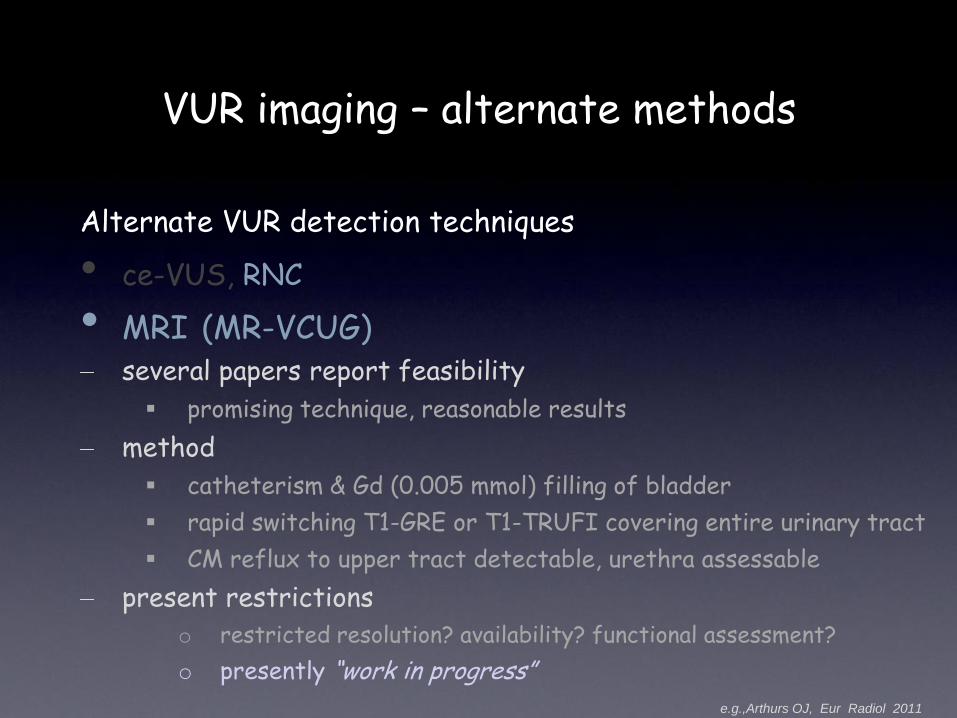
• ce-VUS, RNC
• MRI (MR-VCUG) several papers report feasibility
promising technique, reasonable results
method
catheterism & Gd (0.005 mmol) filling of bladder
rapid switching T1-GRE or T1-TRUFI covering entire urinary tract
CM reflux to upper tract detectable, urethra assessable
present restrictions o restricted resolution? availability? functional assessment?
o presently “work in progress”
VUR imaging – alternate methods
e.g.,Arthurs OJ, Eur Radiol 2011

Imaging methods
• IVU
declining importance in children
NO importance in VUR / RNP / UTI setting any longer
however, has been major imaging tool
initially only comprehensive assessment of upper UT
could evaluate anatomy + function
detection of scaring (Smellie)
easy accessible, standardized, reliable
replaced by US, (DMSA) scintigraphy, MRI
less / no radiation, no / less / other contrast needs …
improved diagnostic capabilities …

Imaging methods - IVU
• If you do it, do it properly
reduce number of films
no zonograms
adapted CM & radiation dose, hydration …
age adapted, adequate filters
o also DR needs to adapted to pediatric needs
initial US to properly plan investigation
individually choose image timing
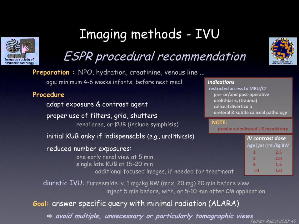
Preparation : NPO, hydration, creatinine, venous line ...
age: minimum 4-6 weeks infants: before next meal
Procedure
adapt exposure & contrast agent
proper use of filters, grid, shutters renal area, or KUB (include symphisis)
initial KUB onky if indispensable (e.g., urolithiasis)
reduced number exposures: one early renal view at 5 min single late KUB at 15-20 min
additional focused images, if needed for treatment
diuretic IVU: Furosemide iv. 1 mg/kg BW (max. 20 mg) 20 min before view
inject 5 min before, with, or 5-10 min after CM application
Goal: answer specific query with minimal radiation (ALARA)
avoid multiple, unnecessary or particularly tomographic views Pediatr Radiol 2010: 40
Imaging methods - IVU
ESPR procedural recommendation
IV contrast dose Age [year] ml/kg BW
1 2.5
2 2.0 3 1.5 >4 1.0
Indications restricted access to MRU/CT pre- or/and post-operative urolithiasis, (trauma) caliceal diverticula ureteral & subtle caliceal pathology
NOTE: previous dedicated US mandatory

Imaging methods
• Renal scintigraphy = gold standard
for assessment of
renal involvement in UTI (?)
scarring
(split) renal function
usually Tc99m DMSA as tracer
better than MAG3 for VUR- / UTI-associated queries
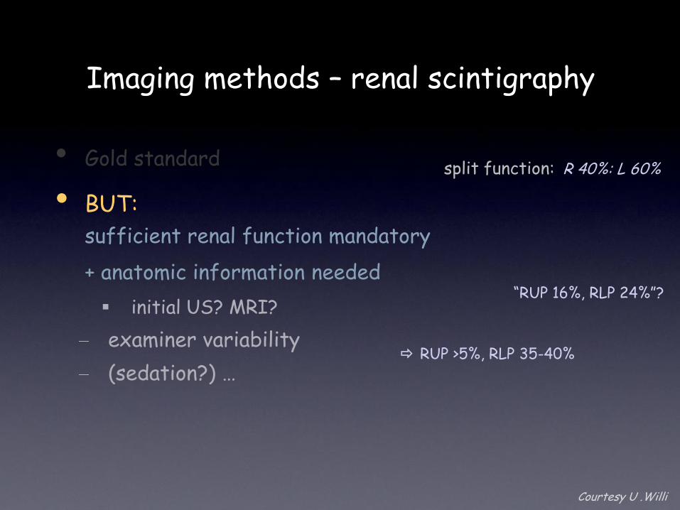
• Gold standard
• BUT:
sufficient renal function mandatory
+ anatomic information needed
initial US? MRI?
examiner variability
(sedation?) …
split function: R 40%: L 60%
“RUP 16%, RLP 24%”?
RUP >5%, RLP 35-40%
Courtesy U .Willi
Imaging methods – renal scintigraphy

• Renal MRI & MRU = future gold standard?
for assessment of
scars, upper UTI, dysplasia, upper UT involvement …
complications, DDx
UT-malformations
(split) function & drainage
GFR? VUR? …
constantly new techniques being introduced
enhanced functional imaging
DWI, perfusion, BOLD … - rTx with VUR & sars?
Imaging methods
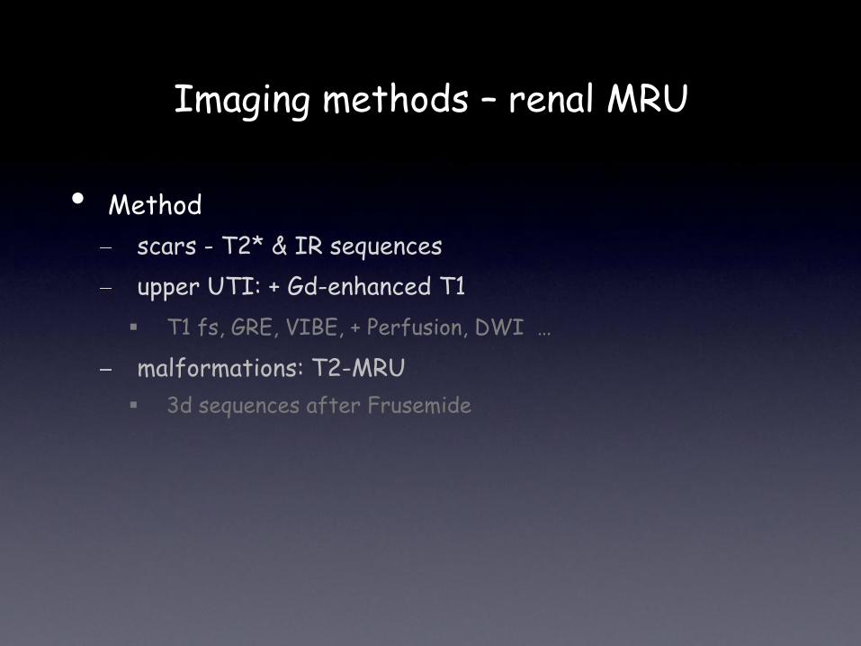
• Method
scars - T2* & IR sequences
upper UTI: + Gd-enhanced T1
T1 fs, GRE, VIBE, + Perfusion, DWI …
malformations: T2-MRU
3d sequences after Frusemide
Imaging methods – renal MRU

• Method
scars - T2* & IR sequences, upper UTI: + Gd-enhanced T1
malformations: T2-MRU
function & drainage
T1 & BOLD & DWI
function assessment possible
split renal function & GFR calculated
ce-MRI essential for complications
Imaging methods – renal MRU

• Method
scars - T2* & IR sequences upper UTI: + Gd-enhanced T1
malformations: T2-MRU, function & drainage
BUT: partially still “work in progress”
restricted availability, costs, expertise, sedation needs …
Imaging methods – renal MRU

Whom ?
When ?
How ?
Dilemma in imaging of UTI (& VUR)
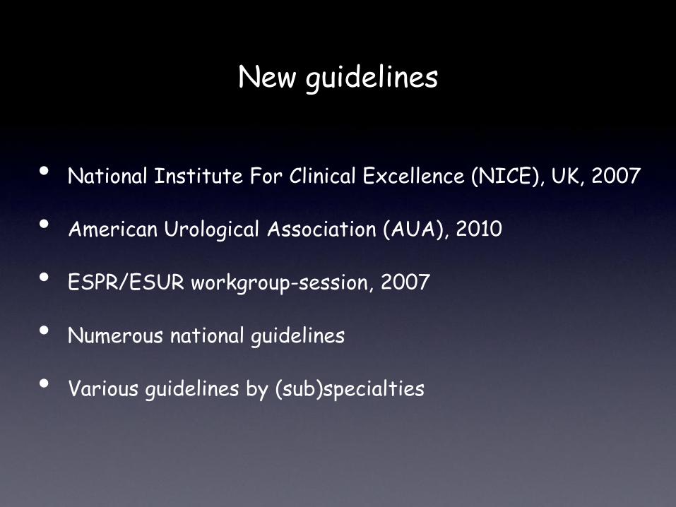
New guidelines
• National Institute For Clinical Excellence (NICE), UK, 2007
• American Urological Association (AUA), 2010
• ESPR/ESUR workgroup-session, 2007
• Numerous national guidelines
• Various guidelines by (sub)specialties

• US
• VCUG / RNC, ce-VUS
• DMSA
How to image?

• US
• VCUG / RNC, ce-VUS
• DMSA
• IVU - no role in UTI
• MRI - no defined place in routine imaging of UTI
mostly for complications or associated malformation …
• CT – only in complications, if no MRI available
DDx, underlying/secondary stones …
How to image?

When to image?

When to image?
Major questions
• early US effective for outcome?
first day? first days?
feasible & realistic
sufficient quality granted?
can US answer relevant questions?
in all? in whom?
only with unclear upper UTI?
with no earlier (fetal, neonatal …) UT screening?
only in complicated UTI ?
When to image?
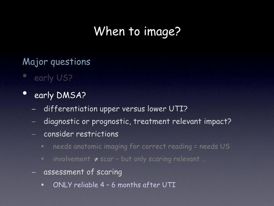
Major questions
• early US?
• early DMSA?
differentiation upper versus lower UTI?
diagnostic or prognostic, treatment relevant impact?
consider restrictions
needs anatomic imaging for correct reading = needs US
involvement scar – but only scaring relevant …
assessment of scaring
ONLY reliable 4 – 6 months after UTI

When to image?
Major questions
• early US? early DMSA?
• early VCUG? other VUR assessment test?
urine should be clear, UTI treated
what for & in whom?
impact on acute treatment? long term relevance?
in all infants with UTI, what about older children?
only with upper or complicated UTI / scaring?
benefit of early VUR assessment
only compliance may be an argument

When to image?
Major questions
• early US? early DMSA?
• early VCUG? other VUR assessment test?
• MRI needed
evident in complication or underlying condition

When to image?
Major questions
• early US? early DMSA? early VCUG? MRI?
• i.e., depends on query & suspicion & compliance & clinic
e.g., in / after upper UTI, with scars, dysplasia …
earliest option for VCUG: as soon as urine is sterile
best at 4–6 weeks after UTI, no emergency … - don’t rush
prompt US & CT/MRI in complications, severe course …
DMSA for scarring after 4-6 mo

When to image?
Major questions
• early US? early DMSA? early VCUG? MRI?
• i.e., depends on query & suspicion & compliance & clinic
e.g., in / after upper UTI, with scars, dysplasia …
PUV, neonatal renal failure …
= early imaging (US + VCUG) = first 24 hours
suspicion of high grade VUR / complex malformation
= neonatal assessment
less urgent, parental compliance? US day 5–7 …, VCUG at all?
don’t rush
DMSA after 3-6 mo

Whom to image?
Whom to image?
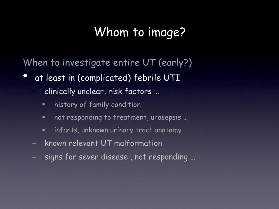
When to investigate entire UT (early?)
• at least in (complicated) febrile UTI
clinically unclear, risk factors …
history of family condition
not responding to treatment, urosepsis …
infants, unknown urinary tract anatomy
known relevant UT malformation
signs for sever disease , not responding …

Whom to image?
When to look for entire UT
• at least in (complicated) febrile UTI
clinically unclear, risk factors …
known relevant UT malformation …
• always performed by comprehensive US as first step
other / further imaging planned according to results

Whom to image?
When to look for VUR
• (recurrent) febrile (complicated) UTI
pathology on DMSA / US
renal involvement / damage
dilatation or bladder pathology
<5 years, therapy implication
relevant UT malformation (DDx)
infravesical obstruction (boys)
megaureter , UPJO …
duplex kidney …

Whom to image?
When to look for VUR
• (recurrent) febrile (complicated) UTI
pathology on DMSA / US
relevant UT malformation (DDx)
lower UT dysfunction
family screening?
neonatal HN?
grade? only boys? when?
therapy implications? …
= in selected patients (groups)

Whom to image?
How to look for VUR
• VCUG
boys / neonates
pre-operatively, complex malformation
• ce-VUS (& RNC)
girls, follow-up, family screening? bed side
exception: indirect RNC for all older patients?
supplemented by VCUG, when positive & surgery planned?
therapy implications? …

European consensus recommendations for VUR
• VCUG infant boys, preoperative
complex malformation
query “urethra” or “diverticula”
• ce-VUG & RNC girls
follow-up
family screening
if RNC + comprehensive US
How to image: When to use what?

girls, all others (infant) boys, PUV, malformation
ce-VUS, RNC, (VCUG) VCUG
+ -
stop
* VCUG in suspected infra-vesical obstruction, para-ostial diverticula, pre-operatively, no ce-VUS / RNC available
-
stop
+
US follow-up + DMSA (fMRI?)
*
VUR?
When to use what? How to image: When to use what for VUR?

Task of (pediatric) radiology
• Know potential diseases & conditions & DDx pathogenesis, origin, history
• Know suitable imaging techniques
potential, risks, & limitation, economical aspects
• Know implications of imaging results
on patient management + prognosis
• Suggest imaging algorithm
adapt individually, follow established guidelines

Discussion
Role of imaging for VUR in combination with UTI
• Remains controversial
depends on therapy consequences
growing knowledge, new concepts …
like a pendulum
• Try to reduce overuse of imaging
invasive (catheter)
radiation (VCUG, RNC)
BUT: lack of approved US-CM (ce-VUS)
without missing important conditions

Role of imaging for VUR in combination with UTI
• Remains controversial
• Try to reduce overuse of imaging
• Avoid missing important conditions
with long term sequalae
goal: prevent harm to the kidney
= if you do invasive imaging
do it right, don’t miss important aspects
Discussion

Role of imaging for VUR in combination with UTI
• controversial, reduce overuse, avoid missing conditions
extensive use of comprehensive US
high quality, extended criteria
post-void check
apply modern methods …
Discussion

Conclusion
• Imaging in UTI remains controversial
still: an important condition, deserves dedicated imaging
though less generous indications than earlier
But: imaging must address all essential aspects
• Properly select patients
based on history
and on initial detailed US findings
with respect to therapeutic consequences
and possible long term sequalae
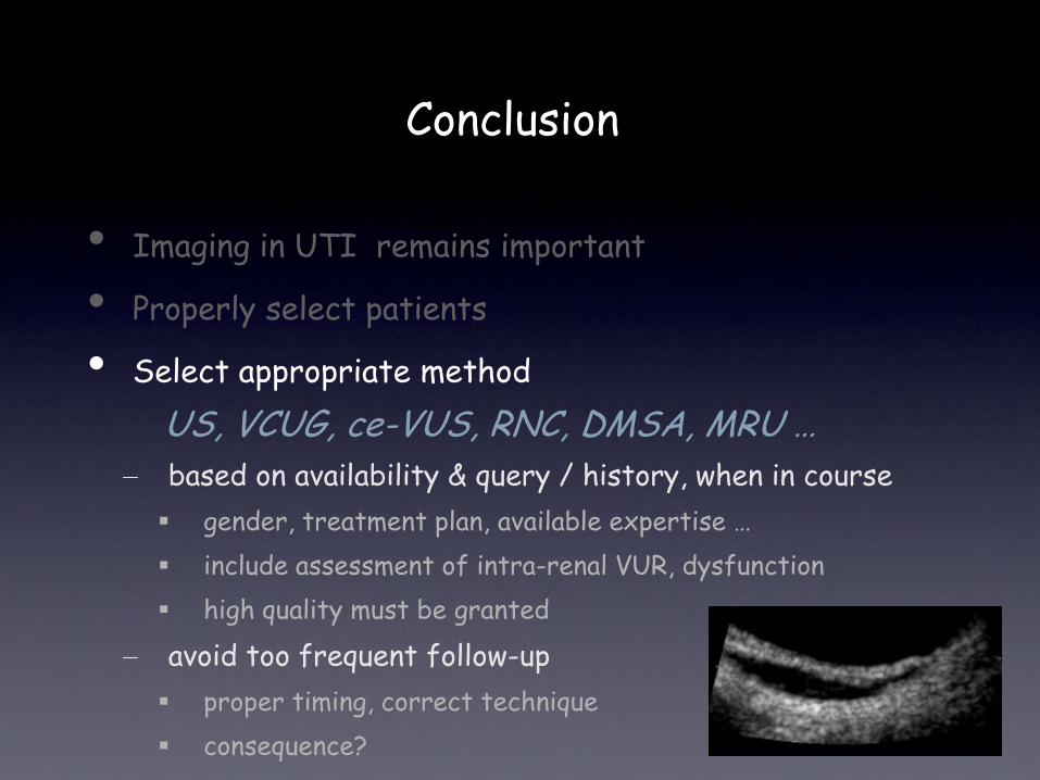
• Imaging in UTI remains important
• Properly select patients
• Select appropriate method
US, VCUG, ce-VUS, RNC, DMSA, MRU … based on availability & query / history, when in course
gender, treatment plan, available expertise …
include assessment of intra-renal VUR, dysfunction
high quality must be granted
avoid too frequent follow-up
proper timing, correct technique
consequence?
Conclusion
„Take away“
• Established “gold standards” exist
not to be dropped light mindedly
individualized imaging approach?
• New imaging concepts
at present complimentary, introduce only when proven
= evaluation of new modalities & algorithms essential
potential, impact on management & outcome
strong research efforts necessary
• If benefit proven, make it available to all
= introduce altered imaging protocols at high quality

• Most important diagnostic tool
• Always first modality
sometimes only investigation in UTI
• Acute phase + follow-up
Riccabona M et al (2008) Imaging recommendations in paediatric uroradiology: minutes of the ESPR workgroup session on urinary tract infection, fetal hydronephrosis, urinary tract ultrasonography and voiding cystourethrography, Barcelona, Spain, June 2007 Pediatr Radiol
(2008) 38:138–145
US
• Most important diagnostic tool
• Always first modality, acute phase + follow-up
• To be performed by pediatrically experienced investigator
• Include (a)CDS, careful assessment
well hydrated child
pre- + post-void imaging
use aCDS = reduces need for DMSA
Peter Brader et al. (2008) Value of comprehensive renal ultrasound in children with acute urinary tract infection for assessment of renal involvement: comparison with DMSA scintigraphy and final diagnosis Eur Radiol 18: 2981–2989
US

• Most important diagnostic tool, always first modality
• Acute + follow-up, experienced investigator, (a)CDS …
• Allows
grading of UTI
detection of obstruction & malformation
assessment of complications & stones & …
evaluation of (evidence of) VUR
US

NICE Guideline
Age <6months
Age 6 months – 3 years
Age > 3 years
responds well to treatment in 48 h atypical UTI 1 recurrent UTI 2
US 3
None
None
US, DMSA, VCUG US, DMSA, VCUG
US + DMSA 4 US + DMSA 4
US
1.Atypical UTI: Non-Escherechia coli UTI: seriously ill, poor urine flow, abdominal or bladder mass, raised creatinine, septicemia, failure to respond to treatment with suitable antibiotics within 48 h
2. Two or more episodes of UTI with acute pyelonephritis/upper urinary tract infection or one episode of UTI with acute pyelonephritis/upper urinary tract infection plus one or more episode of UTI with cystitis/lower urinary tract infection or three or more episodes of UTI with cystitis/lower urinary tract infection
3. If ultrasound is abnormal, consider a VCUG
4. Consider VCUG if dilatation on ultrasound, poor urine flow, non-E. coli infection, family history of VUR
US + DMSA 4
= routine use of imaging for localization of UTI not recommended
But: in young children clinical & laboratory diagnosis can be difficult
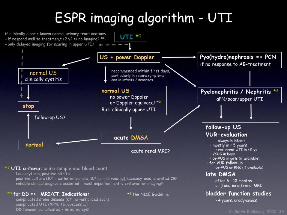
recommended within first days, particularly in severe symptoms and in infants / neonates
UTI *1
US + power Doppler
acute DMSA
normal US no power Doppler or Doppler equivocal *2
But: clinically upper UTI
normal
follow-up US VUR-evaluation - always in infants
• mostly in < 5 years + recurrent UTI in > 5 ys • VCUG in boys • ce-VUS in girls (if available)
- for VUR follow-up ce-VUS or RNC (if available)
late DMSA after 6 - 12 months or (functional) renal MRI
bladder function studies > 4 years, urodynamics
*3 for DD => MRI/CT; Indications: complicated stone disease (CT, un-enhanced scan) complicated UTI (XPN, Tb, abscess ...) DD tumour, complicated / infected cyst
Pyo(hydro)nephrosis => PCN if no response to AB-treatment
normal US clinically cystitis
stop
follow-up US?
Pyelonephritis / Nephritis *3
aPN/scar/upper UTI
*1 UTI criteria: urine sample and blood count Leucocyturia, positive nitrite positive culture (104 = catheter sample, 106 normal voiding), Leucocytosis, elevated CRP reliable clinical diagnosis essential = most important entry criteria for imaging!!
acute renal MRI?
ESPR imaging algorithm - UTI if clinically clear + known normal urinary tract anatomy - if respond well to treatmen,t >2 y? => no imaging? *2 - only delayed imaging for scaring in upper UTI?
Pediatric Radiology, 2008; 38
*2 The NICE Guideline
Coulthard MG (2008) Is reflux nephropathy preventable, and will the NICE childhood UTI guidelines help? Arch Dis Child 93:196–199
Coulthard MG (2007) NICE on childhood UTI: Nasty processes produce nasty guidelines. BMJ 335:463; author reply 463-464
Tse NK et al. (2009) Imaging studies for first urinary tract infection in infants less than 6 months old: can they be more selective? Pediatr Nephrol 24:1699–1703
Saadeh SA, Mattoo TK. (2011/12) Managing urinary tract infections. Pediatr Nephrol
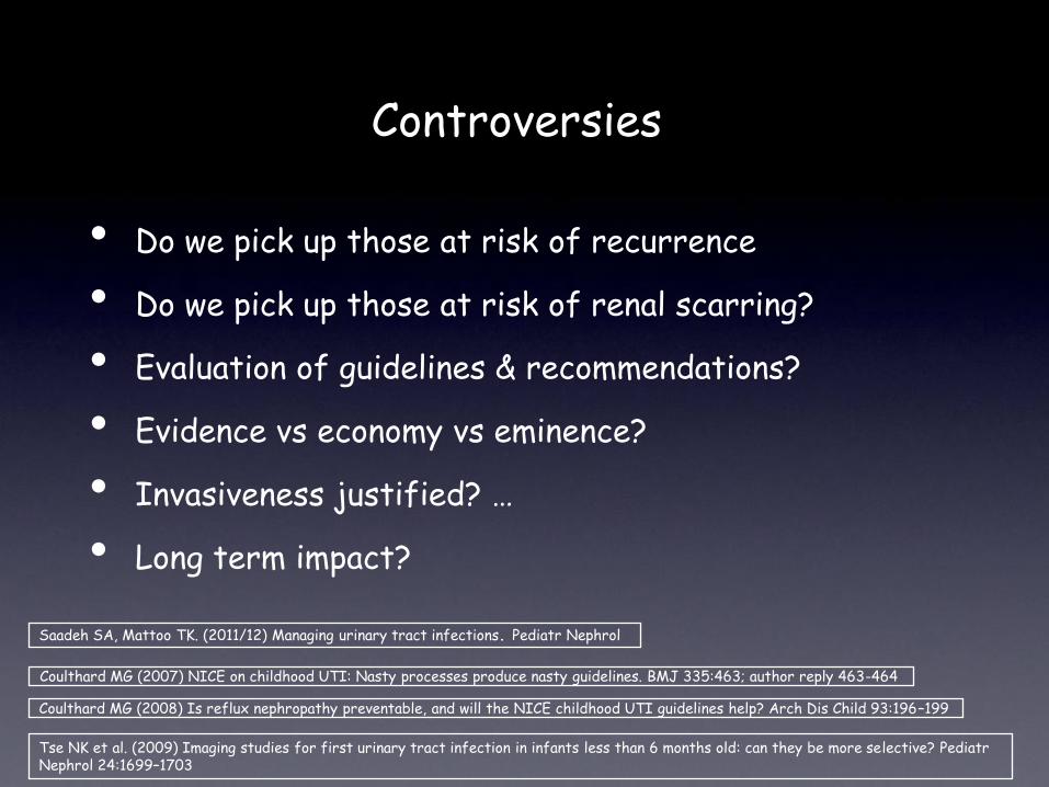
• Do we pick up those at risk of recurrence
• Do we pick up those at risk of renal scarring?
• Evaluation of guidelines & recommendations?
• Evidence vs economy vs eminence?
• Invasiveness justified? …
• Long term impact?
Controversies

Saadeh SA, Mattoo TK. Managing urinary tract infections. Pediatr Nephrol. 2011.
• Pick up those at risk of recurrence & renal scarring?
• Evidence versus economy versus eminence? Invasive?
• Proper evaluation of guidelines & recommendations?
• “”…The best approach for imaging studies in children with UTI is debatable - because of doubtful evidence & concerns over actual value of these studies in altering management & final outcome.”
• “ … In view of all these studies and recommendations, VUR (& UTI) management is a subject of constant debate. The need for higher-quality evidence to guide management is increasing. “
Controversies

Conclusion
• Imaging in UTI remains important, but controversial
to detect underlying pathology in selected patients
to monitor kidneys in order to prevent renal scarring
• Focus moved - from «down-up» to «top-down» approach
with focus on kidney
preferably using non-invasive, non-radiating imaging
• Patients must be carefully selected
for more invasive investigations, particularly older children
(bladder) function will become even more important
• Proper validation of new guidelines needed

Questions?
- welcome!

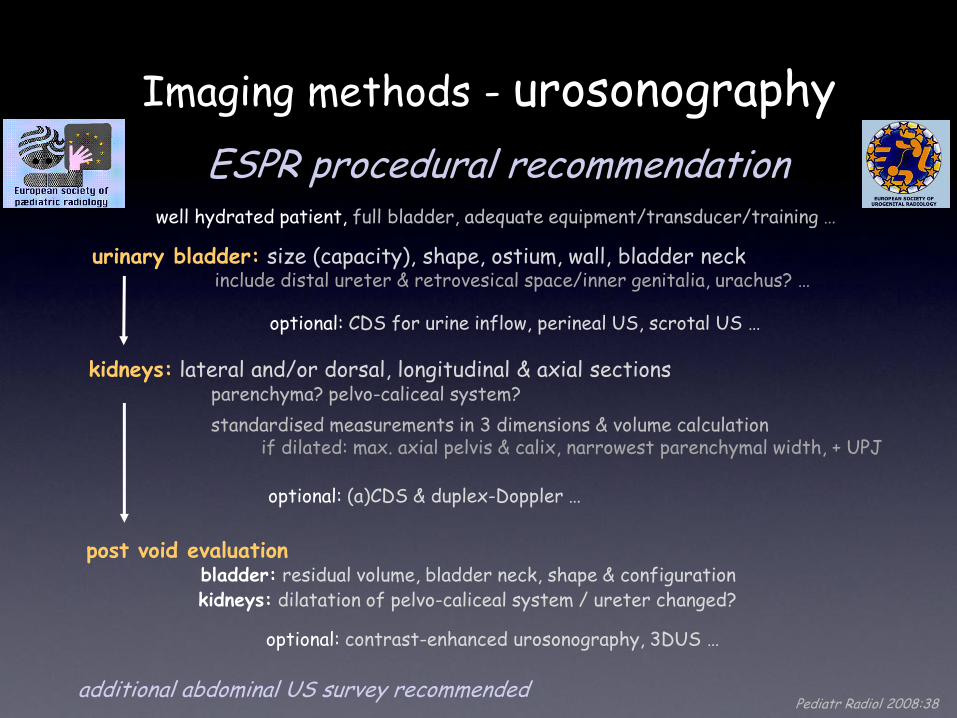
Imaging methods - urosonography
ESPR procedural recommendation well hydrated patient, full bladder, adequate equipment/transducer/training …
urinary bladder: size (capacity), shape, ostium, wall, bladder neck include distal ureter & retrovesical space/inner genitalia, urachus? …
optional: CDS for urine inflow, perineal US, scrotal US …
post void evaluation bladder: residual volume, bladder neck, shape & configuration kidneys: dilatation of pelvo-caliceal system / ureter changed?
optional: (a)CDS & duplex-Doppler …
kidneys: lateral and/or dorsal, longitudinal & axial sections parenchyma? pelvo-caliceal system?
standardised measurements in 3 dimensions & volume calculation if dilated: max. axial pelvis & calix, narrowest parenchymal width, + UPJ
optional: contrast-enhanced urosonography, 3DUS …
Pediatr Radiol 2008:38 additional abdominal US survey recommended

Pediatr Radiol 2008:38
No diet restriction or enema, urine analysis, potentially antibiotics …
catheterism: feeding tube, 4-8 french or suprapubic puncture latex precaution: neuro tube defect, bladder exstrophy …
Bladder filling with radiopaque contrast gravity drip = bottle 30-40 cm above table, watch dripping, AB?
after voiding: ap view of bladder & renal fossae assess contrast drainage form kidney if refluxed
when voiding: remove catheter, unless cyclic VCUG = 3 fillings, 1st y(s) female: 2 spots of distended urethra (slightly oblique) male: 2-3 spots during voiding (ap & high oblique / lateral) include renal fossae during voiding, if VUR => spot film
fluoroscopy: if signs of increased bladder pressure, imminent voiding, urge … bilateral oblique views of distal ureters, include catheter
document VUR, include kidney (spot film, intra-renal reflux)
Note: VUR staging, AB-prophylaxis? …
fluoroscopic view of renal fossae & bladder, initial + early filling
Imaging methods - VCUG
ESPR procedural recommendation

No diet restriction or enema, urine analysis; AB as in VCUG …
Catheterism: feeding tube, 4-8 french, or suprapubic puncture anaesthetic lubricant or coated plaste
Bladder filling with NaCl (only from plastic containers)
During/after voiding: US of bladder & kidneys & urethra supine ± prone, laying or sitting or standing
Peri-/ post-contrast US of bladder + kidneys: continuous, alternating US modalities: fundamental, HI, CDS, contrast specific methods
alternate scans of right & left side during & after filling
Install US contrast medium, e.g., SonoVue ®, 0.5-1.0% of bladder volume slow, US monitoring, potentially fractional administration
VUR diagnosis: echogenic micro-bubbles in ureters or renal pelves
Standard US of bladder & kidneys (supine, ± prone)
Pediatr Radiol 2008: 38
Imaging methods – ce-VUS
ESPR procedural recommendation
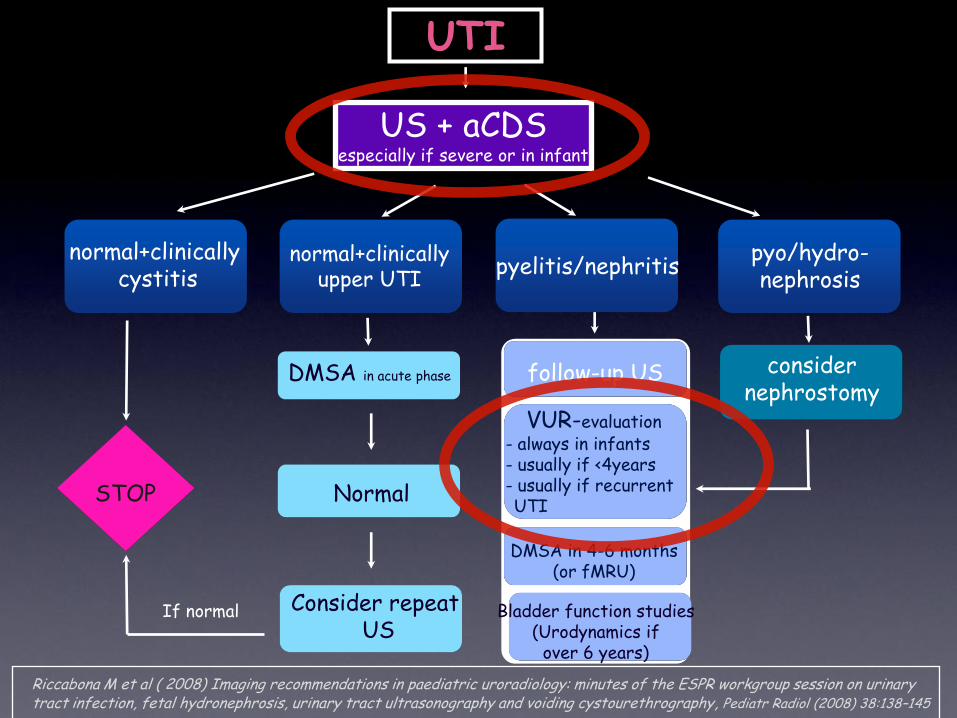
UTI
US + aCDS especially if severe or in infant
STOP
pyelitis/nephritis pyo/hydro- nephrosis
normal+clinically upper UTI
normal+clinically cystitis
DMSA in acute phase
Normal
Consider repeat US
If normal
follow-up US
VUR-evaluation - always in infants - usually if <4years - usually if recurrent UTI
DMSA in 4-6 months (or fMRU)
Bladder function studies (Urodynamics if
over 6 years)
consider nephrostomy
Riccabona M et al ( 2008) Imaging recommendations in paediatric uroradiology: minutes of the ESPR workgroup session on urinary tract infection, fetal hydronephrosis, urinary tract ultrasonography and voiding cystourethrography, Pediatr Radiol (2008) 38:138–145

DMSA
• The main role of DMA is to detect renal scarring 4-6 months post UTI
• Large renal scars can also be seen on US, but US is not as sensitive for renal scarring as DMSA
Ahmed M, Eggleston D, Kapur G, Jain A, Valentini RP, Mattoo TK (2008) Dimercaptosuccinic acid (DMSA) renal scan in the evaluation
of hypertension in children. Pediatr Nephrol 23:435–438