vaccination with dengue virus-like particles induces humoral and cellular immune responses in mice
TRANSCRIPT

RESEARCH Open Access
Vaccination with dengue virus-like particlesinduces humoral and cellular immune responsesin miceShuo Zhang1, Mifang Liang1, Wen Gu1, Chuan Li1, Fang Miao1, Xiaofang Wang1, Cong Jin1, Li Zhang1,Fushun Zhang1, Quanfu Zhang1, Lifang Jiang2, Mengfeng Li2 and Dexin Li1*
Abstract
Background: The incidence of dengue, an infectious disease caused by dengue virus (DENV), has dramaticallyincreased around the world in recent decades and is becoming a severe public health threat. However, there iscurrently no specific treatment for dengue fever, and licensed vaccine against dengue is not available. Vaccinationwith virus-like particles (VLPs) has shown considerable promise for many viral diseases, but the effect of DENV VLPsto induce specific immune responses has not been adequately investigated.
Results: By optimizing the expression plasmids, recombinant VLPs of four antigenically different DENV serotypesDENV1-4 were successfully produced in 293T cells. The vaccination effect of dengue VLPs in mice showed thatmonovalent VLPs of each serotype stimulated specific IgG responses and potent neutralizing antibodies againsthomotypic virus. Tetravalent VLPs efficiently enhanced specific IgG and neutralizing antibodies against all four serotypesof DENV. Moreover, vaccination with monovalent or tetravalent VLPs resulted in the induction of specific cytotoxic T cellresponses.
Conclusions: Mammalian cell expressed dengue VLPs are capable to induce VLP-specific humoral and cellularimmune responses in mice, and being a promising subunit vaccine candidate for prevention of dengue virusinfection.
Keywords: Dengue virus, VLP, Vaccine
BackgroundDengue viruses (DENV) are transmitted among humansby mosquitos, such as Aedes aegypti and Aedes albopic-tus [1]. DENV infection may cause a self-limited febrileillness known as dengue fever (DF), or result in a life-threatening dengue hemorrhagic fever or dengue shocksyndrome (DHF/DSS). It has been estimated that 50-100million cases of DF and 250,000-500,000 cases of DHFoccur annually [2], mainly in tropical and subtropicalregions of the world. Dengue viruses, exist as four sero-types, belong to the family of Flaviviridae, genus Flavi-virus. The virion contains a positive-sense single-strandRNA genome with a long open reading frame coding
for capsid (C), premembrane(prM), and envelope(E)structural proteins, as well as seven non-structural(NS)proteins: NS1, NS2A, NS2B, NS3, NS4A, NS4B, andNS5[3].Because of the widespread geographical distribution
and the severe clinical symptoms, dengue vaccine isurgently needed. However, licensed vaccine is not cur-rently available for prevention of DENV infection. Onemajor reason is the phenomenon of antibody depen-dent-enhancement (ADE), which is known as that a sub-sequent infection with an alternate serotype canenhance severity of dengue disease [1]. One explanationof this phenomenon is that pre-existing non-neutralizingantibodies may enhance capacity of the new infectingDENV to access FcgR bearing cells. Therefore, DENVinfection commonly lacks of antibody cross-protectionamong serotypes. Various strategies have been used to
* Correspondence: [email protected] Key Laboratory for Molecular Virology and Genetic Engineering,Institute for Viral Disease Control and Prevention, China CDC, 155 Chang BaiRoad, Chang Ping District, Beijing 102206, ChinaFull list of author information is available at the end of the article
Zhang et al. Virology Journal 2011, 8:333http://www.virologyj.com/content/8/1/333
© 2011 Zhang et al; licensee BioMed Central Ltd. This is an Open Access article distributed under the terms of the Creative CommonsAttribution License (http://creativecommons.org/licenses/by/2.0), which permits unrestricted use, distribution, and reproduction inany medium, provided the original work is properly cited.

develop dengue vaccine. The most promising candidatesare the live-attenuated tetravalent vaccines of which theclinical trials are in progress [4-7]. One example is theSanofi Pasteur’s dengue vaccine candidate, which isbased on a backbone of yellow fever vaccine (YF 17D)replication genes and incorporates the envelope genes ofthe four dengue virus serotypes, entered its final stage ofclinical development in Australia. However, concernshave been raised about interference in virus replicationamong serotypes [8]. If the replication of four serotypesof vaccine viruses is not balanced, the replication ofnon-dominant serotypes can be interfered by dominantserotypes, which can result in preferential antibodyresponse to the dominant strains and lead to a risk ofdeveloping more serious disease [9]. Thus, an ideal den-gue vaccine should induce neutralizing antibodyresponses against all four serotypes simultaneously andit must be safe to use.To develop an effective and safe dengue vaccine, we
tested the effect of recombinant dengue virus-like parti-cles (VLPs). Virus-like particle vaccine has shown con-siderable promise as vaccine candidate for many viraldiseases [10-13]. VLPs, which are similar to infectiousvirions in the structural and physicochemical features,are non-infectious particles and have advantages insafety and manufacturing. VLPs can be produced inmultiple expression systems such as E.coli, yeast, baculo-virus and mammalian cells. Recombinant VLPs can beefficiently taken up, internalized and processed by anti-gen presenting cells (APCs) [11], and capable to elicitstrong humoral and cellular immune responses againstviruses [14-16]. Recombinant VLPs of flaviviruses havebeen shown to be produced efficiently by co-expressingthe prM and E proteins in the absence of C protein[17-19].In this study, four serotypes of dengue virus-like parti-
cles containing recombinant prM and E proteins weregenerated in mammalian cells, and their immunogeni-city was evaluated in BALB/c mice. The results showedthat monovalent VLPs of each serotype could stimulatespecific IgG and neutralizing antibody against homoty-pic virus, and tetravalent VLPs could induce specificIgG and neutralizing antibodies against all four sero-types of dengue virus. Moreover, vaccination withmonovalent or tetravalent VLPs also resulted in theinduction of specific cellular responses. Therefore, den-gue VLPs can be a potential vaccine candidate for theprevention of dengue infection.
Materials and methodsCells and viruses293T cells (ATCC No.CRL-11268) were cultured inDulbecco’s Modified Eagle Medium (DMEM; Gibco)supplemented with 10% heat-inactivated fetal bovine
serum (FBS), penicillin (100 U/ml) and streptomycin(100 μg/ml) at 37°C with 5% CO2. C6/36 Aedes albopic-tus cells (ATCC No.CRL-1660) were grown at 28°Cwithout CO2 in Eagle’s Minimum Essential Medium(EMEM; Gibco) supplemented with FBS, penicillin andstreptomycin as well.Each serotype of dengue virus was passaged and pro-
pagated in C6/36 cells. The DENV-1 strain GZ01/95and DENV-2 strain ZS01/01 were supplied by theDepartment of Microbiology, Zhongshan School ofMedicine, Sun Yat-sen University, China. DENV-1 strainHawaii, DENV-2 strain NGC, DENV-3 strain H87 andDENV-4 strain H241 were preserved by our laboratory.Strain GZ01/95, ZS01/01, H87 and H241 were used forRNA extraction and then VLPs expression plasmidsconstruction while strain Hawaii, NGC, H87 and H241were used for neutralization analysis. Japanese encepha-litis virus (JEV) strain SA14-14-2 was also propagated inC6/36 cells and mainly used for cDNA cloning.
Construction of DENV VLP expression plasmidsThe QIAamp Viral RNA Kit (Qiagen, Santa Clarita, CA)was used to extract genomic RNA of DENV1-4 and JEVfrom 140 μl C6/36 cells culture supernatant infectedwith each virus. The extracted RNA was subjected toRT using Transcriptor High Fidelity cDNA Synthesis Kit(Roche, Cat. No. 05081955001) to generate cDNA tem-plates for amplification of target genes. The PCR seg-ments were digested with NheI and NotI enzymes andinserted into NheI and NotI sites of pcDNA5/FRTvector.Three types of expression plasmids were constructed
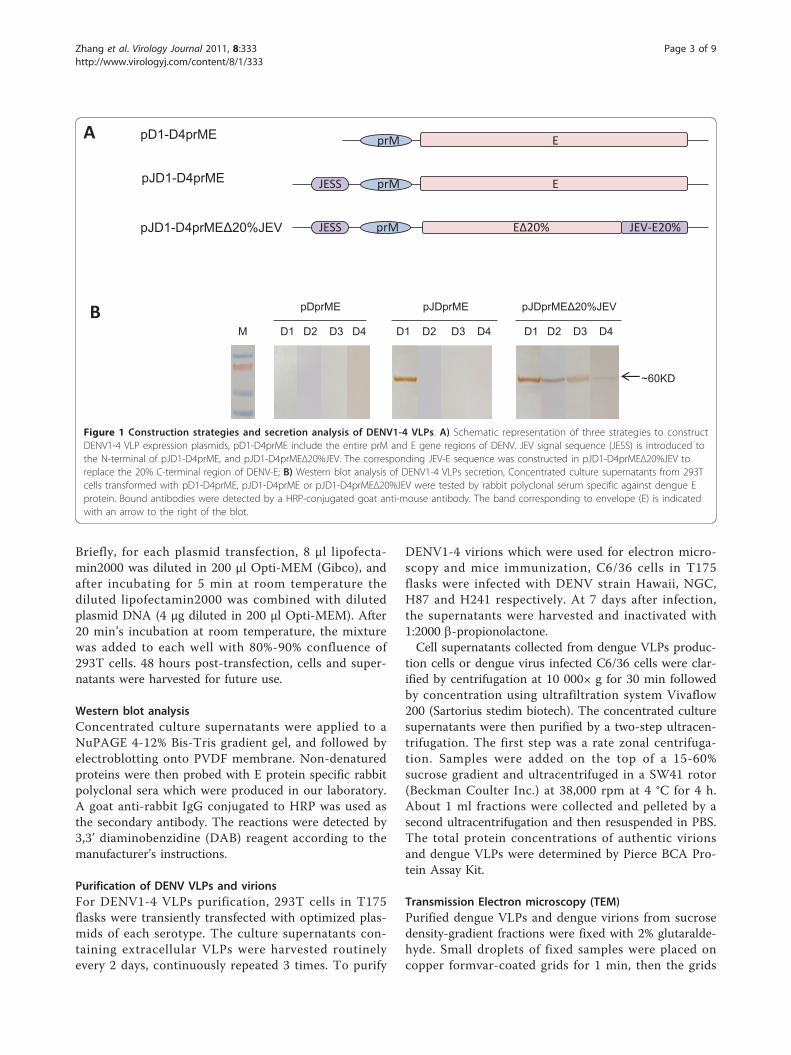
for each serotype of DENV (Figure 1A). The first typeof plasmids was named pD1-D4prME, with entire prMand E genes of each DENV serotype cloned into thepcDNA5/FRT vector. The second type of plasmids waspJD1-D4prME, with entire prM and E genes of eachDENV serotype and an optimized JEV signal sequence(JESS) [17,20,21] gene from SA14-14-2 strain. Tofurther compare the impact of E protein transmem-brane and cytoplasmatic domains, the third type ofplasmids was designed as chimeric constructs pJD1-D4prMEΔ20%JEV, which contained JESS and fulllength of prM, but replaced the 3’ terminal 20% regionof E gene with the corresponding sequence of JEV(strain SA14-14-2).
Transient transfection of 293T cells with DENV VLPexpression plasmids293T cells were prepared in wells of 6-well plates oneday earlier and were transfected with pD1-D4prME,pJD1-D4prME or pJD1-D4prMEΔ20%JEV for each wellusing lipofectamin2000 (Invitrogen, Cat no.11668019)according to instructions supplied by the manufacturer.
Zhang et al. Virology Journal 2011, 8:333http://www.virologyj.com/content/8/1/333
Page 2 of 9
Briefly, for each plasmid transfection, 8 μl lipofecta-min2000 was diluted in 200 μl Opti-MEM (Gibco), andafter incubating for 5 min at room temperature thediluted lipofectamin2000 was combined with dilutedplasmid DNA (4 μg diluted in 200 μl Opti-MEM). After20 min’s incubation at room temperature, the mixturewas added to each well with 80%-90% confluence of293T cells. 48 hours post-transfection, cells and super-natants were harvested for future use.
Western blot analysisConcentrated culture supernatants were applied to aNuPAGE 4-12% Bis-Tris gradient gel, and followed byelectroblotting onto PVDF membrane. Non-denaturedproteins were then probed with E protein specific rabbitpolyclonal sera which were produced in our laboratory.A goat anti-rabbit IgG conjugated to HRP was used asthe secondary antibody. The reactions were detected by3,3’ diaminobenzidine (DAB) reagent according to themanufacturer’s instructions.
Purification of DENV VLPs and virionsFor DENV1-4 VLPs purification, 293T cells in T175flasks were transiently transfected with optimized plas-mids of each serotype. The culture supernatants con-taining extracellular VLPs were harvested routinelyevery 2 days, continuously repeated 3 times. To purify
DENV1-4 virions which were used for electron micro-scopy and mice immunization, C6/36 cells in T175flasks were infected with DENV strain Hawaii, NGC,H87 and H241 respectively. At 7 days after infection,the supernatants were harvested and inactivated with1:2000 b-propionolactone.Cell supernatants collected from dengue VLPs produc-
tion cells or dengue virus infected C6/36 cells were clar-ified by centrifugation at 10 000× g for 30 min followedby concentration using ultrafiltration system Vivaflow200 (Sartorius stedim biotech). The concentrated culturesupernatants were then purified by a two-step ultracen-trifugation. The first step was a rate zonal centrifuga-tion. Samples were added on the top of a 15-60%sucrose gradient and ultracentrifuged in a SW41 rotor(Beckman Coulter Inc.) at 38,000 rpm at 4 °C for 4 h.About 1 ml fractions were collected and pelleted by asecond ultracentrifugation and then resuspended in PBS.The total protein concentrations of authentic virionsand dengue VLPs were determined by Pierce BCA Pro-tein Assay Kit.
Transmission Electron microscopy (TEM)Purified dengue VLPs and dengue virions from sucrosedensity-gradient fractions were fixed with 2% glutaralde-hyde. Small droplets of fixed samples were placed oncopper formvar-coated grids for 1 min, then the grids
prM JEV-E20%JESS
EprM
EprMJESSpJD1-D4prME
pJD1-D4prMEΔ20%JEV
A
B
EΔ20%
M D1 D2 D3 D4 D1 D2 D3 D4 D1 D2 D3 D4
pDprME pJDprMEΔ20%JEVpJDprME
~60KD
Figure 1 Construction strategies and secretion analysis of DENV1-4 VLPs. A) Schematic representation of three strategies to constructDENV1-4 VLP expression plasmids, pD1-D4prME include the entire prM and E gene regions of DENV. JEV signal sequence (JESS) is introduced tothe N-terminal of pJD1-D4prME, and pJD1-D4prMEΔ20%JEV. The corresponding JEV-E sequence was constructed in pJD1-D4prMEΔ20%JEV toreplace the 20% C-terminal region of DENV-E; B) Western blot analysis of DENV1-4 VLPs secretion, Concentrated culture supernatants from 293Tcells transformed with pD1-D4prME, pJD1-D4prME or pJD1-D4prMEΔ20%JEV were tested by rabbit polyclonal serum specific against dengue Eprotein. Bound antibodies were detected by a HRP-conjugated goat anti-mouse antibody. The band corresponding to envelope (E) is indicatedwith an arrow to the right of the blot.
Zhang et al. Virology Journal 2011, 8:333http://www.virologyj.com/content/8/1/333
Page 3 of 9

were stained with sodium phosphotungstate for 1 min(excess samples of each step were removed). At last, thegrids were visualized by TEM.
Mice immunizationFour to six-week-old female BALB/c mice were pur-chased from Chinese Academy of Medical SciencesBreeding Laboratories and were intraperitoneally (i.p.)inoculated with monovalent DENV VLPs (100 μg perdose) or a tetravalent combination (25 μg of each sero-type per dose) in Freund’s complete adjuvant (Sigma)for priming and in Freund’s incomplete adjuvant for twotimes of boosting at an interval of 2 weeks. Equalamount of DENV virions (100 μg for monovalent vac-cine and 25 μg of each serotype for tetravalent vaccine)were used as controls with the same regimen. On days0, 14 and 28, blood samples were collected through tailvein for measurement of serum IgG. At 2 weeks afterthe last inoculation, mice were sacrificed to collectserum for the neutralizing antibodies assay and sepa-rated splenocytes for testing cytotoxic T cell responses.
ELISA to measure serum IgGVLPs specific serum IgG antibodies were titred by thebinding capacity with rEIII protein, a recombinant pro-tein that chimericly expressed DENV1-4 EIII domains ina certain order and previously produced in our labora-tory. IgG titers were measured using enzyme-linkedimmunosorbent assay (ELISA). Briefly, 200 ng purifiedrEIII per well was coated on 96-well plates at 4°C over-night. Then, the plates were blocked with 5% skimmedmilk in PBS for 1 h, and incubated with 2-fold serialdiluted serum samples (starting from 1:50) at 37°C for 1h. Bound IgG was detected by HRP-conjugated goatanti-mouse IgG (Sigma). After addition of 3,3’, 3,5’-tet-ramethylbenzidine (TMB), absorbance was measured at450 nm. The value which exceeds the mean+2 S.D. ofnegative control was considered positive.
Antibody neutralization assayThe neutralization ability of serum antibodies againstDENV was determined using CPE-determination assays.Briefly, mice sera from all groups were heat-inactivatedat 56°C for 30 min, then the sera were two-fold serialdiluted from 1:5 to 1:160 in Eagle’s medium supplemen-ted with 1% heat-inactivated FBS, penicillin and strepto-mycin and mixed with 100TCID50 virus. After 1 hincubation at 37°C, 100 μl of virus-serum mixture wasinoculated to the confluent monolayer of BHK-21 cellsin 96-well plates. Every dilution of each serum was per-formed in quadruplicate. The plates were then incubatedin a CO2 incubator at 37°C for 7 days. The neutraliza-tion titer was expressed as the maximum serum dilution
at which the CPE of the virus was not observed in allfour wells.
Enzyme Linked Immunospot (ELISPOT) AssayThe ELISPOT 96-well plates (BD) were coated with 100μl of anti-mouse IFN-g (5 μg/ml in coating buffer) at 4°C overnight. The following day, plates were washed andblocked with blocking solution for 2 h. Then, 100 μlfreshly isolated splenocytes (5 × 10⁵ cells) from theimmunized mice were added to each well and stimu-lated with DENV VLPs at 37°C for 40 h. After cellswere washed out, biotinylated anti-mouse IFN-g wasadded to each well and incubated for 2 h at room tem-perature. Thereafter, the plates were washed and incu-bated for 1 h at room temperature with streptavidin-HRP. Finally, AEC substrate solution (BD) was addedand spots were counted by ImmunoSpot® Analyzer(Cellular Technology Ltd.).
Statistical analysisStatistical comparisons among groups were analyzed byone way ANOVA using SPSS 11.5. A P value less than0.05 was considered statistically significant.
ResultsProduction of DENV VLPsTo optimize the production of DENV VLPs, three typesof expression plasmids encoding prM and E glycopro-teins were constructed for each serotype (Figure 1A). Eand prM proteins were chosen as two subunits ofrecombinant VLPs, because the former one constitutesthe spikes on DENV membrane surface and is known asthe major protective antigen of DENV to induce neutra-lizing antibodies, and the latter one is also embedded inthe viral envelop and contributes to the stability of Eprotein. To test the secretion of DENV VLPs from tran-sient transfected 293T cells, culture supernatants of293T cells were collected and identified by western blotanalysis for E protein expression (Figure 1B). Cellstransfected with pD1-D4prME plasmids that express fulllength of prM and E proteins could express intracellularproteins (data not shown). However, due to the lack ofsignal sequence, they could not secret VLPs into tissueculture. When adding a JEV signal sequence at the N-terminal of full length of prM and E genes, cells trans-fected with expression plasmid pJD1prME could effec-tively secret VLPs, but cells transfected with pJD2-D4prME still could not secret VLPs. When both carry-ing a N-terminal JEV signal sequence and replacing C-terminal 20% regions of DENV E gene with the corre-sponding region of JEV E gene, all four constructs,pJD1-D4prMEΔ20%JEV, could secrete VLPs into tissueculture. Therefore, we chose pJD1prME and pJD2-
Zhang et al. Virology Journal 2011, 8:333http://www.virologyj.com/content/8/1/333
Page 4 of 9

D4prMEΔ20%JEV to express recombinant DENV1 andDENV2-4 VLPs in 293T cells, respectively.To compare the size and morphology of recombinant
DENV1-4 VLPs and their corresponding serotype ofDENV virions, purified dengue VLPs and virions wereobserved under transmission electron microscopy(TEM) (Figure 2). All four types of recombinant VLPsexhibited as electron-dense spherical particles of 45-55nm size, which were similar to the morphology and sizeof DENV virions. Therefore, by optimizing the expressplasmids and using mammalian 293T cells, we success-fully acquired DENV VLPs which consisted of majorantigenic proteins of the virus and exhibited similarmorphological features as nature virus particles.
DENV VLPs elicited virus specific IgG and neutralizingantibodiesTo evaluate humoral responses induced by recombinantVLPs, BALB/c mice were immunized with monovalentdengue VLPs of each serotype or their correspondingvirion counterparts for three times at two-week inter-vals. Serum samples were collected after 2 weeks ofeach immunization, and analyzed for IgG antibodiesspecific against rEIII, a recombinant protein thatexpressed chimerical EIII domains of all four DENV
serotypes. EIII domain of DENV contains several neu-tralizing epitopes and host cell receptor recognitionsites. As shown in Figure 3A-D, in comparison to thePBS control, mice immunized with either dengue VLPsor virions induced high level of rEIII specific serum IgG.For DENV-1, DENV-2 and DENV-4 VLPs, they inducedeven higher IgG responses than DENV-1, DENV-2 andDENV-4 virions, respectively. Neutralization assaysusing serum collected on day 42 demonstrated thatDENV1-4 VLPs could elicit comparable level of homoty-pic neutralizing antibodies as monovalent dengue virions(Figure 4A).The results that monovalent dengue VLPs could effi-
ciently enhance specific IgG and develop neutralizationantibodies, suggested that the tetravalent formulation ofDENV VLPs, which was constituting of four types ofDENV VLPs at equal amount, might be capable to stimu-late neutralizing antibodies against all four serotypes ofDENV. As generating balanced neutralizing antibodies toeach serotype of a tetravalent dengue vaccine is desired forits safety and efficacy, a tetravalent dengue VLPs combina-tion was applied to BALB/c mice for all priming andboosting immunizations as the regimen used for monova-lent VLPs vaccination. Tetravalent dengue virions and PBSwere again used as controls. The serum samples collected
A C E
FDB 100nm
100nm
100nm
100nm100nm
Figure 2 Morphology and size of dengue VLPs and virions. Purified dengue virions (arrow indicated in A) and VLPs (arrow indicated in C-F)were negatively stained and analyzed by TEM. Scale bar indicates 100 nm. A. Dengue virions, B. Negative control, C. DENV-1 VLPs, D. DENV-2VLPs, E. DENV-3 VLPs, F. DENV-4 VLPs.
Zhang et al. Virology Journal 2011, 8:333http://www.virologyj.com/content/8/1/333
Page 5 of 9

Figure 3 Virus specific IgG were enhanced by DENV VLPs or virions. BALB/c mice were intraperitoneally immunized with 100 μgmonovalent DENV VLPs or virions (A-D), or tetravalent VLPs or virions (E 25 μg of each serotype) for three times at two-week intervals. At day14, 28, 42, sera were collected and ELISA was used to test for rEIII specific IgG. Data were expressed as mean titer with a standard deviation (SD)bar. *indicates statistical significance (P < 0.05).
Zhang et al. Virology Journal 2011, 8:333http://www.virologyj.com/content/8/1/333
Page 6 of 9

at Day 14, 28, and 42 post the initial tetravalent VLPs vac-cination, were analyzed for rEIII specific IgG antibodies.The data showed that tetravalent virions could stimulateIgG antibodies against rEIII protein. Intriguingly, tetrava-lent VLPs could even achieved to a higher level than tetra-valent virions could induce (Figure 3E). Moreover,neutralizing antibodies stimulated by tetravalent VLPsexhibited simultaneous blocking of DENV1-4 infections,and the blocking effect was comparable to tetravalent for-mula of virions (Figure 4B).Therefore, similar as DENV virions, both monovalent
and tetravalent formula of DENV VLPs could effectivelyinduce virus specific IgG and produce high titer of pro-tective neutralizing antibodies in vaccinated mice.
DENV VLPs induced virus-specific T cell responsesLastly, to investigate cellular immune responses trig-gered by dengue VLPs, ELISPOT assay was employed totest dengue specific cytotoxic T cell responses. At day42 after the initial immunization, mice were sacrificed
and spleen cells were collected. Each of the monovalentDENV1-4 VLPs immunized spleen cells were then invitro stimulated with corresponding DENV1-4 VLPs andmonitored for IFN-g production. As depicted in Figure5A, splenocytes from mice immunized with denguemonovalent VLPs exibited specific cytotoxic T cellresponses, and the extent was similar to the effect ofmonovalent DENV virions. Furthermore, dengue tetra-valent VLPs manifested high level of IFN-g to in vitrostimulation with each serotype of DENV1-4 VLPs, andwith no significant differences to the homotypic virions(Figure 5B). These results demonstrated that vaccinationwith either monovalent or tetravalent dengue VLPscould elicit dengue specific cellular immune responses.
DiscussionDespite many years of efforts, an effective dengue vaccinehas not been developed. Various strategies have beenapplied to develop dengue vaccine, such as attenuated
Neu
traliz
ing
antib
ody
titer
-1(G
MT)
Neu
traliz
ing
antib
ody
titer
-1(G
MT)
Figure 4 Serum neutralizing antibody titer of vaccinated mice.BalB/C mice were immunized with A) 100 μg monovalent VLPs orvirions, B) tetravalent VLPs or virions (25 μg of each serotype) forthree times at two weeks interval. At day 42, the serum neutralizingantibodies were assessed using CPE-determination assays. Data ofeach group was expressed as geometric mean titer (GMT) with anS.D. bar (n = 5). *indicates statistical significance (P < 0.05).
Figure 5 Dengue specific T cell responses evaluated byELISPOT assay. BALB/c mice were immunized with DENV1-4 VLPsor virions in either monovalent A) or tetravalent B) formula. Theimmunized mice were sacrificed at day 42 and the collected spleencells were isolated and stimulated in vitro with each of DENV1-4VLPs. ELISPOT assay was performed to test IFN-g production. Themean number of spot forming cells (SFCs)/106 splenocytes wasshown as VLPs-stimulated plus mock-stimulated with an S.D bar.*indicates statistical significance (P < 0.05).
Zhang et al. Virology Journal 2011, 8:333http://www.virologyj.com/content/8/1/333
Page 7 of 9

[22,23], subunit [24,25], chimeric [26,27], and DNA [1,28]vaccines. Previous studies have shown that co-expressedprM and E proteins of dengue virus could produce VLPsin mammalian cells, but the VLPs expression plasmidswere often used as DNA vaccine [17,20,29]. Here we stu-died on recombinant dengue VLP vaccine as a new candi-date. VLPs are similar to infectious virions in bothstructural and biochemical properties but are non-replicat-ing and free of genome. Therefore, due to their preservedimmunogenicity in native forms and better safety, VLPshave been used in many vaccine researches for preventionof viral diseases [11].In order to optimize the production of dengue VLPs,
three types of expression plasmids were constructed.The indirect fluorescent antibody (IFA) assay showed allconstructs could express intracellular VLPs (data notshown). However, the western blot analysis of culturingsupernatant showed that, pD1-D4prME constructs with-out signal peptide could not secret dengue VLPs. Andexcept for pJD1prME, the constructs simply adding aJEV signal sequence could not secret dengue VLPs. Allconstructs which replaced the carboxy-terminal 20% ofDENV E protein with corresponding JEV E proteincould secret VLPs into cell culturing supernatant. Basedon data from these express plasmids, pJD1prME andpJD2-D4prMEΔ20%JEV were identified as optimizedVLP formation constructs for each DENV serotype.These data from different express plasmids indicatedthat i) signal peptide was one of the most important fac-tors that influence downstream protein translocationand topology, thus dictating correct processing of den-gue virus prM and E proteins by the host encoded sig-nalase and endopeptidase [20]. ii) the transmembranedomain of dengue E protein contains a strong ER reten-tion signal [30,31], replacement of the carboxy-terminal20% of DENV E protein with the corresponding regionof JEV provides extracellular secretion of DENV2-4VLPs, but does not produce additional benefit to pro-mote extracellular secretion of DENV-1 VLPs.This was different from previous studies [29,32],
which showed that 20% JEV sequence replacement wasabsolutely necessary for DENV-1 and DENV-2 VLPssecretion; DENV-3 plasmids containing either the full-length DENV-3 E protein gene or the 20% JEV sequencereplacement secreted VLPs to similar levels; WhereasDENV-4 VLPs were secreted to high levels by plasmidscontaining the full-length DENV-4 E protein gene butnot by the chimeric plasmid containing 20% JEV Ereplacement. Considering that dengue viruses of differ-ent serotypes or even among different strains of thesame type, their biological characteristics were not thesame, thus it is essential to use different strategies whenconstructed dengue VLPs expression plasmids. As den-gue particle assembly and secretion is influenced by
interaction of prM and E [29], we inferred that the chi-meric E (80%DENV and 20%JEV) of DENV2-4 couldinteract with DENV2-4 prM, which help to stabilize theinteraction between prM and E and lead to efficientsecretion of VLPs. As for DENV-1 (strain GZ01/95), itsprM and E domains might interact stably enough, andthe replacement of 20% C-terminal region of E wouldnot improve more on this interaction, therefore DENV-1 VLPs showed similar secret pattern betweenpJD1prME and pJD1prMEΔ20%JEV plasmids.The immunogenicity of dengue VLPs was evaluated
using BALB/c mice. The analysis of humoral immuneresponses revealed that dengue VLPs, in either monova-lent or tetravalent formula, induced high levels of rEIII-specific IgG. Because dengue virus-induced neutralizingantibodies can bind to virus and prevent virus frombinding to host cell receptors, therefore inducing neu-tralizing antibodies is particularly important to blockvirus entry into target cells [33]. In this study, either themonovalent or the tetravalent formula of dengue VLPscould efficiently trigger in vivo development of neutra-lizing antibodies. Furthermore, the tetravalent formulaof VLPs was able to simultaneously induce balancedneutralizing antibodies against all four serotypes. Allthese results confirmed that all four serotypes of DENVVLPs prepared in this study preserved the antigenicityof prM and E proteins.One of the marked advantages of VLPs is their ability
to induce cellular immunity [34,35]. Since IFN-g consti-tutes a major mediator of the Th1 cell-mediatedimmune response and has been shown to play a keyrole in antiviral activity against dengue [36], in ourstudy, cellular immune responses were assessed by IFN-g releasing ability of VLPs-stimulated spleen cells.Spleen cells from mice vaccinated with dengue VLPsand virions produced comparable levels of IFN-g after invitro stimulation with dengue VLPs.In conclusion, by optimizing the expression plasmids,
we successfully generated recombinant DENV1-4 VLPs inmammalian cells. Furthermore, the vaccination effect ofVLPs in mice showed that either monovalent or tetrava-lent formula of dengue VLPs could efficiently elicit virusspecific humoral and cellular immune responses. Theseresults supplied evidence that VLP vaccine may serve as apromising strategy for dengue vaccine development.
AcknowledgementsThis work was supported by the grants (2006AA02A223) from National keyprojects of “863” High Technology R&D, Chinese Ministry of Science andTechnology, and National Key Programs for Infectious Diseases, Ministry ofHealth.
Author details1State Key Laboratory for Molecular Virology and Genetic Engineering,Institute for Viral Disease Control and Prevention, China CDC, 155 Chang Bai
Zhang et al. Virology Journal 2011, 8:333http://www.virologyj.com/content/8/1/333
Page 8 of 9

Road, Chang Ping District, Beijing 102206, China. 2Department ofMicrobiology, Zhongshan School of Medicine, Sun Yat-Sen University,Guangzhou 510080, Guangdong, China.
Authors’ contributionsZS performed most of the experiments and involved in manuscriptpreparation. LM coordinated laboratory manipulation and edited themanuscript. GW participated in mice immunization and detection ofhumoral immune responses. LC, MF, WX and ZQ were involved in cellsculture, virus infection and VLPs purification. JC participated in editing of themanuscript. ZL and ZF participated in the detection of cellular immuneresponses. JL and LM provided Chinese strains of dengue viruses and gaveadvices for the project. LD is the project leader and was involved in projectdesign, manipulation, data analysis and finalization of the manuscript. Allauthors read and approved the final manuscript.
Competing interestsThe authors declare that they have no competing interests.
Received: 20 April 2011 Accepted: 30 June 2011Published: 30 June 2011
References1. Ramanathan MP, Kuo YC, Selling BH, et al: Development of a novel DNA
SynCon™ tetravalent dengue vaccine that elicitsimmune responsesagainst four serotypes. Vaccine 2009, 27:6444-53.
2. Centers for Disease Control and Prevention (CDC) DoV-bID-DF. [http://cdc.gov/ncidod/dvbid/dengue], [cited].
3. Chambers TJ, Hahn CS, Galler R, et al: Flavivirus genome organization,expression, and replication. Ann Rev Microbiol 1990, 44:649-88.
4. Sabchareon A, Lang J, Chanthavanich P, et al: Safety and immunogenicityof tetravalent live-attenuated dengue vaccines in Thai adult volunteers:role of serotype concentration, ratio, and multiple doses. Am J Trop MedHyg 2002, 66:264-72.
5. Sabchareon A, Lang J, Chanthavanich P, et al: Safety and immunogenicityof a three dose regimen of two tetravalent live-attenuated denguevaccines in five- to twelve-year-old Thai children. Pediatr Infect Dis J 2004,23:99-109.
6. Edelman R, Wasserman SS, Bodison SA, et al: Phase I trial of 16formulations of a tetravalent live-attenuated dengue vaccine. Am J TropMed Hyg 2003, 69:48-60.
7. Innis BL, Eckels KH: Progress in development of a live-attenuated,tetravalent dengue virus vaccine by the United States Army MedicalResearch and Materiel Command. Am J Trop Med Hyg 2003, 69:1-4.
8. Imoto Jun-ichi, Konishi Eiji: Dengue tetravalent DNA vaccine increases itsimmunogenicity in mice when mixed with a dengue type 2 subunitvaccine or an inactivated Japanese encephalitis vaccine. Vaccine 2007,25:1076-84.
9. Webster PDaniel, Farrar Jeremy, Rowland-Jones Sarah: Progress towards adengue vaccine. The Lancet Infectious Diseases 2009, 9:678-87.
10. Galarza JM, Latham T, Cupo A: Virus-like particle (VLP) vaccine conferredcomplete protection against a lethal influenza virus challenge. ViralImmunol 2005, 18(1):244-51.
11. Li C, Liu F, Liang M, Zhang Q, et al: Hantavirus-like particles generated inCHO cells induce specific immune responses in C57BL/6 mice. Vaccine2010, 28:4294-4300.
12. Weber J, Cheinsong-Popov R, Callow D, et al: Immunogenicity of the yeastrecombinant p17/p24:Ty virus-like particles (p24-VLP) in healthyvolunteers. Vaccine 1995, 13:831-4.
13. Sedlik C, Saron M, Sarraseca J, et al: Recombinant parvovirus-like particlesas an antigen carrier: a novel nonreplicative exogenous antigen to elicitprotective antiviral cytotoxic T cells. Proc Natl Acad Sci USA 1997,94:7503-8.
14. Murata K, Lechmann M, Qiao M, et al: Immunization with hepatitis Cvirus-like particles protects mice from recombinant hepatitis C virus-vaccinia infection. Proc Natl Acad Sci USA 2003, 100:6753-8.
15. Pinto , Ligia A, Castle , et al: HPV-16 L1 VLP vaccine elicits a broad-spectrum of cytokine responses in whole blood. Vaccine 2005,23:3555-64.
16. Akahata Wataru, Yang Zhi-Yong, Andersen Hanne, et al: A virus-like particlevaccine for epidemic Chikungunya virus protects nonhuman primatesagainst infection. Nature Medicine 2010, 16:334-8.
17. Chang GJ, Davis BS, Hunt AR, et al: Flavivirus DNA vaccines: current statusand potential. Ann NY Acad Sci 2001, 951:272-85.
18. Putnak R, Porter K, Schmaljohn C: DNA vaccines for flaviviruses. Adv VirusRes 2003, 61:445-68.
19. Lindenbach BD, Rice CM: Flaviviridae: the viruses and their replication. InFields Virology.. 4 edition. Edited by: Knipe DM, Howley PM. Philadelphia:Lippincott Williams 2001:991-1041.
20. Chang GJ, Hunt AR, Davis B: A single intramuscular injection ofrecombinant plasmid DNA induces protective immunity and preventsJapanese encephalitis in mice. J Virol 2000, 74:4244-52.
21. Lobigs Mario, Lee Eva: Inefficient Signalase Cleavage Promotes EfficientNucleocapsid Incorporation into Budding Flavivirus Membranes. J Virol2004, 78:178-86.
22. Hoke CH Jr, Malinoski FJ, Eckels KH, et al: Preparation of an attenuateddengue 4 (341750 Carib) virus vaccine. II. Safety and immunogenicity inhumans. Am J Trop Med Hyg 1990, 43:219-26.
23. Edelman R, Tacket CO, Wasserman SS, et al: A live attenuated dengue-1vaccine candidate (45AZ5) passaged in primary dog kidney cell cultureis attenuated and immunogenic for humans. J Infect Dis 1994,170:1448-55.
24. Staropoli I, Frenkiel MP, Megret F, et al: Affinity-purified dengue-2 virusenvelope glycoprotein induces neutralizing antibodies and protectiveimmunity in mice. Vaccine 1997, 15:1946-54.
25. Simmons M, Nelson WM, Wu SJ, et al: Evaluation of the protectiveefficacy of a recombinant dengue envelope B domain fusion proteinagainst dengue 2 virus infection in mice. Am J Trop Med Hyg 1998,58:655-62.
26. Bray M, Men R, Lai CJ: Monkeys immunized with intertypic chimericdengue viruses are protected against wild-type virus challenge. J Virol1996, 70:4162-6.
27. Pletnev AG, Men R: Attenuation of the Langat tick-borne flavivirus bychimerization with mosquito-borne flavivirus dengue type 4. Proc NatlAcad Sci USA 1998, 95:1746-51.
28. Kochel T, Wu SJ, Raviprakash K, et al: Inoculation of plasmids expressingthe dengue-2 envelope gene elicit neutralizing antibodies in mice.Vaccine 1997, 15:547-52.
29. Chang GJ, Hunt AR, Holmes DA, et al: Enhancing biosynthesis andsecretion of premembrane and envelope proteins by the chimericplasmid of dengue virus type 2 and Japanese encephalitis virus. Virology2003, 306:170-80.
30. Hsieh SC, Liu IJ, King CC, et al: A strong endoplasmic reticulum retentionsignal in the stem-anchor region of envelope glycoprotein of denguevirus type 2 affects the production of virus-like particles Virology. 2008,374(2):338-50.
31. Hsieh SC, Tsai WY, Wang WK: The length of and nonhydrophobic residuesin the transmembrane domain of dengue virus envelope protein arecritical for its retention and assembly in the endoplasmic reticulum. JVirol 2010, 84:4782-97.
32. Purdy DE, Chang GJ: Secretion of noninfectious dengue virus-likeparticles and identification of amino acids in the stem region involvedin intracellular retention of envelope protein. Virology 2005, 333:239-250.
33. Babu J Pradeep, Pattnaik1 Priyabrata, Gupta Nimesh, et al: Immunogenicityof a recombinant envelope domain III protein of dengue virus type-4with various adjuvants in mice. Vaccine 2008, 26:4655-63.
34. Schirmbeck R, Melber K, Kuhrober A, et al: Immunization with solublehepatitis B virus surface protein elicits murine H-2 class I-restricted CD8+ cytotoxic T lymphocyte responses in vivo. J Immunol 1994, 152:1110-9.
35. Greenstone HL, Nieland JD, de Visser KE, et al: Chimeric papillomavirusvirus-like particles elicit antitumor immunity against the E7 oncoproteinin an HPV16 tumor model. Proc Natl Acad Sci USA 1998, 95(4):1800-5.
36. Valdés I, Bernardo L, Gil L, et al: A novel fusion protein domain III-capsidfrom dengue-2, in a highly aggregated form, induces a functionalimmune response and protection in mice. Virology 2009, 394:249-58.
doi:10.1186/1743-422X-8-333Cite this article as: Zhang et al.: Vaccination with dengue virus-likeparticles induces humoral and cellular immune responses in mice.Virology Journal 2011 8:333.
Zhang et al. Virology Journal 2011, 8:333http://www.virologyj.com/content/8/1/333
Page 9 of 9