vaginal discharge. definition running of white substance and the. than blood nb : normally the...
TRANSCRIPT

Vaginal Discharge
DEFINITION
Running of white substance and the. than blood
NB : Normally the vagina and the introitus are kept moist by the
following:
1. Bartholin gland play the most important role in the lubrication of
vaginal introitus. It secrete a thick mucus which is increased during
sexual intercourse.
2. In the periurethral region of the visible are situated Skerie's ducts
which contribute to the lubrication of the vulval structures.
3. Although the vagina is itself devoid of glands its surfaces is moistened
by the secretion of the cervical glands and to much less extent by
transudation
from its own surface.
Causes
I. Physiological causes: pelvic Congestion :
1. During pregnancy
2. Chronic constipation
3. Sedentary life
4. At the time of ovulation
5. Emotional and sexual upset
II. Pathological Causes:
1. INFLAMMATORY CONDITION: Vulvitis, vaginitis, cervical erosion, tuberculous, endometritis, puerperal sepsis, chronic salpingo-oophritis and pelvic abscess draining vaginally
2. NEOPLASTIC CONDITION:-
Infected cervical polyp Malignant neoplasia in the lower genital tract
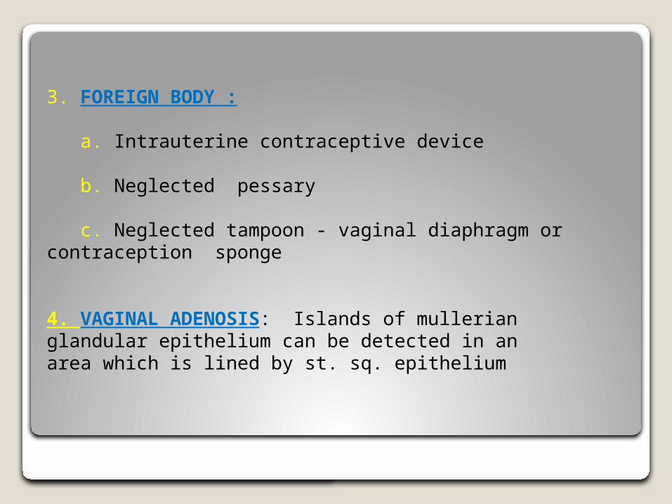
3. FOREIGN BODY :
a. Intrauterine contraceptive device
b. Neglected pessary
c. Neglected tampoon - vaginal diaphragm or contraception sponge
4. VAGINAL ADENOSIS: Islands of mullerian glandular epithelium can be detected in an area which is lined by st. sq. epithelium

5. INTERMITTENT HYDROSALPINX
6 . Ascitis Fluid
7. SMALL VVF OR SI
8. ANTE-PARTUM R.M.
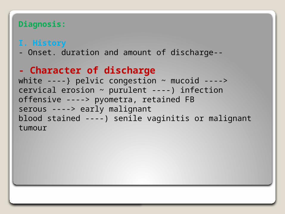
Diagnosis:
I. History - Onset. duration and amount of discharge--
- Character of discharge white ----} pelvic congestion ~ mucoid ----> cervical erosion ~ purulent ----) infection offensive ----> pyometra, retained FB serous ----> early malignant blood stained ----) senile vaginitis or malignant tumour

ASSOCIATED SYMPTOMS:
itching (monilial or trichomonas vaginitis) dysuria - gonoccal infection .previous medication " menstrual troubles last abortion or labour
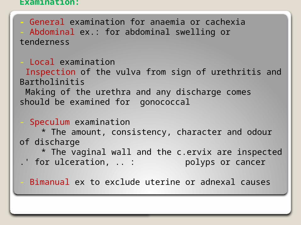
Examination:
- General examination for anaemia or cachexia - Abdominal ex.: for abdominal swelling or tenderness
- Local examination Inspection of the vulva from sign of urethritis and Bartholinitis Making of the urethra and any discharge comes should be examined for gonococcal
- Speculum examination * The amount, consistency, character and odour of discharge * The vaginal wall and the c.ervix are inspected .' for ulceration, .. :
polyps or cancer
- Bimanual ex to exclude uterine or adnexal causes
III. Investigation
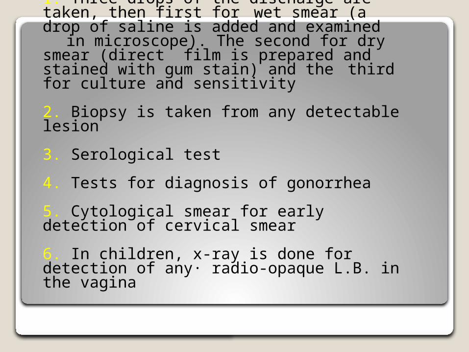
1. Three drops of the discharge are taken, then first for wet smear (a drop of saline is added and
examined in microscope). The second for dry smear (direct film is prepared and stained with gum stain) and
the third for culture and sensitivity
2. Biopsy is taken from any detectable lesion
3. Serological test
4. Tests for diagnosis of gonorrhea
5. Cytological smear for early detection of cervical smear
6. In children, x-ray is done for detection of any· radio-opaque L.B. in the vagina
Treatment Treatment of the cause Non-infective vaginal discharge is managed by
- Minimize pelvic congestion by active exercises and active life
- Pelvic decongestant as glycerine icthyol pessaries - Cleanliness
Monilial Vaginitis
CAUSATIVE FUNGUS:
Candida albican. [gm + ve unicellula. Organism reproduce by budding, spore forming, characterized by pseudo hyphae formation.

Predisposing Factors:
Acidic PH of the vagina (4-5) Pregnancy as vaginal PH more acidic Oral contraceptive pills Antibiotic ---) alternation of normal vaginal flora Diabetes due to high glucose in blood and urine Corticosteroid and other immunosuppressive drugs Clinical manifestation of monilia increased before period and decreased during and after menses

Mode of Infection
Transformed from carrier state into active state.
SymptomsItching Vaginal discharge
Signs:- Sign of scratching and acute inflammation in the vulva - Vagina shows white patches adherent to the mucera its removal leaves bare area with bleeding [ - Vaginal discharge ---> scanty, thick, white curdy with offensive odour Investigation:(1) Wet smear: A drop vaginal discharge is mixed with drop of KoH 10% and examined microscopically (2) Direct film is prepared and stained with M.S. the organism identified by long slender filament ends in small buds
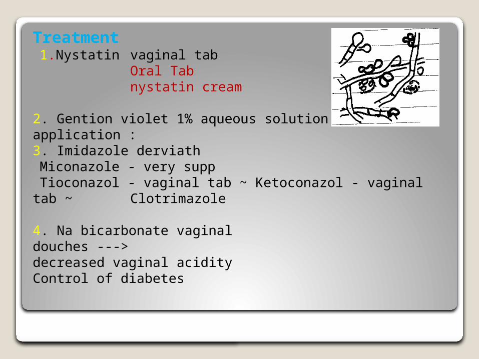
Treatment1.Nystatin vaginal tab
Oral Tabnystatin cream
2. Gention violet 1% aqueous solution local application : 3. Imidazole derviath
Miconazole - very supp Tioconazol - vaginal tab ~ Ketoconazol - vaginal tab ~
Clotrimazole
4. Na bicarbonate vaginal douches ---> decreased vaginal acidityControl of diabetes
Trichomonas Vagincal
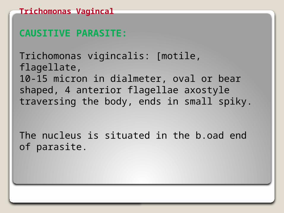
CAUSITIVE PARASITE:
Trichomonas vigincalis: [motile, flagellate, 10-15 micron in dialmeter, oval or bear shaped, 4 anterior flagellae axostyle traversing the body, ends in small spiky.
The nucleus is situated in the b.oad end of parasite.
There are 3 types of trichomonas: 1. Trichomonas vaginalis present in vagina 2. Trichomonas buccalis present in mouth 3. Trichomonas homanis present in rectum and anal
canal
Mode of InfextionSexual contact Bad sexual hygiene - infected cloths, baths of or gloves
Symptoms:1.Vaginal discharge, thin coplus foamy greenish yellow in colour and offensive 2. Dyspnea dyspareunia due to local cystitis 3. itching4. Frequency in micturation due to local cystitiis
Sign :1. Characteristic vaginal discharge2. Vaginal mucosa is congested and granular (Strawberry vaginal
INVESTIGATION:1. Wet film drop of vaginal discharge is mixed with
drop of warm saline and examined
microscopically
2. Dry film ---> direct film is prepared and stained with
Leishman stain
3. Culture Kupferberg's whittington medics
'"
TREATMEIT: 1. Metronidazole (Flagyl) 3 times for 7-10 days
2. Tinidazole (fasigyn) 2 gm single dose
3. During pregnancy ---} clotrimazole vagincal pessary
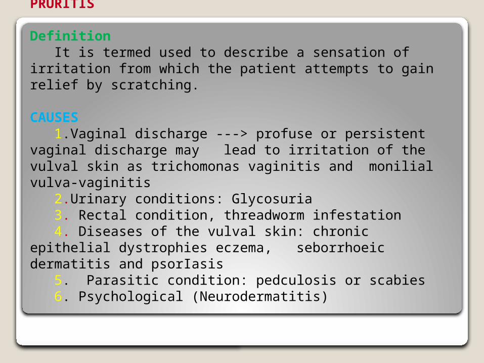
PRURITIS
DefinitionIt is termed used to describe a sensation of
irritation from which the patient attempts to gain relief by scratching.
CAUSES1.Vaginal discharge ---> profuse or persistent vaginal discharge may lead to irritation of the vulval skin as trichomonas vaginitis and monilial vulva-vaginitis 2.Urinary conditions: Glycosuria 3. Rectal condition, threadworm infestation 4. Diseases of the vulval skin: chronic epithelial dystrophies eczema, seborrhoeic dermatitis and psorIasis 5. Parasitic condition: pedculosis or scabies6. Psychological (Neurodermatitis)
Diagnosis:
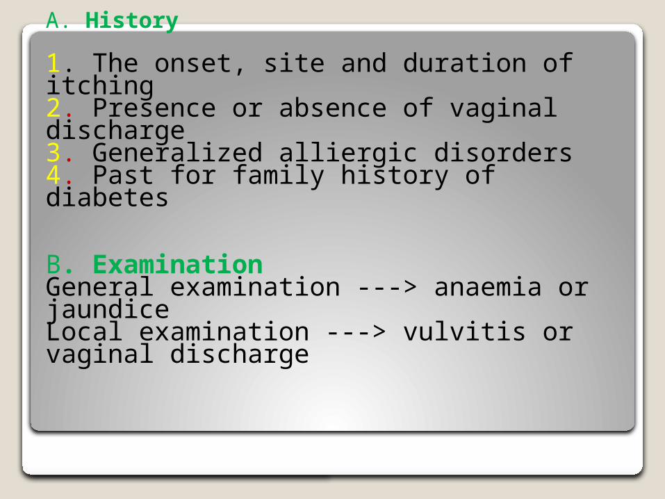
A. History
1. The onset, site and duration of itching 2. Presence or absence of vaginal discharge 3. Generalized alliergic disorders 4. Past for family history of diabetes
B. ExaminationGeneral examination ---> anaemia or jaundice Local examination ---> vulvitis or vaginal discharge
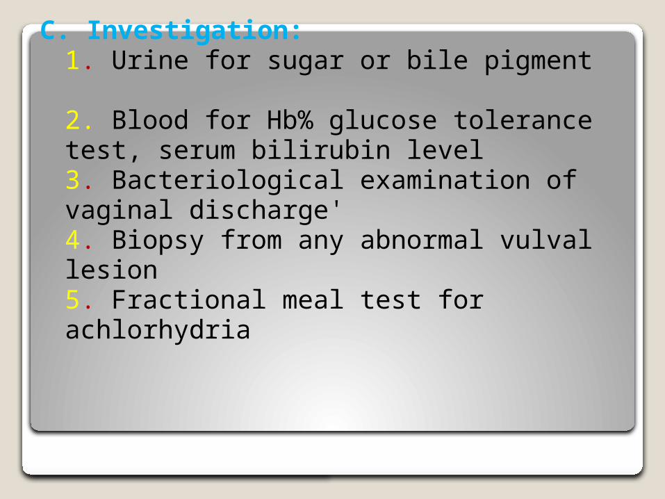
C. Investigation: 1. Urine for sugar or bile pigment 2. Blood for Hb% glucose tolerance test, serum bilirubin level 3. Bacteriological examination of vaginal discharge' 4. Biopsy from any abnormal vulval lesion 5. Fractional meal test for achlorhydria
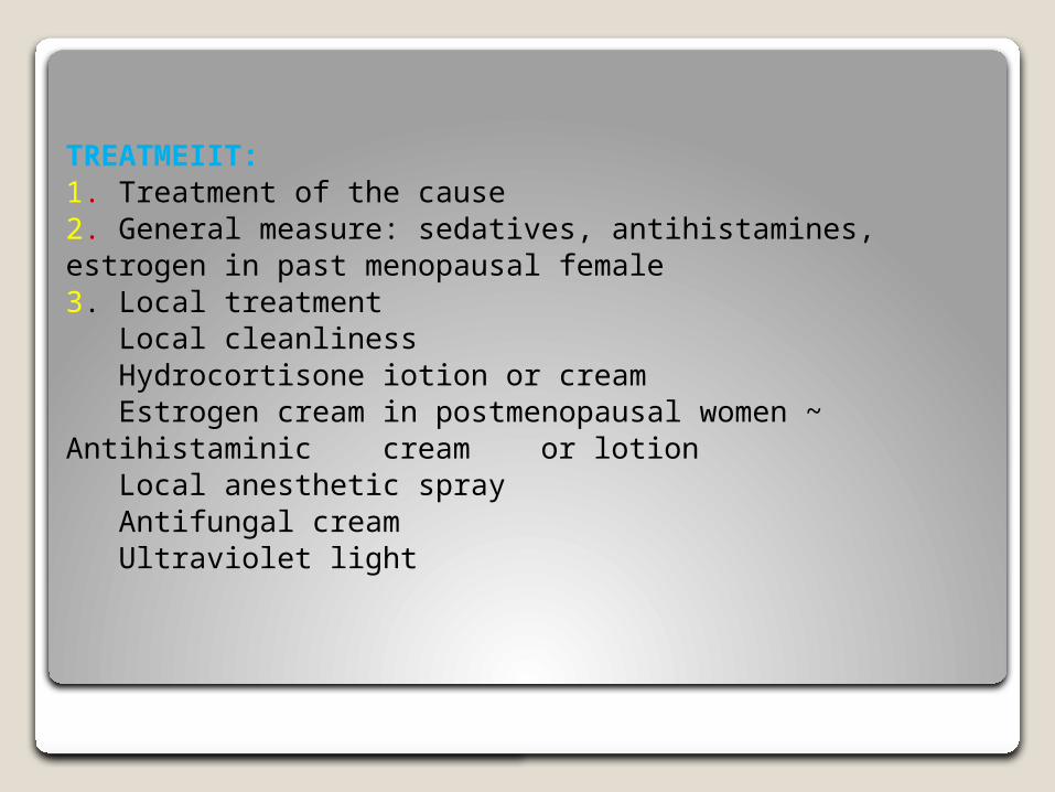
TREATMEIIT: 1. Treatment of the cause 2. General measure: sedatives, antihistamines, estrogen in past menopausal female 3. Local treatment
Local cleanliness Hydrocortisone iotion or cream Estrogen cream in postmenopausal women ~ Antihistaminic cream or lotion Local anesthetic spray Antifungal cream Ultraviolet light
4. Surgical measures (rare)
S.C. infection of 95% alcohol into the vulva
S.C. infiltration with local anesthetic or hydrocortisone
Circular incision around the vulva to cut the nerve fibres
Simple vulvectomy in resistant cases
Chronic Episthelial Dystrophies of the Vulva
DIFINTION:
Disorders in the epithelial growth and nutrition which result in white surface lesion of the vulva.
This Include:1. Atrophic type (lichen sclerosis) 2. Hypertrophic type (leukoplakia) e' or 'eout atypia 3. Mixed type with or without atypia
Atrophic type (lichen sclerosis) Develop after age of menopause but may occur at any age.
CAUSES IF VULVAL DYSTRIIIIIES :
1. Chronic irritation 2. Hormonal defficiency 3. Alternation of hormonal receptivity 4. Disturbance of CT factor 5. Chalone: tissue protein with hormonal like action
It is secreted by the epjdermis and inhibite epjthelial proliferation. Increased the concentration of chalone result in atrophic changes. While decreased the concentration of chalone result in hypertrophic changes.
Clinical Picture: 1. Severe itching 2. Itching of skin ---> scratching, exocriation, secondary infection
MANAGEMEIIT:
Advise the patient to avoid use of pe.futnes, douches and even take shower,
I. Advice patient to use cotton pants
3. Wash the vulva by none irritant soap
4. Biopsy for histopathological study and treatment