validation of a new anatomic severity grading system for
TRANSCRIPT
Yale UniversityEliScholar – A Digital Platform for Scholarly Publishing at Yale
Yale Medicine Thesis Digital Library School of Medicine
January 2018
Validation Of A New Anatomic Severity GradingSystem For Acute CholecystitisKenneth Vera
Follow this and additional works at: https://elischolar.library.yale.edu/ymtdl
This Open Access Thesis is brought to you for free and open access by the School of Medicine at EliScholar – A Digital Platform for ScholarlyPublishing at Yale. It has been accepted for inclusion in Yale Medicine Thesis Digital Library by an authorized administrator of EliScholar – A DigitalPlatform for Scholarly Publishing at Yale. For more information, please contact [email protected].
Recommended CitationVera, Kenneth, "Validation Of A New Anatomic Severity Grading System For Acute Cholecystitis" (2018). Yale Medicine Thesis DigitalLibrary. 3455.https://elischolar.library.yale.edu/ymtdl/3455

Validation of a New Anatomic Severity Grading System for Acute Cholecystitis
A Thesis Submitted to the Yale University School of Medicine
in Partial Fulfillment of the Requirements for the Degree of Doctor of Medicine
by
Kenneth Vera
2018

ii
ABSTRACT
VALIDATION OF A NEW ANATOMIC SEVERITY GRADING SYSTEM FOR
ACUTE CHOLECYSTITIS.
Kenneth Vera B.S. and Kevin Y. Pei M.D. Section of General Surgery, Trauma, and
Surgical Critical Care, Department of Surgery, Yale University School of Medicine, New
Haven, CT.
The American Association for the Surgery of Trauma (AAST) established
anatomic grading in 2015 to facilitate risk stratification and risk adjusted outcomes in
emergency general surgery. This study validates the AAST anatomic grading system for
acute cholecystitis (AC) at a tertiary, academic referral medical center.
This is a retrospective cohort study of 315 patients admitted for AC between 2013
and 2016. Cholecystitis severity was graded based on clinical, imaging, operative, and
pathologic criteria in accordance with the published AAST anatomic grading scale.
Grade I is acute cholecystitis, grade II is gangrenous or emphysematous cholecystitis,
grade III is localized perforation, grade IV and V have regional and systemic peritonitis
respectively. There was very good interrater (2 independent raters) reliability for
anatomic grading, κ=1.00, p<0.005.
Concordance between the AAST grade and outcomes including mortality, length
of stay (LOS), ICU use, and adverse events was assessed using statistical methods.
Incidence of complications, LOS, ICU use, and any adverse event increased with
increasing anatomic grade. When compared to grade I disease, patients with grade II
were more likely to undergo cholecystectomy (Odds Ratio 4.07 [1.93-8.56]), require ICU
use (Odds Ratio 2.41 [1.31 – 4.44]) and develop a complication (Odds Ratio 2.07 [1.22 –
iii
3.53]). Grade III patients were at higher risk of adverse events (Odds Ratio 3.83 [1.34-
10.94]) and ICU use (Odds Ratio 8.07 [2.43-26.80]).
In conclusion, AAST severity grading scores were independently associated with
clinical outcomes in patients with AC. Despite most patients having low grade disease,
complications were common. Therefore, a refinement of the scoring system for
cholecystitis may be necessary for more granular prediction of outcomes at milder levels
of disease.

iv
ACKNOWLEDGEMENTS
I would like to thank Kevin Y. Pei for his support, mentorship, teaching and guidance
over the course of this project. I would also like to thank the Yale General Surgery,
Trauma and Surgical Critical Care section leaders Kevin Schuster and Kimberly Davis
for their support as well as contributions in preparing our manuscript. I would like to
thank the New England Surgical Society for allowing me the opportunity to present the
findings in this thesis at their 2017 meeting. I would also like to thank the editors at
Journal of Trauma and Acute Care Surgery for accepting a manuscript of this thesis for
publication.

1
TABLE OF CONTENTS
Abstract…………………………………………………………………………… ii
Acknowledgements………………………………………………………… ……. iv
Table of Contents…………………………………………………………... ……. 1
Introduction………………………………………………………………… ……. 2
The AAST Scoring System………………………………………………. 2
Validation of the AAST Scoring System………………………………..... 4
Introduction to Cholecystitis…………………………………………........ 7
Diagnosis of Cholecystitis……………………………………………........ 10
Complications of Cholecystitis……………………………………………. 11
Management of Cholecystitis……………………………………………... 12
Statement of Purpose and Hypothesis…………………………………….. 15
Methods……………………………………………………………………………. 17
Inclusion Criteria………………………………………………………….. 17
Data Collection……………………………………………………………. 18
AAST Grading…………………………………………………………….. 20
Statistical Analysis………………………………………………………… 21
Results……………………………………………………………………………… 23
Discussion………………………………………………………………………….. 29
Imaging Scores for AAST Grade………………………………………….. 32
Refining the AAST Scale………………………………………………….. 33
Limitations…………………………………………………………………. 34
Future Directions…………………………………………………………... 34
Conclusions………………………………………………………………………... 37
References…………………………………………………………………………. 38

2
INTRODUCTION
Surgeons routinely care for patients with acute cholecystitis (AC). The
prevalence of gallstones is approximately 10-20% of US adult population and a third will
develop cholecystitis. Laparoscopic cholecystectomy (LC) is one of the most common
surgical procedures, but complexity and outcomes of surgery depend on disease severity.
To that extent, the American Association for the Surgery of Trauma (AAST) introduced
an anatomic severity grading system for emergency general surgery (EGS) diseases in
2014 (1). This objective and uniform system for quantifying anatomic severity has been
proposed for use in research as well as clinical settings. Such a system facilitates
standardized communication of severity in patient management, quality studies, outcome
comparisons, and provider to provider discussion. The AAST anatomic severity grading
system has yet to be validated to patient outcomes for many EGS diseases. The purpose
of this investigation was to validate the system for a cohort of patients admitted to Yale-
New Haven Hospital with AC.
The AAST Scoring System
The AAST anatomic severity grading system is based on the Organ Injury Scale
(OIS) developed in the 1990’s by a designated committee within the AAST. The
committee was charged with developing a set of standardized grading scales from 1-5 for
traumatic injuries to internal organs based on their anatomic description. These organ-
specific scales were formed from expert opinion and subsequently proposed for use in
clinical research (2) and have since been validated in numerous studies following their
introduction. The AAST scoring system was designed in a similar fashion in 2015. A
literature review of all existing scoring systems for individual EGS diseases was
3
performed and a uniform grading system that can be applied to all EGS diseases was then
agreed upon by expert consensus (1).
As of 2016, emergency general surgery care accounts for approximately 7% of all
hospitalizations in the US. The national average cost of a hospitalization and operation
for an EGS disease is $10,744 with the total number of EGS hospitalizations in the US
costing over 28 billion dollars annually. These figures are projected to increase by 45%
by 2060 (3). Standardization of disease severity affords the opportunity to compare
outcomes from various general surgical procedures across different medical centers in the
US when adjusted for comorbid conditions and disease complexity. The need for
standardized assessment is particularly critical in the current era of outcome-based
practice in many facets of medicine. Incorporating a uniform system for describing
disease into the field of emergency general surgery may ultimately lead to improved
outcomes and quality of care.
Criteria-based scoring systems have previously been developed for medical and
surgical diseases including those in EGS. One notable example is the Hinchey score, a
radiographic severity grading system, for diverticulitis. The very first such scale for
cholecystitis was published by a group from Tokyo in 2007 and most recently revised in
2018 (4). Although these and other scales are used across the world for a range of
clinical and research purposes, the AAST scale is the first to have been designed
specifically for use in the field of emergency general surgery.
The AAST advocates its grading system’s wide spectrum of disease severity, its
ease of use, and intuitive application. The grading system is designed to be uniformly
applied across a diverse range of EGS diseases. Furthermore, it only incorporates

4
anatomical data into the grading scale and excludes physiological parameters or patient
comorbidities (1). As the physiological severity of a disease is more often secondary to
its anatomic severity, an anatomically focused grading system is better utilized when
evaluating patient outcomes due to a primary disease process. This is a potential benefit
of AAST over existing grading systems such as the Tokyo system.
The AAST severity scores were not only adopted from the OIS but were also
based on the TNM system used for staging various types of cancers (1). Both TNM and
AAST incorporate gradients from local to wide spread of disease in the range of their
grading. AAST grades I and II are both limited to the organ and are associated with mild
or severe abnormality, respectively. Grades III-V represent anatomical progression from
localized to regional to widespread disease (5). The grading is also consistent with
respect to the progression from modest to severe inflammation. The range of this scale
encompasses almost the entire severity spectrum of any EGS disease. Subsequent studies
validating the scale have adopted a score of 0 for “normal” findings described in
pathology or imaging on retrospective review.
Validation of the AAST Scoring System
The AAST scoring system uses clinical, imaging, operative, and pathological data
to grade the anatomic severity of disease on a scale from 1-5, with 5 being the most
severe. In their most recent report, the AAST provides how the scale can be applied to
individual EGS diseases with 16 diseases described (6). The AAST has advocated for
validation studies using this system to assess its level of applicability to EGS diseases and
the outcomes of patients diagnosed with and admitted for management of the disease. To
date, there have been a handful of validation studies in diseases such as diverticulitis and

5
small bowel obstruction. These studies include both single and multi-center retrospective
studies.
One of the first published studies validating the AAST scale for EGS diseases was
published in 2015 (7). The study aimed to investigate the association between AAST
score and patient outcomes in a retrospective cohort of 512 patients admitted with acute
colonic diverticulitis at a single center. The AAST grades for colonic diverticulitis were
independently associated with adverse outcomes after controlling for patient co-
morbidities. Furthermore, there were no systemic differences in grade assignment
between two graders. A multicenter study including a cohort of 1,105 patients with
diverticulitis from 13 centers was subsequently published by the same investigative group
and again demonstrated an association of disease grade with adverse outcomes and a high
level of interrater reliability (8).
A recent report validated the AAST anatomic severity grading scale for
appendicitis in a population of 334 patients at a single center (9). Their study showed a
significant correlation between severity score and complications including length of stay
(LOS) as well as conversion from a laparoscopic to open operation. Within their cohort,
11.8% of patients with AAST grade 0-2 disease developed a complication versus 54.2%
of patients with grade III-V disease. This was the first report validating the AAST
scoring system for predicting any outcome in appendicitis. In a single-center
retrospective review of 1,099 appendectomies including at least 40 cases from each
AAST severity grade, the AAST was validated to predict symptom duration,
appendectomy duration, as well as cost of care (10).

6
Baghdadi et al published the first report validating the AAST for small bowel
obstruction (11). They studied a retrospective cohort of 351 patients with partial or
complete small bowel obstruction using both the original AAST scoring system and a
modified version of the AAST system incorporating patient physiology and comorbidities
using SIRS criteria and Charlson comorbid scores. The authors argue that physiology is
an inherent part of disease, particularly in the management of patients with small bowel
obstruction, and therefore should be primarily incorporated into disease assessment.
Both the AAST and modified scores showed significant associations between greater
disease severity and greater inpatient complications and extended LOS. However,
neither was superior in predicting these endpoints. Despite a low mortality rate in their
cohort, their modified score better predicted mortality than AAST.
The Tokyo Guidelines (TG) for cholecystitis incorporates anatomical findings as
well as multiple physiological parameters into its approach to diagnosis and severity
assessment. As described in their 2018 revision, grade I or “mild” disease is classified as
cholecystitis in a healthy patient who has no findings of organ dysfunction. Grade II or
“moderate” disease is characterized as having marked inflammation of the gallbladder.
Criteria for grade II disease includes elevated white blood cell count, a tender and
palpable right upper quadrant mass, and duration of disease for 72 hours or more. Grade
III (“severe”) disease is moderate severity accompanied by evidence of organ dysfunction
(4).
The TG has been studied in multiple retrospective cohorts and revised to correct
limitations to its validation. Furthermore, it has evolved from a tool used in research to a
guideline for management and clinical judgement based on disease severity. The
7
management component of the guideline recommends early cholecystectomy with
adjuvant antibiotic therapy for grade I disease and conservative approaches including
medical management with percutaneous cholecystostomy tube in grades II and III (4). A
similar path of evolution is ideally how AAST may become usefully incorporated into the
field of emergency general surgery. However, the distinction between TG and AAST
grading is an important one. Only the anatomic severity of disease contributes to the
AAST grade whereas the inclusion of physiologic parameters in TG may complicate its
ability to compare pure primary disease across patients.
Although physiological variables are significant predictors of outcomes
themselves, statistically controlling for the patient’s physiological state at admission
allows the association between anatomic disease severity and patient outcomes to more
accurately be analyzed. Comorbid conditions such as smoking and hypertension and
social determinants of health such as ethnicity and insurance status can likewise be
controlled for their effect on outcomes. Such an analysis has been consistently
incorporated into published studies validating AAST scales. The AAST scoring system
was designed to assess the extent to which anatomic severity predicts outcomes. It
cannot be determined whether a scale incorporating physiological parameters better
predicts outcomes than one based solely on anatomical severity without first validating
the AAST.
Introduction to Cholecystitis
The clinical presentation of cholecystitis, including its severity and range of
associated complications, can vary within a given patient population. Gallstones are by
far the most common cause of cholecystitis, followed by stenosis of the biliary tract (12).

8
AC may be managed either medically or surgically with a laparoscopic cholecystectomy
(removal of the gallbladder and its contents) being the most common operation used for
treatment (13).
Pathologically, cholecystitis occurs as a result of both cystic duct obstruction and
damage to the gallbladder mucosa. Gallstones may become impacted in the neck of the
gallbladder of the cystic duct and cause mechanical mural irritation as the result of the
gallbladder contracting against the stone. Damage to the mucosa leads to the release of
phospholipases from the epithelial cells lining the gallbladder lumen. Phosopholipase A
catalyzes the production of lysolecithin from lecithin, a normal component of bile.
Lysolecithin further irritates the epithelial lining of the gallbladder and mural distension
and edema leads to epithelial vascular insufficiency. This damage leads to continued
phospholipase release from damaged epithelial cells and propagates the inflammatory
reaction causing AC (12). Due to the stasis of bile proximal to the obstruction at the neck
of the gallbladder, its contents are more prone to a superimposed bacterial infection.
Infection of the gallbladder, and uncommonly the whole biliary system, may complicate
the disease although only 20% of patients with cholecystitis grow a pathogen in bile
cultures (12).
The formation of gallstones is complex and involves an interplay between
secretions of cholesterol into the bile, bile stasis secondary to gallbladder dysmotility, and
crystal nucleation of stones. Oversaturation of bile with cholesterol and low levels of bile
salts enhance stone formation. Age, female gender, obesity, oral contraceptives, parity,
North American Indian ancestry, and consuming a western diet have all been found to be

9
associated with increased risk of gallstones in various studies. Dyslipidemia is also risk
factor and more than 80% of gallstones are cholesterol based (13).
Biliary colic is a steady right upper quadrant or epigastric abdominal pain that
lasts for more than 30 minutes but less than 4 hours. It is sudden in onset and usually
occurs following consumption of a fatty meal. It is caused by an intermittent obstruction
of the gallbladder neck or the cystic duct by gallstones during gallbladder contraction. In
most instances, colic resolves during gallbladder relaxation as the stone falls back.
However, sustained obstruction of the cystic duct can lead to bile stasis, gallbladder
distension with mucosal and endothelial injury, and subsequent activation of
inflammatory mediators leading to AC (14).
Gallstones are highly prevalent in western populations with some studies
reporting prevalence rates as high as 10-15% in adult populations (15). However, some
estimated 1-4% of patients with gallstones will go on to have any serious complications
such as cholecystitis. Furthermore, most patients with gallstones are completely
asymptomatic, with multiple studies reporting digestive symptoms in just less than 10%
of populations positive for sonographic gallstones (14). However, rates of cholecystitis
in this population of patients with symptomatic stones have been reported to be as high as
15% (15). Most gallstone-related complications including cholecystitis can be prevented
with cholecystectomy and thus elective cholecystectomy may be indicated for patients
with frequent digestive symptoms.
Acalculous cholecystitis is pathologically identical to AC but not caused by
gallstones. It accounts for approximately 10% of all cases of AC and usually occurs in
hospitalized patients who are critically ill (16). Pro-inflammatory conditions such as

10
cancers, infections, or trauma can lead to gallbladder ischemia or promote bile stasis.
This can lead to gallbladder endothelial damage which initiates the same
pathophysiological cascade seen in calculous cholecystitis. The clinical features of
acalculous cholecystitis more often include jaundice, hyperbilirubinemia and elevation of
liver transaminases. Patients with acalculous cholecystitis have been observed to have
higher morbidity and mortality rates; this may be partially explained by the comorbid
inflammatory process acalculous cholecystitis may present with (16).
Diagnosis of Cholecystitis
Understanding the diagnosis of cholecystitis is fundamental to evaluating the
severity of the disease. Traditionally, the diagnosis of cholecystitis has been made based
on clinical suspicion supported by lab data and confirmed with imaging findings.
Clinical features of the disease include abdominal pain, nausea, vomiting, fever,
Murphy’s sign (abrupt cessation of inspiratory effort due to elicited pain), and right upper
quadrant or epigastric abdominal tenderness with or without guarding (12). The
differential diagnosis is wide and may also include hepatitis, pancreatitis, peptic ulcer
disease, gallbladder cancer, or Fitz-Hugh-Curtis syndrome.
There are no specific blood tests used to make the diagnosis of AC. However,
laboratory tests can be used to support the diagnosis and/or exclude other etiologies of
pain. Common tests used include white blood cell count, serum bilirubin, lipase levels,
and liver transaminase levels (12).
Imaging studies used in the diagnosis include abdominal ultrasound and
cholescintigraphy. Ultrasonography is the initial imaging modality of choice as it is
11
rapid, taking only minutes to perform, and inexpensive to use. Findings consistent with
AC include gallbladder wall thickening (>4 mm), gallbladder distension, pericholecystic
fluid collections, and pericholecystic fat stranding (17). A meta-analysis of 30 studies on
imaging studies for gallbladder disease found the sensitivity and specific of ultrasound
for diagnosing cholecystitis to be 88% and 80%, respectively (18). A more recent meta-
analysis of 57 studies found the sensitivity and specificity to be 81% and 83%
respectively (17).
Cholescintigraphy, also referred to as a HIDA (Hepatobiliary Iminodiacetic) scan,
may be used to aid in the diagnosis if strong clinical suspicion is present in the context of
an equivocal or indeterminate ultrasound. This study traces the uptake of a technetium
labeled acid administered intravenously to a patient and selectively taken up by
hepatocytes before being excreted into the bile. Prolonged uptake due to cystic duct
obstruction indicates a positive study. Though more sensitive and specific than an
ultrasound, at 96% and 90% respectively, the HIDA scan takes hours to perform and
utilizes more sophisticated personnel and equipment than ultrasound. Additionally, it
provides data solely relevant to gallbladder pathology and is therefore less useful than
ultrasound in examining the liver as an alternative source of right upper quadrant
abdominal pain (17).
Complications of Cholecystitis
Early diagnosis and intervention of AC is important to prevent complications
associated with higher morbidity and mortality. Gangrenous cholecystitis is a more
severe and complicated form of the disease characterized by necrosis secondary to
ischemia and prolonged inflammation of the gallbladder. Gangrenous cholecystitis is
12
considered grade II on the AAST scale. It is more common in patients with greater
comorbidities, such as the elderly and diabetics, and patients with a delayed presentation
(19).
Though uncommon, transmural perforation of the gallbladder may occur leading
to localized abscess formation. This would be grade III disease in the AAST severity
scale. Spillage of gallbladder contents such as pus or bile into the peritoneal cavity may
cause subsequent generalized peritonitis, AAST grade IV disease. In rare cases, a biliary-
enteric fistula may form between the gallbladder and small bowel. Gallstone ileus occurs
when gallstones passed through a biliary-enteric fistula become lodged in the distal small
bowel, most commonly the ileum, and cause obstruction. Sepsis and multiple organ
dysfunction are more likely complicate the patient’s clinical status during these
complications (19). Such a severe case of cholecystitis would be considered grade V on
AAST.
Emphysematous cholecystitis is caused by an intramural or intraluminal infection
of the gallbladder with gas producing organisms. The most commonly isolated offending
pathogens include Clostridium perfringens, Escherichia coli, and Klebsiella.
Emphysematous cholecystitis more commonly affects men, elderly, and diabetic patients.
It is also associated with higher rates of perforation and a mortality rate of up to 15%
(19).
Management of Cholecystitis
Management of cholecystitis can range from conservative to operational.
Conservative approaches to the disease include a course of intravenous antibiotics

13
accompanied by supportive care including intravenous fluids, pain control, and
electrolyte correction. The most common pathogens covered by empiric antimicrobial
therapy include gram negative rods, particularly Escherichia coli, and anaerobes. The
laparoscopic approach to cholecystectomy is associated with decreased morbidity and
mortality as well as shorter LOS. The conversion rates to open surgery are low (13).
The optimal timing of an LC following AC is still an active area of investigation.
Higher complication rates following surgery in the acute setting are a concern as
increased local inflammation may obscure the critical view and dissection of Calot’s
triangle. Because of this concern, some surgeons elect to manage a patient medically in
the acute setting and delay surgery for up to six weeks, even in an otherwise
uncomplicated case (20). However, large population-based analyses and recent meta-
analyses of case-control studies have shown that early LC in the acute setting is superior
to delayed LC, with no differences in complication rates and shorter LOS (20, 21).
However, early cholecystectomy was associated with longer operating times, presumably
due to increased inflammatory changes in the acute setting. Despite the significance of
these findings, these studies do not incorporate anatomic severity of disease in their
analysis due to the lack of a widely adopted scale such as the AAST.
Reports using the TG severity scale also favors early cholecystectomy. A recent
meta-analysis shows that early cholecystectomy can be a feasible treatment alternative to
conservative management for AC in carefully selected candidates with TG grade II and
III disease (22). In a separate investigate report, Loozen et al also found no statistically
significant differences in conversion rates, operating time, perioperative complication
rate, and 30-day mortality between patients with TG grade I and grade II cholecystitis

14
undergoing emergent LC (23). A separate retrospective analysis on a group of 149
patients undergoing emergent LC for AC determined that TG severity grade alone did not
predict whether a patient underwent LC or percutaneous cholecystostomy tube. Those
patients undergoing LC experienced longer LOS though did not experience increased
morbidity or mortality (24). Another recent study did not show statistically significant
differences in complication or conversion rates across TG classification although the
classifications did correlate with LOS (25). While agreeing that early cholecystectomy
should be more widely considered in patients with more severe disease but fewer
comorbidities, these reports also recommend further stratification between grade I and
grade II disease be considered in future TG revisions.
A percutaneous cholecystostomy tube (PCT) is a safe and effective intervention
widely used as a bridging therapy for AC. It is essentially a catheter which drains the
gallbladder contents, placed into the lumen via an ultrasound guided transhepatic
approach. A PCT can provide relief of cholecystitis symptoms for up to 91% of patients
undergoing the procedure (26). It is indicated in patients with severe disease, those with
contraindications to general anesthesia, those who present >72 hours after symptoms
onset, or patients failing medical therapy. Additionally, they are recommended for
elderly patients with multiple co-morbidities who would have a higher likelihood of a
safe and successful LC at some time interval following medical management and
cholecystostomy placement, usually 6-8 weeks. The disadvantage of this therapy is that
rates of tube dysfunction are high with up to 46% of patients experiencing some sort of
dysfunction and over half of these patients requiring re-intervention (26).

15
In elderly patients with multiple chronic comorbidities, PCT may be the only safe
feasible intervention available as the risks of surgery would outweigh the long-term
benefits. Percutaneous gallbladder aspiration has been advocated as an easy to perform
and less invasive alternative therapy to PCT for patients failing initial medical
management. It also avoids drain-related symptoms of discomfort reported by many
patients discharged with a PCT. Percutaneous aspiration has also been shown to provide
comparable outcomes to PCT in retrospective studies as well as randomized controlled
trials and is recommended in cases where PCT may not be available (27, 28).
Statement of Purpose and Hypothesis
The purpose of this original investigation was to retrospectively validate the
AAST anatomic severity grading system using a cohort of patients admitted to a single,
tertiary, high volume center with cholecystitis. A few single center studies have been
published demonstrating independent associations between severity grades and outcomes
in diverticulitis and appendicitis. To our knowledge and to the extent of the published
literature, there has not been a similar investigation concerning the outcomes of patients
with cholecystitis.
Because the AAST grading system was designed to be uniformly applied across a
variety of EGS diseases, we expect to see associations like those previously reported for
other EGS diseases. However, finding a lack of association with the current grading
system would also be equally valuable in gaining insight into how it may be adjusted to
fit the range of severity specific to cholecystitis. Given the current understanding of
cholecystitis and the validation of AAST in other surgical diseases, we hypothesize that
16
the AAST anatomic severity grade in cholecystitis may independently predict higher
likelihood of adverse clinical outcomes.

17
METHODS
This was a retrospective single center cohort study undertaken by Kenneth Vera, medical
student, and Kevin Y. Pei MD at Yale University School of Medicine. This study was
approved by the Human Investigation Committee.
Inclusion Criteria
All patients over eighteen years of age who were admitted to Yale-New Haven
Hospital between August 2013 and August 2016 with a diagnosis of ‘acute cholecystitis’
(575.0 or K81.0), ‘acute on chronic cholecystitis’ (575.12 or K81.2), or ‘cholecystitis
unspecified’ (575.10 or K81.9) based on ICD-9 and ICD-10 codes were included in this
study. Patients were excluded if they were a pregnant, a prisoner, or had advanced
directives limiting care. They were additionally excluded if they had a prior admission
for AC within 90 days. In cases where a patient had multiple admissions for AC on
record, the data and management from their index admission was used. Patients were
excluded if their index admission made note of a recent prior admission for cholecystitis
at another facility or prior admission for cholecystitis at our facility prior to 2013 as
records were not available for access in these cases. Patient who were primarily admitted
to a medical or surgical floor for some other condition but were subsequently admitted as
an EGS consult patient were included so long as they received a new diagnosis of AC or
acalculous cholecystitis (rather than a flare of known chronic cholecystitis) and were
managed as such. Cases of patients in severely critical condition found to have acute
acalculous cholecystitis on imaging within 24-72 hours prior to expiration were excluded.
18
Data Collection
Demographic data collected for each patient included their age, gender, ethnicity,
and insurance status (categorized as either commercial or public). This data was
collected from the electronic medical records (EMR). All other data extraction was
performed by a detailed manual review of individual admission records.
The presence or absence of common co-morbidities was noted based on data in
their admission notes and past diagnoses noted elsewhere on record. Our comorbidities
of interest included whether a patient has is a present smoker, has diabetes, a history of
progressive renal insufficiency or failure, hypertension, chronic dyspnea, or is on chronic
steroids or immunosuppressant medications. Additionally, whether a patient had two or
more SIRS criteria present at admission and/or was septic based on 2016 Sepsis-3 criteria
was noted and counted as a comorbidity (29).
A Sequential Organ Failure Assessment (SOFA) score was calculated based on
the patient’s worse physiological parameters within the initial 24 hours of admission to
the emergency general surgery (EGS) service. We included this score as a covariate in
our analyses to control for the effect of physiology on outcomes. Scores were calculated
as described in the literature and SOFA score component data was available for all
patients (30). Patients breathing on room air were calculated as having 21% FiO2.
Patients documented to be on supplemental oxygen had approximate FiO2 calculated
based on published conversion formulas. Approximate PaO2 was calculated from
documented SaO2 from published conversion formulas (31). GCS was assumed to be 15
unless otherwise noted on documented physical exam. In cases where the patient

19
received an operation within the first 24 hours of admission, the worse parameters prior
to surgery were used.
The outcomes of interest included LOS, whether a patient received operative
management, if they had a complication while admitted, readmission within 30 days, and
ICU use (defined as at least 12 hours of admission to a medical or surgical ICU).
Complications of interest included surgical wound infection or disruption, acute renal
failure, transfusion of blood products during admission, mechanical ventilation for more
than 48 hours, and diagnosis of UTI, pneumonia, or DVT/PE during admission, septic
shock requiring vasopressors, and cardiac arrest or myocardial infarction. Operative
management was cholecystectomy, either index or interval, performed for the indication
of AC. Mortality data was also collected and based on death being reported during the
patient’s hospital stay or within 30 days after discharge if readmitted.
Specific data was additionally collected from the operative records of those
patients who had surgery. This was done to better relate the surgical complexity of this
cohort subset although this data is not included in statistical analysis. This data included
the interval of time between presentation to the hospital and the beginning of the patient’s
operation, the operation time of day and its duration, as well as the training level of the
assisting surgical resident.

20
AAST Grading
Data extraction and disease severity grading were performed by two independent
researchers. AAST grades were assigned to each patient based on clinical, operative,
pathological, and imaging criteria, with a composite of four grades for each of these
criteria. A detailed grading rubric adapted from the AAST’s published 2015 report is
reproduced below (6). The final grade assigned was the highest of any criteria. For those
patients who did not have a cholecystectomy, either index or interval, only clinical and
imaging criteria were available. Grading was based on manual review of data available
in the patient’s EMR including admission history and physical exam, progress notes, as
well as pathology, operative and imaging reports. Pathological grades were recorded for
both patients with index cholecystectomy as well as interval cholecystectomy performed
within 3-12 weeks of conservative management for their index admission of AC.
Interrater reliability of grade assignment was assessed using kappa coefficient with
Fleiss-Cohen quadratic weights.

21
AAST Anatomic Severity Grading Scale as published in Shafi et al 2015.
Clinical Criteria Imaging Criteria Operative Criteria Pathological Criteria
Grade I
RUQ or epigastric pain, Murphy sign, Leukocytosis
Wall thickening, distension,
gallstones or sludge, pericholecystic
fluid, non-visualized GB on HIDA
Localized inflammation, wall
thickening, distension, gallstones
Inflammatory changes without necrosis or
pus
Grade II
RUQ or epigastric pain, Murphy sign, Leukocytosis
Air in GB lumen, wall or biliary tree.
Focal mucosal defects
Distended GB with pus or hydrops,
necrosis/gangrene of wall (non-perforated)
Pus in the GB lumen, wall necrosis,
intramural abscess, epithelial sloughing
Grade III
Localized RUQ peritonitis
Focal transmural defect, extraluminal
fluid collection
Perforated GB wall, bile outside GB but
limited to RUQ
Necrosis with perforation of the GB
wall
Grade IV
Multifocal peritonitis, abdominal
distension, bowel obstruction symptoms
Abscess in RUQ outside of GB,
bilioenteric fistula, gallstone ileus
Pericholecystic abscess, bilioenteric
fistula, gallstone ileus
Necrosis with perforation of the GB
wall
Grade V
Generalized peritonitis
Free intraperitoneal bile
Generalized peritonitis
Necrosis with perforation of the GB
wall
Statistical Analysis
Binary logistic regression analysis was used to model the associations between
AAST grades and their covariate predictors with the occurrence of clinical events. AAST
grade was included as an ordinate independent variable with grade I used as the reference
for comparisons of other severity grades. Separate logistic regression models were made
for each clinical event. C-statistic values were calculated and used for goodness-of-fit
comparisons among the models.
A negative binomial regression model was used to measure the association of
covariate predictor variables and AAST grade with LOS as a continuous dependent
variable. To enforce normality and equal variance assumptions to the negative binomial
22
regression analysis, an interpretation of the original output data was performed where all
data points with Standardized Pearson Residual absolute values > 2 were considered
outliers and excluded from a second otherwise identical regression model. This excluded
11 data points from the original set of 315. The Akaike’s Information Criteria (AIC) and
Bayesian Information Criterion (BIC) values pre- and post-outlier exclusion were used
for goodness-of-fit comparison where smaller-is-better assumptions were applied to
accept the new model. These were 1532 and 1569 for AIC and BIC after outlier
exclusion, compared to 1667 and 1705 originally.
In all models, age and SOFA score were both continuous predictive variables
while sex, race/ethnicity, public insurance status, and presence of comorbidities were
nominal variables. Male sex, non-minority race/ethnicity, having commercial insurance,
and having no comorbidities were the respective reference conditions. The comorbidities
of interest included those described in the data collection as well as having met SIRS or
sepsis criteria at admission. Statistical Analysis was performed with IBM SPSS v.22
statistical software.

23
RESULTS
A final set of 315 patients was included in data analysis. A breakdown of
demographic, LOS and morbidity data is shown in Table 1.
Table 1: Patient Demographics and Morbidity Average age 61.5 Average LOS [SD] 5.1 days [5.6] Minority race/ethnicity* 32% Median LOS [IQR] 3 days [2-6] Female 55% ICU use 71 (22%) Commercial Insurance 38% Average ICU days [SD] 1 [3.47] Government Insurance 60% SIRS or Sepsis 146 (46%) Uninsured 2%
*Defined as self-identifying as black or hispanic (non-white)
A breakdown of disease severity by final AAST grade is shown in Table 2.
Notably, nearly 94% of cases were grade I or grade II. No cases meeting grade IV were
identified and only three were identified as grade V. There was very good interrater
reliability between two independent reviewers for anatomic grading, κ=1.00, p<0.005.
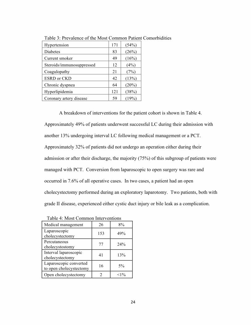
The prevalence for each of our comorbidities of interest is shown in Table 3.
The most common co-morbidities were hypertension, diabetes, and hyperlipidemia. The
prevalence for each of our complications of interest is shown in Table 3. The most
common complications were acute renal injury (AKI), transfusion, or readmission.
Table 2: AAST Grade Distribution Grade Number of Patients Percentage
I 219 (69%) II 75 (25%) III 18 (5%) IV 0 - V 3 (1%)
24
Table 3: Prevalence of the Most Common Patient Comorbidities Hypertension 171 (54%) Diabetes 83 (26%) Current smoker 49 (16%) Steroids/immunosuppressed 12 (4%) Coagulopathy 21 (7%) ESRD or CKD 42 (13%) Chronic dyspnea 64 (20%) Hyperlipidemia 121 (38%) Coronary artery disease 59 (19%)
A breakdown of interventions for the patient cohort is shown in Table 4.
Approximately 49% of patients underwent successful LC during their admission with
another 13% undergoing interval LC following medical management or a PCT.
Approximately 32% of patients did not undergo an operation either during their
admission or after their discharge, the majority (75%) of this subgroup of patients were
managed with PCT. Conversion from laparoscopic to open surgery was rare and
occurred in 7.6% of all operative cases. In two cases, a patient had an open
cholecystectomy performed during an exploratory laparotomy. Two patients, both with
grade II disease, experienced either cystic duct injury or bile leak as a complication.
Table 4: Most Common Interventions Medical management 26 8% Laparoscopic cholecystectomy 153 49%
Percutaneous cholecystostomy 77 24%
Interval laparoscopic cholecystectomy 41 13%
Laparoscopic converted to open cholecystectomy 16 5%
Open cholecystectomy 2 <1%

25
The average time from hospital presentation to time of cholecystectomy for those
patients receiving LC during their index admission was 25.2 hours (standard deviation of
20.9 hours). Approximately 13% of patients underwent interval LC following medical
management or a PCT. The average time from the date of admission for cholecystitis to
date of cholecystectomy for those patients was 67 days (standard deviation of 45 days).
Almost all of these operations involved trainees in a seven-year academic general surgery
residency with the average post-graduate year of training being 4.6 years (standard
deviation of 1.7 years) for the resident trainees scrubbed into the procedure.
Approximately 74% of all operations occurred between the hours of 0700 and 1700. The
average operation length was 105 minutes (standard deviation of 42 minutes).
Table 5 shows the incidence of clinical events by final AAST grade. The
proportional incidence of any complication, adverse event (defined as either death or
readmission), ICU use, and median LOS trended upward with increasing severity grade.
Note that complications in this table are listed as a composite outcome. Table 6 shows a
breakdown of the complications observed. The most common complications were acute
kidney injury, readmission within 30 days and receiving a blood product transfusion.
Table 5: AAST Grade and Clinical Events
Grade I Grade II Grade III Grade V
n=219 n=75 n=18 n=3
Complications 70 (32%) 37 (49%) 12 (67%) 2 (67%) Surgery 142 (65%) 58 (77%) 10 (56%) 2 (67%) ICU Use 34 (16%) 23 (31%) 12 (67%) 2 (67%) 30d readmission 31 (14%) 6 (8%) 7 (58%) 2 (67%) Median LOS (IQR) 3 (1-5) 4 (2-7) 6.5 (5-8) 11 Any adverse event 90 (41%) 39 (52%) 15 (83%) 3 (100%)

26
Table 6: Most Common Complications Wound infection 6 3% Acute kidney injury 77 24% Transfusion 37 12% Urinary tract infection 24 8% Pneumonia 20 6% Deep venous thromboembolism or pulmonary embolus
7 2%
Septic shock 28 9% Cardiac arrest 16 5% Mechanical ventilation 29 9% 30-day readmission 45 14% Death 24 8%
Table 7 shows the association between the included covariates and clinical events
reported in Odds Ratio (Incidence Risk Ratio for LOS). Age independently predicted the
occurrence of a complication and the occurrence of non-operative management.
Increasing age was also associated with a greater incidence of longer LOS. SOFA score
independently predicted higher incidence of a complication, ICU use, non-operative
management, and occurrence of an adverse event (readmission or death). The presence
of any comorbidity predicted ICU use during admission. Female sex, minority race, and
having public insurance were largely non-predictive of the outcomes of interest except
for public insurance status predicting non-operative management.

27
Table 7: Association Between Clinical Events and Predictors Reported as Odds Ratios with 95% CIs (Incidence Risk Ratios with 95% CIs for LOS Predictor Complications LOS Surgery ICU Use Adverse Event
Age 1.04 (1.02 - 1.06)*
1.01 (1.00 - 1.02)*
0.96 (0.94 - 0.97)* 1.02 (1.00 - 1.04) 1.01 (0.99 - 1.03)
Female sex 0.90 (0.52 - 1.57) 0.87 (0.67 - 1.13)
1.05 (0.60 - 1.84) 0.65 (0.33 - 1.28) 1.39 (0.75 - 2.57)
Minority race 1.83 (0.95 - 3.5) 1.06 (0.78 -
1.44) 0.89 (0.47 - 1.71) 1.83 (0.82 - 4.05) 1.93 (0.98 - 3.83)
Public insurance 1.18 (0.66 - 2.09) 1.26 (0.96 -
1.64) 0.55 (0.31 - 0.98)* 1.22 (0.60 - 2.49) 1.11 (0.59 - 2.09)
SOFA 1.73 (1.44 - 2.09)*
1.14 (1.07 - 1.23)*
0.68 (0.58 - 0.79)* 1.83 (1.51 - 2.21) 1.44 (1.24 - 1.67)*
Comorbidity 1.73 (0.78 - 3.80) 1.39 (0.98 - 1.97)
1.44 (0.68 - 3.09) 6.1 (1.3 - 28.74)* 1.37 (0.57 - 3.28)
C-statistic 0.83 (0.78 - .88) N/A 0.80 (0.75 - 0.85) 0.88 (0.84 - 0.92) 0.77 (0.71 - 0.84)
*Statistically significant finding with p < 0.05.
Table 8 shows the risk-adjusted associations between AAST grade and the
occurrence of clinical events reported as Odds Ratios (Incidence Ratio for LOS) with
grade I as the reference range. Grade II disease was associated with four times the odds
of operative management. Grade III disease was associated with almost four times the
odds of an adverse event (readmission or death) and over eight times the odds of ICU
use. Grade III disease was also associated with higher incidence of longer LOS.
Although grade V disease appeared to be associated with greater odds of complications
or ICU use, these were not statistically significant. The notable width of the grade V
confidence intervals is likely explained by the small size of the group in our sample. All
three cases of grade V disease experienced death and the odds ratio of this grade having
an adverse event is not reportable as there is no variability in the outcome.

28
Table 8: Risk-Adjusted Association Between AAST and Clinical Events Reported as Odds Ratios with 95% CIs (Incidence Risk Ratios with 95 % CIs for LOS
Outcome Grade I Grade II Grade III Grade V Adverse Event† Ref 0.58 (0.26 - 1.27) 3.83 (1.34 - 10.94)* -
Complications Ref 1.52 (0.80 - 2.89) 2.22 (0.70 - 7.06) 3.09 (0.23 - 41.41) Surgery Ref 4.07 (1.93 - 8.56)* 1.34 (0.45 - 4.03) 1.02 (0.08 - 12.75) ICU Use Ref 1.86 (0.87 - 4.01) 8.07 (2.43 - 26.90)* 8.37 (0.65 -107.92) Length of Stay Ref 1.06 (0.78 - 1.44) 1.73 (1.03 - 2.92)* 1.03 (0.60 - 1.78) *Statistically significant finding with p < 0.05. †Either death or readmission within 30 days
29
DISCUSSION
This is the first single-center study designed to validate the AAST severity
grading system for cholecystitis. Our results show that higher grades of severity, notably
grade III in our sample, is independently associated with multiple patient outcomes after
controlling for covariate predictors such as age, sex, and physiological status.
A great majority of patients in our population had low grade disease with 94%
percent of our population having grade I or grade II disease. Notably, there were no
patients with grade IV disease included in our data set. Although the AAST scale from I-
V offers a rating for a wide range of severity, our data reflect a common clinical
observation that cholecystitis tends to be milder disease associated with low overall rates
of mortality. This reflects the modern era where we are diagnosing and managing
cholecystitis earlier with the widespread adoption of sensitive imaging techniques for
diagnosis and less invasive modalities for treatment (32). The implications for AAST are
that the current scale for AC may need to be modified prior to being further validated in
larger samples.
Other recent studies validating AAST grading scales report a similar finding of
low-grade disease being over-represented in their retrospective data sets. Savage et al
reported 74% of their 512 patient cohort with diverticulitis to have had grade I or II
disease (7). Hernandez et al reported 78% of their 334 patient cohort with appendicitis to
have grade I or II disease (9). Shafi et al reported two thirds of their 1,105 multicenter
patient cohort to have had grade I or II disease (8). Collins et al reported 91% of their
1,099 patient cohort with appendicitis to have grade I-III disease. This
overrepresentation presents a potential challenge for validation of the current scales. It

30
has been proposed by Shafi et al that oversampling patients with severe disease,
especially in multicenter studies, would better select a cohort representing the spectrum
of disease severity. In our study, patients with severe disease meeting eligibility criteria
were included in the analysis but it was also observed that patients with higher severity
scores more often met exclusion criteria.
An alternative to oversampling patients with severe disease would be to modify
the current scale such that it more discretely classifies low-grade disease while
consolidating the higher grades together. As the scale is currently designed, each of the
five levels of severity is distinguishable from others anatomically and a gradient of
morbidity follows accordingly. For instance, gallbladder perforation is grade III by
definition. Because perforation more often leads to localized abscess or peritonitis, the
step up from grade II to grade III is intuitively associated with increased morbidity and
mortality. However, perforation leading to peritonitis is a rare event and most of the
patients in our cohort cluster in grades I and II. Increasing resolution of the grading
system at this mild end of the scale may allow the AAST grading of cholecystitis to better
fit the spectrum of naturally occurring disease and thus make the tool to more clinically
meaningful. We expect the milder spectrum of severity seen in our patient cohort to be
similar to the range of disease encountered in a larger, multicenter study. Additionally,
consolidation of the high-grade scores could be considered to simplify the existing AAST
system given the rarity of severe grade IV and V disease.
Interestingly, the occurrence of complications was not significantly associated
with AAST grade, unlike in reports validating AAST for other EGS diseases. Although
an explanation for this is not clear from our data, this endpoint may again be better

31
validated after inclusion of cases of higher grade severity. We assessed the same
outcomes as those recorded in large database registries such as the American College of
Surgeons (ACS) National Surgical Quality Improvement Project (NSQIP). Outcomes
such as sepsis, deep vein thrombosis, and pneumonia may not be as applicable to low risk
procedures such as LC. In general, complications arising from cholecystectomy,
particularly when laparoscopic, is rare (33). The confidence interval width for the
complications associations, as well as others, suggests a degree of uncertainty in our data
that is yet to be resolved.
Hernandez et al argues that the AAST scoring systems would be easier validated
for the mortality end-point in diseases with greater odds of death (9). Our data would
agree with this theory as death was rare in our data set, only occurring in 24 of our 315
patients. Only a single death was noted in the 1,105 diverticulitis patients included in
Shafi et al and therefore was not an analyzed nor validated outcome in their work (8).
The higher rate of death in our smaller data set may be due to inclusion of admitted
patients diagnosed with acalculous cholecystitis who have greater morbidity from other
causes. Future analysis of our data would consider excluding or separating this subtype
of patients, further necessitating the inclusion of high grade cases in larger samples of
patients.
32
Imaging Scores for AAST Grade
Many of the criteria proposed in the original AAST severity scale for appendicitis
rely on intraoperative and postoperative pathological findings and thus cannot be utilized
pre-operatively to predict need for operative management. Validation of the AAST scale
as a tool which grades severity without operative or pathological information would make
the scale useful tool in a real time clinical setting where making decisions with limited
information is custom. Hernandez et al therefore proposed that an imaging based AAST
grade, I-AAST, be first validated (8).
The I-AAST used by Hernandez et al assigned an anatomic severity grade of I-V
based on objective CT findings including the size of appendiceal thickening, presence of
periappendiceal edema, free intraperitoneal fluid, or presence of a localized abscess or
phlegmon. A score of 0 was to indicate an appendix which appears normal on CT. They
then compared the I-AAST grade with the final AAST reported from operative and
pathological findings. They found no significant mean difference between the scores and
had a coefficient of repeatability of 0.9 on Bland-Altman analysis and an ordinal [kappa]
coefficient of 0.73 (95% CI, 0.64-0.81) on sensitivity analysis (8). These findings
support a strong correlation between I-AAST and the final AAST grades.
A similar comparison between an imaging based AAST score and the final AAST
can be made for cholecystitis using ultrasound findings. Some limitations will be
expected, however. Ultrasound findings in cholecystitis may be more subjective and open
to interpretation than CT findings for appendicitis. There is no standard imaging severity
for cholecystitis and this may perhaps be a worthy subject of future study. Anatomical
parameters, such as millimeters of wall thickening, are less consistently reported in

33
sonographic assessments for cholecystitis. This may be because it is more difficult to
accurately assess. Furthermore, the quality of ultrasound images would be expected to
vary based on technique and patient factors and be less consistent than the quality of
images obtained in cross section CT scanning performed for appendicitis. Nonetheless,
the AAST imaging scale warrants validation for cholecystitis for the same reasons
addressed by Hernandez et al in their study on appendicitis. This is a future aim of this
body of work.
Refining the AAST Scale
The currently proposed AAST scores include a component of the grade based
purely on clinical criteria, such as presence or absence of fever, abdominal tenderness
and peritoneal signs (6). The utility of this component must be considered given that the
AAST’s intention is to have an anatomically based scale for disease severity.
Furthermore, the clinical component fails to consider the possible complexity of the
disease presentation.
For instance, a patient who is septic from a pneumonia may develop acalculous
cholecystitis secondary to the inflammation and shock associated with their respiratory
disease. While their clinical criteria would meet a grade V score, their gallbladder
findings on ultrasound may only meet a grade I score. This discrepancy may ultimately
be overlooked as the AAST recommend final scores be the highest of the component
scores. Although such a complex presentation of AC such as this would likely have
increased complications associated with a higher score, these complications would not be
primarily due to the patient’s gallbladder disease.

34
The validation of this component of the scale must then be considered separately
from imaging, operative, and pathology based scores, as was done in Hernandez et al. A
future analysis of clinically based AAST may be performed after first validating imaging
based AAST scores with final pathological and operative grades for cholecystitis.
Excluding the clinical component of the scale entirely may be considered given that it is
the most inherently subjective of the four components. Furthermore, it may prove to be
an unnecessary component should the imaging based grades better correlate with the
pathological grades.
Limitations
The data presented in this work is from 315 patients admitted to a single academic
center over the last 3 years. A larger, multicenter population of patients with AC is still
needed to make a more widely generalizable validation the AAST scale. This work is
also retrospective and is subject to inherent limitations from this design. Notably, our
AAST grading may be affected by information bias as we are applying a new scale to
existing data on a disease. Because the records on our patients did not have the AAST
scale in mind when created, the anatomical descriptions in our records must be fit to the
descriptions in the scale. Misclassification of patient’s severity grade may therefore
occur based on misinterpretation of descriptions. This bias may be avoided in future
prospective studies which deliberately record AAST grades in patient records.
Future Directions
The refinement of our patient sample is an important future direction. The use of
ICD codes is sensitive enough to capture all the cases of interest but the exclusion criteria

35
may not be specific enough. Excluding cases of admitted patients diagnosed with
cholecystitis would allow the tool to better assess acute calculous cholecystitis.
However, this exclusion would be weighed against a loss of statistical power in our
whole sample and make the grading scale validations less generalizable to in-patients
who often have acalculous cholecystitis. At many medical centers the same EGS service
providers admit patients with cholecystitis whether they are presenting to the emergency
department or admitted to the floor (32). Given a large enough sample, separate analyses
between the two patient types may distinguish which AC patients the AAST is best used
for and what management approaches are most appropriate.
The inclusion of two reviewers to have a comparison between two sets of AAST
grades provides valuable insight into the reliability and ease of using the grading system.
Future studies on the concordance of multiple reviewers’ scores would benefit in
determining how precise the instrument is across providers at different medical centers
and across providers and trainees with varying levels of experience. As previously
mentioned, we would ideally like to validate the AAST scores based on imaging against
the AAST scores based on operatory and pathological findings. Finally, we are interested
in analyzing whether it’s the imaging, clinical, operative, or pathological findings which
most often determine final AAST scores.
Future outcomes based research using the AAST scale may be valuable after
validation of the scale to common outcomes included in this report. As previously
mentioned, the timing of cholecystectomy for AC is an active area of investigation.
Investigating the association between AAST grade with the difficulty, duration and
timing of surgery may be valuable in identifying which patients most benefit from early

36
cholecystectomy. Such data may also be used to validate the AAST as a system to guide
management.
The mapping of EGS disease severity from existing ICD-9 and ICD-10 codes is
an important future direction for research in AAST. This involves capturing AAST
severity grades from disease descriptions included in ICD billing codes. This is proposed
as a very quick, automated way of categorizing disease severity in currently existing
patient databases that may be performed as an alternative to manual detailed chart review
such as the review described in this thesis and previous AAST validation studies. More
centers with less resources would be able to grade their patients and be compared. As
AAST severity-standardized outcomes comparisons across 12 centers has been published
as proof-of-concept (8), a more automated way of grading patients in existing registries
will be needed to gather multicenter quality improvement metrics in the future.
Utter et al has published a conceptual approach that has described how, in the
language used in current ICD-10 coding descriptions, five EGS diseases can be mapped
into four categories of severity, seven into three categories, and four into two categories.
Furthermore, when compared to ICD-9 codes, two diseases mapped into discontinuous
categories of grades. Their work suggests the resolution of current ICD codes is too
limited for accurate mapping to be accomplished solely using codes. Furthermore, the
increased resolution of ICD-10 codes does not comport with finer mapping of AAST
severity grades. Ultimately, the ability to use ICD codes to grade disease depends on
incorporating the AAST disease descriptions into the code and is not easily achieved
given the current complexity in ICD coding. Nonetheless, Utter et al argues that mapping
ICD codes to AAST grades is a good starting point for manual review (34).

37
Conclusions
Higher grades of AAST anatomic severity were independently associated with
significant clinical outcomes in patients with cholecystitis. Distinctions within low grade
disease were less significant than comparisons of low versus high grade disease.
Enhanced granularity in the lower grades of cholecystitis may enable AAST to better
predict outcomes in larger, multicenter patient samples with a wide range of disease. The
continued validation of AAST will facilitate better risk stratification and quality
improvement in emergency general surgery.
38
REFERENCES
1. Shafi S., Aboutanos M.B., Agarwal S., Brown C.V., and Crandall M.L. et al. 2013.
Emergency general surgery: definition and estimated burden of disease. J Trauma
Acute Care Surg. 74(4):1092-1097.
2. Moore E.E., Cogbill T.H., Jurkovich G.J., Shackford S.R., and Malangoni M.A. et al.
1995. Organ Injury Scaling. J Trauma. 38(3):323-324.
3. Ogola G.O., Gale S.C., Haider A., and Shafi S. 2015. The financial burden of
emergency general surgery. J Trauma Acute Care Surg. 79(3):444-448.
4. Masamichi Y., Hata J., Takada T., Strasberg S.M., and Asbun H.J. et al. 2018. Tokyo
Guidelines 2018: diagnostic criteria and severity grading of acute cholecystitis. J
Hepatobiliary Pancreat Sci. 25(1):41-54.
5. Crandall M.L., Agarwal S., Muskat P., Ross S., and Savage S.A. et al. 2014.
Application of a uniform anatomic grading system to measure disease severity in
eight emergency general surgical illnesses. J Trauma Acute Care Surg. 77(5):705-
708.
6. Tominaga G.T., Staudenmayer K.L., Shafi S., Schuster K.M., and Savage S.A. et al.
2016. The American Association for the Surgery of Trauma grading scale for 16
emergency general surgery conditions. J Trauma Acute Care Surg. 81(3):593-602.
7. Savage S.A., Klekar C.S., Priest E.L., Crandall M.L., Rodriguez B.C., and Shafi S.
2015. Validating a new grading scale for emergency general surgery diseases. J Surg
Res. 196(2):264-269.
8. Shafi S., Priest E., Crandall M.L., Klekar C.S., and Nazim A. et al. 2016. Multicenter
validation of American Association for the Surgery of Trauma grading system for
acute colonic diverticulitis and its use for emergency general surgery quality
improvement program. J Trauma Acute Care Surg. 80(3):405-411.
39
9. Hernandez M.C., Aho J.M., Habermann E.B., Choudhry A.J., Morris D.S., and
Zielinski M.D. 2017. Increased anatomic severity predicts outcomes: validation of
the AAST’s emergency general surgery score in appendicitis. J Trauma Acute Care
Surg. 82(1):73-79.
10. Collins C.M., Davenport D.L., Talley C.L., Bernard A.C. 2018. Appendicitis grade,
operative duration, and hospital cost. J Am Coll Surg. S1072-7515(18)30027-9.
11. Baghdadi Y.M.K., Morris D.S., Choudhry A.J., Thiels C.A., and Khasawneh M.A. et
al. 2016. Validation of the anatomic severity score developed by the American
Association for the Surgery of Trauma in small bowel obstruction. J Surg Res.
204(2):428-434.
12. Indar A. 2002. Acute cholecystitis. BMJ. 325(7365):639-643.
13. Gurusamy K.S. and Davidson B.R. 2014. Gallstones. BMJ. 348(2669).
14. Halldestam I., Enell E.L., Kullman E. and Borch K. 2004. Development of symptoms
and complications in individuals with asymptomatic gallstones. Br J Surg.
91(6):734-738.
15. Muhrbeck O and Ahlberg J. 1995. Prevalence of gallstone disease in a Swedish
population. Scand J Gastroenterol. 30(11):1125-1128.
16. Huffman J.L and Schenker S. 2010. Acute acalculous cholecystitis: a review. Clin
Gastroenterol Hepatol. 8(1):15-22.
17. Kiewiet J.J., Leeuwenburgh M.M., Bipat S., Bossuyt P.M., and Stoker J.M. et al.
2012. A systematic review and meta-analysis of diagnostic performance of imaging
in acute cholecystitis. Radiology. 264(3):708-720.
40
18. Shea J.A., Berlin J.A., Escarce J.J., Clarke J.R., and Kinosian B.P. et al. 1994.
Revised estimates of diagnostic test sensitivity and specificity in suspected biliary
tract disease. Arch Intern Med. 154(22):2573-2581.
19. Wu J.M., Lee C.Y. and Wu Y.M. 2010. Emphysematous cholecystitis. Am J Surg.
200(4):53-54.
20. Cao A.M., Eslick G.D. and Cox M.R. 2015. Early cholecystectomy is superior to
delayed cholecystectomy for acute cholecystitis: a meta-analysis. J Gastrointest
Surg. 19(5):848-857.
21. Banz V., Gsponer T., Candinas D., and Güller U. 2011. Population-based analysis of
4113 patients with acute cholecystitis. Annals of Surgery. 254(6):964-970.
22. Loozen C.S., van Ramshorst B., van Santvoort H.C., and Boerma D. 2017. Early
cholecystectomy for acute cholecystitis in the elderly population: a systematic
review and meta-analysis. Dig Surg. 34(5):371-379.
23. Loozen C.S., Blessing M.M., van Ramshorst B., van Santvoort H.C. and Boerma D.
2017. The optimal treatment of patients with mild and moderate acute cholecystitis:
time for a revision of the Tokyo Guidelines. Surg Endosc. 31(10)3858-3863.
24. Amirthalingam V., Low J.K., Woon W. and Shelat V. 2016. Tokyo Guidelines 2013
may be too restrictive and patients with moderate and severe acute cholecystitis can
be managed by early cholecystectomy too. Surg Endosc. 31(7):2892-2900.
25. Massoumi R.L., Trevino C.M. and Webb T.P. 2017. Postoperative complications of
laparoscopic cholecystectomy for acute cholecystitis: a comparison to the ACS-
NSQIP Risk Calculator and the Tokyo Guidelines. World J Surg. 41(4):935-939.
26. Alvino D.M.L., Fong Z.V., McCarthy C.J., Lillemoe K.D. and Mueller P.R. et al.
2017. Long-term outcomes following percutaneous cholecystostomy tube placement
for treatment of acute calculous cholecystitis. J Gastrointest Surg. 21(5):761-769.

41
27. Komatsu S., Tsuchida S., Tsukamoto T., Wakahara T., and Ashitani H. et al. 2016.
Current role of percutaneous transhepatic gallbladder aspiration: from palliative to
curative management for acute cholecystitis. J Hepatobiliary Pancreat Sci.
23(11):708-714.
28. Haas I., Lahat E., Griton Y., Shmulevsky P., and Shichman S. et al. 2015.
Percutaneous aspiration of the gall bladder for the treatment of acute cholecystitis: a
prospective study. Surg Endosc. 30(5):1948-1951.
29. Shankar-Hari M., Phillips G.S., Levy M.L., Seymour C.W., and Liu V.X. et al.
Developing a new definition and assessing new clinical criteria for septic shock
(Sepsis-3). JAMA. 315(8):775-87.
30. Vincent J.L., Moreno R., Takala J., Willatts S., and De Mendonca A. et al. 1996. The
SOFA (Sepsis-related Organ Failure Assessment) score to describe organ
dysfunction/failure. Intensive Care Med. 22(7):707-710.
31. Davis M.D., Walsh B.K., Sittig S.E. and Restrepo R.D. 2013. AARC clinical practice
guideline: blood gas analysis and hemoximetry: 2013. Respir Care. 58(10):1694-
1703.
32. Ansaloni L., Pisano M., Coccolini F., Peitzmann A.B. and Fingerhut A. et al. 2016.
WSES guidelines on acute calculous cholecystitis. World J Emerg Surg. 11(25).
33. Flum D.R., Koepsell T., Heagerty P., Sinanan M., and Dellinger E.P. 2001. Common
bile duct injury during laparoscopic cholecystectomy and the use of intraoperative
cholangiography: adverse outcome or preventable error? Arch Surg. 136(11):1287-
92.
34. Utter G.H., Miller P.R., Mowery N.T., Tominaga G.T., and Gunter O. et al. 2015.
ICD-9-CM and ICD-10-CM mapping of the AAST emergency general surgery
disease severity grading systems. J Trauma Acute Care Surg. 78(5):1059-1065.