validation of an anatomy-specific finite element model of colle's fracture
TRANSCRIPT
S456 Poster P-83 Injury and Forensic Biomechanics
VALIDATION OF AN ANATOMY-SPECIFIC FINITE ELEMENT MODEL OF COLLE’S FRACTURE
Peter VARGA1, Sebastian BAUMBACH2, Dieter PAHR1, Philippe K. ZYSSET1
1Institute of Lightweight Design and Structural Biomechanics, Vienna University of Technology, Austria; 2Medical University of Vienna, Austria
Introduction Colle's fracture is the most common type of wrist fracture that occurs when falling onto the outstretched hand and represents an early hallmark of osteoporosis. The goal of this study is to develop a patient specific pQCT-image based finite element model of the distal radius and evaluate its biomechanical predictions using an experimental model of Colle’s fracture. Methods The experimental model consisted in a quasi-static, axial compression of formalin fixed distal radii. First, cartilage was removed from the articular surface. The bones were cut 4cm proximal to the third distal side, aligned with a dorsal inclination of 2-3° and embedded 4cm proximally, respectively a few mm distally using cement. The embedded radii were scanned with pQCT at 82 m isotropic resolution (XtremeCT, Scanco Medical AG, Switzerland) and loaded in compression using a servo-hydraulic testing system (MiniBionix 858, MTS Systems Corp., USA) with 1mm/s displacement rate after 10 preconditioning cycles. At this stage, 7 radii were prepared and successfully tested. Smooth FE models of the radii were then built based on the pQCT images, distinguishing cortical and trabecular bone domains and assigning homogenized anisotropic material properties with experimentally validated density and fabric-based relationships [Pahr, 2006]. The mesh included quadratic solid elements. In a first approach, linear analyses (ABAQUS 6.6, HKB, RI, USA) were performed with boundary conditions in rigorous accordance to the mechanical tests. Results A typical experimental result can be seen on Fig. 1.
Figure 1: Loading curve with stiffness and strength.
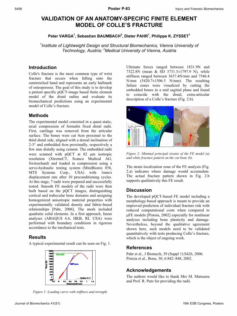
Ultimate forces ranged between 1831.9N and 7322.8N (mean & SD 3731.3±1797.9 N), while stiffness ranged between 3637.4N/mm and 7546.4 N/mm (5420.7±1506.5 N/mm). The resulting failure zones were visualized by cutting the embedded bones in a mid sagittal plane and found to coincide with the distal, extra-articular description of a Colle’s fracture (Fig. 2.b).
Figure 2: Minimal principal strains of the FE model (a) and white fracture pattern on the cut bone (b). The strain localization zone of the FE analysis (Fig. 2.a) indicates where damage would accumulate. The actual fracture pattern shown in Fig. 2.b supports qualitatively this FE result. Discussion The developed pQCT-based FE model including a morphology-based approach is meant to provide an improved prediction of individual fracture risk with reduced computational costs when compared to
FE models [Pistoia, 2002] especially for nonlinear analyses including bone plasticity and damage. Nevertheless, beyond the qualitative agreement shown here, such models need to be validated quantitatively with tests producing Colle’s fracture, which is the object of ongoing work. References Pahr et al., J Biomech, 39 (Suppl 1):S426, 2006. Pistoia et al., Bone, 30, 6:842–848, 2002. Acknowledgements The authors would like to thank Mrs M. Matsuura and Prof. R. Putz for providing the radii.
Journal of Biomechanics 41(S1) 16th ESB Congress, Posters