var2csa serology to detect plasmodium falciparum
TRANSCRIPT

Pregnant women constitute a promising sentinel group for continuous monitoring of malaria transmission. To identify antibody signatures of recent Plasmodium falciparum expo-sure during pregnancy, we dissected IgG responses against VAR2CSA, the parasite antigen that mediates placental
sequestration. We used a multiplex peptide-based suspen-sion array in 2,354 samples from pregnant women from Mozambique, Benin, Kenya, Gabon, Tanzania, and Spain. Two VAR2CSA peptides of limited polymorphism were im-munogenic and targeted by IgG responses readily boosted during infection and with estimated half-lives of <2 years. Seroprevalence against these peptides reflected declines and rebounds of transmission in southern Mozambique dur-ing 2004–2012, reduced exposure associated with use of preventive measures during pregnancy, and local clusters of transmission that were missed by detection of P. falci-parum infections. These data suggest that VAR2CSA se-rology can provide a useful adjunct for the fine-scale esti-mation of the malaria burden among pregnant women over time and space.
Agile malaria surveillance and response systems that can be sustained over time are needed for the optimal
design of control programs (1,2). Rates of Plasmodium fal-ciparum infection among pregnant women are sensitive to changes in transmission (3,4) and correlate well with infec-tion in infants (5) and children (6,7). Thus, passive detection of malaria cases at maternal health care services constitutes a promising approach to providing contemporary data on the levels, and changes in levels, of malaria burden in the popu-lation for successful malaria control and elimination (8).
After exposure to P. falciparum parasites that seques-ter in the placenta (9), antibodies against VAR2CSA, a multidomain variant antigen of the P. falciparum erythro-cyte membrane protein 1 family, develop in pregnant wom-en (10). VAR2CSA is expressed on the surface of infected erythrocytes and mediates placental sequestration of para-sites through binding to chondroitin sulfate A (11). Levels of antibodies against VAR2CSA are affected by variables that influence the risk for P. falciparum exposure (12–14) and mirror malaria trends during pregnancy (3). Moreover,
VAR2CSA Serology to Detect Plasmodium falciparum
Transmission Patterns in Pregnancy Ana Maria Fonseca, Raquel González, Azucena Bardají, Chenjerai Jairoce,
Maria Rupérez, Alfons Jiménez, Llorenç Quintó, Pau Cisteró, Anifa Vala, Charfudin Sacoor, Himanshu Gupta, Jennifer Hegewisch-Taylor, Joe Brew, Nicaise Tuikue Ndam, Simon Kariuki,
Marta López, Carlota Dobaño, Chetan E. Chitnis, Peter Ouma, Michael Ramharter, Salim Abdulla, John J. Aponte, Achille Massougbodji, Valerie Briand, Ghyslain Mombo-Ngoma, Meghna Desai,
Michel Cot, Arsenio Nhacolo, Esperança Sevene, Eusebio Macete, Clara Menéndez, Alfredo Mayor
Emerging Infectious Diseases • www.cdc.gov/eid • Vol. 25, No. 10, October 2019 1851
Author affiliations: ISGlobal, Hospital Clinic–Universitat de Barcelona, Barcelona, Spain (A.M. Fonseca, R. González, A. Bardají, M. Rupérez, A. Jiménez, L. Quintó, P. Cisteró, H. Gupta, J. Hegewisch-Taylor, J. Brew, C. Dobaño, J.J. Aponte, C. Menéndez, A. Mayor); ICBAS–Universidade do Porto, Porto, Portugal (A.M. Fonseca); Centro de Investigação em Saúde da Manhiça, Maputo, Mozambique (R. González, A. Bardají, C. Jairoce, M. Rupérez, A. Vala, C. Sacoor, C. Dobaño, J.J. Aponte, A. Nhacolo, E. Sevene, E. Macete, C. Menéndez, A. Mayor); Universidade Eduardo Mondlane, Maputo (E. Sevene); CIBER Epidemiología y Salud Pública, Madrid, Spain (R. González, A. Jiménez, A. Mayor, A. Bardají); Université d’Aboméy Calavi, Cotonou, Benin (N.T. Ndam, A. Massougbodji); Kenya Medical Research Institute, Kisumu, Kenya (S. Kariuki, P. Ouma); BCNatal–Barcelona Center for Maternal-Fetal and Neonatal Medicine–Hospital Clínic and Hospital Sant Joan de Deu, Barcelona (M. López); Vrije Universiteit Amsterdam, Amsterdam, the Netherlands (J. Brew); MERIT, Institut de Recherche pour le Développement, Paris, France (N.T. Ndam, V. Briand, M. Cot); Institut Pasteur, Paris (C.E. Chitnis); Institute of Tropical Medicine, University of Tübingen, Tübingen, Germany (G. Mombo-Ngoma); Bernhard Nocht Institute for Tropical Medicine, Hamburg, Germany (M. Ramharter, G. Mombo-Ngoma); University Medical Center Hamburg-Eppendorf, Hamburg (M. Ramharter); Ifakara Health Institute, Dar es Salaam, Tanzania (S. Abdulla); Centre de Recherches Médicales de Lambaréné, Lambaréné, Gabon (G. Mombo-Ngoma); Centers for Disease Control and Prevention, Atlanta, Georgia, USA (M. Desai)
DOI: https://doi.org/10.3201/eid2510.181177

RESEARCH
levels of VAR2CSA IgG at delivery correlate with the risk for malaria in the offspring (14), suggesting the value of these antibodies for pinpointing areas of high malaria trans-mission (15). Because VAR2CSA antibodies persist after the infection is cleared (16), they can provide a sensitive adjunct for P. falciparum monitoring, especially in areas of low malaria endemicity, where the chances of detecting an-tibodies are higher than those of detecting the parasite (17).
The utility of serosurveillance depends mainly on spe-cific properties of the antigen, including immunogenicity, polymorphism, cross-reactivity, and longevity of the anti-bodies. Because different VAR2CSA domains elicit IgG re-sponses with varying magnitudes and dynamics (16,18,19), we hypothesized that short-lived antibodies against immu-nogenic nonpolymorphic VAR2CSA epitopes would en-able a fine-scale estimation of recent P. falciparum trans-mission during pregnancy (17). We examined plasma from pregnant women living in areas in which P. falciparum transmission varied from high to low and absent (Benin, Gabon, Mozambique, Kenya, Tanzania, and Spain) against a quantitative suspension array containing VAR2CSA and general parasite antigens. We first selected IgG responses that were rapidly acquired after P. falciparum infection, did persist in circulation, and were sensitive to the level of parasite exposure in pregnant women from Mozambique and Spain. We then used the serologic assay to quantify the relationship of VAR2CSA antibody responses with P. falciparum infection as well as with temporal, spatial, and intervention-driven changes in malaria burden among pregnant women.
Methods
Study Sites, Population, and ProceduresWe included in our study pregnant women who partici-pated in 3 clinical trials of intermittent preventive treat-ment during pregnancy (IPTp) during 2003–2005 in Mo-zambique (NCT00209781) (20) and during 2010–2012 in Mozambique, Benin, Gabon, Kenya, and Tanzania (NCT00811421) (21,22). Participants were recruited at their first antenatal visit, and all received a long-lasting insecticide-treated bed net. During 2003–2005, all received 2 doses of sulfadoxine/pyrimethamine (20); during 2010–2012, they received 2 doses of mefloquine or sulfadoxine/pyrimethamine if they were not HIV infected (21) and 3 doses of mefloquine or placebo plus daily cotrimoxazol prophylaxis if they were HIV infected (22). At delivery, tissue samples from the maternal side of the placenta, as well as 50 μL peripheral and placental dried blood spots (DBS), were collected. Peripheral and placental blood from pregnant women in Mozambique and Benin were also col-lected into EDTA Vacutainer tubes (Becton Dickinson, https://www.bd.com) and centrifuged; plasma was stored
at –20°C. From a subset of pregnant women in Mozam-bique who delivered during 2011–2012, peripheral blood samples were also collected at the first antenatal visit and before administration of the second IPTp dose. We geo-coded the households of women in Mozambique by using a global information system. Clinical malaria episodes were treated according to national guidelines at the time of the study (20–22). DBS and plasma samples were also col-lected from 49 pregnant women never exposed to P. fal-ciparum who delivered in 2010 at the Hospital Clinic of Barcelona (Barcelona, Spain).
The study was approved by the ethics committees from the Hospital Clínic of Barcelona, the Comité Consultatif de Déontologie et d’Éthique from the Institut de Recherche pour le Développement (Marseille, France), the Centers for Disease Control and Prevention (Atlanta, GA, USA), and national ethics review committees from each malaria-en-demic country participating in the study. Written informed consent, which included permission to test for immune markers by using stored biological samples, was obtained from all participants.
Laboratory DeterminationsAt recruitment, we assessed HIV serostatus by using rap-id diagnostic tests according to national guidelines and hemoglobin level at delivery by using following mobile devices on capillary blood samples: HemoCue (Danaher, http://www.hemocue.com), Hemocontrol (EKF Diagnos-tics, http://www.ekfdiagnostics.com), and KX analyzer (Sysmex, http://www.sysmex.com). Thick and thin blood films and placental biopsy samples were checked for Plas-modium spp. according to standard, quality-controlled pro-cedures (3). We tested blood on filter paper for the presence of P. falciparum in duplicate by means of a real-time quan-titative PCR (qPCR) targeting 18S ribosomal DNA (3).
Antibody MeasurementsWe measured IgG in plasma (Benin and Mozambique) or on DBS (Gabon, Kenya, and Tanzania) in appropriate con-ditions for plasma elution (19) by using the xMAP tech-nology and the Luminex 100/200 System (https://www.luminexcorp.com) for 37% of pregnant women participat-ing in the clinical trials with samples available. We con-structed 2 multiplex suspension array panels (Appendix 1, https://wwwnc.cdc.gov/EID/article/25/10/18-1177-App1.pdf) (19), 1 including P. falciparum recombinant proteins (VAR2CSA Duffy binding-like recombinant domains DBL3X, DBL5Ɛ, and DBL6Ɛ; apical membrane antigen 1 [AMA1]; and 19-kDa fragment of the merozoite surface protein-1 [MSP119], from 3D7 strain) and 1 consisting of synthetic peptides (25 VAR2CSA peptides covering con-served and semiconserved regions of VAR2CSA and a cir-cumsporozoite peptide [pCSP]) (19). To assess unspecific
1852 Emerging Infectious Diseases • www.cdc.gov/eid • Vol. 25, No. 10, October 2019

VAR2CSA Serology and P. falciparum Transmission
IgG recognition, we used bovine serum albumin in both arrays (19). Procedures for reconstitution of DBS and qual-ity control, bead-based immunoassay, data normalization, and definition of seropositivity cutoffs are described in Appendix 1.
var2csa Sequencing and 3D Protein ModelingWe used DNA extracted from 50 DBS that were P. falci-parum positive by qPCR for Sanger sequencing of var2csa PCR amplification products covering peptides of interest (Appendix 1). Sequence variability with respect to the pep-tide included in the array was assessed after amino acid alignment, and a 3D model of the DBL1X-ID1 region was developed by using Chimera version 1.5.3 (https://www.cgl.ucsf.edu; Appendix 1).
Definitions and Statistical AnalysesWe included in the analysis pregnant women for whom all information was available for IPTp, date of delivery, HIV status, age, parity, and antibody responses. We classified women as primigravid (first pregnancy) or multigravid (>1 previous pregnancy) and categorized age as <20, 20–24, or >25 years (13). Anemia was defined as hemoglobin level at delivery <11 mg/L. We compared proportions by using the Fisher exact test. We used univariate regression mod-els to evaluate the association of log-transformed IgG lev-els (linear) and seropositivity (logistic) with study periods (2004–2005 and 2010–2012) and country, P. falciparum
infection, parity, anemia, and IPTp intervention, taking into account potential confounding variables (HIV and age) in multivariate models. We assessed the modification of the associations by HIV infection or parity by including inter-action terms into the regression models. To control the false discovery rate in the selection of antigens, we computed adjusted p values (q-values) by using the Simes procedure (23). We used multilevel mixed-effect linear regression analysis to estimate half-life and time to double (T2×) IgG levels in the longitudinal cohort of pregnant women from Mozambique (Appendix 1). We identified spatial clusters of P. falciparum infection and seropositivity as well as the most likely hotspots by using the Ward hierarchical clus-ter analysis and Kulldorff spatial scan method (Appendix 1). We performed statistical analyses by using Stata/SE software version 12.0 (StataCorp, https://www.stata.com), R statistics software version 3.2.1 (https://www.r-project.org), and Graphpad Prism version 6 (https://www.graph-pad.com).
Results
Study Participants and P. falciparum PrevalenceStudy participants consisted of 2,354 pregnant women (Ta-ble; Appendix 1 Figure 2) recruited during 2004–2005 (n = 146) and 2010–2012 (n = 2,208) in the context of IPTp clin-ical trials (20–22). Among them, 993 were from Mozam-bique, 854 from Benin, 131 from Gabon, 296 from Kenya,
Emerging Infectious Diseases • www.cdc.gov/eid • Vol. 25, No. 10, October 2019 1853
Table. Participants in study of VAR2CSA serologic testing to detect Plasmodium falciparum transmission patterns, by country and HIV status*
Variable
HIV-uninfected, no. (%)
HIV-infected, no. (%) 2004–2005
2010–2012 2004–2005
2010–2012 Mozambique,
n = 65 Mozambique,†
n = 485 Benin, n = 854
Gabon, n = 131
Tanzania, n = 31
Mozambique, n = 81
Mozambique,† n = 362
Kenya, n = 296
Parity
Primigravid 17 (26)
181 (37) 188 (22) 38 (29) 16 (52)
28 (35)
46 (13) 22 (7) Multigravid 48 (74)
304 (63) 666 (78) 93 (71) 15 (48)
53 (65)
316 (87) 274 (93)
Age, y
<20 19 (29)
181 (37) 86 (10) 42 (32) 5 (16)
27 (33)
41 (11) 15 (5) 20–24 17 (26)
123 (25) 281 (33) 45 (34) 14 (45)
26 (32)
84 (23) 96 (32)
>25 29 (45)
181 (37) 487 (57) 44 (34) 12 (39)
28 (35)
237 (65) 185 (62) IPTp
Sulfadoxine/ pyrimethamine
65 (100)
151 (31) 288 (34) 55 (42) 11 (35)
81 (100)
0 0
Mefloquine 0
334 (69) 566 (66) 76 (58) 20 (65)
0
178 (49) 139 (47) Placebo‡ 0
0 0 0 0
0
184 (51) 157 (53)
Microscopy§¶
Positive 9 (14)
13 (3) 110 (15) 3 (2) 1 (3)
8 (10)
8 (2) 15 (5) Negative 56 (86)
468 (97) 616 (85) 125 (98) 30 (97)
73 (90)
323 (98) 268 (95)
qPCR¶#
Positive 16 (25)
28 (6) 332 (46) 9 (10) 0
21 (26)
13 (4) 22 (8) Negative 49 (75)
424 (94) 393 (54) 80 (90) 31 (100)
60 (74)
314 (96) 251 (92)
*IPTp, intermittent preventive treatment during pregnancy; qPCR, quantitative PCR. †40% (196/485) of HIV-uninfected and 12% (43/362) of HIV-infected participants were pregnant women with samples collected also at recruitment and second IPTp administration. ‡All HIV-infected women who received placebo were also receiving cotrimoxazol prophylaxis. §Maternal microscopic infection defined by the presence of P. falciparum parasites in peripheral blood or in placenta on microscopic or histologic examination, respectively. ¶Not determined: 179 microscopy and 196 qPCR. #Maternal qPCR-positive infection was defined by a positive result on qPCR testing in peripheral or placental blood.

RESEARCH
31 from Tanzania, and 49 from Spain. The baseline charac-teristics of the women selected for this trial were similar to those of the 6,216 women participating in the randomized clinical trials (Appendix 2 Table 1, https://wwwnc.cdc.gov/EID/article/25/10/18-1177-App2.xlsx).
The study areas represented 5 sites in sub-Saharan Africa with different intensities of malaria transmission. Prevalence of P. falciparum infection detected by qPCR at delivery, in either peripheral or placental blood (aver-aged for 2010–2012), among HIV-uninfected women was 46% (332/725) in Benin, 10% (9/89) in Gabon, and 6% (28/452) in Mozambique and among HIV-infected women was 8% (22/273) in Kenya and 4% (13/327) in Mozam-bique (Table). The prevalence of P. falciparum infection among pregnant women in Mozambique decreased from 25% (37/146) in 2004–2005 to 2% (3/176) in 2010 and in-creased to 6% (4/72) in 2012. A subset of 239 pregnant women from Mozambique recruited during 2011–2012 was followed during pregnancy; prevalence of P. falciparum infection detected by qPCR was 16% (38/239) at first ante-natal visit (mean gestational age ± SD, 20.7 ± 5.45 weeks), 3% (8/239) at the second IPTp administration (25.9 ± 4.98 weeks), and 5% (13/239) at delivery (38.4 ± 2.26 weeks).
P. falciparum infection was detected at unscheduled visits for 2% (5/239) of the women. Overall, P. falciparum in-fection was detected at any of these time points for 21% (49/239) of the women.
P. falciparum–Specific Antibody Profiles and Parasite Exposure during PregnancyMean antiparasite IgG levels in pregnant women from Mo-zambique delivering from 2010 through 2012 were above levels against bovine serum albumin plus 3 SD and higher than IgG levels in pregnant women from Spain except for DBL6Ɛ and 3 of 25 VAR2CSA peptides (Figure 1, panel A; Appendix 2 Table 3). Five VAR2CSA peptides, DBL6Ɛ, and pCSP were recognized by IgG from >5% of the preg-nant women from Spain who had never been exposed to P. falciparum (Figure 1, panel B), suggesting unspecific recognition; thus, these peptides were excluded from sub-sequent analysis. To further narrow down the VAR2CSA peptide candidates, we compared IgG levels in pregnant women from Mozambique delivering in 2004–2005 and 2010–2012, a period when P. falciparum prevalence as-sessed by qPCR at delivery in peripheral or placental blood dropped from 25% to 5% (Figure 1, panel C) (3). This
1854 Emerging Infectious Diseases • www.cdc.gov/eid • Vol. 25, No. 10, October 2019
Figure 1. Plasmodium falciparum VAR2CSA IgG in malaria-exposed and -nonexposed pregnant women. A) nMFI measured in pregnant women from Mozambique and Spain. Red dashed line represents the mean nMFI from bovine serum albumin + 3 SDs. B) Seroprevalence among pregnant women from Spain (blue) and Mozambique (black). Asterisks indicate antigens recognized by pregnant women from Mozambique at levels above IgG against bovine serum albumin + 3 SDs and above levels in pregnant women from Spain (q-value <0.05 by Simes procedure) and those antigens poorly recognized by pregnant women from Spain (seroprevalence <5%). C, D) Prevalence of P. falciparum infection in peripheral and placental blood by quantitative PCR (C) and nMFIs (D) among pregnant women from Mozambique delivering in 2004–2005 and 2010–2012. Red lines represent the geometric mean and T-bars the 95% CI. Asterisks indicate antigens recognized by IgG whose levels dropped between 2004 and 2012, as assessed by linear regression adjusted by intermittent preventive treatment during pregnancy, parity, age, and HIV status (q-value <0.05 by Simes procedure). nMFI, normalized median fluorescent intensity.

VAR2CSA Serology and P. falciparum Transmission
decline in infection rates was mirrored by drops of IgG levels against 10 of the 18 previously selected VAR2CSA peptides (p1, p5, p8, p10, p12, p20, p27, p36, p38, p39) (Figure 1, panel D; Appendix 2 Table 4).
Acquisition and Decay of IgG Responses against VAR2CSAWe assessed the dynamics of IgG responses in a longitu-dinal cohort of 239 pregnant women from Mozambique (Figure 2, panel A). At delivery, compared with uninfected women, the 49 (21%) women infected with P. falciparum
during pregnancy had higher IgG levels against the 10 down-selected peptides (Figure 2, panel B; Appendix 2 Table 5). At delivery, seroprevalence rates for p1 (23%), p5 (26%), p8 (26%), and p39 (31%) antibodies were above the cumulative prevalence of P. falciparum infection dur-ing pregnancy (Figure 2, panel C; Appendix 2 Table 5). No difference in IgG levels was observed between primigravid and multigravid women (Figure 2, panel D; Appendix 2 Table 5). T2× after P. falciparum infection ranged from 0.45 years (95% CI 0.31–0.80 years) for p5 to 1.07 years (95% CI 0.60–5.23 years) for p27 (Figure 2, panel E; Appendix
Emerging Infectious Diseases • www.cdc.gov/eid • Vol. 25, No. 10, October 2019 1855
Figure 2. IgG responses during pregnancy against selected VAR2CSA antigens and polymorphism in target sequences in serologic study of Plasmodium falciparum in pregnant women. A) P. falciparum prevalence by quantitative PCR (qPCR) in 239 pregnant women from Mozambique at recruitment, second administration of IPTp, and delivery. Cumulative prevalence at delivery refers to peripheral or placental infection detected by microscopy, qPCR, or histology at any time point. B) Ratio of nMFIs at delivery in women from Mozambique infected during pregnancy compared with uninfected women. Error bars indicate 95% CIs. C) Seroprevalence at delivery, showing the cumulative prevalence of infection during pregnancy (red dashed line) and the prevalence at delivery by qPCR (light blue line). D) Ratio of nMFIs at delivery in multigravid compared with primigravid women, adjusted by IPTp, parity, age, and HIV status. Error bars indicate 95% CIs. E) IgG dynamics during pregnancy with estimates of time to double (T2x) and half-life (T1/2) obtained from linear mixed-effect regression model. Red points represent P. falciparum infection, dark gray lines the seropositivity cutoff, red lines the fitted-estimation, and dashed lines the 95% CI. F) Space-feeling representation of DBL1X-ID1 showing p5 (blue) and p8 (red). G) Logo representation of p5 and p8 sequences obtained from 50 P. falciparum isolates (20 from Mozambique, 10 from Benin, 10 from Gabon, and 10 from Kenya). IPTp, intermittent preventive treatment during pregnancy; nMFI, normalized median fluorescent intensity.

RESEARCH
2 Table 6). IgG half-life among seropositive women at recruitment without evidence of P. falciparum infection during follow-up ranged from 0.55 (95% CI 0.38–1.02) years for p8 to 3.66 (95% CI 0.98–∞) years for p1 (Fig-ure 2, panel E; Appendix 2 Table 6). Among recombinant antigens, IgG DBL5Ɛ showed the lowest T2× (0.31 [95% CI 0.21–0.61] years) and half-life (0.66 [95% CI 0.42–1.65] years), whereas AMA1 IgG showed the highest T2× (1.76 [95% CI 0.76–∞] years) and half-life (4.18 [95% CI 1.86–∞] years).
Among the down-selected VAR2CSA peptides (p1, p5, p8, and p39), IgG against p5 (51 amino acids) and p8 (48 amino acids) showed the lowest half-lives (0.55 [95% CI 0.38–1.02] years for p8; 1.33 [95% CI 0.65–∞] years for p5) and the largest increase in women exposed to P. falci-parum during pregnancy compared with uninfected women (adjusted ratio [AR]p5 2.15 [95% CI 1.39–3.31] and ARp8 2.17 [95% CI 1.46–3.23]; Figure 2, panel B; Appendix 1 Figure 5; Appendix 2 Table 5). IgG levels and seropreva-lence rates at delivery for p5 and p8 were higher among pregnant women with active or past malaria infection than among women with no parasite or pigment in the placenta, as assessed by histologic examination (Appendix 2 Table 7). 3D modeling mapped both sequences on the exposed surface of DBL1X-ID1 region of VAR2CSA (Figure 2, panel F). Amino acid variability obtained from 50 P. falci-parum isolates collected at study sites was 5% ± 2 SD for p5 sequences and 16% ± 5 SD for p8 sequences, compared with the consensus peptide sequence included in the array (Figure 2, panel G; Appendix 1 Figures 3, 4).
Performance of Selected VAR2CSA Peptides for Assessing Spatial and Temporal Differences in P. falciparum ExposureIn pregnant women from Mozambique at delivery, p5 and p8 seroprevalence rates, as well as the composite of both (p5+8), decreased from 2004–2005 to 2010 (adjusted odds ratio [AOR]p5+8 0.27 [95% CI 0.11–0.68]), followed by an increase from 2010 to 2012 (AORp5+8 2.49 [95% CI 1.34–4.61]; Figure 3, panel A; Appendix 2 Table 8). This decrease and subsequent increase mirrored P. falciparum prevalence by qPCR. HIV infection and parity did not modify the associations observed (p value for interaction >0.05 for all cases; Appendix 2 Table 8). Similar to P. fal-ciparum prevalence determined by qPCR, seroprevalence rates were the highest in HIV-uninfected women from Be-nin, followed by those from Gabon (AORp5+8 0.31 [95% CI 0.21–0.47]) and Mozambique (AORp5+8 0.21 [95% CI 0.16–0.28]; Figure 3, panel B; Appendix 2 Table 9). At delivery, pregnant women living in an area from Tanzania where no P. falciparum infection was detected by qPCR were sero-negative against p5, p8, and p5+8 antibodies; 42% were seropositive against AMA1 and 48% were seropositive
against MSP119 antibodies (Figure 3, panel B). Among HIV-infected women, seroprevalence rates for p8 and p5+8 were lower in Mozambique than in Kenya (AORp5+8 0.58 [95% CI 0.38–0.88]; Figure 3, panel C; Appendix 2 Table 9). p5 and p5+8 seroprevalence rates were higher among anemic than among nonanemic women (AORp5+8 1.26 [95% CI 1.03–1.55]; Figure 3, panel D; Appendix 2 Table 10). Seroprevalence rates were lower among HIV-uninfect-ed women who received IPTp with mefloquine than among those who received sulfadoxine/pyrimethamine (AORp5+8 0.74 [95% CI 0.59–0.94]; Figure 3, panel E; Appendix 2 Table 11). Seroprevalence rates among HIV-infected wom-en were lower among those who received mefloquine than among those who received placebo, although differences were not significant (AORp5+8 0.76 [95% CI 0.50–1.15]; Figure 3, panel F; Appendix 2 Table 11).
Geographic Patterns of P. falciparum Transmission through VAR2CSA Serologic TestingSpatial geocoordinates were available for 698 pregnant women from Mozambique residing in Manhiça District (southern Mozambique). Geographic areas experiencing significantly higher seroprevalence rates than would be ex-pected by chance were observed for p5 (radius 2.82 km; p = 0.024) and p5+8 (radius 1.06 km; p = 0.049) but not for MSP119 and AMA1 (Figure 4; Appendix 2 Table 12). The distribution of HIV infection, parity, age, and IPTp was similar among women inside and outside the serologic hotspot (p>0.05; Appendix 2 Table 12).
DiscussionRoutine P. falciparum testing of easily accessible pregnant women at maternal healthcare services has the potential to offer a rapid, consistent, and cost-effective method for evaluating the malaria burden in different communities and tracking progress of interventions. IgGs against 2 VAR2C-SA peptides, selected according to their ability to maximize the information about recent P. falciparum exposure dur-ing pregnancy, reflected differences in malaria burden over time and space in multiple settings in Africa and changes in parasite rates associated with the use of different preventive regimens. Overall, our results indicate that in areas with well-attended maternal healthcare services, this pregnancy-specific serologic test may serve as a useful sentinel sur-veillance tool for flagging changes in malaria burden and progress in the path toward elimination.
p5 (51 amino acids) is localized in the DBL1X domain and p8 (48 amino acids) in the ID1 region of VAR2CSA. Limited diversity (5%) of p5 sequence was observed in P. falciparum isolates from a variety of regions of Africa, in accordance with estimates from previous studies for the DBL1X domain (24). p8 corresponds to a more diverse (16%) variant of the ID1 region in VAR2CSA (25). Both
1856 Emerging Infectious Diseases • www.cdc.gov/eid • Vol. 25, No. 10, October 2019

VAR2CSA Serology and P. falciparum Transmission
peptides are exposed on the DBL1X-ID1 N terminal region of VAR2CSA (18,26) and recognized by IgG from malar-ia-exposed pregnant women at levels higher than those of pregnant women from Spain and men from Mozambique (19). IgG responses against both VAR2CSA peptides in-creased with P. falciparum infection during pregnancy. Moreover, higher risk for anemia among p5 and p5+8 sero-responders support these antibodies as markers of recent in-fection, which adversely affects the women’s health (3). In contrast to the slow decay of IgG responses against AMA1, the half-life of IgG against p5 and p8 was <2 years, the
average time reported in Mozambique for a second preg-nancy to occur (27). The short half-life of p5 and p8 IgG, together with the similar IgG levels in multigravid and pri-migravid women, suggests that antibodies acquired during one pregnancy are not maintained over multiple pregnan-cies; thus, antibodies can be used as a reliable indicator of recent exposure for pregnant women, regardless of parity.
Seroprevalence rates for p5, p8, and the composite of both peptides (p5+8) mirrored trends in P. falciparum preva-lence among pregnant women from Mozambique deliver-ing during 2004–2012 (3), a temporal pattern that was also
Emerging Infectious Diseases • www.cdc.gov/eid • Vol. 25, No. 10, October 2019 1857
Figure 3. IgG seroprevalence against VAR2CSA selected antigens according to study period, country, anemia status and intermittent preventive treatment group in serologic study of Plasmodium falciparum in pregnant women. A) Pregnant women from Mozambique delivering during different periods. B) HIV-uninfected pregnant women from Benin, Gabon, Mozambique, and Tanzania. C) HIV-infected pregnant women from Kenya and Mozambique. D) Nonanemic (NA) and anemic (A) pregnant women. E) HIV-uninfected pregnant women receiving mefloquine (MQ) or sulfadoxine/pyrimethamine (SP) as intermittent preventive treatment during pregnancy (IPTp). F) HIV-infected pregnant women receiving MQ or placebo (PL) as IPTp. Maternal microscopic infection was defined by the presence of P. falciparum parasites in peripheral blood or in placenta on microscopic or histologic examination, respectively. Maternal quantitative PCR (qPCR)–positive infection was defined by a positive result on qPCR of peripheral or placental blood. P-values were obtained from multivariate regression models adjusted for HIV, parity, age, IPTp, and country when applicable. T-bars represent 95% CIs. *Crude p<0.05; **adjusted p<0.05. B, Benin; G, Gabon; K, Kenya; M, Mozambique; OM, optical microscopy; T, Tanzania.

RESEARCH
observed for PfPR2–10 (28). Trends were similar among HIV-uninfected and infected women, suggesting that impairment of P. falciparum–specific antibody responses driven by vi-ral infection (29) may not affect short-lived IgG responses against p5 and p8. Seroprevalence also reflected the bur-den of malaria among pregnant women residing in a vari-ety of settings in Africa, as well as reductions in infection rates resulting from the use of mefloquine as IPTp among HIV-uninfected women (21). Similar trends, although not statistically significant, were observed among HIV-infect-ed women receiving cotrimoxazol prophylaxis alone or in combination with mefloquine (22), possibly because of the longer duration of protection provided by 3 IPTp doses in HIV-infected women compared with the 2 doses in HIV-uninfected women. We also found that pregnant women liv-ing in an area from Tanzania where no P. falciparum infec-tion was detected by qPCR as well as pregnant women from Spain never exposed to malaria were seronegative against p5 and p8, suggesting that pregnancy-specific serology might be used to confirm the eventual interruption of transmission.
Geographic distribution of pregnant women from Mozambique who were seropositive against p5 and p5+8 revealed a serologic hotspot in an area close to the river and sugar cane plantations, where the density of anopheline mosquitoes can be expected to be higher. In contrast, anti-bodies against MSP119 and AMA1 were not able to identify these malaria transmission patterns because of saturation of antibody responses after lifelong exposure to P. falci-parum. These results support the value of using VAR2CSA serologic testing to amplify signals of recent exposure and
suggest its potential to trigger targeted interventions to per-sons living in close proximity to passively detected sero-positive pregnant women.
Our study has several limitations. First, the peptide ar-ray we used may have missed some conformational nonlin-ear epitopes. Second, different transmission dynamics and host genetic factors may affect the acquisition and decay of antibodies (16). Third, steeper decay of antibodies may be observed out of pregnancy when infecting parasites express non-VAR2CSA variants. Fourth, the reduction of data from median fluorescence intensity to seroprevalence to simplify the serologic information of the assay may reduce the depth of serologic information. Developing alternative mathemat-ical models that use antibody levels (30) may increase the sensitivity to detect temporal and spatial changes in malar-ia transmission. Fifth, small numbers of pregnant women from malaria-free areas in Tanzania and Spain limit the generalizability of our data to support pregnancy-specific serologic testing as a tool to confirm interruption of trans-mission. Last, antibody assessments in this study were con-ducted mainly at delivery; further studies should assess the performance of this testing at antenatal visits or soon after delivery (i.e., during infant immunization). Future research is needed to describe the relationship between pregnancy-specific serologic testing and malaria transmission in the general population and its value for confirming interruption of malaria transmission and providing early signals of P. falciparum resurgence after local elimination.
In summary, this study shows that IgG against 2 VAR2CSA peptides from the DBL1X-ID1 domain reveal
1858 Emerging Infectious Diseases • www.cdc.gov/eid • Vol. 25, No. 10, October 2019
Figure 4. Geographic patterns of Plasmodium falciparum infection and IgG seropositivity in pregnant women living in southern Mozambique. Geographic distribution of seropositive pregnant women (HIV-uninfected and HIV-infected) living in Manhiça District, Mozambique, who delivered during 2010–2012 and for whom microscopy, quantitative PCR (qPCR), and spatial geocoordinates were available. Distribution shows pregnant women with and without P. falciparum infection at delivery, either in peripheral or in placental blood, detected by microscopy or histology (A) or qPCR (B), as well as AMA1 (C), MSP119 (D), p5 (E), p8 (F), and p5+8 (G) seropositive and seronegative pregnant women at delivery. Grey dots indicate seronegative women, blue dots indicate seropositive women, red dots indicate seropositive women selected by the hotspot cluster algorithm; red circles indicate the most likely hotspot (continuous line p<0.05, dashed line p>0.05). Maps were generated by using OpenStreetMap (https://www.openstreetmap.org). Hotspot NE, not estimated because of low/high prevalence of seropresponders.

VAR2CSA Serology and P. falciparum Transmission
temporal and spatial differences in malaria burden among pregnant women and reductions in exposure associated with the use of preventive measures during pregnancy. These antibodies enable the identification of local clusters of transmission that are missed by detection of P. falci-parum infections. Our results suggest that inferring recent exposure through VAR2CSA serologic testing would am-plify signals of ongoing malaria transmission and increase the power to detect changes, either natural or driven by deliberate efforts, as well as malaria hotspots, among preg-nant women (2). Moreover, peptides such as p1 targeted by long-lasting IgG responses may be useful for capturing past changes in transmission by sampling women of child-bearing age and relating seroprevalence with the number and timing of previous pregnancies. Operationally suitable serologic tests (31) capable of detecting antibodies against VAR2CSA synthetic peptides may be used in programmat-ic environments to stratify areas based on malaria burden, measure the effects of interventions, and document year-to-year changes in transmission.
AcknowledgmentsWe thank the women from Benin, Gabon, Kenya, Mozambique, Tanzania, and Spain who participated in the study, as well as the staff of the hospitals, clinical officers, field supervisors, and data manager. We also thank Laura Puyol, Diana Barrios, Gemma Moncunill, Pau Cisteró, Lazaro Mussacate, Nelito Ernesto Jose, and Ana Rosa Manhiça for their contribution to the collection and organization of sample shipment and Joe Campo, Aida Valmaseda, Marta Vidal, Eduard Rovira-Vallbona, Pedro Aide, Beatriz Galatas, Patrick G.T. Walker, and Peter Gething for providing important inputs for standardization of the Luminex technology, the design of protein 3D models, the interpretation of results and PfPR2-10 data.
This study was supported by the Malaria Eradication Scientific Alliance; the European and Developing Countries Clinical Trials Partnership; the Malaria in Pregnancy Consortium; and grants from Banco de Bilbao, Vizcaya, Argentaria Foundation (BBVA 02-0), Instituto de Salud Carlos III (PS09/01113, PI13/01478, and CES10/021-I3SNS, to A. Mayor), and Ministerio de Ciencia e Innovacion (RYC-2008-02631, to C.D.), the Fundação para a Ciência e Tecnologia (SFRH/BD/51696/2011, to A.M.F.), the Department of Science & Technology, Government of India (SB/OS/PDF-043/2015-16, to H.G.) and the Department d’Universitats I Recerca de la Generalitat de Catalunya (AGAUR; 2017SGR664, to A.M.).
The Centro de Investigacao em Saude da Manhica receives core support from the Spanish Agency for International Cooperation and Development. The Malaria in Pregnancy Consortium is funded by a grant from the Bill & Melinda Gates Foundation to the Liverpool School of Tropical Medicine and the Malaria Eradication Scientific Alliance is funded by a grant from the
Barcelona Institute of Global Health. ISGlobal is a member of the CERCA Programme, Generalitat de Catalunya.
A patent application has been filed for the use of p5 and p8 for serologic surveillance (US 376 patent application no. 62523828, filed on June 23, 2017, by A.M.).
About the AuthorDr. Fonseca is a doctoral fellow at ISGlobal, Barcelona Institute for Global Health, Hospital Clínic, Universitat de Barcelona, Barcelona, Spain. Her research focus includes malaria immune epidemiology in pregnant women and strategies to improve malaria surveillance in disease-endemic regions experiencing a low burden of infection.
References 1. World Health Organization. The world malaria report. Geneva:
The Organization; 2018. p. 32. 2. Mogeni P, Omedo I, Nyundo C, Kamau A, Noor A, Bejon P,
et al. on behalf of the Hotspot Group authors. Effect of transmission intensity on hotspots and micro-epidemiology of malaria in sub-Saharan Africa [cited 2019 Aug 2]. https://bmcmedicine. biomedcentral.com/articles/10.1186/s12916-017-0887-4
3. Mayor A, Bardají A, Macete E, Nhampossa T, Fonseca AM, González R, et al. Changing trends in P. falciparum burden, immunity, and disease in pregnancy. N Engl J Med. 2015; 373:1607–17. http://dx.doi.org/10.1056/ NEJMoa1406459
4. Ndam NT, Mbuba E, González R, Cisteró P, Kariuki S, Sevene E, et al. Resisting and tolerating P. falciparum in pregnancy under different malaria transmission intensities. BMC Med. 2017;15:130. http://dx.doi.org/10.1186/s12916-017-0893-6
5. Willilo RA, Molteni F, Mandike R, Mugalura FE, Mutafungwa A, Thadeo A, et al. Pregnant women and infants as sentinel populations to monitor prevalence of malaria: results of pilot study in Lake Zone of Tanzania. Malar J. 2016;15:392. http://dx.doi.org/10.1186/s12936-016-1441-0
6. Walton GA. On the control of malaria in Freetown, Sierra Leone; control methods and the effects upon the transmission of Plasmodium falciparum resulting from the reduced abundance of Anopheles gambiae. Ann Trop Med Parasitol. 1949;43:117–39. https://doi.org/10.1080/00034983.1949.11685399
7. van Eijk AM, Hill J, Noor AM, Snow RW, ter Kuile FO. Prevalence of malaria infection in pregnant women compared with children for tracking malaria transmission in sub-Saharan Africa: a systematic review and meta-analysis. Lancet Glob Health. 2015;3:e617–28. http://dx.doi.org/10.1016/S2214-109X(15)00049-2
8. Ataíde R, Mayor A, Rogerson SJ. Malaria, primigravidae, and antibodies: knowledge gained and future perspectives. Trends Parasitol. 2014;30:85–94. http://dx.doi.org/10.1016/j.pt.2013.12.007
9. Brabin BJ, Romagosa C, Abdelgalil S, Menéndez C, Verhoeff FH, McGready R, et al. The sick placenta—the role of malaria. Placenta. 2004;25:359–78. http://dx.doi.org/ 10.1016/ j.placenta.2003.10.019
10. Salanti A, Staalsoe T, Lavstsen T, Jensen AT, Sowa MP, Arnot DE, et al. Selective upregulation of a single distinctly structured var gene in chondroitin sulphate A-adhering Plasmodium falci-parum involved in pregnancy-associated malaria. Mol Microbiol. 2003;49:179–91. https://doi.org/10.1046/j.1365-2958.2003.03570.x
11. Fried M, Duffy PE. Adherence of Plasmodium falciparum to chondroitin sulfate A in the human placenta. Science. 1996; 272:1502–4. https://doi.org/10.1126/science.272.5267.1502
Emerging Infectious Diseases • www.cdc.gov/eid • Vol. 25, No. 10, October 2019 1859

RESEARCH
12. Aitken EH, Mbewe B, Luntamo M, Kulmala T, Beeson JG, Ashorn P, et al. Antibody to P. falciparum in pregnancy varies with intermittent preventive treatment regime and bed net use. PLoS One. 2012;7:e29874. https://doi.org/10.1371/ journal.pone.0029874
13. Mayor A, Kumar U, Bardají A, Gupta P, Jiménez A, Hamad A, et al. Improved pregnancy outcomes in women exposed to malaria with high antibody levels against Plasmodium falciparum. J Infect Dis. 2013;207:1664–74. http://dx.doi.org/10.1093/infdis/jit083
14. Serra-Casas E, Menéndez C, Bardají A, Quintó L, Dobaño C, Sigauque B, et al. The effect of intermittent preventive treatment during pregnancy on malarial antibodies depends on HIV status and is not associated with poor delivery outcomes. J Infect Dis. 2010;201:123–31. http://dx.doi.org/10.1086/648595
15. Bejon P, Williams TN, Liljander A, Noor AM, Wambua J, Ogada E, et al. Stable and unstable malaria hotspots in longitudinal cohort studies in Kenya. PLoS Med. 2010;7:e1000304. http://dx.doi.org/10.1371/journal.pmed.1000304
16. Fowkes FJ, McGready R, Cross NJ, Hommel M, Simpson JA, Elliott SR, et al. New insights into acquisition, boosting, and longevity of immunity to malaria in pregnant women. J Infect Dis. 2012;206:1612–21. http://dx.doi.org/10.1093/infdis/jis566
17. Drakeley C, Cook J. Chapter 5: Potential contribution of sero‐epidemiological analysis for monitoring malaria control and elimination: historical and current perspectives. Adv Parasitol. 2009;69:299–352.
18. Andersen P, Nielsen MA, Resende M, Rask TS, Dahlbäck M, Theander T, et al. Structural insight into epitopes in the pregnancy-associated malaria protein VAR2CSA. PLoS Pathog. 2008;4:e42. http://dx.doi.org/10.1371/journal.ppat.0040042
19. Fonseca AM, Quinto L, Jiménez A, González R, Bardají A, Maculuve S, et al. Multiplexing detection of IgG against Plasmodium falciparum pregnancy-specific antigens. PLoS One. 2017;12:e0181150. https://doi.org/10.1371/journal.pone.0181150
20. Menéndez C, Bardají A, Sigauque B, Romagosa C, Sanz S, Serra-Casas E, et al. A randomized placebo-controlled trial of intermittent preventive treatment in pregnant women in the context of insecticide treated nets delivered through the antenatal clinic. PLoS One. 2008;3:e1934. https://doi.org/10.1371/ journal.pone.0001934
21. González R, Mombo-Ngoma G, Ouédraogo S, Kakolwa MA, Abdulla S, Accrombessi M, et al. Intermittent preventive treatment of malaria in pregnancy with mefloquine in HIV- negative women: a multicentre randomized controlled trial. PLoS Med. 2014;11:e1001733. http://dx.doi.org/10.1371/ journal.pmed.1001733
22. González R, Desai M, Macete E, Ouma P, Kakolwa MA, Abdulla S, et al. Intermittent preventive treatment of malaria in pregnancy
with mefloquine in HIV-infected women receiving cotrimoxazole prophylaxis: a multicenter randomized placebo-controlled trial. PLoS Med. 2014;11:e1001735. http://dx.doi.org/10.1371/ journal.pmed.1001735
23. Simes R. An improved Bonferroni procedure for multiple tests of significance. Biometrika. 1986;73:751–4. https://doi.org/10.1093/biomet/73.3.751
24. Bockhorst J, Lu F, Janes JH, Keebler J, Gamain B, Awadalla P, et al. Structural polymorphism and diversifying selection on the pregnancy malaria vaccine candidate VAR2CSA. Mol Biochem Parasitol. 2007;155:103–12. http://dx.doi.org/10.1016/ j.molbiopara.2007.06.007
25. Doritchamou J, Sabbagh A, Jespersen JS, Renard E, Salanti A, Nielsen MA, et al. Identification of a major dimorphic region in the functionally critical N-terminal ID1 domain of VAR2CSA. PLoS One. 2015;10:e0137695. https://doi.org/10.1371/ journal.pone.0137695
26. Nunes-Silva S, Gangnard S, Vidal M, Vuchelen A, Dechavanne S, Chan S, et al. Llama immunization with full-length VAR2CSA generates cross-reactive and inhibitory single-domain antibodies against the DBL1X domain. Sci Rep. 2014;4:7373. http://dx.doi.org/10.1038/srep07373
27. Mandomando IM, Macete EV, Ruiz J, Sanz S, Abacassamo F, Vallès X, et al. Etiology of diarrhea in children younger than 5 years of age admitted in a rural hospital of southern Mozambique. Am J Trop Med Hyg. 2007;76:522–7. https://doi.org/10.4269/ajtmh.2007.76.522
28. Bhatt S, Weiss DJ, Cameron E, Bisanzio D, Mappin B, Dalrymple U, et al. The effect of malaria control on Plasmodium falciparum in Africa between 2000 and 2015. Nature. 2015;526:207–11. http://dx.doi.org/10.1038/nature15535
29. Naniche D, Serra-Casas E, Bardají A, Quintó L, Dobaño C, Sigauque B, et al. Reduction of antimalarial antibodies by HIV infection is associated with increased risk of Plasmodium falciparum cord blood infection. J Infect Dis. 2012;205:568–77. http://dx.doi.org/10.1093/infdis/jir815
30. Sepúlveda N, Stresman G, White MT, Drakeley CJ. Current mathematical models for analyzing anti-malarial antibody data with an eye to malaria elimination and eradication. J Immunol Res. 2015;2015:738030. https://doi.org/10.1155/2015/738030
31. Welch RJ, Anderson BL, Litwin CM. Rapid immunochromato-graphic strip test for detection of anti-K39 immunoglobulin G antibodies for diagnosis of visceral leishmaniasis. Clin Vaccine Immunol. 2008;15:1483–4. https://doi.org/10.1128/CVI.00174-08
Address for correspondence: Alfredo Mayor, ISGlobal, Hospital Clínic, Universitat de Barcelona, Carrer Rosselló 153 (CEK Bldg), E-08036 Barcelona, Spain; email: [email protected]
1860 Emerging Infectious Diseases • www.cdc.gov/eid • Vol. 25, No. 10, October 2019

Page 1 of 14
Article DOI: https://doi.org/10.3201/eid2510.181177
VAR2CSA Serology to Detect Plasmodium falciparum Transmission Patterns
Appendix 1
P. falciparum antigens
A total of 46 peptides (length between 35 and 65 aminoacids) from conserved and semi-
conserved regions of the VAR2CSA protein were designed after alignment by Clustal W of 18
VAR2CSA full-length sequences from P. falciparum isolates of different geographic origins
(Asia, Africa, Central and South America) (1). The 25 peptides analyzed (3 peptides from
DBL1X, 3 from DBL2X, 5 from DBL3X, 2 from DBL4Ɛ, 5 from DBL5Ɛ, 2 from DBL6Ɛ and 5
from NTS and ID regions) were previously selected based on highly recognized by IgGs from
plasma of malaria exposed pregnant women compared with Spanish individuals and malaria
exposed Mozambican men (1) (Appendix Table 2). A circumsporozoite peptide (pCSP) of 64
aminoacids (NVDP[NANP]15) was also analyzed. Peptides were synthetized by Gl Biochem
(Xangai, China) and median purity was estimated as 79% (range: 71%–91%) by high
performance liquid chromatography (HPLC) and mass spectrometry. The Duffy binding-like
recombinant domains (DBL3X, DBL5Ɛ and DBL6Ɛ) and the P. falciparum general antigens
(AMA1 and MSP119) were all produced at ICGEB, New Delhi, India. Clostridium tetani, tetanus
toxin was purchased from Santa Cruz Biotechnology (Dallas, Texas) to control for the amount of
IgGs eluted from dried blood spots (DBS).
Bead-based immunoassay
Two multiplex suspension array panels were constructed to quantify IgG responses
against P. falciparum recombinant proteins and synthetic peptides, using the xMAP technology
and the Luminex® 100/200 System (Luminex® Corp., Austin, Texas). MagPlex® microspheres
(magnetic carboxylated polystyrene microparticles, 5.6 μm) with different spectral signatures
were selected for each protein (DBL3X, DBL5Ɛ, DBL6Ɛ, AMA1 and MSP119), peptide (25

Page 2 of 14
VAR2CSA peptides and pCSP), tetanus toxin and bovine serum albumin. Antigens were
covalently coupled to beads following a modification of the Luminex® Corporation protocol (1).
Briefly, 200 μl of beads (2,5x10⁶) were transferred into a 1.5 mL eppendorf tube and
resuspended by sonication and vortexing. The supernatant was removed after precipitation of the
beads by magnetic separation during 60 seconds. Beads were washed twice with 250 μl of
distilled water and pellets were resuspended in 80 μl of activation buffer (0.1 M NaH2PO4, pH
6.2). Sulfo-NHS (N-hydroxysulfosuccinimide) and EDC (1-Ethyl- 3-[3-dimethylaminopropyl]
carbodiimide hydrochloride; Pierce, Thermo Fisher Scientific Inc., Rockford, IL) were
simultaneously added to reaction tubes at 5 mg/mL each in activation buffer, and reaction tubes
were incubated at room temperature with gentle agitation, protected from light for 20 minutes.
Activated beads were washed twice with 250 μl of coupling buffer (MES 50 mM, 2-[N-
morpholino] ethanesulfonic acid monohydrate pH 5, Sigma-Aldrich). Antigen amounts in the
coupling reaction for one million beads were determined after a titration experiment, and were as
follows: 2 μg of tetanus toxin, 4 μg of DBL3X, DBL5Ɛ, DBL6Ɛ, AMA1, 8 μg of MSP119, 170
μg of each peptide and for bovine serum albumin a 1% solution in PBS (Phosphate-buffered
saline) and volume was adjusted with coupling buffer until a maximum of 400 μl. Beads and
antigens were vortexed, sonicated and then incubated over night at 4°C in the dark, with shaking.
Coupled beads were blocked with 500 μl of 1% BSA in PBS for 30 minutes on a shaker at room
temperature, avoiding light and then washed twice with 500 μl of assay buffer (1% BSA, 0.05%
sodium azide in PBS filtrated) and resuspended in 400 μl of the same buffer. Beads were
quantified on a Guava PCA desktop cytometer (Guava, Hayward, CA), and stored at 4°C in the
dark. Protein and peptide multiplex arrays were prepared by pooling together equal volumes of
coated beads.
Immediately before use, stock suspensions of antigen-coated microspheres were
thoroughly resuspended by vortexing and sonication. Frozen plasma or the product of DBS
elution were thawed at room temperature, mixed by vortexing, and spun at 16000 g for 5 minutes
to remove particles. 50 μl of diluted microspheres (1000 microspheres/analyte/well) were added
to a 96-well Mylar flat-bottom plate following the addition of 50 μl of diluted sample in
duplicates to a final concentration of 1:400 for protein array and 1:100 for peptide array and
incubated for 1 hour in agitation, protected from light at room temperature. After incubation, the
plates were washed three times with 200 μl of washing buffer (0.05% Tween 20 in PBS) by

Page 3 of 14
pelleting in a magnetic 96 well separator. 100 μl of biotinylated anti-human IgG (Sigma, Tres
Cantos, Spain) diluted 1:1000 in assay buffer was added to each well, and plates were incubated
for 45 minutes in agitation, protected from light at room temperature. After the incubation
period, the plates were washed and 100 μl of streptavidin-conjugated R-phycoeryhtrin
(Invitrogen, Carlsbad, CA) at a 1:1000 dilution in assay buffer was added and incubated for 25
minutes. Finally the plates were washed as before and the beads were resuspended in 100 μl of
assay buffer and analyzed using the Luminex® 100/200 System.
A hyperimmune plasma pool composed by 23 plasmas from P. falciparum infected
Mozambican pregnant women was tested to determine if the coupling was effective and was
included in each assay plate as positive control, in addition to blanks (wells without sample) to
assess background levels. A minimum of 50 microspheres were read per spectral signature and
results were exported as crude median fluorescent intensity (MFI). Duplicates were averaged and
background MFIs were subtracted. A total of 224 plates were analyzed and the intra-assay
variation (mean CV of replicates from 20 plasma samples per plate) ranged from 1.4% to 7.3%
for the protein array and from 2.5% to 12.4% for the peptide array. The inter-assay variation
(variability of positive pool between 224 plates) was 5% for the protein array and 26% for the
peptide array (1). To assure the validity of the luminex plates, a quality control was performed on
the MFI values of the positive control pool. Results were plotted in Levey Jenning Charts
(Appendix Figure 1) and five plates from the peptide array fell out of −2 standard deviation (SD)
and +2SD and were re-analyzed (2). Results were normalized (nMFI) to account for plate-to-
plate variation by dividing the background subtracted MFI of each sample by the value of the
positive pool in the same plate and multiplying by the median of positive pools in all plates.
Definition of IgG seropositivity
Seropositivity cutoffs were obtained using finite mixture models (FMM) for pregnancy-
specific malaria antigens (VAR2CSA peptides and recombinant domains). FMM can be applied
in scenarios of heterogeneous IgG distributions where a subgroup of the study population is
seronegative and another seropositive avoiding additional sampling of malaria never exposed
pregnant women. However, seropositivity cutoffs to general malaria antigens (AMA1, MSP119
and pCSP) were obtained from means plus three SD of IgG levels from never exposed pregnant

Page 4 of 14
women as a consequence of absence of heterogeneous IgG distribution against P. falciparum
general antigens in malaria exposed adult pregnant women (1).
Reconstitution of dried blood spots
Antibodies were eluted from a total of 880 DBS from Gabon (n = 310), Kenya (n = 408)
and Tanzania (n = 162) as previously described (1,3). Briefly, four spots of 3 mm in diameter
were cut from the filter papers using a punch (McGill® round punch, 3 mm) and transferred to
individual wells of a 96-well polystyrene U-bottom plate. Antibodies were eluted with 200 μl
Luminex® assay buffer (1% BSA, 0.05% sodium azide in filtrated PBS) at room temperature
overnight with gentle mixing which, assuming a hematocrit of 50%, gives a concentration of
eluted blood proteins equivalent to a 1:50 plasma dilution (3,4).
To assess the quality of the elution, hemoglobin levels in the eluted DBS were measured
by spectrophotometry (wavelengths 415, 380 and 450) and calculated using the Harboe method
with the Allen correction (Hb [mg/l] = 167.2 x A415 – 83.6 x A380 – 83.6 x A450) x dilution
factor). Three criteria to discard DBS improperly eluted were followed, as previously described
(1). First, 259 reddish-brown spots against a pale background were discarded after visual
examination of reconstituted spots.(3) Second, 10 DBS were also discarded because hemoglobin
levels measured in the elutions were below the upper quartile of hemoglobin value among
samples considered with inappropriate visual aspect (hemoglobin upper quartile = 7.4m/l).
Finally, 153 samples were also discarded because anti-tetanus antibodies measured in the
elutions were below the lowest quartile obtained from anti-tetanus IgG among samples with
appropriate visual aspect and hemoglobin levels (anti-tetanus lowest quartile = [11563,5 nMFI]).
Finally, 458/880 samples (131 from Gabon, 296 from Kenya and 31 from Tanzania) were
considered as correctly eluted.
Sequencing of var2csa p5 and p8 in P. falciparum isolates
A total of 50 P. falciparum isolates collected on filter paper (Whatman 903) from
infected individuals in Mozambique (n = 20), Benin (n = 10), Gabon (n = 10) and Kenya (n = 10)
were selected for DNA sequencing. A half of the filter paper containing a 25 μL of blood drop
was used for DNA extraction using a QIAamp DNA Mini kit (Qiagen), as per the manufacturer's

Page 5 of 14
instructions. Finally, DNA was eluted in 100 μL of AE buffer given in the kit. The presence of P.
falciparum infection was detected using a previously described method (5,6).
We designed a single polymerase chain reaction (PCR) based assay to amplify purified
DNA templates using 2720 Thermal Cycler (Applied Biosystems) followed by Sanger
sequencing for var2csa gene. In brief, a 25 μl reaction was set up, containing 0.5 μM of each
forward (p5_F- 5aaggtgtgggaagttattac-3) and reverse (p8_R- 5attagttaaagatgcaagtact-3)
primers, 1x HOT FirePol Master Mix (Solis BioDyne; Cat. No. 04–27–00125) and 5 μl of
template DNA. The reaction volume was raised by PCR-grade water. The template DNA was
denatured at 95°C for 15 minutes in a thermocycler, followed by 35 cycles of amplification
(95°C for 1 minute, 54°C for 1 minute, and 72°C for 1 minute) and a final extension at 72°C for
10 minutes. A reaction using 5 μl of PCR-grade water instead of template DNA was included as
a negative control. PCR products were run on 1.5% agarose (Invitrogen) gels in 1× TBE buffer
(Tris/Borate/EDTA; Thermo Fisher Scientific) to determine the presence and size of the
amplified DNA. PCR products were visualized using a ultraviolet trans-illuminator. The PCR
primer set was also tested with human gDNA to check their specificity. The expected size of the
PCR was 960 bp covering amino acid positions 220 to 539 of var2csa gene. PCR products were
quantified using EPOCH Biotech system. Approximately 1200 ng of PCR products were sent to
Genewiz, following safety instructions for the accurate shipment of PCR amplicons. To sequence
the p5 and p8 fragments of var2csa gene, three new sequencing primers were used (Seq_p5_R -
5ccatttcttcacacattcac-3; Seq_p8_F - 5gggtgatccttatttcgcagaa-3; Seq_p8_R-
5cgcaagaaatcttggcaacaaca-3) along with PCR p5_F primer. This allowed us to sequence 960 bp
var2csa amplicons in both directions. The bi-directional sequencing with PCR p5_F and
Seq_p5_R, and Seq_p8_F and Seq_p8_R primer sets covered 220–335 and 389–499 aa
respectively.
The variations in the test sequences were identified by sequence alignment (Blastn,
NCBI:https://blast.ncbi.nlm.nih.gov) against reference sequence of 3D7 (PF3D7_1200600)
retrieved from PlasmoDB. The nucleotide sequences obtained from field isolates were translated
using ExPASy online tool (http://web.expasy.org/translate/) and represented as a logo figure
using the Weblogo Version 2.8.2 online tool (http://weblogo.berkeley.edu).

Page 6 of 14
VAR2CSA DBL1X-ID1 3D model
The 3D-structure of DBL1X-ID1 was obtained by submitting the 3D7 sequence, with
domain limits defined by Bockhorst and colleagues (2007) (7) to the HHPred server
(http://toolkit.tuebingen.mpg.de/hhpred). The structure with highest HHPred score,
corresponding to the DBL1α domain of the VarO strain (Protein Data Bank [PDB] 2yk0 (8)),
was selected for homology modeling in MODELER based on the default alignment. Molecular
graphics were generated in UCSF Chimera version 1.5.3 (9).
IgG dynamic analysis through mixed-effects regression models
Summary statistics according to the longitudinal design were calculated by trimester.
Time-at-risk was estimated using gestational age at recruitment as the time when subjects first
came under observation, and gestational age at delivery as the latest time under which the
subjects were both under observation and at risk. Antibody levels were analyzed assuming a
lognormal distribution, and therefore they were described by the geometric means and the
overall, between and within-subjects standard deviations.
The crude and adjusted effect of P. falciparum infection on antibody levels was analyzed
using log-linear mixed-effects regression models incorporating Gaussian random intercepts. This
resulted in an estimate of the rates of antibody dynamics (boosting or decay), assuming a single
exponential model. Time to 2-fold increase were calculated from the estimated rates and the
boundaries at 95% confidence interval (CI) obtained from mixed-effects models for women with
P. falciparum infection at follow-up (10,11). Similarly, half-lives were calculated from models
including women who were seropositive at recruitment with no P. falciparum infection at
follow-up (10,11). Where the boosting rate is a negative value (rate below 1) or the decay rate is
a positive value (rate above 1), the calculated time to 2-fold increase or half-life was reported as
infinity. Statistical comparisons were performed at two-sided significance level of 0.05 and 95%
CI were calculated for all estimations.
Consider our longitudinal dataset consisting of antibody measurements of 239 pregnant
women on three successive gestational ages. Because we were not really interested in these
particular 239 women per se, we treated them as a random sample from a larger population and

Page 7 of 14
modeled the between-woman variability as a random effect, as a random-intercept term at the
woman level. We thus fitted the model:
ln(Cij) = β0 + β1Tij + υj + εij
where Cij are the concentrations for i = 1 .. Three measurement of gestational age (T) and
j = 1 .... 239 women. The fixed portion of the model, β0 + β1Tij, simply states that we wanted one
overall regression line representing the population average. The random effect υj serves to shift
this regression line up or down according to each woman. Back-transforming the measurements
to the original scale we obtain the following overall regression line:
Cij = eβ0
+ β1
Tij = eβ
0 eβ
1T
ij = C0 eβ
1T
ij
where C0 is the baseline concentration. We can estimate the time required to obtain a
value Δ times Cij
ΔCij = C0 eβ1
(Tij
+tΔ
) = C0 eβ
1T
ij eβ
1tΔ = Cij e
β1tΔ
and therefore
Δ = eβ1
tΔ
ln(Δ) = β1tΔ
tΔ = ln(Δ) ⁄ β1
In particular, for half-life Δ = 1/2 and then t1/2 = ln(1/2) ⁄ β1 = -ln (2) ⁄ β1. Similarly, time
to 2-fold increase can be calculated ad t2x = ln (2) ⁄ β1.
Geospatial analysis
Spatial hotspots of P. falciparum infection and seropositivity among pregnant women
from Mozambique living in Manhiça district were designated using hierarchical cluster analysis
with Ward's minimum variance method (12). Kulldorff spatial scan method (13–15) was used to
identify the most likely hotspot for P. falciparum infection and seropositivity, setting our
significance threshold (α) as 0.05. Hotspots of greater size than one cluster were identified by
consecutively aggregating nearest-neighboring areas until a proportion of the total study
population was included. To ascertain statistical significance, we employed Monte Carlo
sampling, using Poisson likelihoods, and following the original Kulldorff method for

Page 8 of 14
identification of clusters (16). Analysis were performed using the R statistical software (version
3.2.1) (17) and maps were generated using OpenStreetMap (18). The key R packages used were
SpatialEpi (19), deldir (20), geosphere (21), rgeos (22) raster (23) and leaflet (24).
References
1. Fonseca AM, Quinto L, Jiménez A, González R, Bardají A, Maculuve S, et al. Multiplexing detection
of IgG against Plasmodium falciparum pregnancy-specific antigens. PLoS One.
2017;12:e0181150. PubMed https://doi.org/10.1371/journal.pone.0181150
2. Kerkhof K, Canier L, Kim S, Heng S, Sochantha T, Sovannaroth S, et al. Implementation and
application of a multiplex assay to detect malaria-specific antibodies: a promising tool for
assessing malaria transmission in Southeast Asian pre-elimination areas. Malar J. 2015;14:338.
PubMed https://doi.org/10.1186/s12936-015-0868-z
3. Corran PH, Cook J, Lynch C, Leendertse H, Manjurano A, Griffin J, et al. Dried blood spots as a
source of anti-malarial antibodies for epidemiological studies. Malar J. 2008;7:195. PubMed
https://doi.org/10.1186/1475-2875-7-195
4. Stewart L, Gosling R, Griffin J, Gesase S, Campo J, Hashim R, et al. Rapid assessment of malaria
transmission using age-specific sero-conversion rates. PLoS One. 2009;4:e6083. PubMed
https://doi.org/10.1371/journal.pone.0006083
5. Mayor A, Serra-Casas E, Bardají A, Sanz S, Puyol L, Cisteró P, et al. Sub-microscopic infections and
long-term recrudescence of Plasmodium falciparum in Mozambican pregnant women. Malar J.
2009;8:9. 10.1186/1475-2875-8-9 PubMed https://doi.org/10.1186/1475-2875-8-9
6. Taylor SM, Mayor A, Mombo-Ngoma G, Kenguele HM, Ouédraogo S, Ndam NT, et al. A quality
control program within a clinical trial Consortium for PCR protocols to detect Plasmodium
species. J Clin Microbiol. 2014;52:2144–9. PubMed http://dx.doi.org/10.1128/JCM.00565-14
7. Bockhorst J, Lu F, Janes JH, Keebler J, Gamain B, Awadalla P, et al. Structural polymorphism and
diversifying selection on the pregnancy malaria vaccine candidate VAR2CSA. Mol Biochem
Parasitol. 2007;155:103–12. PubMed http://dx.doi.org/10.1016/j.molbiopara.2007.06.007
8. Vigan-Womas I, Guillotte M, Juillerat A, Hessel A, Raynal B, England P, et al. Structural basis for the
ABO blood-group dependence of Plasmodium falciparum rosetting. PLoS Pathog.
2012;8:e1002781. PubMed https://doi.org/10.1371/journal.ppat.1002781

Page 9 of 14
9. Pettersen EF, Goddard TD, Huang CC, Couch GS, Greenblatt DM, Meng EC, et al. UCSF Chimera—a
visualization system for exploratory research and analysis. J Comput Chem. 2004;25:1605–12.
PubMed http://dx.doi.org/10.1002/jcc.20084
10. Wipasa J, Suphavilai C, Okell LC, Cook J, Corran PH, Thaikla K, et al. Long-lived antibody and B
Cell memory responses to the human malaria parasites, Plasmodium falciparum and Plasmodium
vivax. PLoS Pathog. 2010;6:e1000770. PubMed http://dx.doi.org/10.1371/journal.ppat.1000770
11. Kinyanjui SM, Conway DJ, Lanar DE, Marsh K. IgG antibody responses to Plasmodium falciparum
merozoite antigens in Kenyan children have a short half-life. Malar J. 2007;6:82. PubMed
https://doi.org/10.1186/1475-2875-6-82
12. Murtagh F, Legendre P. Ward’s hierarchical clustering method: clustering criterion and agglomerative
algorithm. J Classif. 2011;31:274–95. 10.1007/s00357-014-9161-z
https://doi.org/10.1007/s00357-014-9161-z
13. Kulldorff M. Spatial scans statistics: models, calculations, and applications. Scan Statistics and
Applications. 1999;303–22.
14. Lawson AB, Kleinman KP. Spatial and syndromic surveillance for public health. West Sussex,
England (UK); John Wiley & Sons; 2005.
15. Lin P-SK, Kung YH, Clayton M. Spatial scan statistics for detection of multiple clusters with
arbitrary shapes. Biometrics. 2016;72:1226–34. PubMed https://doi.org/10.1111/biom.12509
16. Kulldorff M, Nagarwalla N. Spatial disease clusters: detection and inference. Stat Med. 1995;14:799–
810. PubMed https://doi.org/10.1002/sim.4780140809
17. Kusi KA, Bosomprah S, Dodoo D, Kyei-Baafour E, Dickson EK, Mensah D, et al. Anti-sporozoite
antibodies as alternative markers for malaria transmission intensity estimation. Malar J.
2014;13:103. PubMed https://doi.org/10.1186/1475-2875-13-103
18. Ramm JF, Topf J, Chilton S. OpenStreetMap: Using and Enhancing the Free Map of the World.
Cambridge, UK: UIT Cambridge Limited; 2010).
19. Kim AY, Wakefield J. SpatialEpi: methods and data for spatial epidemiology. R package version
1.2.2 [cited 2017 Nov 5]. https://CRAN.R-project.org/package=SpatialEpi
20. Turner R. Deldir: Delaunay triangulation and Dirichlet (Voronoi) tessellation. R package version 0.1–
14 [cited 2017 Nov 5]. https://CRAN.R-project.org/package=deldir, (2017).

Page 10 of 14
21. Hijmans RJ. Geosphere: spherical trigonometry. R package version 1.5–5 [cited 2017 Nov 5].
https://CRAN.R-project.org/package=geosphere, (2016).
22. Bivand R, Rundel C. rgeos: interface to geometry engine - open source (GEOS). R package version
0.3–19 [cited 2017 Nov 5]. https://CRAN.R-project.org/package=rgeos, (2016).
23. Hijmans RJ. raster: Geographic Data Analysis and Modeling. R package version 2.5–8 [cited 2017
Nov 5]. https://CRAN.R-project.org/package=raster, (2016).
24. Cheng J, Karambelkar B, Xie Y. Leaflet: create interactive Web maps with the JavaScript Leaflet
Library. R package version 1.1.0 [cited 2017 Nov 5]. https://CRAN.R-
project.org/package=leaflet, (2017).
Appendix Figure 1. Levey Jenning Charts plotted for the quality control of the Luminex assay. Data
analysis for a quality control of the MFI values obtained from the hyperimmune plasma pool (positive
pool) for the A) protein array and B) peptide array. The dots represent each positive pool per plate. If
these dots fell out of the mean of positive pool +/− two standard deviation (SD) (red area), these plates
were rejected and re-analyzed.
Page 11 of 14
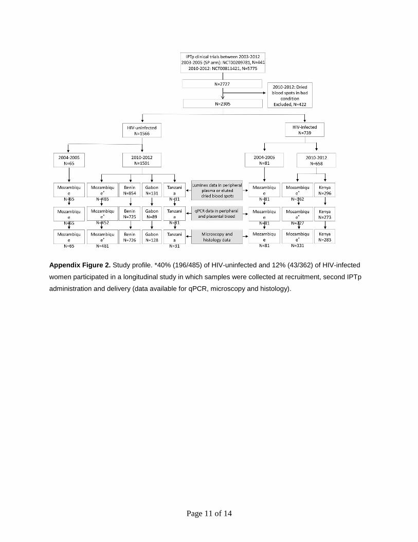
Appendix Figure 2. Study profile. *40% (196/485) of HIV-uninfected and 12% (43/362) of HIV-infected
women participated in a longitudinal study in which samples were collected at recruitment, second IPTp
administration and delivery (data available for qPCR, microscopy and histology).

Page 12 of 14
Appendix Figure 3. VAR2CSA peptide p5 sequences from P. falciparum isolates from Mozambique,
Benin, Kenya and Gabon. Grey fill means conserved regions, red indicate aminoacid change, blue
indicate nucleotide change but same aminoacid and positions with two aminoacids correspond to mixed
infections. Samples Mç1–10, B1–10, K1–10 and G1 were collected from pregnant women in Mozambique
(Mç), Benin (B), Kenya (K) and Gabon (G) who were participating in the clinical trial (2010–2012).
Samples MTP1–10 we obtained from Northern Mozambique (Montepuez).

Page 13 of 14
Appendix Figure 4. VAR2CSA peptide p8 sequences from P. falciparum isolates from Mozambique,
Benin, Kenya and Gabon. Grey fill means conserved regions, red indicate aminoacid change and blue
indicate nucleotide change but same aminoacid. Samples Mç1–10, B1–10, K1–10 and G1 were collected
from pregnant women in Mozambique (Mç), Benin (B), Kenya (K) and Gabon (G) who were participating
in the clinical trial (2010–2012). Samples MTP1–10 we obtained from Northern Mozambique
(Montepuez).

Page 14 of 14
Appendix Figure 5. Diagram of peptide selection.