variation in the anatomy of the ligamentum arteriosum in a...
TRANSCRIPT

Variation in the anatomy of the ligamentum arteriosum in a South
African sample
ORIGINAL ARTICLE Eur. J. Anat. 22 (2): 119-125 (2018)
Kerri Keet1, Geney Gunston
1, Rachel Alexander
2
1Division of Clinical Anatomy and Biological Anthropology, Department of Human Biology, University of Cape Town, Cape Town, South Africa, 2School of Medical and Health Sciences, Edith Cowan University, Perth, Western Australia
SUMMARY
The ligamentum arteriosum is a remnant of the ductus arteriosus, which connects the aortic arch and the pulmonary trunk during fetal life. Variation in the anatomy of the ligamentum arteriosum, its connections with the aorta and pulmonary trunk and the course of the left recurrent laryngeal nerve relative to the ligamentum arteriosum were investi-gated. Dissection of the superior mediastinum was performed on 40 cadavers. The anatomy of the ligamentum arteriosum and its relationship to the aortic arch, pulmonary trunk and left recurrent laryn-geal nerve were documented. The dimensions of the ligamentum arteriosum were measured with a caliper. Ligamenta arteriosa in which presence of a lumen was suspected were examined histological-ly.
Variation in the structure and size of the ligamen-ta arteriosa was found to be common. A ‘line’ on the luminal surface of the aorta at the attachment site of the ligamentum arteriosum was observed in 26%. A shallow fossa or depression was found on the luminal surface of the pulmonary trunk in all but one individual. The left recurrent laryngeal nerve was situated lateral to the ligamentum arteri-osum in 97%. Variation in the anatomy of the liga-mentum arteriosum was found to be common, whereas variation in the position of the left recur-rent laryngeal nerve was rare. This information is relevant for surgeons to avoid accidental injury to variant structures. Remnants, in the form of ‘lines’
or depressions, of the anatomical association be-tween the ductus arteriosus and the aorta and pul-monary trunk were present.
Key words: Ligamentum arteriosum – Aortic arch – Pulmonary trunk – Left recurrent laryngeal nerve – Anatomical variation – Ductus arteriosus
INTRODUCTION
The ligamentum arteriosum is a remnant of the ductus arteriosus, a small artery that connects the arch of the aorta and the pulmonary trunk during embryonic and fetal life. The ductus arteriosus di-rects blood from the pulmonary trunk into the aorta to bypass the pulmonary circulation. Increased oxygen levels in the blood after birth cause the smooth muscle wall of the ductus arteriosus to constrict, closing off this pathway. The ductus arte-riosus subsequently degenerates into a fibrous ligamentum arteriosum, which is located between the arch of the aorta and the pulmonary trunk in the superior mediastinum (Abrams, 1958; Wiyono et al., 2008). Occasionally the ductus arteriosus fails to close, resulting in a patent ductus arteriosus (PDA). PDA is diagnosed clinically when the ductus arteriosus is still open in term infants older than three months of age (Borow et al., 1981; Forsey et al., 2009).
Postnatal anatomical changes in the ligamentum arteriosum are also known to occur, and include calcification and bone formation. Calcification of the ligamentum arteriosum has been observed in both children and adults during computed tomog-raphy (CT) imaging (Wimpfheimer et al., 1996, cited in Hong et al., 2012).
119
Submitted: 30 August, 2017. Accepted: 20 December, 2017.
Corresponding author: Kerri Keet. University of Cape Town,
Department of Human Biology, Room 3.11, Anatomy Building,
Faculty of Health Sciences, Anzio Road, Observatory, 7925,
Cape Town, South Africa. Phone: 021 406 7707.
E-mail: [email protected]

Variation in the anatomy of the ligamentum arteriosum
120
The length and width of the ligamentum arterio-sum are known to vary between individuals, while remnants of the embryological connection between the ductus arteriosus and associated arteries may still be seen in the adult. Gambu et al. (2009, un-published observations) and Schwab et al. (2013, unpublished thesis) described a ridge on the lu-minal surface of the aorta that corresponded to the position of the ligamentum arteriosum. A dimple-like feature has also been observed on the luminal surface of the pulmonary trunk in an adult cadaver (Bhatnagar et al., 1996). This dimple corresponded to the site of attachment of the ligamentum arterio-sum and was considered to be a remnant of the opening of the ductus arteriosus into the pulmonary trunk (Bhatnagar et al., 1996). No additional re-ports on the presence or prevalence of this dimple have been found.
The ligamentum arteriosum has a close anatomi-cal relationship with the left recurrent laryngeal nerve (LRLN)(Higgins,1927). The LRLN is a branch of the left vagus nerve and arises as the nerve crosses over the aortic arch. The LRLN loops around the aortic arch on the lateral side of the ligamentum arteriosum, after which it passes through the aortopulmonary window and travels upwards in the tracheoesophageal groove in the neck towards the larynx (Higgins, 1927; Lardinois et al., 1999; Nakahira et al., 2001).
Knowledge of variations in the relationship be-tween the ligamentum arteriosum and the left re-current laryngeal nerve is relevant for surgeons who operate on structures in the neck and medias-tinum. Injury to the laryngeal nerves can, for exam-ple, result in permanent hoarseness of the voice (Higgins, 1927). In addition, changes in the liga-mentum arteriosum such as calcification or the presence of an opening may be observed on radi-ographs and could create confusion if not correctly identified as a feature of the ligamentum arterio-sum. Calcification of the ligamentum arteriosum may be incorrectly diagnosed as a pathological mediastinal calcification, or even cancer (Bisceglia and Donaldson, 1991).
There have been few studies documenting the anatomy of the ligamentum arteriosum and its rela-tionship to other structures, such as the left recur-rent laryngeal nerve and the luminal surfaces of the aorta and pulmonary trunk. Studies to date have been undertaken in Europe, the United States, the Middle East and Asia, with no studies, to our knowledge, reported in Africa.
The aim of this study was thus to investigate and document the following: anatomical changes in the ligamentum arteriosum, such as calcification or patency; variations in the dimensions of the liga-mentum arteriosum; the presence of any remnants of the association between the ductus arteriosus and the aorta and pulmonary trunk; and finally, any variation in the position of the left recurrent laryn-geal nerve relative to the ligamentum arteriosum.
MATERIALS AND METHODS
A cross-sectional study of embalmed cadavers was undertaken during the year 2010 at the Uni-versity of Cape Town’s Faculty of Health Sciences. The lower neck region and the superior mediasti-num were dissected and examined for evidence of variation in the ligamentum arteriosum.
The sample comprised 40 individuals, of which 27 were males and 13 were females. Ages ranged from 29 to 96 years.
The cadavers in this study were utilised in the dissection course for medical students. Any cadav-ers in which the ligamentum arteriosum, aortic arch, pulmonary trunk or the LRLN had been dam-aged by prior dissection were excluded from this study. Individuals with atherosclerotic plaques ob-structing the luminal surface of the aorta or the pul-monary artery were also excluded.
For each individual, the ligamentum arteriosum, aortic arch, pulmonary trunk, left vagus nerve and the left recurrent laryngeal nerve were identified, exposed and cleared of connective tissues by means of standard dissection techniques.
The anatomy of the ligamentum arteriosum and the course of the left recurrent laryngeal nerve was documented by means of photographs and written descriptions. The aortic arch and the pulmonary trunk, with the ligamentum arteriosum connecting them, were excised with scissors and removed from individuals for further examination. These
Fig 1. The length (**) of the ligamentum arteriosum (LA) was measured between its attachment points to the aor-ta and pulmonary trunk (PT) and the width (*) was measured at its midpoint. Abbreviations: ASC A: as-cending aorta, BCT: brachiocephalic trunk, RSA: right subclavian artery, RCC: right common carotid, LCC: left common carotid, LSA: left subclavian artery, DSC A: descending aorta, RPA: right pulmonary artery, LPA: left pulmonary artery.

K. Keet et al.
121
structures were stored in 4% formalin and labelled with a number that corresponded to that of the ca-daver from which they were removed.
The dimensions of each ligamentum arteriosum were measured with an electronic digital Vernier caliper (ORIGIN 0-150 mm). The total length of the ligamentum arteriosum between its attachment to the aorta and pulmonary trunk was measured on the ventral side (Fig. 1). The width of each liga-mentum arteriosum was measured at its midpoint. The measurements were repeated three times and the average was recorded to reduce intra-observer error. Statistica™ Version 10 was used to deter-mine any correlation between the lengths and widths of the ligamentum arteriosum.
To determine whether the ligamentum arteriosum was obliterated or patent, a piece of fishing line (Speed Spin 100m) 0.60 mm in diameter was gen-tly probed into the ligamentum arteriosum at the luminal aspect of its pulmonary and aortic attach-ment sites. The ligamentum arteriosum was then incised approximately midway along its length with scissors to assess macroscopically for the pres-ence of a lumen.
The wall of the aortic arch was opened by means of an incision that extended along its ventral as-pect to the origin of the left subclavian artery. The luminal surface was exposed and examined for any evidence of thickenings or ridges associated with the ligamentum arteriosum.
The pulmonary trunk was also opened by an inci-sion along its ventral aspect to examine the lu-minal surface at the site of attachment of the liga-mentum arteriosum. Any markings or ridges in the aortic arch or pulmonary trunk were documented and photographed.
Histological examination of two ligamenta arterio-sa in which the presence of a lumen was suspect-ed was undertaken. A section of the ligamentum arteriosum was removed from the mid-region using a scalpel with size 10 blade. The samples were processed and embedded in separate wax blocks. Sections measuring 4 µm were cut using a Reichert-Jung Autocut 2040 microtome. The sec-
tions were stained with Haemotoxylin and Eosin (H&E) and Elastin von Gieson's (EVG).
The slides were viewed by four observers using a Nikon multiheader light and fluorescent micro-scope on 4x, 10x and 40x objective magnifications, with eyepiece magnification of 10x.
This study was part of a larger project that was initiated in 2005 and approved by the Ethics Com-mittee on May 18, 2005. An amended ethics ap-proval was obtained on July 2, 2008 to include the current study.
RESULTS AND DISCUSSION
As the cadavers had already been dissected by medical students during routine dissection practi-cal sessions, some of the relevant structures had been damaged or removed in some individuals. Table 1 shows the total number of individuals, and their sex, in which the various structures were ex-amined. Ligamentum Arteriosum
The ligamentum arteriosum was observed pass-ing between the pulmonary trunk and the aortic arch in all of the individuals.
Obliterated ligamenta arteriosa were observed in 26 individuals (72.2%), calcified ligamenta arterio-sa were observed in six individuals (16.7%), and in four individuals (11.1%), an opening, which was too small for reliable measurements, was observed in the mid-region (Table 2). In these latter four indi-viduals, however, the ligamentum arteriosum ap-peared to be closed at both the aortic and pulmo-nary ends and therefore could not be considered as a patent ductus arteriosus. Histological exami-nation of two of these ligamenta arteriosa revealed that the opening may be an artefact in one individ-ual as no endothelial cells were observed lining the opening. In the second individual however, endo-thelial cells were observed, suggesting that the opening may be a remnant where the intimal sur-face of the ductus arteriosus did not unite fully in the middle, thereby leaving a small opening. This description of an opening has previously been doc-umented by Ho and Anderson, 1978. Jager and Wollenman (1942) noted that a microscopic lumen may remain for several months or longer after clo-sure at birth. They observed that the aortic and pulmonary ends of the ductus arteriosus close be-fore the central portion does and thus, the micro-
Total Male Female
Ligamentum arteriosum
36
23
13
Length of ligamentum arterio-sum
30 17 13
Width of ligamentum arteriosum 31 18 13
Luminal surface of the aortic arch
39 27 12
Luminal surface of pulmonary trunk
33 20 13
Left recurrent laryngeal nerve 30 20 10
Table 1. Total number of cadavers with sex and anatom-ical structures examined.
Ligamentum arteriosum Number of males (%)
Number of females (%)
Obliterated 18 (78.3) 8 (61.5)
Calcified 3 (13.0) 3 (23.1)
Opening in mid-region 2 (8.7) 2 (15.4)
Table 2. Prevalence of characteristics of the ligamentum arteriosum with respect to sex.

Variation in the anatomy of the ligamentum arteriosum
122
scopic lumen does not appear to be pathological in nature.
There did not appear to be any association be-tween calcification of the ligamentum arteriosum and age, as calcified ligamenta arteriosa were found in individuals who ranged from 37 to 96 years. Calcification has been previously reported in children and adults, although it is suggested that it is not atherosclerotic in aetiology in children, as appears to be the case in older individuals (Bisceglia and Donaldson, 1991; Wimpfheimer et al., 1996, cited in Hong et al., 2012). Hong et al. (2012) found calcification to be more prevalent in children (37.8%) than in adults (11.2%) on multi-section spiral CT. Other reports have shown calci-fication to be more common in adults, although these studies were done using conventional CT, which has a lower image quality and is therefore less sensitive than multi-section spiral CT (Bisceglia and Donaldson, 1991; Wimpfheimer et al., 1996, cited in Hong et al., 2012).
The prevalence of calcification in this study was lower than the 48% reported by Wimpfheimer et al. (1996, cited in Hong et al., 2012). These authors, however, used CT imaging, which may be more likely to detect calcification than direct observation as is the case in this study.
Calcified ligamenta arteriosa were equally distrib-uted among males and females. Other studies have however shown that calcification is more common in females than in males (Wimpfheimer et al., 1996, cited in Hong et al., 2012). The differ-ence in the findings of this study and other studies may be a reflection of sample size. Wimpfheimer et al. (1996) study comprised 402 individuals, whereas only 36 individuals were examined in this
study. No evidence of bone in the ligamentum arterio-
sum was found in this study. The lengths of the ligamenta arteriosa ranged from 3.48 mm to 31.28 mm, with a mean length of 14.65 mm (standard deviation 7.33 mm), which is similar to those de-scribed by Bhatnagar et al. (1996) for adults (range: 8 - 40 mm, mean 15.47 mm). The range of lengths in infants was recorded as 7.5 - 11 mm, with a mean value of 9.5 mm (Bhatnagar et al., 1996), indicating that variation in the length of the ligamentum arteriosum is normal.
The widths of the ligamenta arteriosa ranged from 1.54 mm to 4.58 mm, with a mean width of 2.86 mm (standard deviation 0.87 mm). These measurements are similar to those recorded by Delmas and Eralp (1953, cited in Bhatnagar et al., 1996), who reported a mean width of 2.94 mm (range: 1.50-5.00 mm). Given that a range in size of the ligamentum arteriosum in individuals has also been reported in the literature and that all the ligamenta arteriosa were obliterated, variation in length and width does not appear to influence clo-sure of the ductus arteriosus.
No significant correlation was found between the lengths and the widths of the ligamentum arterio-sum (p > 0.05) (p = 0.177). Aortic Ridge
With regards to visible remnants of the relation-ship between the ductus arteriosus and the aorta, no pronounced ridges on the luminal surface of the aorta, as described by Gambu et al. (2009, un-published observations) and Schwab et al. (2013, unpublished thesis) were observed, although a slight elevation or ‘line’ was present in ten individu-als (25.6%) (Fig. 2). The ‘line’ was observed in seven males (26.0%) and three females (25.0%). The ‘line’ was at the site of attachment of the liga-mentum arteriosum in six individuals, and was just distal to this position in four individuals. Table 3 shows the position of the ‘line’ with respect to the sex of the individuals.
The ‘line’ found was neither as prominent nor as prevalent as the ridge described by Gambu et al. (2009, unpublished observations) and Schwab et al. (2013, unpublished thesis), who report its pres-ence as 74% and 100% in their respective sam-ples. These differences could be a reflection of sample size and the ages of the individuals exam-ined. For example, the previous studies comprised
Fig 2. The position of the line (indicated by an arrow) on the luminal surface of the aorta at the site of attachment of the ligamentum arteriosum. Abbreviations: ASC A: ascending aorta, LCC: left common carotid artery, LSA: left subclavian artery, DSC A: descending aorta.
Position of the aortic ‘line’ Number of males (%)
Number of females (%)
At site of the ligamentum arteriosum
4 (57.1) 2 (66.7)
Distal to site of the ligamen-tum arteriosum
3 (42.9) 1 (33.3)
Table 3. Position of the aortic ‘line’ with respect to sex.
K. Keet et al.
123
large mortuary samples that included infants, chil-dren and adults that had not been embalmed, un-like the sample in this study.
The ‘line’ was predominantly found in younger individuals, with 60% of individuals with the ‘line’ being below the age of 50 years. Six individuals were under the age of 50 years (33 to 47 years) and three were older than 50 (56 to 60 years), while the age of one individual was not known. This is consistent with the observations of Gambu et al. (2009, unpublished observations), in which the ridge was predominantly found in younger indi-viduals, with a prevalence of 84% in individuals aged 11-25 years, 71% in individuals aged 26-50 years, and 53% in those aged 50 years and older. Schwab et al. (2013, unpublished thesis) observed the ridge in all of the infants and children that they studied. Owing to its high prevalence in neonates and young individuals, these authors suggest that the ridge may be a normal anatomical feature that is present before birth and regresses with age.
Prevalence of the ‘line’ in this study with respect to sex also differed to that of the ridge (Gambu et al., 2009, unpublished observations). The ‘line’ was
observed with equal prevalence in males and fe-males, whereas Gambu et al. (2009, unpublished observations) found the ridge to be more prevalent in males. Their study, however, consisted of a larg-er sample of 150 individuals, which could account for this variation. Dimple on luminal surface of pulmonary trunk
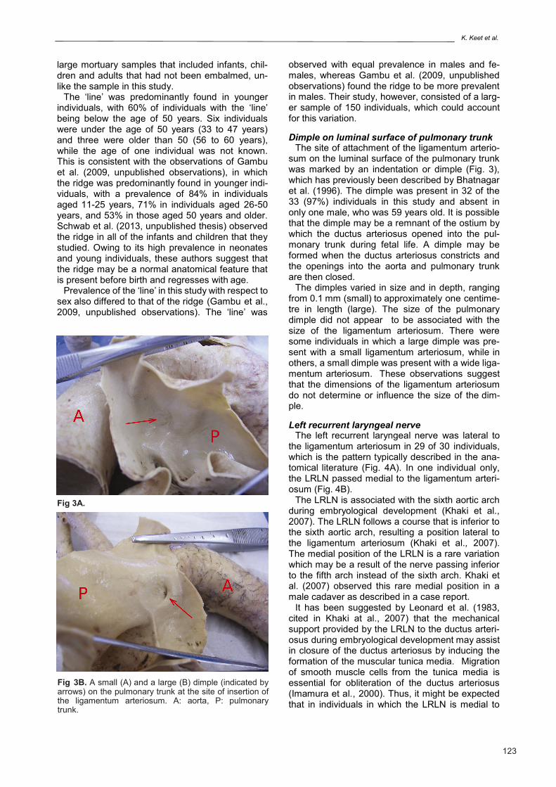
The site of attachment of the ligamentum arterio-sum on the luminal surface of the pulmonary trunk was marked by an indentation or dimple (Fig. 3), which has previously been described by Bhatnagar et al. (1996). The dimple was present in 32 of the 33 (97%) individuals in this study and absent in only one male, who was 59 years old. It is possible that the dimple may be a remnant of the ostium by which the ductus arteriosus opened into the pul-monary trunk during fetal life. A dimple may be formed when the ductus arteriosus constricts and the openings into the aorta and pulmonary trunk are then closed.
The dimples varied in size and in depth, ranging from 0.1 mm (small) to approximately one centime-tre in length (large). The size of the pulmonary dimple did not appear to be associated with the size of the ligamentum arteriosum. There were some individuals in which a large dimple was pre-sent with a small ligamentum arteriosum, while in others, a small dimple was present with a wide liga-mentum arteriosum. These observations suggest that the dimensions of the ligamentum arteriosum do not determine or influence the size of the dim-ple. Left recurrent laryngeal nerve
The left recurrent laryngeal nerve was lateral to the ligamentum arteriosum in 29 of 30 individuals, which is the pattern typically described in the ana-tomical literature (Fig. 4A). In one individual only, the LRLN passed medial to the ligamentum arteri-osum (Fig. 4B).
The LRLN is associated with the sixth aortic arch during embryological development (Khaki et al., 2007). The LRLN follows a course that is inferior to the sixth aortic arch, resulting a position lateral to the ligamentum arteriosum (Khaki et al., 2007). The medial position of the LRLN is a rare variation which may be a result of the nerve passing inferior to the fifth arch instead of the sixth arch. Khaki et al. (2007) observed this rare medial position in a male cadaver as described in a case report.
It has been suggested by Leonard et al. (1983, cited in Khaki at al., 2007) that the mechanical support provided by the LRLN to the ductus arteri-osus during embryological development may assist in closure of the ductus arteriosus by inducing the formation of the muscular tunica media. Migration of smooth muscle cells from the tunica media is essential for obliteration of the ductus arteriosus (Imamura et al., 2000). Thus, it might be expected that in individuals in which the LRLN is medial to
Fig 3B. A small (A) and a large (B) dimple (indicated by arrows) on the pulmonary trunk at the site of insertion of the ligamentum arteriosum. A: aorta, P: pulmonary trunk.
Fig 3A.

Variation in the anatomy of the ligamentum arteriosum
124
the ductus arteriosus, the ductus arteriosus would remain open in the absence of the support offered by the nerve. The ligamentum arteriosum was, however, obliterated in the single individual in which the LRLN passed medially to the ligamen-tum arteriosum in this study.
Although variation is present at very low frequen-cy, knowledge that the LRLN may be situated on the medial side of the ligamentum arteriosum is clinically important, as the nerve may be damaged during surgery on the aorta or ligamentum arterio-sum (Khaki et al., 2007).
Limitations and recommendations for future studies
A relatively small sample of adult cadavers that had been previously dissected by medical students was available for the study. In addition, these ca-davers had been fixed in formalin, which is known to cause tissue shrinkage (Jonmarker et al., 2006). This may have affected certain parameters such as the dimensions of the ligamentum arteriosum. Ex-amination of non-embalmed tissues would provide useful comparative material.
Further studies on a larger and more wide-ranging population, particularly with respect to age, would enhance current knowledge of variations in these structures. Additional research is necessary to in-vestigate whether calcification of the ligamentum
arteriosum is associated with calcification of other structures, such as the valves of the heart or the aorta.
Conclusion
Variations in the anatomy of the ligamentum arteri-osum and the left recurrent laryngeal nerve exist in South African samples. Knowledge of these varia-tions is of relevance to surgeons who operate on the mediastinum and thorax. Changes in the liga-mentum arteriosum such as calcification and the presence of an opening may be observed on radi-ographs and could create confusion if not identified as a feature of the ligamentum arteriosum. The presence of an opening in the mid-region of the ligamentum arteriosum does not necessarily mean it is patent as aortic and pulmonary ends may be closed. Clinicians also need to be aware that varia-tions in the dimensions of the ligamentum arterio-sum are not uncommon.
ACKNOWLEDGEMENTS
The body donors and their families for their gen-erous gift. National Research Foundation and the University of Cape Town for funding the research.
REFERENCES
Fig 4A. Lateral position (A) and medial position (B) of the left recurrent laryngeal nerve. Key: Light blue: aortic arch, black: brachiocephalic trunk, white: left common carotid artery, dark blue: left subclavian artery, orange: left vagus nerve, pink: left recurrent laryngeal nerve, yellow: ligamentum arteriosum. Fig 4B.

K. Keet et al.
125
ABRAMS HL (1958) Persistence of fetal ductus function after birth: the ductus arteriosus as an avenue of es-cape. Circulation, 18: 206-226.
BHATNAGAR KP, WAGNER CE, AUSTIN EH (1996) An unusually long ligamentum arteriosum. Ann Anat, 178(5): 467-470.
BISCEGLIA M, DONALDSON JS (1991) Calcification of the ligamentum arteriosum in children: a normal find-ing on CT. Am J Roentgenol, 156(2): 351-352.
BOROW KM, HESSEL SJ, SLOSS LJ (1981) Fistulous aneurysm of ductus arteriosus. Br Heart J, 45: 467-470.
FORSEY JT, ELMASRY OA, MARTIN RP (2009) Patent arterial duct. Orphanet J of Rare Dis, 4: 17. https://ojrd.biomedcentral.com/articles/10.1186/1750-1172-4-17
GAMBU M, DA SILVA R, LIEBENBERG L, GUNSTON G, ALEXANDER R (2009) Gross anatomy and histolo-gy of an aortic ridge. Unpublished observations.
Higgins CC (1927) Surgical anatomy of the recurrent laryngeal nerve with especial reference to thyroid sur-gery. Ann Surg, 85(6): 827-838.
HO SY, ANDERSON RH (1979) Anatomical closure of the ductus arteriosus: a study in 35 specimens. J Anat, 128(4): 829-836.
HONG GS, GOO HW, SONG JW (2012) Prevalence of ligamentum arteriosum calcification on multi-section spiral CT and digital radiography. Int J Cardiovasc Imaging, 28(1): 61-67.
IMAMURA S, NISHIKAWA T, HIRATSUKA E, TAKAO A, MATSUOKA R (2000) Behavior of smooth muscle cells during arterial ductal closure at birth. J Histochem Cytochem, 48(1): 35-44.
JAGER BV, WOLLENMAN Jr OJ (1942) An anatomical study of the closure of the ductus arteriosus. Am J Pathol, 18(4): 595-613.
JONMARKER S, VALDMAN A, LINDBERG A, HELL-STRÖM M, EGEVAD L (2006) Tissue shrinkage after fixation with formalin injection of prostatectomy speci-mens. Virchows Archiv, 449(3): 297-301.
KHAKI AA, TUBBS RS, SHOJA MM, ZARRINTAN S (2007) An unusual course of the left recurrent larynge-al nerve. Clin Anat, 20(3): 344-346.
LARDINOIS D, GUGGER M, BALMER MC, RIS HB (1999) Left recurrent laryngeal nerve palsy associated with silicosis. Eur Respir J, 14(3): 720-722.
NAKAHIRA M, NAKATANI H, TAKEDA T (2001) Left vocal cord paralysis associated with long-standing patent ductus arteriosus. Am J Neuroradiol, 22(4): 759-761.
SCHWAB P (2013) The aortic-ridge in children under eleven years of age. (Honours thesis). University of Cape Town, South Africa.
WIYONO SA, WITSENBURG M, DE JAEGERE PPT, ROOS-HESSELINK JW (2008) Patent ductus arterio-sus in adults: Case report and review illustrating the spectrum of the disease. Neth Heart J, 16(7-8): 255-259.