vascular access complications. introduction after decades of success in dialysis research and...
TRANSCRIPT
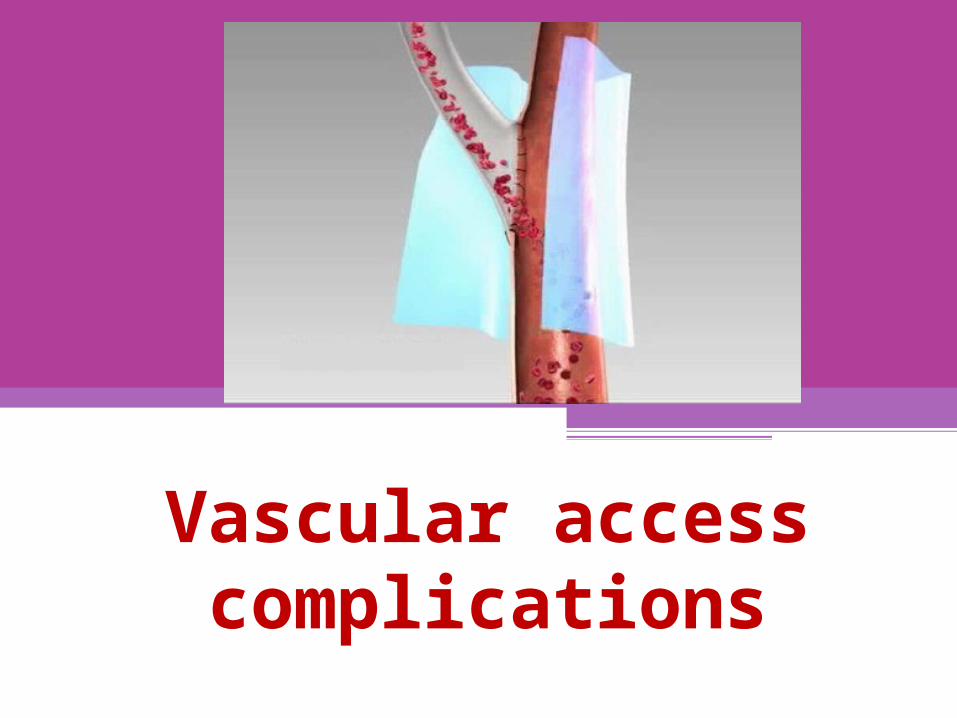
Vascular access complications
Introduction
• After decades of success in dialysis research and treatment, the prompt availability of a well functioning vascular access (VA) for dialysis remains a disturbing problem
• VA complications account for 16-25% of hospital admissions in hemodialysis (HD) patients
• HD VA dysfunction is a major cause of morbidity and hospitalization among the HD population
1. El Minshawy et al. The Journal of Vascular Access 2004; 5: 76-82)
2. Ravani P, et al. Am J Kidney Dis 2002; 40: 1264-76.3. Dhingra RK et al ,. Kidney Int 2001; 60: 1443-51.4. Roy-Chaudhury P, et al J Invasive Cardiol 2003; 15: A25-30.

The Problem •Access sites are the Achilles’ heel of the
hemodialysis therapy•Limited number of sites on the body•Consistent surveillance of the access site,
while desired, does not routinely occur due to
Time Staffing Patient absence Because……

Consequences of the Problem
•25% of deaths in HD patients are infection-related.
1. Feldman HI, Held PJ, Hutchinson JT, Stoiber E, Hartigan MF, Berlin JE 1993. Kidney Int 43:1091-1096.
2. Chazan JA, London MR, Pono L 1990. Am J Kid Dis 6:523-525.
3. Feldman HI, Kobrin S, Wasserstein A 1996 J Am Soc Nephrol 7:523-535.
4. Dr. Larry Spergel presentation
Vascular access complications account for ~30% of hospital admissions in chronic HD programs.2
Infection rate increases with temporary catheter use.
Nearly 75% of HD patients are hospitalized for vascular access-related problem within 2 years.1
Estimates on the cost of hospitalization for vascular access problems range from $2 billion3 per year to $3 billion4, representing 10% to 15% of Medicare ESRD expenditures.

5
K/DOQI
http://www.kidney.org/PROFESSIONALS/kdoqi/guideline_upHD_PD_VA/index.htm
•Promotes new standards of care in order to treat all forms of kidney disease and reduce the number of dialysis patients.
National (USA) Kidney Foundation Kidney Disease Outcomes Quality Initiative
6
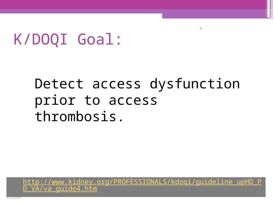
K/DOQI Goal:
• Detect access dysfunction prior to access thrombosis.
http://www.kidney.org/PROFESSIONALS/kdoqi/guideline_upHD_PD_VA/va_guide4.htm

7
K/DOQI Suggested Technology
•Indicator dilution (Transonic) flow measurements are the preferred method of A-V graft and fistula surveillance.
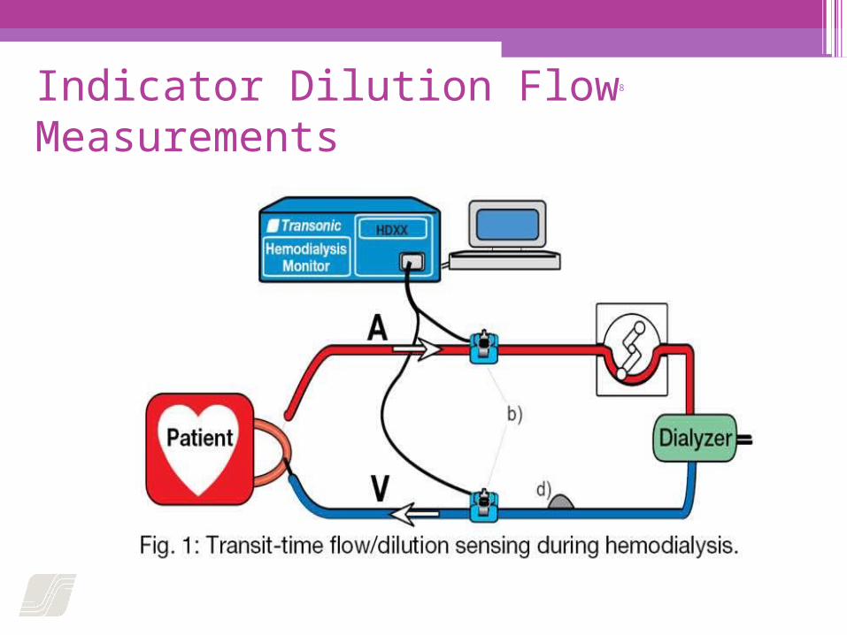
8Indicator Dilution Flow Measurements

9Vascular Access Management Tools
Intraoperative Flowmeter Endovascular Flowmeter
Hemodialysis Monitor

10
•Dialysis Adequacy▫Delivered Blood Flow▫Recirculation
•Vascular Access Flow
•Cardiac Output
HD Monitor Parameters

11Access Flow Measurement (Blood Lines are Reversed)
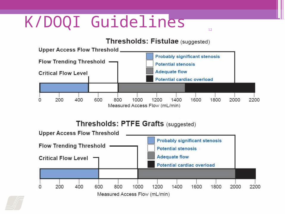
12K/DOQI Guidelines

13K/DOQI Guidelines for Monitoring AV Grafts and Fistulas
•Access flow < 600 ml/min, the patient should be referred for fistulagram.
•Access flow < 1000ml/min that has decreased by > 25% over 4 months should be referred for fistulagram.

14
Monthly Access Flow Trending
Documented InterventionK/DOQI Guideline: <600 ml/min

15
Clinical Results
•Gambro Study
•McCarley Study

16Gambro Study
– 44% decrease in thrombosis. – Significant improvements in clinical
outcomes.–Decrease in hospitalizations.
An 18-month Gambro Study of 254 patients using a multidisciplinary approach to vascular access care resulted in:
Reference: Duda, CR, Spergel, LM, Holland, J, Tucker, T, Bosch JP, Bander, SJ. “A Multidisciplinary Vascular Access Care Program (VACS) Enables Implementation of Dialysis Outcomes Quality Initiative (DOQI)”, JASN Abstracts, Vol. 10, p. 206A, 1999.
17Vascular Access Blood Flow Monitoring Reduces Access Morbidity and Costs
Patricia McCarley, Rebecca L. Wingard, Yu Shyr, William Pettus, Raymond M. Hakim, and T. Alp Ikizler
Vanderbilt University Medical Center, Dialysis Clinics, Inc., Renal Care Group, Inc.
Kidney International, Vol. 60 (2001), pp. 1164-1172

18Three-Phase Study
Phase Duration Monitoring Technique
I 11 Months None
II 12 Months Dynamic V.P.
III 10 Months Transonic HD01
132 Hemodialysis Patients with A-V grafts or fistulas.

19

20

21Europe
•“Objective monitoring of access function should be performed regularly by measuring access flow.”
European best practice guidelines on hemodialysis Guideline 5. Surveillance of Vascular Access. ERA/EDTA. Nephrol Dial Transplant, 2007; 22(Suppl 2): ii99. Transonic Reference # HD7450A
22Japan
•“Regular monitoring of shunt flow in haemodialysis patients has become extremely important.”
“Clinical Evaluation of New Non-Invasive Shunt Flow Measurement Device.” Satoshi YAMAGUCHI1, Noriko OKUMURA1, Izumi AMANO1 Department of Blood Purification, Tenri Hospital. 42nd Annual Meeting of the Japanese Society for Artificial Organs; October 6, 2004

23
Flow-QC in HD
Preserve an Access - Save a Live

NKF-KDOQI clinical practice guidelines for vascular access: update 2006.
Vascular Access Work Group. Clinical practice guidelines for vascular access. Am J Kidney Dis 2006 Jul;48 Suppl 1:S248-73
Guideline 5. Treatment of Fistula Complications • Appropriate interventions for access dysfunction
may result in an increased duration of survival of the AVF.
▫ 5.1 Problems developing in the early period after AVF construction (first 6 months) should be promptly addressed.
▫ 5.1.1 Persistent swelling of the hand or arm should be expeditiously evaluated and the underlying pathology should be corrected. [B]
▫ 5.1.2 A program should be in place to detect early access dysfunction, particularly delays in maturation. The patient should be evaluated no later than 6 weeks after access placement. [B]

Guideline 5
Summary of Physical Examination

First step in assessing dysfunction

5.2 Intervention:
• Intervention on a fistula should be performed for the presence of: ▫ 5.2.1 Inadequate flow to support the prescribed
dialysis blood flow. [B] ▫ 5.2.2 Hemodynamically significant venous stenosis.
[B] ▫ 5.2.3 Aneurysm formation in a primary fistula.
Postaneurysmal stenosis that drives aneurysm also should be corrected. The aneurysmal segment should not be cannulated. [B]
▫ 5.2.4 Ischemia in the access arm. [B]

5.3 Indications for preemptive percutaneous angioplasty (PTA): • A fistula with a greater than 50% stenosis in either
the venous outflow or arterial inflow, in conjunction with clinical or physiological abnormalities, should be treated with PTA or surgical revision. [B]
• 5.3.1 Abnormalities include reduction in flow, increase in static pressures, access recirculation preempting adequate delivery of dialysis, or abnormal physical findings. [B]

5.4 Stenosis
• 5.4 Stenosis, as well as the clinical parameters used to detect it, should return to within acceptable limits following intervention. [B]
• 5.5 Thrombectomy of a fistula should be attempted as early as possible after thrombosis is detected, but can be successful even after several days. [B]

5.6 Access evaluation for ischemia:
▫5.6.1 Patients with an AVF should be assessed on a regular basis for possible ischemia. [B]
▫5.6.2 Patients with new findings of ischemia should be referred to a vascular access surgeon emergently. [B]

5.7 Infection:
•Infections of primary AVFs are rare and should be treated as subacute bacterial endocarditis with 6 weeks of antibiotic therapy. Fistula surgical excision should be performed in cases of septic emboli. [B]

Guideline 6. Treatment of Arteriovenous Graft Complications
•Appropriate management and treatment of AVG complications may improve the function and longevity of the vascular access.

6.1 Extremity edema: • Patients with extremity edema that persists beyond 2
weeks after graft placement should undergo an imaging study (including dilute iodinated contrast) to evaluate patency of the central veins.
• The preferred treatment for central vein stenosis is PTA.
• Stent placement should be considered in the following situations: ▫6.1.1 Acute elastic recoil of the vein (>50%
stenosis) after angioplasty. [B]
▫6.1.2 The stenosis recurs within a 3-month period. [B]

6.2 Indicators of risk for graft rupture:
•Any of the following changes in the integrity of the overlying skin should be evaluated urgently:
▫6.2.1 Poor eschar formation. [B] ▫6.2.2 Evidence of spontaneous bleeding. [B]
6.2.3 Rapid expansion in the size of a pseudoaneurysm. [B]
▫6.2.4 Severe degenerative changes in the graft material. [B]

6.3 Indications for revision/repair: • 6.3.1 AVGs with severe degenerative changes
or pseudoaneurysm formation should be repaired in the following situations: ▫6.3.1.1 The number of cannulation sites
are limited by the presence of a large (or multiple) pseudoaneurysm(s). [B]
▫6.3.1.2 The pseudoaneurysm threatens the viability of the overlying skin. [B]
▫6.3.1.3 The pseudoaneurysm is symptomatic (pain, throbbing). [B]
▫6.3.1.4 There is evidence of infection. [B]

6.4 Treatment of stenosis without thrombosis: •Stenoses that are associated with AVGs
should be treated with angioplasty or surgical revision if the lesion causes a greater than 50% decrease in the luminal diameter and is associated with the following clinical/physiological abnormalities:▫6.4.1 Abnormal physical findings. [B] ▫6.4.2 Decreasing intragraft blood flow
(<600 mL/min). [B] 6.4.3 Elevated static pressure within the graft. [B]

6.5 Outcomes after treatment of stenosis without thrombosis:
• After angioplasty or surgical revision of a stenosis, each institution should monitor the primary patency of the AVG. Reasonable goals are as follow:
• 6.5.1 Angioplasty: ▫ 6.5.1.1 The treated lesion should have less than 30%
residual stenosis and the clinical/physiological parameters used to detect the stenosis should return to acceptable limits after the intervention. [B]
▫ 6.5.1.2 A primary patency of 50% at 6 months. [B]
• 6.5.2 Surgical revision: ▫ 6.5.2.1 The clinical/physiological parameters used to
detect the stenosis should return to acceptable limits after the intervention. [B]
▫ 6.5.2.2 A primary patency of 50% at 1 year. [B]
6.6 If angioplasty of the same lesion is required more than 2 times within a 3-month period, the patient should be considered for surgical revision if the patient is a good surgical candidate.
•6.6.1 If angioplasty fails, stents may be useful in the following situations:
6.6.1.1 Surgically inaccessible lesion. [B] 6.6.1.2 Contraindication to surgery. [B] 6.6.1.3 Angioplasty-induced vascular
rupture. [B]
6.7 Treatment of thrombosis and associated stenosis:
•Each institution should determine which procedure, percutaneous thrombectomy with angioplasty or surgical thrombectomy with AVG revision, is preferable based upon expediency and physician expertise at that center.
• 6.7.1 Treatment of AVG thrombosis should be performed urgently to minimize the need for a temporary HD catheter. [B]
• 6.7.2 Treatment of AVG thrombosis can be performed by using either percutaneous or surgical techniques. Local or regional anesthesia should be used for the majority of patients. [B]
• 6.7.3 The thrombectomy procedure can be performed in either an outpatient or inpatient environment. [B]
• 6.7.4 Ideally, the AVG and native veins should be evaluated by using intraprocedural imaging. [B]
• 6.7.5 Stenoses should be corrected by using angioplasty or surgical revision. [B]
• 6.7.6 Methods for monitoring or surveillance of AVG abnormalities that are used to screen for venous stenosis should return to normal after intervention. [B]

6.8 Outcomes after treatment of AVG thrombosis: • After percutaneous or surgical thrombectomy, each
institution should monitor the outcome of treatment on the basis of AVG patency. Reasonable goals are as follows: ▫6.8.1 A clinical success rate of 85%; clinical success
is defined as the ability to use the AVG for at least 1 HD treatment. [B]
▫6.8.2 After percutaneous thrombectomy, primary patency should be 40% at 3 months. [B]
▫6.8.3 After surgical thrombectomy, primary patency should be 50% at 6 months and 40% at 1 year. [B]

6.9 Treatment of AVG infection:
• Superficial infection of an AVG should be treated as follows:
▫ 6.9.1 Initial antibiotic treatment should cover both gram-negative and gram-positive microorganisms. [B]
▫ 6.9.1.1 Subsequent antibiotic therapy should be based upon culture results.
▫ 6.9.1.2 Incision and drainage may be beneficial.
▫ 6.9.2 Extensive infection of an AVG should be treated with appropriate antibiotic therapy and resection of the infected graft material. [B]
Guideline 7. Prevention and Treatment of Catheter and Port Complications • Catheters and ports are essential tools for
providing urgent and, in some cases, long-term vascular access.
• Prevention and early treatment of complications should greatly reduce associated morbidity and mortality. ▫ 7.1 Catheters and ports should be evaluated
when they become dysfunctional. Dysfunction is defined as failure to attain and
maintain an extracorporeal blood flow of 300 mL/min or greater at a prepump arterial pressure more negative than –250 mm Hg. [B]
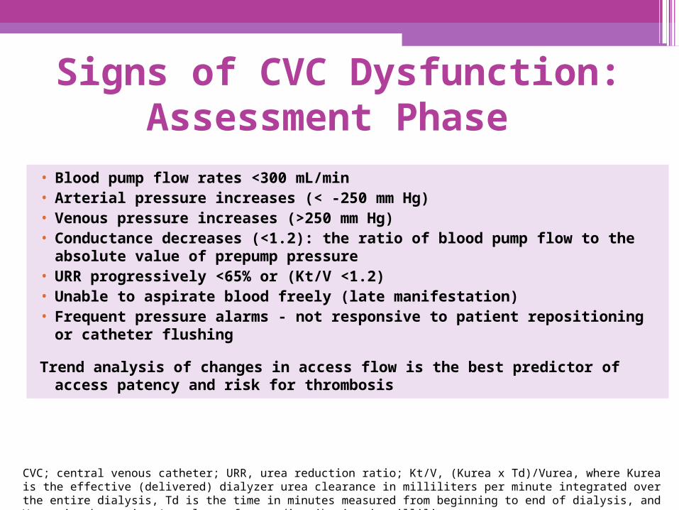
Signs of CVC Dysfunction: Assessment Phase
• Blood pump flow rates <300 mL/min • Arterial pressure increases (< -250 mm Hg) • Venous pressure increases (>250 mm Hg) • Conductance decreases (<1.2): the ratio of blood pump flow to the
absolute value of prepump pressure • URR progressively <65% or (Kt/V <1.2) • Unable to aspirate blood freely (late manifestation) • Frequent pressure alarms - not responsive to patient repositioning
or catheter flushing
Trend analysis of changes in access flow is the best predictor of access patency and risk for thrombosis
CVC; central venous catheter; URR, urea reduction ratio; Kt/V, (Kurea x Td)/Vurea, where Kurea is the effective (delivered) dialyzer urea clearance in milliliters per minute integrated over the entire dialysis, Td is the time in minutes measured from beginning to end of dialysis, and Vurea is the patient's volume of urea distribution in milliliters
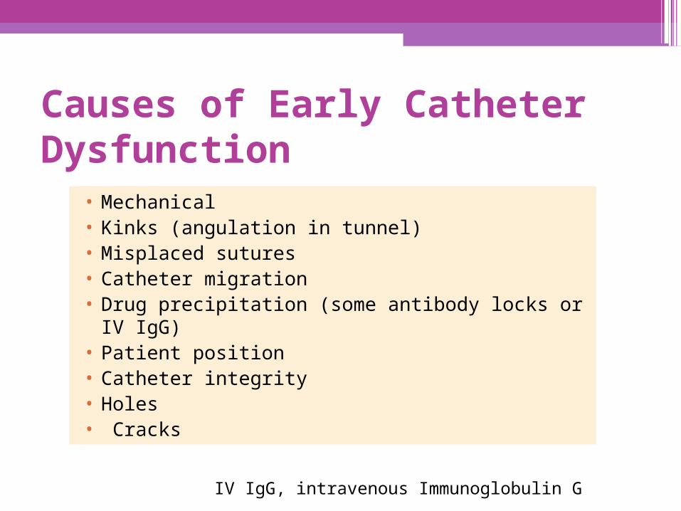
Causes of Early Catheter Dysfunction
• Mechanical • Kinks (angulation in tunnel) • Misplaced sutures • Catheter migration • Drug precipitation (some antibody locks or IV
IgG) • Patient position • Catheter integrity • Holes• Cracks
IV IgG, intravenous Immunoglobulin G

Available Thrombolytics • Streptokinase
▫ Highly antigenic ▫ Low fibrin affinity
• Urokinase ▫ Available for PE treatment ▫ No longer manufactured (11/2004)
• Reteplase ▫ Used in treatment of AMI ▫ Must be aliquoted and frozen
• Ateplase, tPA ▫ High fibrin specificity ▫ FDA approved ▫ Available in single dose vials ▫ No antigenicity

•7.2 The exception is pediatric or smaller adult catheters that are not designed to have flows in excess of 300 mL/min. [B]

Dysfunctional or nonfunctional catheter or port
•7.3 Methods that should be used to treat a dysfunctional or nonfunctional catheter or port include: ▫7.3.1 Repositioning of a malpositioned
catheter. [B] ▫7.3.2 Thrombolytics, using either an
intraluminal lytic, intradialytic lock protocol, or an intracatheter thrombolytic infusion or interdialytic lock. [B]
▫7.3.3 Catheter exchange with sheath disruption, when appropriate. [B]
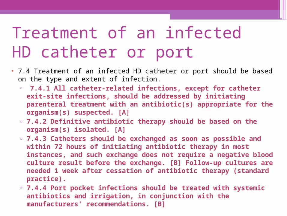
Treatment of an infected HD catheter or port• 7.4 Treatment of an infected HD catheter or port should be based on the
type and extent of infection.▫ 7.4.1 All catheter-related infections, except for catheter exit-
site infections, should be addressed by initiating parenteral treatment with an antibiotic(s) appropriate for the organism(s) suspected. [A]
▫ 7.4.2 Definitive antibiotic therapy should be based on the organism(s) isolated. [A]
▫ 7.4.3 Catheters should be exchanged as soon as possible and within 72 hours of initiating antibiotic therapy in most instances, and such exchange does not require a negative blood culture result before the exchange. [B] Follow-up cultures are needed 1 week after cessation of antibiotic therapy (standard practice).
▫ 7.4.4 Port pocket infections should be treated with systemic antibiotics and irrigation, in conjunction with the manufacturers' recommendations. [B]
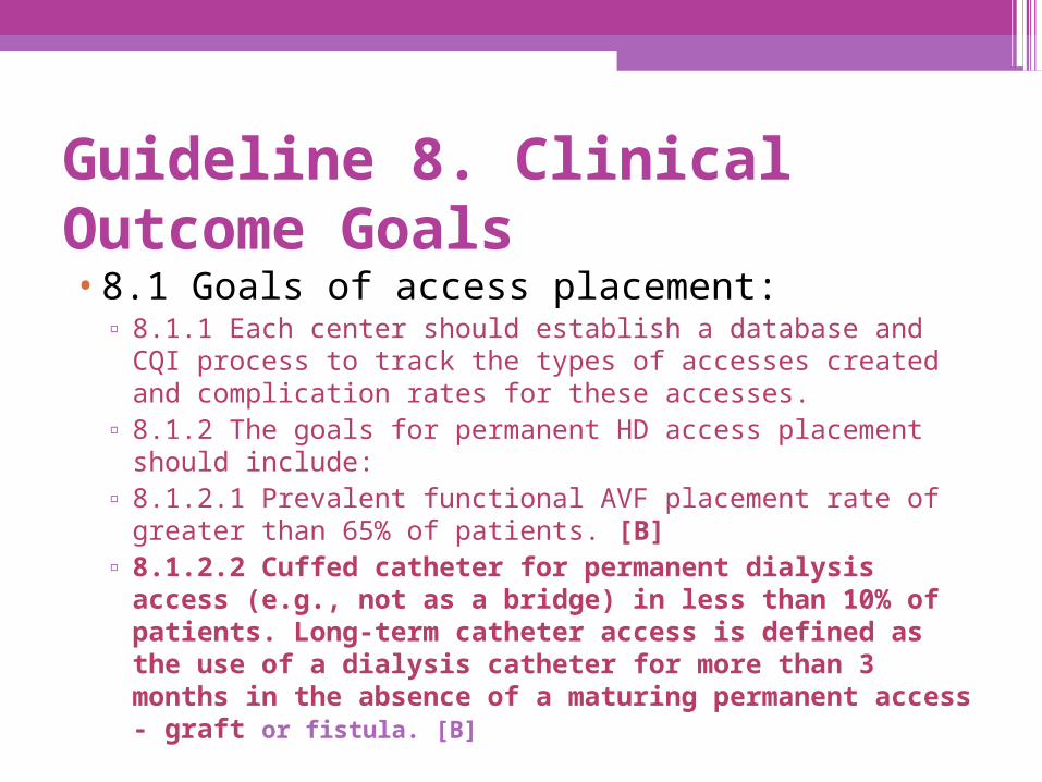
Guideline 8. Clinical Outcome Goals •8.1 Goals of access placement:
▫ 8.1.1 Each center should establish a database and CQI process to track the types of accesses created and complication rates for these accesses.
▫ 8.1.2 The goals for permanent HD access placement should include:
▫ 8.1.2.1 Prevalent functional AVF placement rate of greater than 65% of patients. [B]
▫ 8.1.2.2 Cuffed catheter for permanent dialysis access (e.g., not as a bridge) in less than 10% of patients. Long-term catheter access is defined as the use of a dialysis catheter for more than 3 months in the absence of a maturing permanent access - graft or fistula. [B]

Primary access failure rates of HD accesses•8.2 The primary access failure rates of
HD accesses in the following locations and configurations should not be more than the following:
•8.2.1 Forearm straight grafts: 15%. [B] •8.2.2 Forearm loop grafts: 10%. [B] •8.2.3 Upper-arm grafts: 5%. [B] •8.2.4 Tunneled catheters with blood
flow less than 300 mL/min: 5%. [B]
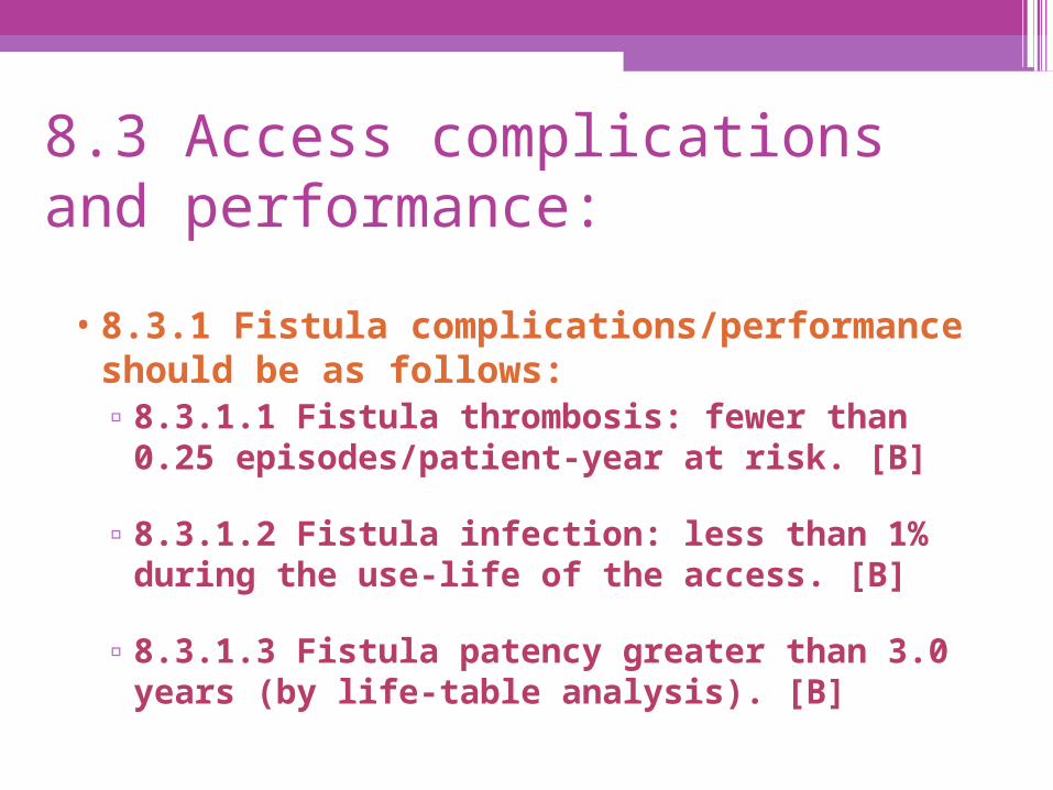
8.3 Access complications and performance:
• 8.3.1 Fistula complications/performance should be as follows: ▫8.3.1.1 Fistula thrombosis: fewer than 0.25
episodes/patient-year at risk. [B]
▫8.3.1.2 Fistula infection: less than 1% during the use-life of the access. [B]
▫8.3.1.3 Fistula patency greater than 3.0 years (by life-table analysis). [B]

Graft complications/ performance
•8.3.2 Graft complications/ performance should be as follows: ▫8.3.2.1 Graft thrombosis: fewer than 0.5
thrombotic episodes/patient-year at risk. [B]
▫ 8.3.2.2 Graft infection: less than 10% during the use-life of the access. [B]
▫8.3.2.3 Graft patency greater than 2 years (by life-table analysis). [B]
▫8.3.2.4 Graft patency after PTA: longer than 4 months. [B]

Catheter complications/performance
• 8.3.3 Catheter complications/performance should be as follows:
▫ 8.3.3.1 Tunneled catheter-related infection less than 10% at 3 months and less than 50% at 1 year. [B]
▫ 8.3.3.2 The cumulative incidence of the following insertion complications should not exceed 1% of all catheter placements: [B]
Pneumothorax requiring a chest tube Symptomatic air embolism Hemothorax Hemomediastinum Hematoma requiring evacuation
▫ 8.3.4 Cumulative patency rate of tunneled cuffed catheters (TCCs): Not specified. [B]

8.4 Efficacy of corrective intervention: • The rate of certain milestones after correction of
thrombosis or stenosis should be as follows: ▫ 8.4.1 AVF patency after PTA: greater than 50%
unassisted patency at 6 months (and <30% residual stenosis postprocedure or lack of resolution of physical findings postprocedure); AVF patency following surgery: greater than 50% unassisted patency at 1 year. [B]
▫ 8.4.2 AVG patency after PTA: please refer to CPG 6.5.1▫ AVG patency after surgery: please refer to CPG 6.5.2 ▫ AVG after either PTA or surgery: greater than 90% with
postprocedure restoration of blood flow and greater than 85% postprocedure ability to complete 1 dialysis treatment. Please refer to CPG 6.8. [B]
▫ 8.4.3 Surgical correction is set to a higher standard because of the use of venous capital. [B]
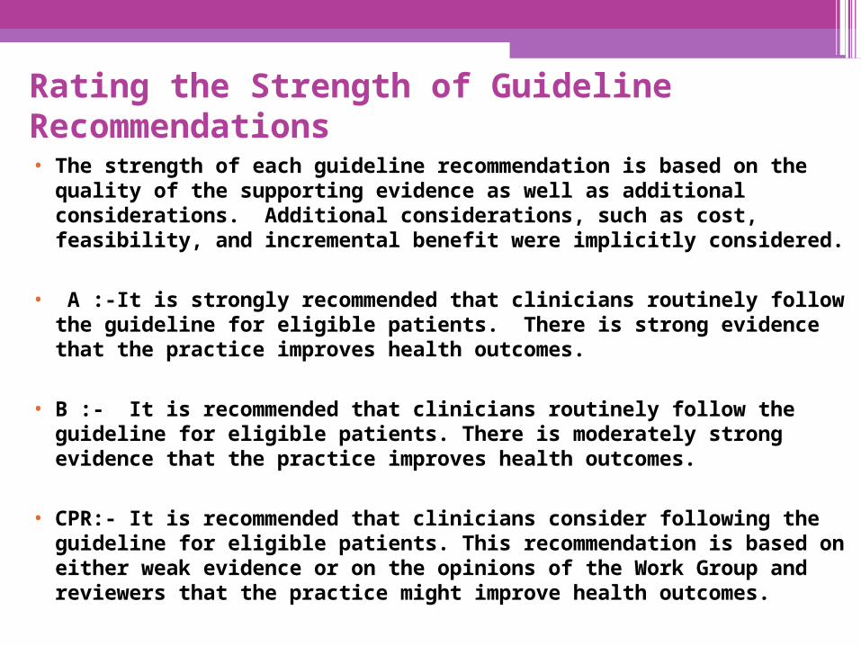
Rating the Strength of Guideline Recommendations • The strength of each guideline recommendation is based on the
quality of the supporting evidence as well as additional considerations. Additional considerations, such as cost, feasibility, and incremental benefit were implicitly considered.
• A :-It is strongly recommended that clinicians routinely follow the guideline for eligible patients. There is strong evidence that the practice improves health outcomes.
• B :- It is recommended that clinicians routinely follow the guideline for eligible patients. There is moderately strong evidence that the practice improves health outcomes.
• CPR:- It is recommended that clinicians consider following the guideline for eligible patients. This recommendation is based on either weak evidence or on the opinions of the Work Group and reviewers that the practice might improve health outcomes.