vascular mild cognitive impairment tuscany study t he term vascular cognitive impairment refers to...
TRANSCRIPT

1
The term vascular cognitive impairment refers to cognitive impairment, of any degree, associated with cerebrovascu-
lar diseases, among which small vessel disease (SVD) is the most frequent cause.1,2
Montreal Cognitive Assessment (MoCA) has been proposed as a screening tool in vascular cognitive impairment because, differently from the widely used mini mental state examina-tion (MMSE), it includes attentional, psychomotor speed, and executive tasks.3–6
Diffusion tensor imaging (DTI) is an MRI technique able to detect changes in white matter microstructure that are not evi-denced on conventional MRI, but may have a clinical effect.7
We assessed whether white matter microstructural damage as measured with DTI in patients with mild cognitive impair-ment (MCI) and SVD is more strongly reflected by MoCA than MMSE performances. If this holds true, it would support the hypothesis that MoCA is more suited than MMSE as a cogni-tive screening tool to assess patients with MCI related to SVD.
MethodsThe Vascular Mild Cognitive Impairment Tuscany (VMCI-Tuscany) study is a multicenter, prospective, observational study designed to estimate the effect of a large set of clinical, neuroimaging, and bi-ological markers of SVD in predicting the transition from MCI to dementia.8 To be included, patients had (1) MCI (Winblad criteria)9
Background and Purpose—Montreal Cognitive Assessment (MoCA) has been proposed as a screening tool in vascular cognitive impairment. Diffusion tensor imaging is sensitive to white matter microstructural damage. We investigated if diffusion tensor imaging-derived indices are more strongly associated with performances on MoCA or on the widely used mini mental state examination in patients with mild cognitive impairment and small vessel disease.
Methods—Mild cognitive impairment patients with moderate/severe degrees of white matter hyperintensities on MRI were enrolled. Lacunar infarcts, cortical atrophy, medial temporal lobe atrophy and median values of mean diffusivity and fractional anisotropy of the cerebral white matter were studied and correlated with cognitive tests performances.
Results—Seventy-six patients (mean age 75.1±6.8 years, mean years of education 8.0±4.3) were assessed. In univariate analyses, a significant association of both MoCA and mini mental state examination scores with age, education, cortical atrophy, and medial temporal lobe atrophy was found, whereas mean diffusivity and fractional anisotropy were associated with MoCA. In partial correlation analyses, adjusting for all demographic and neuroimaging variables, both mean diffusivity and fractional anisotropy were associated only with MoCA (mean diffusivity: r= −0.275, P=0.023; fractional anisotropy: r=0.246, P=0.043).
Conclusions—In patients with mild cognitive impairment and small vessel disease, diffusion tensor imaging-measured white matter microstructural damage is more related to MoCA than mini mental state examination performances. MoCA is suited for the cognitive screening of patients with small vessel disease. (Stroke. 2015;46:00-00.)
Key Words: cerebral small vessel disease ◼ diffusion tensor imaging
White Matter Microstructural Damage in Small Vessel Disease Is Associated With Montreal Cognitive Assessment
But Not With Mini Mental State Examination PerformancesVascular Mild Cognitive Impairment Tuscany Study
Marco Pasi, MD; Emilia Salvadori, PhD; Anna Poggesi, MD, PhD; Laura Ciolli, MD, PhD; Alessandra Del Bene, MD, PhD; Sandro Marini, MD; Serena Nannucci, MD;
Francesca Pescini, MD, PhD; Raffaella Valenti, MD; Andrea Ginestroni, MD, PhD; Nicola Toschi, PhD; Stefano Diciotti, PhD; Mario Mascalchi, MD, PhD;
Domenico Inzitari, MD; Leonardo Pantoni, MD, PhD; for the VMCI Study Investigators
Received September 24, 2014; final revision received October 20, 2014; accepted October 21, 2014.From the Department of NEUROFARBA, Neuroscience Section, University of Florence, Italy (M.P., E.S., A.P., L.C., A.D.B., S.M., S.N., F.P., R.V.,
D.I.); “Mario Serio” Department of Experimental and Clinical Biomedical Sciences, University of Florence, Italy (A.G., M.M.); Medical Physics Section, Department of Biomedicine and Prevention, University of Rome “Tor Vergata”, Italy (N.T.); Department of Electrical, Electronic, and Information Engineering “Guglielmo Marconi”, University of Bologna, Cesena, Italy (S.D.); and Stroke Unit and Neurology, Azienda Ospedaliero Universitaria Careggi, Florence, Italy (L.P.).
The online-only Data Supplement is available with this article at http://stroke.ahajournals.org/lookup/suppl/doi:10.1161/STROKEAHA.114. 007553/-/DC1.
Correspondence to Leonardo Pantoni, MD, PhD, Stroke Unit and Neurology, Azienda Ospedaliero Universitaria Careggi, Largo Brambilla 3, 50134 Firenze, Italy. E-mail [email protected]
© 2014 American Heart Association, Inc.
Stroke is available at http://stroke.ahajournals.org DOI: 10.1161/STROKEAHA.114.007553
Brief Report
by guest on June 9, 2017http://stroke.ahajournals.org/
Dow
nloaded from
by guest on June 9, 2017http://stroke.ahajournals.org/
Dow
nloaded from
by guest on June 9, 2017http://stroke.ahajournals.org/
Dow
nloaded from
by guest on June 9, 2017http://stroke.ahajournals.org/
Dow
nloaded from
by guest on June 9, 2017http://stroke.ahajournals.org/
Dow
nloaded from
by guest on June 9, 2017http://stroke.ahajournals.org/
Dow
nloaded from
by guest on June 9, 2017http://stroke.ahajournals.org/
Dow
nloaded from
by guest on June 9, 2017http://stroke.ahajournals.org/
Dow
nloaded from
by guest on June 9, 2017http://stroke.ahajournals.org/
Dow
nloaded from

2 Stroke January 2015
and (2) moderate to severe degrees of white matter hyperintensities (WMH) on MRI, (modified Fazekas scale).10 The local ethics com-mittee approved the study and informed consent was obtained from all participants.
At baseline, demographic variables (age, education, and sex) were collected, and both MoCA and MMSE were administered. For cut-off values and correction of age and education effect, we used norms validated in the Italian population.5,6 Conventional MRI features in-cluded lacunar infarcts, WMH, global cortical atrophy, and medial temporal lobe atrophy. Median values of mean diffusivity (MD) and fractional anisotropy of the cerebral white matter were used as DTI-derived indices (Figure). Other details of study methodology, clinical and MRI protocol are presented in the online-only Data Supplement.
Statistical analysis included adjusted partial correlation analysis between MoCA, MMSE, and DTI-derived indices (see online-only Data Supplement).
ResultsAt baseline, 76 patients had both clinical and DTI assessment (Table 1; see online-only Data Supplement).
Univariate analyses showed a significant association of both MoCA and MMSE with age, education, cortical global atrophy, and medial temporal lobe atrophy, whereas no associ-ation emerged with WMH and lacunar infarcts. MD and frac-tional anisotropy only correlated with MoCA score (Table 2).
In partial correlation analysis between MoCA, MMSE, and DTI-derived indices, adjusted for demographics and con-ventional MRI variables, only MoCA proved significantly associated with MD (r=−0.275, P=0.023) and fractional anisotropy (r=0.246, P=0.043). No significant correlation was observed between MMSE- and DTI-derived indices (MD: r=−0.107, P=0.385; fractional anisotropy: r=0.219, P=0.073).
Concerning MoCA subtests, correlation analysis showed a significant association between MD and visuoexecutive (ρ=−0.372, P=0.001) and attentional (r
bp=−0.259, P=0.026)
tasks (Table in the online-only Data Supplement). DiscussionIn our sample of patients with MCI and SVD, white matter microstructural damage, as evaluated by DTI-derived indi-ces, was related to MoCA but not to MMSE performances, supporting the hypothesis that MoCA is more sensitive to the presence of subtle SVD.
One limitation of the study is that our cohort might not be purely vascular as imaging markers of neurodegeneration, are present. This reflects the frequent coexistence of vascular and degenerative mechanisms in the aging brain.
We found no statistically significant difference between patients with moderate and severe WMH in terms of MMSE or MoCA scores. This maybe due to a loss of accuracy in discriminating among patients subgroup using cognitive screening tests once a certain degree of SVD is reached. However, patients with severe WMH had on average a 2-point lower score on MoCA in comparison with those with moderate WMH.
Consistent with our hypothesis and results, other studies showed that DTI-derived indices correlated with executive dysfunction in patients with SVD,7 whereas conflicting results derive from previous studies comparing MoCA and MMSE in patients with SVD.11
Our data confirm the hypothesis that microstructural damage related to SVD is more expressed by MoCA than
Figure. Automatic white matter segmentation mask in 1 patient with Fazekas grade 2 white matter hyperintensities on fluid-atten-uated inversion recovery.
Table 1. Demographic, Clinical, and MRI Characteristics
n=76Mean±SD or
Percentage (%)
Age (y) 75.1±6.8
Education (y) 8.0±4.3
Sex
Female 44.7
Male 55.3
MMSE score 26.1±3.3
MMSE performance (impaired) 14.6
MoCA score 18.9±5.7
MoCA performance (impaired) 26.7
Lacunar infarcts
n=0 28.9
n=1–3 32.9
n>3 38.2
Fazekas
Grade 2 48.7
Grade 3 51.3
Global atrophy
Grade 1 18.4
Grade 2 67.1
Grade 3 14.5
MTA 3.1±0.9
Median MD cerebral WM 0.82×10⁻³±0.37×10⁻⁴
Median FA cerebral WM 0.4±0.2
FA indicates fractional anisotropy; MD, mean diffusivity; MMSE, mini mental state examination; MoCA, Montreal cognitive assessment; MTA, medial temporal lobe atrophy; and WM, white matter.
by guest on June 9, 2017http://stroke.ahajournals.org/
Dow
nloaded from

Pasi et al MoCA and WM Microstructural Damage 3
MMSE performances and that MoCA is a suited screening tool for patients with SVD. This is probably because of the psychometrical structure of MoCA, in particular the pres-ence of items reflecting executive functions and psychomo-tor speed.
AcknowledgmentsVascular Mild Cognitive Impairment Tuscany (VMCI-Tuscany) study participants are reported in supplemental material.
Source of FundingVascular Mild Cognitive Impairment Tuscany (VMCI-Tuscany) study is funded by Tuscany region. Dr Salvadori is currently supported by a project funded by Tuscany region and Health Ministry (Grant num-ber: RF-2010-2321706, Principal Investigator: Dr Pantoni).
DisclosuresNone.
References 1. O’Brien JT, Erkinjuntti T, Reisberg B, Roman G, Sawada T, Pantoni L,
et al. Vascular cognitive impairment. Lancet Neurol. 2003;2:89–98. 2. Pantoni L. Cerebral small vessel disease: from pathogenesis and clinical
characteristics to therapeutic challenges. Lancet Neurol. 2010;9:689–701. 3. Hachinski V, Iadecola C, Petersen RC, Breteler MM, Nyenhuis DL,
Black SE, et al. National Institute of Neurological Disorders and Stroke-Canadian Stroke Network vascular cognitive impairment harmonization standards. Stroke. 2006;37:2220–2241.
4. Conti S, Bonazzi S, Laiacona M, Masina M, Coralli MV. Montreal Cognitive Assessment (MoCA)-Italian version: regression based norms and equivalent scores. [published online ahead of print August 20, 2014]. Neurol Sci. doi: 10.1007/s10072-014-1921-3. http://link.springer.com/article/10.1007%2Fs10072-014-1921-3. Accessed October 7, 2014.
5. Measso G, Cavarzeran F, Zappala G, Lebowittz BD, Crook TH, Pirazzolo FJ, et al. The mini-mental state examination: normative study of an Italian random sample. Dev Neuropsychol. 1993;9:77–85.
6. Pendlebury ST, Cuthbertson FC, Welch SJ, Mehta Z, Rothwell PM. Underestimation of cognitive impairment by mini-mental state examination versus the Montreal Cognitive Assessment in patients with transient ischemic attack and stroke: a population-based study. Stroke. 2010;41:1290–1293.
7. O’Sullivan M, Morris RG, Huckstep B, Jones DK, Williams SC, Markus HS. Diffusion tensor MRI correlates with executive dysfunction in patients with ischaemic leukoaraiosis. J Neurol Neurosurg Psychiatry. 2004;75:441–447.
8. Poggesi A, Salvadori E, Pantoni L, Pracucci G, Cesari F, Chiti A, et al. Risk and determinants of dementia in patients with mild cognitive impairment and brain subcortical vascular changes: a study of clinical, neuroimaging, and biological markers-The VMCI-Tuscany Study: ratio-nale, design, and methodology. Int J Alzheimers Dis. 2012;2012:608013.
9. Winblad B, Palmer K, Kivipelto M, Jelic V, Fratiglioni L, Wahlund LO, et al. Mild cognitive impairment–beyond controversies, towards a con-sensus: report of the International Working Group on Mild Cognitive Impairment. J Intern Med. 2004;256:240–246.
10. Pantoni L, Basile AM, Pracucci G, Asplund K, Bogousslavsky J, Chabriat H, et al. Impact of age-related cerebral white matter changes on the transition to disability – the LADIS study: rationale, design and methodology. Neuroepidemiology. 2005;24:51–62.
11. Xu Q, Cao WW, Mi JH, Yu L, Lin Y, Li YS. Brief screening for mild cog-nitive impairment in subcortical ischemic vascular disease: a comparison study of the Montreal Cognitive Assessment with the Mini-Mental State Examination. Eur Neurol. 2014;71:106–114.
Table 2. Univariate Analysis: Association Between Demographic, Neuroimaging Variables, and MMSE and MoCA Score
MMSE P Values MoCA P Values
Age −0.246* 0.032 −0.435* <0.001
Education (y) 0.428* <0.001 0.522* <0.001
Sex
Female 25.3±3.5 0.041† 17.6±6.0 NS†
Male 26.8±2.9 19.9±5.4
Lacunar infarcts
n=0 25.2±4.0 NS§ 19.5±5.6 NS§
n=1–3 26.6±3.3 18.0±6.0
n>3 26.4±2.5 19.2±5.7
Fazekas
Grade 2 26.5±3.1 NS† 20.1±5.6 NS†
Grade 3 25.8±3.4 17.8±5.7
Global atrophy
Grade 1 28.3±1.2 0.004§ 23.5±2.8 0.006§
Grade 2 26.0±3.1 18.0±5.7
Grade 3 24.1±4.5 17.6±6.4
MTA −0.284* 0.013 −0.293* 0.011
Median MD cerebral WM −0.210* NS −0.415* <0.001
Median FA cerebral WM 0.176* NS 0.251* 0.030
FA indicates fractional anisotropy; MD, median diffusivity; MMSE, mini mental state examination; MoCA, Montreal cognitive assessment; MTA, medial temporal lobe atrophy; NS, not significant; and WM, white matter.
*Pearson r.†Student t test.§ANOVA.
by guest on June 9, 2017http://stroke.ahajournals.org/
Dow
nloaded from

Stefano Diciotti, Mario Mascalchi, Domenico Inzitari and Leonardo PantoniSerena Nannucci, Francesca Pescini, Raffaella Valenti, Andrea Ginestroni, Nicola Toschi,
Marco Pasi, Emilia Salvadori, Anna Poggesi, Laura Ciolli, Alessandra Del Bene, Sandro Marini,Performances: Vascular Mild Cognitive Impairment Tuscany Study
Montreal Cognitive Assessment But Not With Mini Mental State Examination White Matter Microstructural Damage in Small Vessel Disease Is Associated With
Print ISSN: 0039-2499. Online ISSN: 1524-4628 Copyright © 2014 American Heart Association, Inc. All rights reserved.
is published by the American Heart Association, 7272 Greenville Avenue, Dallas, TX 75231Stroke published online November 13, 2014;Stroke.
http://stroke.ahajournals.org/content/early/2014/11/13/STROKEAHA.114.007553World Wide Web at:
The online version of this article, along with updated information and services, is located on the
http://stroke.ahajournals.org/content/suppl/2016/04/06/STROKEAHA.114.007553.DC2 http://stroke.ahajournals.org/content/suppl/2014/11/13/STROKEAHA.114.007553.DC1
Data Supplement (unedited) at:
http://stroke.ahajournals.org//subscriptions/
is online at: Stroke Information about subscribing to Subscriptions:
http://www.lww.com/reprints Information about reprints can be found online at: Reprints:
document. Permissions and Rights Question and Answer process is available in the
Request Permissions in the middle column of the Web page under Services. Further information about thisOnce the online version of the published article for which permission is being requested is located, click
can be obtained via RightsLink, a service of the Copyright Clearance Center, not the Editorial Office.Strokein Requests for permissions to reproduce figures, tables, or portions of articles originally publishedPermissions:
by guest on June 9, 2017http://stroke.ahajournals.org/
Dow
nloaded from

Supplemental material
White matter microstructural damage in SVD is associated with MoCA but not with MMSE
performances: VMCI-Tuscany Study
Methodology and study protocol of the VMCI-Tuscany Study The Vascular Mild Cognitive Impairment (VMCI) Tuscany Study is a multicenter, prospective, observational study, carried out in the Tuscany region of Italy, and aimed at estimating the role of a large set of clinical, cognitive, neuroimaging, and biological markers of SVD as independent predictors of the transition from MCI to dementia. According to the study protocol, at baseline, each enrolled patient undergoes an extensive clinical, functional and neuropsychological assessment, an MRI examination, and the collection of blood samples. MRI assessment MRI protocol Patients were examined on a 1.5 T system (Intera, Philips Medical System, Best, The Netherlands) with 33 mT/m gradients capability and a head coil with SENSE technology. The examination protocol was previously described and included a sagittal T1 sequence, an axial FLAIR sequence, and an axial single-shot echo planar imaging sequence for DTI (diffusion sensitizing gradients applied along 15 non-collinear directions using b value of 0 (b0 image) and 1000 s/mm2) [1]. Conventional MRI features For the purpose of the present investigation the following features were evaluated: a) lacunar infarcts visible on MRI were defined as hypointense lesions on T1 imaging with corresponding hyperintense lesion on FLAIR images with a diameter <20 mm; lacunar infarcts were classified as absent, 1-3, and >3; b) deep WMH on FLAIR graded according to the modified Fazekas scale [2]. A score of 2 (moderate) was attributed to beginning confluent lesions and a score of 3 (severe) to large confluent lesions [2]; d) global cortical atrophy using the scale of Pasquier et al. that assesses sulci opening of sulci and narrowing of gyri [3]. Scores 0–3 represent absent, mild, moderate, and severe cortical atrophy, respectively; e) medial temporal lobe atrophy (MTA) assessed by means of the Scheltens' scale [4]. This scale assesses coronal T1-weighted images acquired parallel to the brainstem. Scores 0–4 indicate progressive medial temporal volume loss. The above MRI features were visually evaluated in all patients by one experienced neurologist (AP). Patients with incidental non-lacunar infarcts in the cerebral cortex, cerebellum, or brainstem were excluded to avoid a possible confounding effect on DTI analysis and also to have a more homogenous patients sample. DTI and analysis of microstructural damage of cerebral WM Diffusion-weighted images were corrected for head motion and eddy current distortions using FDT (FMRIB’s Diffusion Toolbox 2.0), part of FSL 5.0.2 [5] after which brain tissue was segmented using BET, also part of FSL [6]. The b-matrix was reoriented by applying the rotational part of the affine transformation employed in the eddy-correction step [7]. A tensor model was fitted to the raw data using a constrained nonlinear least squares procedure implemented in the software package CAMINO, and residual non-positive definite tensors (in isolated regions where the nonlinear algorithm failed to converge, mainly located at the edge of the brain) were removed by tensor interpolation in the log-euclidean domain [8]. Mean diffusivity (MD) and fractional anisotropy (FA) maps were then computed from the estimated tensor field. The segmentation method employed to obtain cerebral WM masks was previously detailed [9]. Briefly, WM segmentation on T1-weighted images was carried out using FAST 4, part of FSL [10]. To reduce partial volume effects, a preliminary WM mask was obtained by retaining only those

voxels which had a tissue class probability equal to or above 0.75. In order to select identical cerebral regions across subjects, a standard space (MNI152 average normal brain) WM mask was mapped onto each subject's native space and multiplied by each subject's WM mask (Figure 1). Resulting WM masks were successively mapped onto native diffusion space by applying intra-subject affine transformations (12 degrees of freedom) between the T1 and b0 images [11] in order to compute WM-wide MD and FA statistics for each subject. The inter-subject agreement of our WM segmentation was assessed by using inverse transformations to map single subject WM masks to standard space, averaging and calculating descriptive statistics of the resultant agreement image. The inter-subject agreement was substantial (median 87%, mode: 97%, s.d. 19%). While several metrics can be used to evaluate the MD and FA properties of brain tissue [12], in the present study we employed median values of MD and FA to characterize the microstructural properties of cerebral WM. Statistical analysis Descriptive analyses were used to briefly characterize the baseline sample in terms of demographic, clinical and neuroimaging features. Bivariate statistical analyses (independent samples t test, ANOVA, Pearson’s r) were used to exclude statistical significant differences between the group composed by the excluded patients for DTI data unavailability and presence of non-lacunar infarcts and the final sample (data not shown). The considered variables were age, education, gender, MoCA, MMSE, lacunar infarcts, WMH, global cortical atrophy, MTA, and median value of MD and FA of the cerebral WM. Bivariate statistical analyses (independent samples t test, ANOVA, Pearson’s r) were used to evaluate the association of demographic and neuroimaging variables, with MoCA and MMSE total score. The considered variables were: demographic characteristics (age, education level, and gender), conventional MRI features (lacunar infarcts, WM hyperintensities, global cortical atrophy, MTA), DTI-derived features (median value of MD and FA of the cerebral WM), and MoCA and MMSE total score. To evaluate which test was more strongly associated with DTI-derived indices we performed a model of partial correlation analysis adjusting for demographic variables (age, education level, and gender) and conventional MRI features (lacunar infarcts, WMH, global cortical atrophy, MTA). In order to evaluate if psychometrical structure of MoCA influenced the possible association with DTI parameters, correlation analysis (Spearman’s Rho and biserial point rbp) were performed for each single subtest (visuoexecutive, naming, digit span, attention, calculation, language, verbal fluency, abstraction, recall, and orientation). All data analyses were performed using SPSS 20.
Definition of the final sample included in this study From December 2011 to March 2013, 104 patients were enrolled in the VMCI-Tuscany Study in the Florence center, where the MRI protocol included also DTI evaluation. For the purposes of the present study, 7 patients were excluded because DTI imaging was not available for technical reasons, 21 patients were excluded for the presence of non-lacunar infarcts in the cerebral cortex, cerebellum or brainstem. No statistically significant differences between excluded and final sample patients were found in terms of age, education, gender, MoCA, MMSE, and MRI features.
Supplemental references: 1) Mascalchi M, Ginestroni A, Toschi N, Poggesi A, Cecchi P, Salvadori E, et al. VMCI Tuscany
Investigators. The burden of microstructural damage modulates cortical activation in elderly subjects with MCI and leuko-araiosis. A DTI and fMRI study. Hum Brain Mapp. 2014;35:819-830.
2) Pantoni L, Basile AM, Pracucci G, Asplund K, Bogousslavsky J, Chabriat H, et al. Impact of age-related cerebral white matter changes on the transition to disability -- the LADIS study: rationale, design and methodology. Neuroepidemiology. 2005;24:51-62.
3) Pasquier F, Leys D, Weerts JG, Mounier-Vehier F, Barkhof F, Scheltens P. Inter- and intraobserver reproducibility of cerebral atrophy assessment on MRI scans with hemispheric infarcts. Eur Neurol. 1996;36:268-272
4) Scheltens P, Pasquier F, Weerts JG, Barkhof F, Leys D. Qualitative assessment of cerebral atrophy on MRI: inter- and intra-observer reproducibility in dementia and normal aging. Eur Neurol. 1997;37:95-99.
5) Smith S, Jenkinson M, Woolrich MW, Beckmann CF, Behrens TE, Johansen-Berg H, et al. Advances in functional and structural MR image analysis and implementation as FSL. NeuroImage. 2004;23:208-219.
6) Smith S. Fast robust automated brain extraction. Hum Brain Mapp. 2002;17:143-155. 7) Leemans A, Jones DK. The B-matrix must be rotated when correcting for subject motion in DTI
data. Magn Reson Med. 2009;61:1336–1349. 8) Arsigny V, Fillard P, Pennec X, Ayache N. Log-euclidean metrics for fast and simple calculus
on diffusion tensors. Magn Reson Med. 2006;56:411–421. 9) De Stefano N, Battaglini M, Stromillo ML, Zipoli V, Bartolozzi ML, Guidi L, et al. Brain
damage as detected by magnetization transfer imaging is less pronounced in benign than in early relapsing multiple sclerosis. Brain. 2006;129:2008-2016.
10) Zhang Y, Brady M, Smith S. Segmentation of brain MR images through a hidden Markov random field model and the expectation maximization algorithm. IEEE Trans Med Imaging 2001;20:45–57.
11) Jenkinson M, Smith S. A global optimisation method for robust affine registration of brain images. Med Image Anal 2001;5:143–156
12) Della Nave R, Foresti S, Pratesi A, Ginestroni A, Inzitari M, Salvadori E, et al. Whole-brain histogram and voxel-based analyses of diffusion tensor imaging in patients with leukoaraiosis: correlation with motor and cognitive impairment. Am J Neuroradiol AJNR. 2007;28:1313-1319.

Supplemental table I. Association between each MoCA subtest and DTI parameters
MoCA subtest Median MD cerebral WM
Median FA cerebral WM
Visuoexecutive -0.372*, p=0.001 0.279*, p=0.015
Naming -0.148*, p=0.206 0.026*, p=0.825
Digit Span -0.255*, p=0.028 0.185*, p=0.114
Attention -0.259§, p=0.026 0.147§, p=0.212
Calculation -0.077*, p=0.514 0.061*, p=0.608
Language -0.141*, p=0.232 0.156*, p=0.185
Verbal fluency -0.095§, p=0.421 0.030§, p=0.797
Abstraction -0.239*, p=0.040 0.139*, p=0.237
Recall -0.241*, p=0.039 0.057*, p=0.628
Orientation -0.138*, p=0.238 0.001*, p=0.997
MD: mean diffusivity FA: fractional anisotropy * Spearman rho § biserial point rbp
Appendix. List of participating centers and personnel in the VMCI-Tuscany. University of Florence: (Coordinating Center): Domenico Inzitari (Study coordinator), Rosanna Abbate, Maria Boddi, Francesca Cesari, Laura Ciolli, Mirella Coppo, Alessandra Del Bene, Stefano Diciotti, Andrea Ginestroni, Betti Giusti, Anna Maria Gori, Sandro Marini, Mario Mascalchi, Serena Nannucci, Leonardo Pantoni, Marco Pasi, Francesca Pescini, Anna Poggesi, Giovanni Pracucci, Emilia Salvadori, Raffaella Valenti. University of Pisa: Ubaldo Bonuccelli, Paolo Cecchi, Alberto Chiti, Mirco Cosottini, Giovanni Orlandi, Cristina Pagni, Gabriele Siciliano, Gloria Tognoni. University of Siena: Antonio Federico, Nicola De Stefano, Ilaria Di Donato, Maria Teresa Dotti, Patrizia Formichi, Claudia Gambetti, Antonio Giorgio, Francesca Rossi, Laura Stromillo, Enza Zicari. Tuscany Region: Arezzo (Paolo Zolo, Alessandro Tiezzi); Empoli (Elisabetta Bertini, Stefania Brotini, Leonello Guidi, Maria Lombardi, Stefania Mugnai, Antonella Notarelli); Florence (Laura Bracco, Massimo Cadelo, Renzo Cisbani, Luciano Gabbani, Guido Gori, Lorella Lambertucci, Luca Massacesi, Enrico Mossello, Marco Paganini, Maristella Piccininni, Francesco Pinto, Claudia Pozzi, Sandro Sorbi, Gaetano Zaccara); Grosseto (Tiziano Borgogni, Mario Mancuso, Roberto Marconi); Lucca (Monica Mazzoni, Marco Vista); Livorno (Giuseppe Meucci, Giovanna Bellini); Massa Carrara (Luciano Gabrielli); Pisa (Cristina Frittelli, Renato Galli, Gianna Gambaccini); Pistoia (Stefano Bartolini, Carlo Biagini, Veronica Caleri, Paola Vanni); Prato (Donatella Calvani, Carla Giorgi, Stefano Magnolfi, Pasquale Palumbo, Carlo Valente); Siena (Alessandro Rossi, Rossana Tassi, Stefania Boschi); Viareggio (Filippo Baldacci).
iris
20 Stroke 日本語版 Vol. 10, No. 1
脳小血管病における白質の微細構造の障害はモントリオール認知評価検査と関連するが,ミニメンタルステート検査の成績とは関連しない
Vascular Mild Cognitive Impairment Tuscany 研究 White Matter Microstructural Damage in Small Vessel Disease Is Associated With Montreal
Cognitive Assessment But Not With Mini Mental State Examination Performances Vascular Mild Cognitive Impairment Tuscany Study
Marco Pasi, MD; Emilia Salvadori, PhD; Anna Poggesi, MD, PhD, et al. Department of NEUROFARBA, Neuroscience Section, University of Florence, Italy.
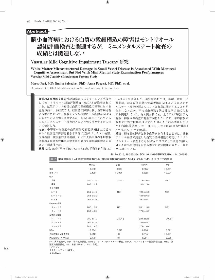
背景および目的: 血管性認知障害のスクリーニング手段としてモントリオール認知評価検査(MoCA)が推奨されている。拡散テンソル画像は白質の微細構造の障害に対する感度が高い。本研究では,軽度認知障害と脳小血管病を有する患者において,拡散テンソル画像による指標がMoCAのスコアとより強く関連するか,あるいは汎用されているミニメンタルステート検査のスコアと強く関連するかについて検討した。 方法: 中等度から重度の白質高信号病変がMRI 上で認められた軽度認知障害患者を本研究に登録した。ラクナ梗塞,皮質萎縮,側頭葉内側部萎縮,および大脳白質の平均拡散係数および異方性比率の中央値を調べて認知機能検査のスコアと関連付けた。 結果: 患者 76例(平均年齢:75.1 ± 6.8歳,平均教育年数:8.0
± 4.3 年)を評価した。単変量解析では,年齢,教育,皮質萎縮,および側頭葉内側部萎縮がMoCAとミニメンタルステート検査の両方のスコアと有意に関連することが明らかになったが,平均拡散係数と異方性比率はMoCAとのみ関連していた。偏相関分析でも,全ての人口統計学的変数と神経画像検査の変数で調整したところ,平均拡散係数および異方性比率はいずれもMoCAとのみ関連していた(平均拡散係数: r = - 0.275, p = 0.023;異方性比率: r = 0.246, p = 0.043)。 結論: 軽度認知障害と脳小血管病を有する患者では,拡散テンソル画像で測定した白質の微細構造の障害はミニメンタルステート検査よりもMoCAのスコアとの関連が強い。MoCAは小血管病を有する患者の認知機能のスクリーニングに適している。
Stroke 2015; 46:262-264. DOI: 10.1161/STROKEAHA. 114. 007553.
Abstract
表 2 単変量解析:人口統計学的変数および神経画像検査の変数とMMSEおよびMoCAスコアとの関連
MMSE p 値 MoCA p 値
年齢 - 0.246 * 0.032 - 0.435 * < 0.001
教育(年) 0.428 * < 0.001 0.522 * < 0.001
性別
女性 25.3 ± 3.5 0.041 † 17.6 ± 6.0 NS †
男性 26.8 ± 2.9 19.9 ± 5.4
ラクナ梗塞
n = 0 25.2 ± 4.0 NS § 19.5 ± 5.6 NS §
n = 1 ~ 3 26.6 ± 3.3 18.0 ± 6.0
n > 3 26.4 ± 2.5 19.2 ± 5.7
Fazekas 分類
グレード 2 26.5 ± 3.1 NS † 20.1 ± 5.6 NS †
グレード 3 25.8 ± 3.4 17.8 ± 5.7
全体的な萎縮
グレード 1 28.3 ± 1.2 0.004 § 23.5 ± 2.8 0.006 §
グレード 2 26.0 ± 3.1 18.0 ± 5.7
グレード 3 24.1 ± 4.5 17.6 ± 6.4
MTA - 0.284 * 0.013 - 0.293 * 0.011
大脳白質のMD中央値 - 0.210 * NS - 0.415 * < 0.001
大脳白質の FA中央値 0.176 * NS 0.251 * 0.030
FA:異方性比率,MD: 平均拡散係数,MMSE:ミニメンタルステート検査,MoCA:モントリオール認知評価検査,MTA:側頭葉内側部萎縮,NS:有意ではない,WM:白質。 * ピアソン r 。 † ステューデント t 検定 。 § ANOVA 。
Stroke-J_v10-i1_ab.indd 20Stroke-J_v10-i1_ab.indd 20 16-Jun-15 10:25:4116-Jun-15 10:25:41