vegf activates divergent intracellular signaling...
TRANSCRIPT
DEVELO
PMENT
2201RESEARCH ARTICLE
INTRODUCTIONThe vertebrate retina is an excellent model with which to studymolecular mechanisms governing development of multipotentneural progenitor cells in the central nervous system. Asdevelopment proceeds, retinal progenitor cells undergo cell-intrinsicchanges, which generally define the competence state for variouscell fate decisions (Cayouette et al., 2003; Harris, 1997; Hatakeyamaand Kageyama, 2004; Lillien and Wancio, 1998). Progenitor cellsare additionally influenced by extracellular signals in the localenvironment, which is continuously altered by accumulatingpostmitotic neurons. Existing evidence supports that both cell-intrinsic molecules and cell-extrinsic cues are important contributingfactors to retinal progenitor behaviors (Cepko et al., 1996; Liveseyand Cepko, 2001; Perron and Harris, 2000; Yang, 2004).
During vertebrate retinogenesis, the retinal ganglion cell (RGC)is the first type of neuron to emerge from the proliferative neuralepithelium (Prada et al., 1991; Young, 1985). Nascent postmitoticRGC neurons begin differentiating immediately at the ventricularsurface and the cell bodies of these RGCs translocate to the innerretina to form the ganglion cell layer (Waid and McLoon, 1995). Theaccumulation of postmitotic RGCs occurs in a center to peripherydirection throughout the retina to generate a neurogenic wave(McCabe et al., 1999). A cell-intrinsic factor, the basic helix-loop-
helix transcription factor Ath5, crucially dictates the competence ofearly retinal progenitors to give rise to RGCs (Brown et al., 2001;Kay et al., 2001; Mu and Klein, 2004; Wang et al., 2001; Yang et al.,2003). Among the cell-extrinsic cues that influence RGCdevelopment is the secreted molecule sonic hedgehog (SHH), whichis produced by differentiated RGCs and regulates two importantaspects of RGC genesis. First, SHH signals emanating frompostmitotic RGCs are necessary for the propagation of theneurogenic wave towards the peripheral retina (Masai et al., 2005;Neumann and Nuesslein-Volhard, 2000). Second, behind theneurogenic wave front, SHH signals derived from the accumulatingRGCs suppress the emergence of additional RGCs from thecompetent retinal progenitor pool (Wang et al., 2005; Zhang andYang, 2001a). In addition to diffusible signals, cell contact-mediatedDELTA-NOTCH signaling among progenitor cells cruciallycontrols RGC genesis. Either elevated DELTA signal or constitutiveNOTCH receptor activity decreases the number of RGCs during theearly neurogenic period (Ahmad et al., 1997; Austin et al., 1995;Dorsky et al., 1995; Dorsky et al., 1997).
The secreted protein vascular endothelial growth factor (VEGFA)plays crucial roles during development by signaling through tyrosinekinase receptors (Carmeliet et al., 1996; Millauer et al., 1993).Among these, the cognate high-affinity receptor FLK1(KDR/VEGFR2) mediates the effects of VEGF in vasculogenesis,angiogenesis and hematopoiesis (Shalaby et al., 1995; Shalaby et al.,1997). Accumulating evidence also suggests that VEGF and FLK1function in the nervous system (Carmeliet, 2003; Palmer et al., 2000;Weinstein, 2005; Yang and Cepko, 1996). The VEGF family ofligands have been shown to promote cell proliferation and neuronaldifferentiation in the cortex, enhance sensory and motoneuronsurvival, and control axonal guidance (Azzouz et al., 2004; Fabel etal., 2003; Jin et al., 2002; Le Bras et al., 2006; Rosenstein et al., 2003;
VEGF activates divergent intracellular signaling componentsto regulate retinal progenitor cell proliferation and neuronaldifferentiationTakao Hashimoto, Xiang-Mei Zhang, Brenden Yi-kuang Chen* and Xian-Jie Yang†
During vertebrate neurogenesis, multiple extracellular signals influence progenitor cell fate choices. The process by whichuncommitted progenitor cells interpret and integrate signals is not well understood. We demonstrate here that in the avascularchicken retina, vascular endothelial growth factor (VEGF) secreted by postmitotic neurons acts through the FLK1 receptor presenton progenitor cells to influence cell proliferation and commitment. Augmenting VEGF signals increases progenitor cell proliferationand decreases retinal ganglion cell genesis. Conversely, absorbing endogenous VEGF ligand or disrupting FLK1 activity attenuatescell proliferation and enhances retinal ganglion cell production. In addition, we provide evidence that VEGF signals transmitted bythe FLK1 receptor activate divergent intracellular signaling components, which regulate different responses of progenitor cells.VEGF-induced proliferation is influenced by the MEK-ERK pathway, as well as by the basic helix-loop-helix factor HES1. By contrast,VEGF-dependent ganglion cell suppression does not require MEK-ERK activation, but instead relies on VEGF-stimulated HES1activity, which is independent of NOTCH signaling. Moreover, elevated HES1 expression promotes progenitor cell proliferation andprevents overproduction of retinal ganglion cells owing to the loss of VEGF or sonic hedgehog (SHH), another signal that suppressesganglion cell development. Based on previous and current findings, we propose that HES1 serves as a convergent signaling nodewithin early retinal progenitor cells to integrate various cell-extrinsic cues, including VEGF and SHH, in order to control cellproliferation and neuronal specification.
KEY WORDS: VEGF, FLK1, Retina, Development, Proliferation, Retinal ganglion cells, HES, ERK, Sonic hedgehog (SHH), Chicken
Development 133, 2201-2210 (2006) doi:10.1242/dev.02385
Jules Stein Eye Institute and Department of Ophthalmology, Molecular BiologyInstitute, University of California, David Geffen School of Medicine, 100 Stein PlazaLos Angeles, CA 90095, USA.
*Present address: Integrated Program in Molecular, Cellular and Biophysical Studies,Institute for Cancer Genetics, Columbia University, College of Physicians andSurgeons, New York, NY 10032, USA†Author for correspondence (e-mail: [email protected])
Accepted 5 April 2006
DEVELO
PMENT
2202
Schwarz et al., 2004; Sondell et al., 1999; Sun et al., 2006).Nevertheless, owing to the proximity between the vasculature andneuronal cells, it remains a challenge to understand the precisefunction of VEGF and FLK1 in the nervous system, as VEGF signalsmay act directly upon neuronal cells or indirectly upon endothelialcells embedded within the neural tissue to influence neuronal cellpopulations (Louissaint, Jr et al., 2002; Shen et al., 2004).
We have shown previously that at the onset of retinaldifferentiation in mouse and chicken, expression of the FLK1receptor commences in progenitor cells residing in the central retinaand subsequently spreads to the peripheral retina, thus suggesting anevolutionarily conserved function of FLK1-mediated signals duringvertebrate retinogenesis (Hashimoto et al., 2003; Yang and Cepko,1996). In this study, we have taken advantage of the chicken retina,which is completely devoid of blood vessels throughoutdevelopment (De Schaepdrijver et al., 1989), to investigate thepotential role of FLK1 receptor-mediated VEGF signaling duringthe early stages of retinal neurogenesis. By perturbing VEGF signalsas well as FLK1 receptor function, we demonstrate that VEGFsignals mediated by the FLK1 receptor directly modulate thebehavior of uncommitted retinal progenitor cells. We also show thatdistinct intracellular signaling components mediate the effects ofVEGF on progenitor proliferation and neuronal differentiation.Moreover, we reveal a common downstream signal integrationmechanism for several extrinsic cues, including VEGF, SHH andNOTCH, all of which participate in the regulation of earlyprogenitor fate decisions.
MATERIALS AND METHODSChicken embryosWhite Leghorn chicken eggs were purchased from Charles River SPAFASand incubated at 38°C in a rotating humidified incubator. Embryos werestaged according to Hamburger and Hamilton (HH stage) (Hamburger andHamilton, 1951).
In situ hybridizationChicken FLK1 cDNA (GenBank AY382882) (Hashimoto et al., 2003) wasused as DNA template to generate digoxigenin-labeled RNA probes for insitu hybridization as described (Yang and Cepko, 1996).
Retinal culturesFor explant cultures, the central retina without the peripheral one-pupillarydiameter was incubated at 37°C in 5% CO2 in basal medium consisting of50% F12/50% DMEM (JRH), 10 mM HEPES, 50 U/ml penicillin and 50�g/ml streptomycin (Gibco). Explants were either cultured on polycarbonatefilter discs (Costar) floating on the medium, or on hydrophilicpolytetrafluoroethylene culture plate inserts (Millipore). Recombinanthuman VEGF165 (R&D Systems) was used at 100 ng/ml in the basal mediumcontaining 1� N2 supplement (Gibco), or 1% fetal calf serum and 0.2%chicken serum (Sigma); except for the dose effect analyses, which wereperformed in the basal medium containing 1� N2 supplement. To testeffects of sFLK1 or control alkaline phosphatase (AP), half of the culturemedium containing 1� N2 supplement was replaced with conditionedmedia. To test effects of inhibitors, 10 �M U0126 (Cell SignalingTechnology), 10 �M SU1498 (Calbiochem) or 200 nM cyclopamine(Toronto Research Chemicals) were used.
For collagen gel cultures, dissociated retinal cells were cast at a densityof 4000 cells/�l in 1.2 mg/ml rat type 1 collagen (BD Biosciences), 100 mMHEPES in basal medium containing 1� N2 supplement. Explant andcollagen gel dissociations were performed as described (Ezzeddine et al.,1997; Zhang and Yang, 2001a).
Expression vectors and cellsThe VEGF expression construct (pCMV-VEGF-IRES-GFP) contained theCMV promoter followed by the murine VEGF164 cDNA (Yang and Cepko,1996), the internal ribosomal entry site (IRES), and the enhanced green
fluorescent protein (GFP) sequence. The expression construct (pCMV-FLK1Ext-AP) encoding the FLK1 extracellular domain and AP fusionprotein (sFLK1) was constructed from a murine FLK1 cDNA clone(Matthews et al., 1991) and the APtag-2 vector (Cheng et al., 1995).Conditioned media containing sFLK1, VEGF or AP were collected between24 to 72 hours after Lipofectamine transfection of HEK293T cells withpCMV-FLK1Ext-AP, pCMV-VEGF-IRES-GFP, or APtag-4. For cellimplantation, 0.5 �l of transfected HEK 293T cells resuspended at 2�105
cells/ml in DMEM with 10 mM HEPES was injected intravitreally at HHstage 17 (Zhang and Yang, 2001a).
The mutant HES1 cDNA (pCAG-dnHES1) (Hirata et al., 2002) wascloned downstream of the chicken �-actin promoter with a CMV enhancer(CAG) (Niwa et al., 1991). The wild-type murine HES1 cDNA wasexpressed from the LTR promoter of the avian retrovirus RCAS (Hughes etal., 1987).
The FLK1 siRNA construct (phU6A-FLK1i) was created by PCR cloningof the human U6 promoter (Fitzgerald et al., 2001) and oligonucleotidescontaining 29 nucleotides specific to chicken FLK1 (nucleotides 185-213)as a palindrome. A target construct (pCMV-GFP-cFLK1) was made toexpress from the CMV promoter a chimeric mRNA of GFP and a partialchicken FLK1 fragment (nucleotides 55-232) after the GFP stop codon.
Viral stocks and injectionAn avian replication competent retroviral vector (RCAS-FLK1-DN-FLAG)encoding a mutant murine FLK1 with a deletion of the intracellular proteinkinase domain (Millauer et al., 1994) was constructed using pMFG-FLK1and the RCAS(A) viral vector (Hughes et al., 1987). The viral vectorexpressing siRNA for chicken FLK1 were constructed by inserting the U6-FLK1i cassette and a CMV-GFP expression cassette into the RCAS viralvector. An RCAS vector containing the CMV-GFP cassette and U6 promoterwithout the FLK1 sequence was used as control. Viral stocks were producedas described (Yang, 2002). Concentrated viral stocks were injected into theoptic vesicle at HH stage 10 or the subretinal space at HH stage 17 asdescribed (Zhang and Yang, 2001b).
ElectroporationRetinal explants were electroporated in 1 �g/�l of a cDNA expressionplasmid and 0.1 �g/�l of a plasmid with the CAG promoter driving GFP(pAS-CAG-G1) in PBS using an ECM 830 Square Wave ElectroporationSystem (BTX) at 10 V/mm for three 50 mseconds pulses with 950 msecondsintervals. Electroporated explants were further cultured as described.
RT-PCRReverse transcriptions were carried out using total RNAs from E6 retinalexplants treated with or without 100 ng/ml VEGF for 24 hours andSuperScript II Reverse Transcriptase (Invitrogen) (Hashimoto et al., 1997).Primers used were: for cyclin D1, XJY456 (5�-gccaagcaaacccattagaa-gaagtcctc) and XJY457 (5�-cctgctcgccctcggtgtc); and for glyceraldehyde-3-phosphate dehydrogenase (GAPDH), XJY361 (5�-ccatcaagtccacaacacggtt-gctgta) and XJY362 (5�-gtcttatgaccactgtccatgccatcac). PCRs were carriedout using AmpliTaq polymerase (Roche) at 2.5 mM final MgCl2 at 94°C for5 minutes, followed by 25 cycles of 94°C for 10 seconds, 60°C for 5 secondsand 72°C for 60 seconds.
ImmunoblottingWestern blots were performed using standard protocols. Cells or retinaltissues were lysed in 1% IGEPAL CA-630 (Sigma), 0.1% SDS, 150 mMNaCl, 2 mM EDTA, 50 mM Tris-Cl (pH 8.0). Blots were incubated withantibodies against AP (Zymed), VEGF (SantaCruz), FLAG (Sigma), GFP(Molecular Probe), �-tubulin (Sigma) or phospho-ERK1/2 (Cell Signaling),followed by secondary antibodies conjugated with horseradish peroxidase(HRP) and detected by enhanced chemiluminescence (ECL plus,Amersham).
ImmunostainingTo label proliferating cells, 100 �g of 5-bromo-2�-deoxyuridine (BrdU) wasapplied in ovo for 3 hours or retinal cultures were incubated with 25 �MBrdU for the last 3 or 6 hours of the culture period. Immunostaining ofcryosections was performed as described (Zhang and Yang, 2001a) using
RESEARCH ARTICLE Development 133 (11)
DEVELO
PMENT
antibodies against BrdU (Amersham), VEGF (SantaCruz), proliferating cellnuclear antigen (PCNA; Sigma), neurofilament 145 (NF 145; Chemicon),NF 200 (Sigma), GFP (Molecular Probes), Islet1/2 [clone 39.4D5;Developmental Studies Hybridoma Bank (DSHB)], AP-2� (clone 3B5;DSHB) or Brn3a (Chemicon). Sections were then incubated withbiotinylated secondary antibodies with HRP detection (Vector Laboratories)using 3,3�-diaminobenzidine (Sigma), or binding to Alexa 488-, Alexa 594-(Molecular Probes) or Texas Red- (Jackson ImmunoResearch Laboratories)conjugated antibodies. For nuclear staining, 1 �g/ml of 4�,6-diamidino-2-phenylindole (DAPI; Roche) was used.
Photomicrographs were captured using a Nikon E800 microscopeequipped with a SPOTII digital camera. Confocal images of 1 �m opticalsections were obtained using a Zeiss LSM 410 confocal laser-scanningsystem attached to a Zeiss Axiovert 135M microscope. Quantification ofdissociated retinal cells stained for various cell markers was performed usingImagePro PLUS software (Media Cybernetics).
Statistical analysisIn general, 300-1000 cells were counted per experimental condition that wasrepeated five to nine times (n�5). The quantified data are expressed asmean±s.e.m. For pairwise analyses (Figs 2-7 and Fig. 8D,E), the Wilcoxonsigned-rank test was used. For comparison of multiple groups (Fig. 8A-C),the ANOVA analysis was performed followed by the Fisher’s ProtectedLeast Significance Difference (PLSD) test. P values less than 0.05 wereconsidered to be statistically significant.
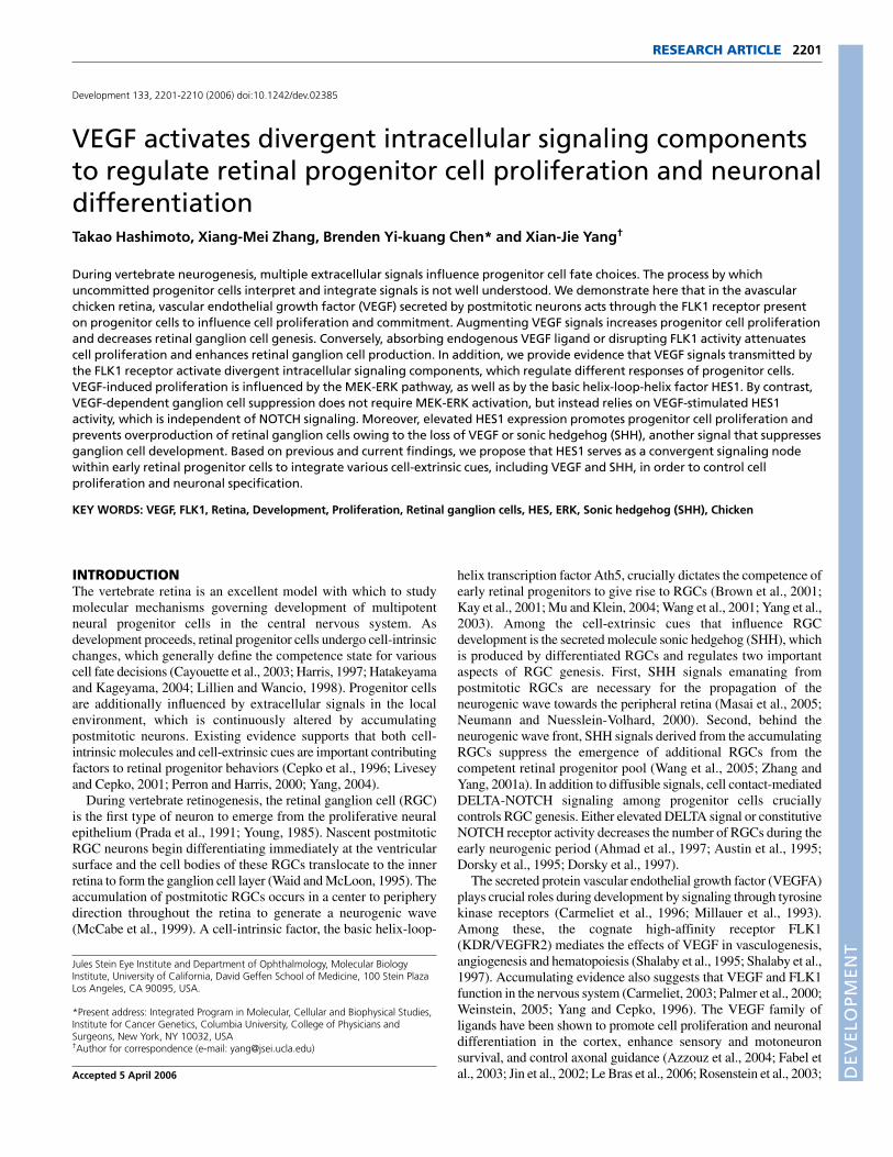
RESULTSThe avascular chicken retina expresses VEGF andFLK1 in complementary patternsIn the chicken retina, RGC differentiation initiates at embryonic day2 (E2) and peaks at E6 (Prada et al., 1991). At E5, VEGFimmunostaining signals were mainly detected in the inner retina(Fig. 1A), partially co-localized with cell nuclei expressing the LIMhomeodomain proteins Islet1 and 2 (Islet1/2), markers of RGCs atearly stages of retinogenesis (Zhang and Yang, 2001a) (Fig. 1B,C).
By E6, VEGF expression appeared as a protein gradient with themost intense staining signals in the ganglion cell layer (Fig. 1D-F).At E6, FLK1 mRNA was detected in the majority of cells in theventricular zone (Fig. 1G), overlapping with progenitor cells labeledby the proliferating cell nuclear antigen (PCNA) (Fig. 1H) and BrdU(Fig. 1I). Therefore, during the peak period of RGC production,VEGF is produced by the first emerging postmitotic neuronsopposing the proliferative zone occupied by FLK1-expressingprogenitor cells. This complementary expression pattern suggeststhat RGC-derived VEGF may act in a paracrine fashion to signalprogenitor cells through the FLK1 receptor.
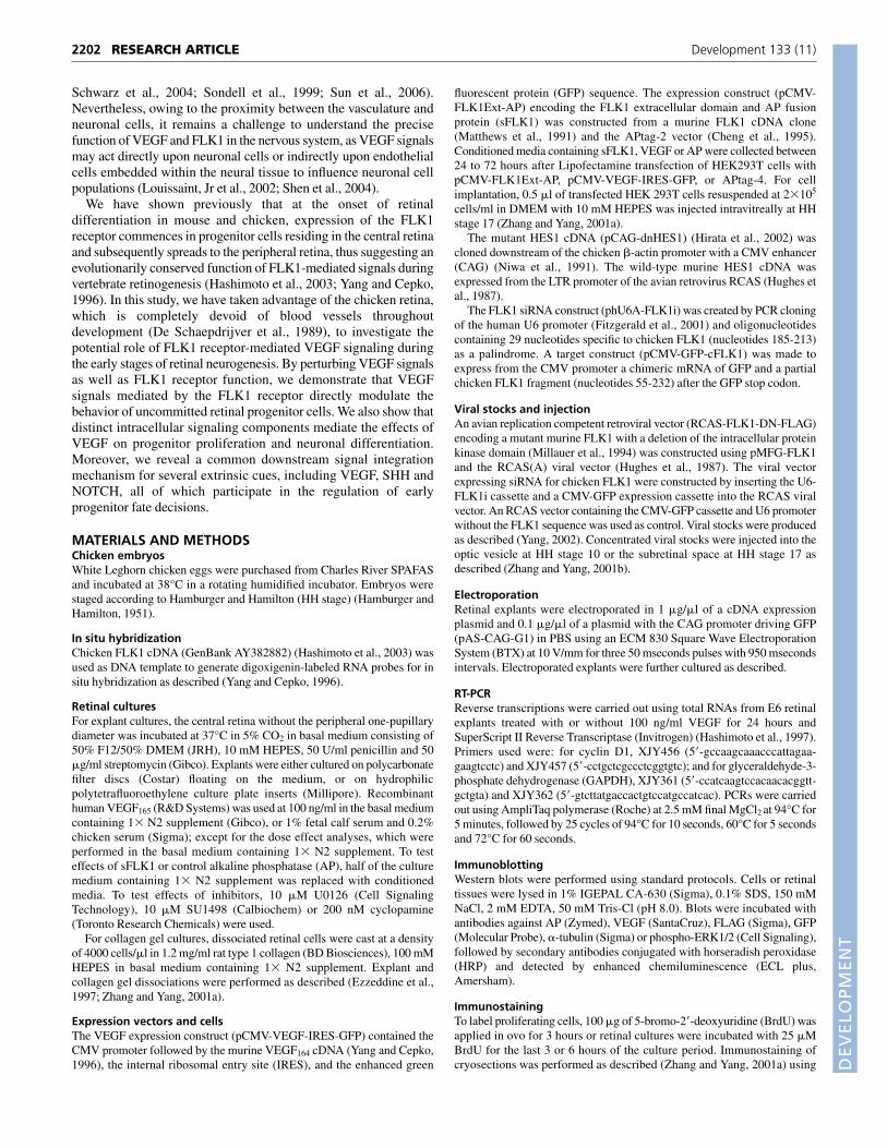
VEGF promotes proliferation of retinal progenitorcellsTo unravel potential roles of VEGF-FLK1 signaling duringretinogenesis, we examined whether VEGF influences retinalprogenitor cell proliferation. Addition of recombinant VEGF to E5retinal explants cultured for 48 hours in vitro enhanced BrdUincorporation in a dose-dependent manner (Fig. 2A). ExogenousVEGF at 10 ng/ml and 100 ng/ml resulted in 38.7% and 67.4%increases in BrdU labeling, respectively, within the last 6 hours ofculture compared with controls. To examine whether endogenousVEGF plays a mitogenic role, we used a diffusible form of the FLK1receptor consisting of the extracellular domain fused to alkalinephosphatase (sFLK1), which has been shown to bind and blockVEGF signals (Fig. 2B) (Tessler et al., 1994). In contrast toexogenous VEGF stimulation, treatment with sFLK1 resulted in adecrease in BrdU incorporation in retinal explants cultured in vitrofrom E5 to E7 (Fig. 2E-G).
We next examined effects of VEGF levels on cell proliferation invivo by performing intravitreal implantation of transfected HEKcells that produce secreted AP, sFLK1 or VEGF (Fig. 2B,C).Compared with the contralateral non-implanted eyes at E5,
2203RESEARCH ARTICLEVEGF signaling in developing chicken retina
Fig. 1. Expression of VEGF and FLK1 duringchicken retinogenesis. (A-F) Images of E5 and E6retinal sections show double immunostainingagainst VEGF (A,D), Islet1/2 (B,E) and the respectivemerged images (C,F). (G-I) Images of E6 sectionsshow in situ hybridization for FLK1 (G), andimmunostaining for PCNA (H) and BrdU (I, in ovolabeling for 3 hours). Scale bars: 50 �m. gcl,ganglion cell layer; vz, ventricular zone.
DEVELO
PMENT
2204
implanting AP-secreting cells did not alter BrdU incorporation,whereas implanting sFLK1-producing cells decreased BrdU+ cellsfrom 24.2 to 16.0% (P<0.03) (Fig. 2H). Conversely, implantingVEGF-producing cells resulted in an increase of BrdU incorporationfrom 28.1 to 30.6% (P<0.04) compared with the contralateral eyes(Fig. 2H).
To test if the proliferative effect of VEGF was mediated by theFLK1 receptor in vivo, we infected the chicken optic vesicle at E1.5(HH stage 10) with an avian retrovirus (RCAS) that expressed adominant-negative FLK1 mutant (FLK1-DN) lacking theintracellular tyrosine kinase domain (Fig. 2D) (Millauer et al., 1994).At E5, compared with control RCAS-AP virus (Fekete and Cepko,1993) infected retinas, the FLK1-DN virus infection reduced BrdU+
cells from 24.3 to 21.1% (P<0.04; average infection rate: 85.6%)(Fig. 2I).
Together, these results indicate that exogenous VEGF promotesearly retinal cell proliferation and that endogenous VEGF serves asa mitogen during early retinogenesis. Moreover, the data indicatethat the mitogenic effect of VEGF in the retina is mediated by theFLK1 receptor in vivo.
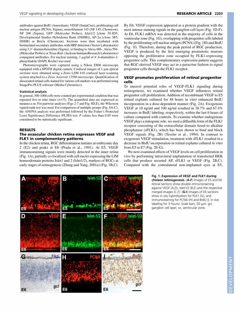
VEGF suppresses differentiation of retinalganglion cellsWe also analyzed if VEGF plays a role in neurogenesis, as maximalFLK1 expression in the chicken retina coincides with the peakperiod of RGC production (Hashimoto et al., 2003; Yang and Cepko,1996). In E5 retinal explants cultured for 48 hours in vitro,exogenous VEGF suppressed expression of the RGC markerneurofilament (NF) in a dose-dependent manner (Fig. 3A). VEGFat 100 ng/ml resulted in 67.5% decrease of NF+ cells compared withthe control (from 6.5 to 2.1%). Furthermore, sFLK1 treatmentcaused an increased number of Islet1/2+ cells, whereas supplementof VEGF suppressed differentiation of Islet1/2+ neurons in vitro(Fig. 3C-E).
To examine whether altered VEGF levels in vivo affected RGCdevelopment, we implanted intravitreally VEGF- or sFLK1-producing HEK cells at E2.5 (HH stage 17) and performed RGCmarker analyses at E5. Compared with contralateral non-implanted
RESEARCH ARTICLE Development 133 (11)
Fig. 2. Influence of VEGF on retinal cell proliferation.(A) Effects of VEGF concentrations on BrdU incorporation invitro. E5-E7 explants were labeled with BrdU for the last 6hours (n=4 or 5). (B-D) Western blots show sFLK1 (B), VEGF(C) or FLK1-DN (D) expression. Culture media of transfectedHEK cells (B,C) or infected DF-1 cell extracts (D) wereprobed with antibodies against AP (B), VEGF (C) or FLAGtag (D). Controls used were CMV-AP (B,C) or RCAS-AP virus(D). Arrowheads indicate bands with expected molecularweights. (E-G) Immunostaining show effects of AP (E),sFLK1 (F) or VEGF (G) in E5-E7 explants on BrdU labeling forthe last 3 hours. Scale bars: 50 �m. (H) Effects of sFLK1-and VEGF-producing cells on BrdU incorporation (3 hours)at E5 in vivo. Black and white bars represent implanted andthe contralateral non-implanted eyes, respectively (n=5 or 6;*P<0.03). (I) Effect of FLK1-DN expression on BrdUincorporation in vivo. Retinas infected at HH stage 10 withRCAS-AP (control) or RCAS-FLK1-DN virus were harvestedat E5 and labeled with BrdU for 3 hours (n=5; *P<0.04).
Fig. 3. Influence of VEGF on retinal ganglion cell differentiation.(A) Effects of VEGF concentrations on development of NF+ cells in E5-E7 explants in vitro (n=5). (B) Effects of sFLK1- and VEGF-producingcells on percentages of NF+ cells in vivo at E5. Black and white barsrepresent implanted and the contralateral non-implanted eyes,respectively (n=6; *P<0.03). (C-E) Immunostaining show effects of AP(C), sFLK1 (D) or VEGF (E) on Islet1/2+ cells in E5-E7 explants. Scale bars:50 �m.

DEVELO
PMENT
eyes, VEGF-secreting cell implantation decreased NF+ cells from13.3 to 10.6% (P<0.03) (Fig. 3B). By contrast, absorption ofendogenous VEGF by sFLK1 increased NF+ cells from 14.3 to18.3% in vivo (P<0.03) (Fig. 3B). These results show that VEGFsignals negatively regulate RGC genesis in vitro and in vivo.
VEGF acts through FLK1 to regulate progenitorproliferation and ganglion cell genesisTo confirm that the effects of VEGF in the developing retina aremediated by the FLK1 receptor, we performed small interferingRNA (siRNA) ‘knockdown’ of the chicken FLK1. An RCASretroviral vector encoding the human U6 promoter and a smallhairpin RNA specifically targeting the chicken FLK1 (FLK1i) wasproduced (RCAS-FLK1i, Fig. 4A). The efficiency of siRNAknockdown was determined by co-transfection of the FLK1iconstruct and an artificial target mRNA that encoded GFP with itsstop codon, followed by a 178 nucleotide untranslated sequence ofchicken FLK1 mRNA, including the FLK1i target sequence.Analyses of GFP fluorescence and western blots showed that theFLK1i construct, but not the control U6 promoter construct, causeda 60-70% reduction of the target GFP-FLK1 in transfected HEKcells (Fig. 4B-E).
To inhibit FLK1 expression in vivo, E2.5 (HH stage 17) chickenretinas were infected with RCAS-FLK1i or a control virus encodingonly the U6 promoter and the CMV-GFP cassette. Double
immunostaining showed that at E6.5, a higher proportion of RCAS-FLK1i virus infected cells were labeled positive for the Islet1/2 markerin the RGC layer compared with the control virus-infected retinas(Fig. 4F-K). As RCAS viruses infected only proliferating cells, theseresults suggest that inhibiting FLK1 receptor expression with siRNAin retinal progenitor cells bias them towards the RGC fate.
We also quantified the effects of FLK1i on retinal cellproliferation and RGC differentiation using retinal explantselectroporated with the RCAS-FLK1i or the control viral construct.Compared with controls, FLK1i-expressing GFP+ cells showedreduced BrdU labeling from 20.2 to 13.6% (P<0.03) (Fig. 4L). Inaddition, FLK1i expression caused increases of Islet1/2+ cells from21.5 to 30.7%, and of NF+ cells from 33.9 to 58.7% (P<0.03),respectively (Fig. 4M,N). By contrast, expression of FLK1i did notsignificantly affect amacrine cell differentiation during the E5 to E7culture period as indicated by staining of the AP-2� marker (West-Mays et al., 1999) (data not shown). These results further supportthat the effects of VEGF on retinal proliferation and RGCdifferentiation are both mediated by the FLK1 receptor.
Divergent intracellular transduction machineriesmediate VEGF-FLK1 signalingAs VEGF may trigger multiple signaling events in neuronal cells(Zhu et al., 2003), we next examined the intracellular mechanismsresponsible for the observed VEGF effects. We first tested if the
2205RESEARCH ARTICLEVEGF signaling in developing chicken retina
Fig. 4. Effects of RNAi-mediated FLK1 knockdown.(A) A schematic represents the RCAS viral vector (white)encoding the U6 promoter and the CMV-GFP cassette.The shFLK1 RNA (FLK1i) contains a 29 nucleotideantisense (AS, red) and a complementary sense (S, blue)sequence of chicken FLK1 followed by transcriptiontermination sequence (Term). (B-E) Efficiency of FLK1iknockdown in vitro. Fluorescence images show HEKcells co-transfected with the GFP-FLK1 chimeric targetand the control U6 (B) or U6-FLK1i (C) plasmid.Western blots (D) show GFP levels in HEK cells co-transfected with the GFP-FLK1 chimeric target andcontrol U6 or U6-FLK1i plasmid. Blots were probedagainst GFP (top) and AP (bottom), which serves as atransfection efficiency control. Optical densities of GFPsignals (E) were normalized against AP signals (n=6;*P<0.03). (F-K) Effects of FLK1i knockdown on RGCdevelopment in vivo. Confocal images show retinasinfected with the control RCAS (F-H) or RCAS-FLK1i (I-K) viruses at HH stage 17 and immunostained at E6.5for GFP (F,I), Islet1/2 (G,J) and the respective mergedimages (H,K). White arrows indicate co-stained cells.Scale bars: 50 �m. (L-N) Quantification of FLK1iknockdown effects in vitro. Explants wereelectroporated at E5 with the control RCAS or theRCAS-FLK1i construct, and labeled with BrdU for 3hours before dissociation at 48 hours. Percentages ofBrdU+ (L), Islet1/2+ (M) or NF+ (N) cells amongtransected GFP+ cells are shown (n=6; *P<0.03).

DEVELO
PMENT
2206
MEK-ERK signaling cascade was involved. In the E5 retina, anendogenous low level of phospho-ERK was detectable by westernblot (Fig. 5A). Treatment with 100 ng/ml VEGF for 10 minutesresulted in a twofold increase of ERK phosphorylation (Fig. 5A,B),indicating that VEGF signaling further activated the MEK-ERKcascade. Inclusion of the protein kinase inhibitor U0126, whichspecifically blocks activities of the MEK1 and MEK2 kinases thatact upstream of the ERK1/2 (Favata et al., 1998), significantlyreduced both endogenous and VEGF-induced ERK phosphorylation(Fig. 5A,B). Consistent with the observed effect of VEGF onproliferation, RT-PCR assays also detected enhanced cyclin D1expression in the retinas treated with VEGF (Fig. 5C).
In the E5 to E7 cultures of retinal explants, exposure to VEGFincreased BrdU incorporation from 37.0 to 47.0% (P<0.03).Addition of U0126 completely blocked VEGF-enhanced cellproliferation, but did not affect the endogenous level of BrdUincorporation (Fig. 5D). By contrast, U0126 treatment had noeffect on VEGF-dependent RGC suppression, as shown by theaverage 38% decrease of NF+ cells (Fig. 5E) and 45% decrease ofIslet1/2+ cells (Fig. 5F). Therefore, the effect of VEGF on cellproliferation requires activation of the MEK-ERK pathway,whereas VEGF-mediated RGC inhibition does not rely on MEK-ERK signaling.
HES1 activity is involved in both VEGF-dependentRGC suppression and progenitor cell proliferationAs DELTA-NOTCH signaling plays a crucial role in controllingRGC specification, we tested if perturbing the activity of the knownNOTCH signal effector HES1 (Hatakeyama and Kageyama, 2004;Tomita et al., 1996) had cell-autonomous effects on RGCdifferentiation. The wild-type or a dominant-negative mutant HES1(dnHES1) (Hirata et al., 2002; Ström et al., 1997) was co-
transfected with a GFP-expressing construct into E5 retinal explantsby electroporation. Among transfected E7 cells, forced expressionof HES1 resulted in a reduction of Islet1/2+ cells from 21.0 to12.9% (P<0.03), whereas misexpression of dnHES1 caused anincrease of Islet1/2+ cells from 17.3 to 28.1% (P<0.03) (Fig. 6A,B).Analyses of transfected cells also revealed effects of perturbingHES1 activity on cell proliferation, with misexpression of HES1resulting in a moderate but significant increase of BrdU-labeledcells from 20.1 to 24.5% (P<0.03) and dnHES1 causing a dramaticreduction of BrdU incorporation in retinal explants from 22.3 to10.9% (P<0.03) (Fig. 6C,D). These results demonstrate that HES1plays a dual role in cell proliferation and cell fate specificationduring early retinogenesis.
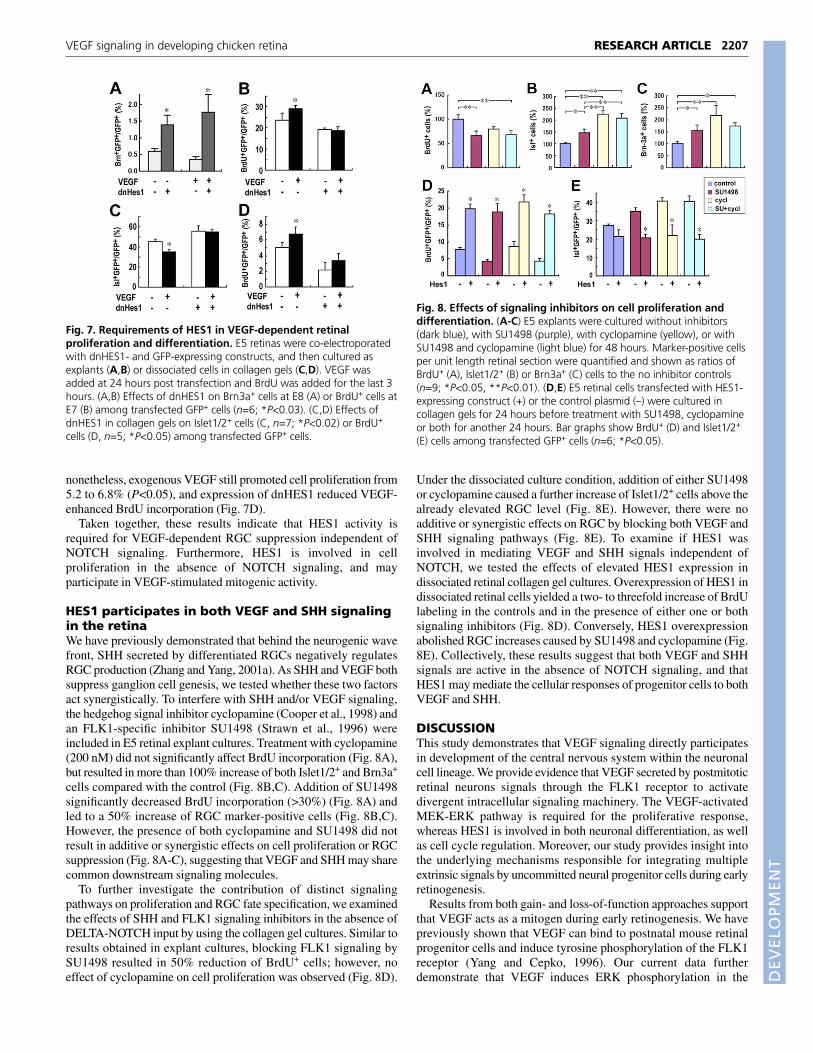
In order to delineate whether HES1 activity is involved in VEGF-dependent progenitor cell proliferation and RGC fate suppression,we introduced the dnHES1-expressing construct into E5 retinas, andthen cultured the explants in the presence or absence of VEGF.Forced expression of dnHES1 resulted in an increase of RGCs at E8,as detected by Brn3a, a marker for more mature RGCs (Liu et al.,2000; Xiang et al., 1995), with or without exogenous VEGF (Fig.7A). Furthermore, unlike control cells that showed elevated BrdUincorporation because of VEGF stimulation from 23.6 to 29.7%(P<0.03), misexpression of dnHES1 abolished VEGF-induced cellproliferation (Fig. 7B).
To evaluate if HES1 activity involved in VEGF signaling isindependent of DELTA-NOTCH interactions, we used a collagengel culture system in which cell-cell contacts among dissociatedretinal cells were minimized. Consistent with previous observations(Austin et al., 1995), the proportion of progenitor cells adopting theRGC fate is greatly increased in gel cultures owing to the eliminationof NOTCH signaling (Fig. 7C). Addition of VEGF still resulted ina 22% reduction of RGCs from 45.2 to 35.3% (P<0.02), suggestingthat the effect of VEGF on neuronal specification did not depend oncell-cell contacts (Fig. 7C). Among dnHES1-expressing cells,however, the suppression of RGCs by VEGF was eradicated,indicating the direct involvement of HES1 in VEGF-dependentRGC fate regulation (Fig. 7C). In the gel culture, the overall cellproliferation rate was lower than in retinal explant cultures;
RESEARCH ARTICLE Development 133 (11)
Fig. 5. Effects of MEK-ERK inhibition on VEGF-dependentproliferation and differentiation. (A) Western blots of VEGF-inducedphospho-ERK. E6 retinas were cultured with or without U0126 for 60minutes before VEGF was added for 10 minutes. Blots were probedwith phospho-ERK1/2 (top) and �-tubulin (bottom) antibodies.(B) Quantification of optical densities of phospho-ERK signals werenormalized against �-tubulin signals (n=4). (C) RT-PCR detection ofcyclin D1 transcripts in E6 retinas cultured for 24 hours in the presenceor absence of VEGF. Ratios of cyclin D1 and GAPDH products areshown (n=8). (D-F) Quantifications of U0126 effects on VEGF-dependent cell proliferation (D) or differentiation (E,F). E5 explants werecultured for 24 hours and BrdU labeled for the last 3 hours (n=6;*P<0.03).
Fig. 6. Influence of HES1 activity on retinal proliferation anddifferentiation. E5 retinas were co-electroporated with HES1- ordnHES1-expressing construct and a GFP-expressing construct, andcultured as explants for 48 hours with BrdU present for the last 3 hours.Effects of HES1 (A,C) or dnHES1 (B,D) on Islet1/2+ (A,B) or BrdU+ (C,D)cells among transfected GFP+ cells are shown (n=6; *P<0.03).
DEVELO
PMENT
nonetheless, exogenous VEGF still promoted cell proliferation from5.2 to 6.8% (P<0.05), and expression of dnHES1 reduced VEGF-enhanced BrdU incorporation (Fig. 7D).
Taken together, these results indicate that HES1 activity isrequired for VEGF-dependent RGC suppression independent ofNOTCH signaling. Furthermore, HES1 is involved in cellproliferation in the absence of NOTCH signaling, and mayparticipate in VEGF-stimulated mitogenic activity.
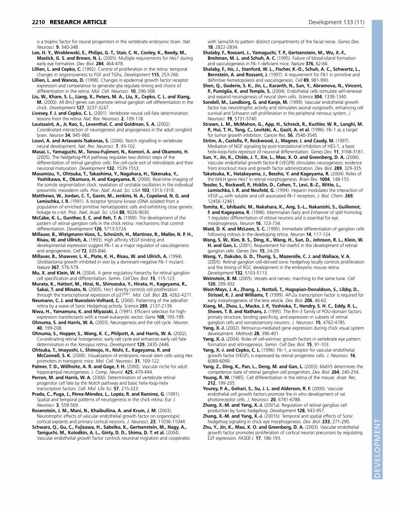
HES1 participates in both VEGF and SHH signalingin the retinaWe have previously demonstrated that behind the neurogenic wavefront, SHH secreted by differentiated RGCs negatively regulatesRGC production (Zhang and Yang, 2001a). As SHH and VEGF bothsuppress ganglion cell genesis, we tested whether these two factorsact synergistically. To interfere with SHH and/or VEGF signaling,the hedgehog signal inhibitor cyclopamine (Cooper et al., 1998) andan FLK1-specific inhibitor SU1498 (Strawn et al., 1996) wereincluded in E5 retinal explant cultures. Treatment with cyclopamine(200 nM) did not significantly affect BrdU incorporation (Fig. 8A),but resulted in more than 100% increase of both Islet1/2+ and Brn3a+
cells compared with the control (Fig. 8B,C). Addition of SU1498significantly decreased BrdU incorporation (>30%) (Fig. 8A) andled to a 50% increase of RGC marker-positive cells (Fig. 8B,C).However, the presence of both cyclopamine and SU1498 did notresult in additive or synergistic effects on cell proliferation or RGCsuppression (Fig. 8A-C), suggesting that VEGF and SHH may sharecommon downstream signaling molecules.
To further investigate the contribution of distinct signalingpathways on proliferation and RGC fate specification, we examinedthe effects of SHH and FLK1 signaling inhibitors in the absence ofDELTA-NOTCH input by using the collagen gel cultures. Similar toresults obtained in explant cultures, blocking FLK1 signaling bySU1498 resulted in 50% reduction of BrdU+ cells; however, noeffect of cyclopamine on cell proliferation was observed (Fig. 8D).
Under the dissociated culture condition, addition of either SU1498or cyclopamine caused a further increase of Islet1/2+ cells above thealready elevated RGC level (Fig. 8E). However, there were noadditive or synergistic effects on RGC by blocking both VEGF andSHH signaling pathways (Fig. 8E). To examine if HES1 wasinvolved in mediating VEGF and SHH signals independent ofNOTCH, we tested the effects of elevated HES1 expression indissociated retinal collagen gel cultures. Overexpression of HES1 indissociated retinal cells yielded a two- to threefold increase of BrdUlabeling in the controls and in the presence of either one or bothsignaling inhibitors (Fig. 8D). Conversely, HES1 overexpressionabolished RGC increases caused by SU1498 and cyclopamine (Fig.8E). Collectively, these results suggest that both VEGF and SHHsignals are active in the absence of NOTCH signaling, and thatHES1 may mediate the cellular responses of progenitor cells to bothVEGF and SHH.
DISCUSSIONThis study demonstrates that VEGF signaling directly participatesin development of the central nervous system within the neuronalcell lineage. We provide evidence that VEGF secreted by postmitoticretinal neurons signals through the FLK1 receptor to activatedivergent intracellular signaling machinery. The VEGF-activatedMEK-ERK pathway is required for the proliferative response,whereas HES1 is involved in both neuronal differentiation, as wellas cell cycle regulation. Moreover, our study provides insight intothe underlying mechanisms responsible for integrating multipleextrinsic signals by uncommitted neural progenitor cells during earlyretinogenesis.
Results from both gain- and loss-of-function approaches supportthat VEGF acts as a mitogen during early retinogenesis. We havepreviously shown that VEGF can bind to postnatal mouse retinalprogenitor cells and induce tyrosine phosphorylation of the FLK1receptor (Yang and Cepko, 1996). Our current data furtherdemonstrate that VEGF induces ERK phosphorylation in the
2207RESEARCH ARTICLEVEGF signaling in developing chicken retina
Fig. 7. Requirements of HES1 in VEGF-dependent retinalproliferation and differentiation. E5 retinas were co-electroporatedwith dnHES1- and GFP-expressing constructs, and then cultured asexplants (A,B) or dissociated cells in collagen gels (C,D). VEGF wasadded at 24 hours post transfection and BrdU was added for the last 3hours. (A,B) Effects of dnHES1 on Brn3a+ cells at E8 (A) or BrdU+ cells atE7 (B) among transfected GFP+ cells (n=6; *P<0.03). (C,D) Effects ofdnHES1 in collagen gels on Islet1/2+ cells (C, n=7; *P<0.02) or BrdU+
cells (D, n=5; *P<0.05) among transfected GFP+ cells.
Fig. 8. Effects of signaling inhibitors on cell proliferation anddifferentiation. (A-C) E5 explants were cultured without inhibitors(dark blue), with SU1498 (purple), with cyclopamine (yellow), or withSU1498 and cyclopamine (light blue) for 48 hours. Marker-positive cellsper unit length retinal section were quantified and shown as ratios ofBrdU+ (A), Islet1/2+ (B) or Brn3a+ (C) cells to the no inhibitor controls(n=9; *P<0.05, **P<0.01). (D,E) E5 retinal cells transfected with HES1-expressing construct (+) or the control plasmid (–) were cultured incollagen gels for 24 hours before treatment with SU1498, cyclopamineor both for another 24 hours. Bar graphs show BrdU+ (D) and Islet1/2+
(E) cells among transfected GFP+ cells (n=6; *P<0.05).

DEVELO
PMENT
2208
chicken retina, and that the stimulatory effect of VEGF onproliferation involves the MEK-ERK signaling pathway. Eventhough the MEK1/2 inhibitor U0126 effectively blocked bothendogenous and VEGF-induced ERK phosphorylation, U0126 wasunable to eliminate all cell proliferation. This result suggests thatrobust proliferation during early retinogenesis is dependent onmultiple cell-extrinsic cues, which may trigger signaling eventsother than MEK-ERK activation to regulate the cell cycle (Anchanet al., 1991; Dyer and Cepko, 2001; Jensen and Wallace, 1997;Lillien and Cepko, 1992; Ohnuma and Harris, 2003). Other knownsignals that regulate cell proliferation in the nervous system includeWnt and hedgehog molecules, which are not thought to depend uponMEK activation (Chesnutt et al., 2004; Kenney et al., 2004). We didnot detect altered retinal cell death using TUNEL under conditionsthat either elevate or diminish VEGF signaling (data not shown).Therefore, the observed effects of VEGF are unlikely to be due toenhanced cell survival or reduced apoptosis.
Intriguingly, interfering with the activity of HES1, one of theeffectors of NOTCH signaling (Louvi and Artavanis-Tsakonas,2006), also affected VEGF-induced cell proliferation. Elegantmolecular genetic studies have established that HES1 acts as atranscription repressor to maintain neural progenitor cell potentialand regulate differentiation (Hatakeyama et al., 2004; Kageyama etal., 2005). In the mouse retina, loss of HES1 causes premature cellcycle exit and neuronal differentiation, whereas misexpression ofHES1 results in the formation of undifferentiated precursor-like cellsand Müller glia (Furukawa et al., 2000; Lee et al., 2005; Takatsukaet al., 2004; Tomita et al., 1996). These results are consistent withour observations that manipulating HES1 activity affectsproliferation in the chicken retina. The most likely target of thednHES1 in the chicken retina is the chicken Hairy2 protein, whichshares sequence homologies and functional similarities with themammalian HES1 (Jouve et al., 2000). The dnHES1 mayadditionally inhibit chicken Hairy1, which also acts as a NOTCHsignaling effector (Jouve et al., 2000). Although the precise role ofHES1 in cell cycle regulation remains to be determined, recentevidence suggests that HES1 may directly repress transcription ofthe cell cycle inhibitor p27kip1 (Murata et al., 2005). In thedeveloping Drosophila eye imaginal disc, NOTCH signaling isrequired for the G1-S phase transition of the cell cycle and isresponsible for triggering the onset of proliferation by multiplesignaling pathways (Baonza and Freeman, 2005). However, ourresults from dissociated retinal cultures demonstrate that theinvolvement of HES1 in cell proliferation can also be independentof NOTCH signaling. Therefore, HES1 emerges as an importanteffector for other cell-extrinsic cues in addition to thetransmembrane DELTA ligands.
It has been reported that VEGF promotes rhodopsin expression inneonatal rat retinal monolayer cultures (Yourey et al., 2000). Here,we reveal a novel function of VEGF in modulating production of thefirst-born retinal neurons. Our results demonstrate that VEGF actssimultaneously to promote progenitor proliferation and suppressganglion cell production. Experiments in the Xenopus retina havesuggested that cells receiving a proliferative signal are unable toeffectively execute a differentiation pathway (Ohnuma et al., 2002).We thus examined whether the mitogenic and neurogenic effects ofVEGF in the chicken retina are related to each other. Our findingsindicate that VEGF-dependent suppression of RGC does not involveactivation of the MEK-ERK cascade, but instead is dependent onHES1 activity. Because HES1 activity is involved in both VEGFeffects, at present the mitogenic effect of VEGF can only be partiallyuncoupled from its effect on neuronal differentiation. The exact
mechanism of how VEGF signaling leads to altered HES1 activityis currently unknown. Because VEGF-triggered FLK1phosphorylation can activate multiple intracellular signalingcomponents (Carmeliet, 2003), it is plausible that FLK1 signalingmay influence HES1 activity at both the transcriptional and post-transcriptional levels. The results of MEK1/2 inhibitor U0126indicate that VEGF-enhanced HES1 function does not requireMEK-ERK activation, and thus is probably related to signalingevents diverged upstream of MEK.
Current evidence supports that both cell-cell interactions andsecreted signals participate in vertebrate RGC fate specification.NOTCH signaling among early retinal progenitor cells plays animportant role in controlling RGC formation (Ahmad et al., 1997;Austin et al., 1995; Dorsky et al., 1995; Dorsky et al., 1997),presumably through the downstream effectors HES1 and Hes5(Hatakeyama and Kageyama, 2004). We have identified SHH as anRGC-derived secreted signal that negatively regulates RGC genesisbehind the neurogenic wave front (Zhang and Yang, 2001a). Ourmodel is consistent with a recent study showing that conditionalremoval of the SHH gene from the peripheral retina results inenhanced local RGC genesis (Wang et al., 2005). Like FLK1, theSHH receptor patched is expressed by mouse and chicken retinalprogenitors (Jensen and Wallace, 1997; Zhang and Yang, 2001a).Thus, molecular evidence clearly indicates that early retinalprogenitor cells receive multiple extrinsic cues, including DELTA1,SHH and VEGF, which are mediated by distinct cell surfacereceptors and canonical intracellular signal transduction pathways.
How do uncommitted progenitor cells integrate and respond tomultiple environmental signals? Results presented in this studysuggest that HES1 protein plays a key role in mediating VEGFeffects, independent of NOTCH signaling. Our on-going studiesshow that SHH signaling enhances HES1 activity in the chicken
RESEARCH ARTICLE Development 133 (11)
Fig. 9. A model of signal convergence in retinal progenitor cells.DELTA-NOTCH signaling yields the NOTCH intracellular domain (NICD)and activates HES1 transcription. SHH signaling through patched (PTC)and Smoothened (SMO) reduces Gli repressor (GliRep) and facilitates theaccumulation of the Gli activator (GliAct). SHH signaling upregulatesHES1 by unknown mechanism(s). VEGF stimulation causes theactivation of the MEK-ERK cascade and enhances HES1 activity withoutthe involvement of MEK1/2 function. The promotion of cellproliferation may require independent inputs from both MEK-ERK andHES1, whereas suppression of RGC specification mainly involves HES1activity. Therefore, HES1 serves as a node of signal convergence tointegrate inputs of multiple cell-extrinsic cues.
Flk1
Hes1
MEK1/2
ERK1/2
SmoPtc
GliGliAct Rep
Cell cycle re-entry
Progenitor responses
Neuronal differentiation
Notch
VEGF Delta Shh
?
?
NICD
Extrinsic cues
?
?

DEVELO
PMENT
retina (X.-M.Z., T.H. and X.-J.Y., unpublished). Partial reduction ofSHH in the mouse retina also leads to a reduction of HES1 mRNA(Wang et al., 2005). Moreover, Hes1 knockout mice exhibitprecocious ATH5 expression and RGC defects (Lee et al., 2005).The lack of synergistic or additive effects of VEGF and SHH on cellproliferation and RGC suppression may reflect that these twopathways share common downstream signaling components that arelimiting or tightly regulated. Based on these lines of evidence, wepropose a model in which HES1 not only acts as an effector forNOTCH signals, but also serves as a signaling node for theconvergence of VEGF and SHH signals during early retinogenesis(Fig. 9). Our model suggests that the negative regulation of RGCgenesis by NOTCH, SHH and VEGF signaling all involves HES1activity. However, the precise function of HES1 in cell cycleprogression remains to be further defined. To date, our perturbationresults indicate that VEGF but not SHH influences proliferation inthe early developing chicken retina. As SHH signaling aloneprovides enhanced HES1 but not ERK activation, it is possible thatcell cycle re-entry may require simultaneous inputs from bothactivated ERK and HES1. Alternatively, HES1 may play an essentialrole in regulating cell cycle progression independently of ERKactivation. Our model proposes that levels of HES1 activity in earlyretinal progenitor cells reflect the integrated inputs of multipleextrinsic cues, and critically control progenitor cell fate decisions.Because HES1 protein has been shown to have a short half-life andHES1 gene transcription is auto-repressed by HES1 protein itself(Hirata et al., 2002), it is expected that HES1 protein levels arehighly dynamic (Masamizu et al., 2006; Ohtsuka et al., 2006).Future studies on mechanisms that transduce or relay divergentsignals to HES1 within the neural progenitor cells and how HES1interfaces with multiple signals to control the cell cycle will becrucial to further our understanding of neuronal cell fatedetermination.
We thank Drs I. Lemischka, J. Flanagan and R. Kageyama for reagents, and DrFei Yu for statistical expertise. This work was in part supported by a grant fromJapan Eye Bank Association to T.H., and by grants from the Research toPrevent Blindness Foundation, the Karl Kirchgessner Foundation, theFoundation Fighting Blindness and the National Eye Institute to X.-J.Y.
ReferencesAhmad, I., Dooley, C. M. and Polk, D. L. (1997). Delta-1 is a regulator of
neurogenesis in the vertebrate retina. Dev. Biol. 185, 92-103.Anchan, R. M., Reh, T. A., Angello, J., Balliet, A. and Walker, M. (1991). EGF
and TGF-� stimulate retinal neuroepithelial cell proliferation in vitro. Neuron 6,923-936.
Austin, C. P., Feldman, D. E., Ida, J. A., Jr and Cepko, C. L. (1995). Vertebrateretinal ganglion cells are selected from competent progenitors by the action ofNotch. Development 121, 3637-3650.
Azzouz, M., Ralph, G. S., Storkebaum, E., Walmsley, L. E., Mitrophanous, K.A., Kingsman, S. M., Carmeliet, P. and Mazarakis, N. D. (2004). VEGFdelivery with retrogradely transported lentivector prolongs survival in a mouseALS model. Nature 429, 413-417.
Baonza, A. and Freeman, M. (2005). Control of cell proliferation in theDrosophila eye by Notch signaling. Dev. Cell 8, 529-539.
Brown, N. L., Patel, S., Brzezinski, J. and Glaser, T. (2001). Math5 is requiredfor retinal ganglion cell and optic nerve formation. Development 128, 2497-2508.
Carmeliet, P. (2003). Blood vessels and nerves: common signals, pathways anddiseases. Nat. Rev. Genet. 4, 710-720.
Carmeliet, P., Ferreira, V., Breier, G., Pollefeyt, S., Kieckens, L., Gertsenstein,M., Fahrig, M., Vandenhoeck, A., Harpal, K., Eberhardt, C. et al. (1996).Abnormal blood vessel development and lethality in embryos lacking a singleVEGF allele. Nature 380, 435-439.
Cayouette, M., Barres, B. A. and Raff, M. (2003). Importance of intrinsicmechanisms in cell fate decisions in the developing rat retina. Neuron 40, 897-904.
Cepko, C. L., Austin, C. P., Yang, X., Alexiades, M. and Ezzeddine, D. (1996).Cell fate determination in the vertebrate retina. Proc. Natl. Acad. Sci. USA 93,589-595.
Cheng, H.-J., Nakamoto, M., Bergemann, A. D. and Flanagan, J. G. (1995).Complementary gradients in expression and binding of ELF-1 and Mek4 indevelopment of the topographic retinotectal projection map. Cell 82, 371-381.
Chesnutt, C., Burrus, L. W., Brown, A. M. C. and Niswander, L. (2004).Coordinate regulation of neural tube patterning and proliferation by TGF� andWNT activity. Dev. Biol. 274, 334-347.
Cooper, M. K., Porter, J. A., Young, K. E. and Beachy, P. A. (1998). Teratogen-mediated inhibition of target tissue response to Shh signaling. Science 280,1603-1607.
De Schaepdrijver, L., Simoens, P., Lauwers, H. and De Geest, J. P. (1989).Retinal vascular patterns in domestic animals. Res. Vet. Sci. 47, 34-42.
Dorsky, R. I., Rapaport, D. H. and Harris, W. A. (1995). Xotch inhibits celldifferentiation in the Xenopus retina. Neuron 14, 487-496.
Dorsky, R. I., Chang, W. S., Rapaport, D. H. and Harris, W. A. (1997).Regulation of neuronal diversity in the Xenopus retina by Delta signalling. Nature385, 67-70.
Dyer, M. A. and Cepko, C. L. (2001). Regulating proliferation during retinaldevelopment. Nat. Rev. Neurosci. 2, 333-342.
Ezzeddine, Z. D., Yang, X., DeChiara, T., Yancopoulos, G. and Cepko, C. L.(1997). Postmitotic cells fated to become rod photoreceptors can be respecifiedby CNTF treatment of the retina. Development 124, 1055-1067.
Fabel, K., Fabel, K., Tam, B., Kaufer, D., Baiker, A., Simmons, N., Kuo, C. J.and Palmer, T. D. (2003). VEGF is necessary for exercise-induced adulthippocampal neurogenesis. Eur. J. Neurosci. 18, 2803-2812.
Favata, M. F., Horiuchi, K. Y., Manos, E. J., Daulerio, A. J., Stradley, D. A.,Feeser, W. S., Van Dyk, D. E., Pitts, W. J., Earl, R. A., Hobbs, F. et al. (1998).Identification of a novel inhibitor of mitogen-activated protein kinase kinase. J.Biol. Chem. 273, 18623-18632.
Fekete, D. M. and Cepko, C. L. (1993). Replication-competent retroviral vectorsencoding alkaline phosphatase reveal spatial restriction of viral geneexpression/transduction in the chick embryo. Mol. Cell. Biol. 13, 2604-2613.
Fitzgerald, K. M., Hashimoto, T., Hug, T. E., Cibis, G. W. and Harris, D. J.(2001). Autosomal dominant inheritance of a negative electroretinogramphenotype in three generations. Am. J. Ophthalmol. 131, 495-502.
Furukawa, T., Mukherjee, S., Bao, Z.-Z., Morrow, E. M. and Cepko, C. L.(2000). rax, Hes1, and notch1 promote the formation of Müller glia by postnatalretinal progenitor cells. Neuron 26, 383-394.
Hamburger, V. and Hamilton, H. L. (1951). A series of normal stages in thedevelopment of the chick embryo. J. Morphol. 88, 49-92.
Harris, W. A. (1997). Cellular diversification in the vertebrate retina. Curr. Opin.Genet. Dev. 7, 651-658.
Hashimoto, T., Inazawa, J., Okamoto, N., Tagawa, Y., Bessho, Y., Honda, Y.and Nakanishi, S. (1997). The whole nucleotide sequence and chromosomallocalization of the gene for human metabotropic glutamate receptor subtype 6.Eur. J. Neurosci. 9, 1226-1235.
Hashimoto, T., Zhang, X.-M. and Yang, X.-J. (2003). Expression of the Flk1receptor and its ligand VEGF in the developing chick central nervous system.Gene Expr. Patterns 3, 109-113.
Hatakeyama, J. and Kageyama, R. (2004). Retinal cell fate determination andbHLH factors. Semin. Cell Dev. Biol. 15, 83-89.
Hatakeyama, J., Bessho, Y., Katoh, K., Ookawara, S., Fujioka, M., Guillemot,F. and Kageyama, R. (2004). Hes genes regulate size, shape and histogenesisof the nervous system by control of the timing of neural stem cell differentiation.Development 131, 5539-5550.
Hirata, H., Yoshiura, S., Ohtsuka, T., Bessho, Y., Harada, T., Yoshikawa, K.and Kageyama, R. (2002). Oscillatory expression of the bHLH factor Hes1regulated by a negative feedback loop. Science 298, 840-843.
Hughes, S. H., Greenhouse, J. J., Petropoulos, C. J. and Sutrave, P. (1987).Adaptor plasmids simplify the insertion of foreign DNA into helper-independentretroviral vectors. J. Virol. 61, 3004-3012.
Jensen, A. M. and Wallace, V. A. (1997). Expression of Sonic hedgehog and itsputative role as a precursor cell mitogen in the developing mouse retina.Development 124, 363-371.
Jin, K., Zhu, Y., Sun, Y., Mao, X. O., Xie, L. and Greenberg, D. A. (2002).Vascular endothelial growth factor (VEGF) stimulates neurogenesis in vitro and invivo. Proc. Natl. Acad. Sci. USA 99, 11946-11950.
Jouve, C., Palmeirim, I., Henrique, D., Beckers, J., Gossler, A., Ish-Horowicz,D. and Pourquié, O. (2000). Notch signalling is required for cyclic expression ofthe hairy-like gene HES1 in the presomitic mesoderm. Development 127, 1421-1429.
Kageyama, R., Ohtsuka, T., Hatakeyama, J. and Ohsawa, R. (2005). Roles ofbHLH genes in neural stem cell differentiation. Exp. Cell Res. 306, 343-348.
Kay, J. N., Finger-Baier, K. C., Roeser, T., Staub, W. and Baier, H. (2001).Retinal ganglion cell genesis requires lakritz, a Zebrafish atonal Homolog.Neuron 30, 725-736.
Kenney, A. M., Widlund, H. R. and Rowitch, D. H. (2004). Hedgehog and PI-3kinase signaling converge on Nmyc1 to promote cell cycle progression incerebellar neuronal precursors. Development 131, 217-228.
Le Bras, B., Barallobre, M.-J., Homman-Ludiye, J., Ny, A., Wyns, S., Tammela,T., Haiko, P., Karkkainen, M. J., Yuan, L., Muriel, M. P. et al. (2006). VEGF-C
2209RESEARCH ARTICLEVEGF signaling in developing chicken retina
DEVELO
PMENT
2210
is a trophic factor for neural progenitors in the vertebrate embryonic brain. Nat.Neurosci. 9, 340-348.
Lee, H. Y., Wroblewski, E., Philips, G. T., Stair, C. N., Conley, K., Reedy, M.,Mastick, G. S. and Brown, N. L. (2005). Multiple requirements for Hes1 duringearly eye formation. Dev. Biol. 284, 464-478.
Lillien, L. and Cepko, C. (1992). Control of proliferation in the retina: temporalchanges in responsiveness to FGF and TGF�. Development 115, 253-266.
Lillien, L. and Wancio, D. (1998). Changes in epidermal growth factor receptorexpression and competence to generate glia regulate timing and choice ofdifferentiation in the retina. Mol. Cell. Neurosci. 10, 296-308.
Liu, W., Khare, S. L., Liang, X., Peters, M. A., Liu, X., Cepko, C. L. and Xiang,M. (2000). All Brn3 genes can promote retinal ganglion cell differentiation in thechick. Development 127, 3237-3247.
Livesey, F. J. and Cepko, C. L. (2001). Vertebrate neural cell-fate determination:lessons from the retina. Nat. Rev. Neurosci. 2, 109-118.
Louissaint, A., Jr, Rao, S., Leventhal, C. and Goldman, S. A. (2002).Coordinated interaction of neurogenesis and angiogenesis in the adult songbirdbrain. Neuron 34, 945-960.
Louvi, A. and Artavanis-Tsakonas, S. (2006). Notch signalling in vertebrateneural development. Nat. Rev. Neurosci. 7, 93-102.
Masai, I., Yamaguchi, M., Tonou-Fujimori, N., Komori, A. and Okamoto, H.(2005). The hedgehog-PKA pathway regulates two distinct steps of thedifferentiation of retinal ganglion cells: the cell-cycle exit of retinoblasts and theirneuronal maturation. Development 132, 1539-1553.
Masamizu, Y., Ohtsuka, T., Takashima, Y., Nagahara, H., Takenaka, Y.,Yoshikawa, K., Okamura, H. and Kageyama, R. (2006). Real-time imaging ofthe somite segmentation clock: revelation of unstable oscillators in the individualpresomitic mesoderm cells. Proc. Natl. Acad. Sci. USA 103, 1313-1318.
Matthews, W., Jordan, C. T., Gavin, M., Jenkins, N. A., Copeland, N. G. andLemischka, I. R. (1991). A receptor tyrosine kinase cDNA isolated from apopulation of enriched primitive hematopoietic cells and exhibiting close geneticlinkage to c-kit. Proc. Natl. Acad. Sci. USA 88, 9026-9030.
McCabe, K. L., Gunther, E. C. and Reh, T. A. (1999). The development of thepattern of retinal ganglion cells in the chick retina: mechanisms that controldifferentiation. Development 126, 5713-5724.
Millauer, B., Wizigmann-Voos, S., Schnürch, H., Martinez, R., Møller, N. P. H.,Risau, W. and Ullrich, A. (1993). High affinity VEGF binding anddevelopmental expression suggest Flk-1 as a major regulator of vasculogenesisand angiogenesis. Cell 72, 835-846.
Millauer, B., Shawver, L. K., Plate, K. H., Risau, W. and Ullrich, A. (1994).Glioblastoma growth inhibited in vivo by a dominant-negative Flk-1 mutant.Nature 367, 576-579.
Mu, X. and Klein, W. H. (2004). A gene regulatory hierarchy for retinal ganglioncell specification and differentiation. Semin. Cell Dev. Biol. 15, 115-123.
Murata, K., Hattori, M., Hirai, N., Shinozuka, Y., Hirata, H., Kageyama, R.,Sakai, T. and Minato, N. (2005). Hes1 directly controls cell proliferationthrough the transcriptional repression of p27Kip1. Mol. Cell. Biol. 25, 4262-4271.
Neumann, C. J. and Nuesslein-Volhard, C. (2000). Patterning of the zebrafishretina by a wave of Sonic Hedgehog activity. Science 289, 2137-2139.
Niwa, H., Yamamura, K. and Miyazaki, J. (1991). Efficient selection for high-expression transfectants with a novel eukaryotic vector. Gene 108, 193-199.
Ohnuma, S. and Harris, W. A. (2003). Neurogenesis and the cell cycle. Neuron40, 199-208.
Ohnuma, S., Hopper, S., Wang, K. C., Philpott, A. and Harris, W. A. (2002).Co-ordinating retinal histogenesis: early cell cycle exit enhances early cell fatedetermination in the Xenopus retina. Development 129, 2435-2446.
Ohtsuka, T., Imayoshi, I., Shimojo, H., Nishi, E., Kageyama, R. andMcConnell, S. K. (2006). Visualization of embryonic neural stem cells using Hespromoters in transgenic mice. Mol. Cell. Neurosci. 31, 109-122.
Palmer, T. D., Willhoite, A. R. and Gage, F. H. (2000). Vascular niche for adulthippocampal neurogenesis. J. Comp. Neurol. 425, 479-494.
Perron, M. and Harris, W. A. (2000). Determination of vertebrate retinalprogenitor cell fate by the Notch pathway and basic helix-loop-helixtranscription factors. Cell. Mol. Life Sci. 57, 215-223.
Prada, C., Puga, J., Pérez-Méndez, L., Lopéz, R. and Ramírez, G. (1991).Spatial and temporal patterns of neurogenesis in the chick retina. Eur. J.Neurosci. 3, 559-569.
Rosenstein, J. M., Mani, N., Khaibullina, A. and Krum, J. M. (2003).Neurotrophic effects of vascular endothelial growth factor on organotypiccortical explants and primary cortical neurons. J. Neurosci. 23, 11036-11044.
Schwarz, Q., Gu, C., Fujisawa, H., Sabelko, K., Gertsenstein, M., Nagy, A.,Taniguchi, M., Kolodkin, A. L., Ginty, D. D., Shima, D. T. et al. (2004).Vascular endothelial growth factor controls neuronal migration and cooperates
with Sema3A to pattern distinct compartments of the facial nerve. Genes Dev.18, 2822-2834.
Shalaby, F., Rossant, J., Yamaguchi, T. P., Gertsenstein, M., Wu, X.-F.,Breitman, M. L. and Schuh, A. C. (1995). Failure of blood-island formationand vasculogenesis in Flk-1-deficient mice. Nature 376, 62-66.
Shalaby, F., Ho, J., Stanford, W. L., Fischer, K.-D., Schuh, A. C., Schwartz, L.,Bernstein, A. and Rossant, J. (1997). A requirement for Flk1 in primitive anddefinitive hematopoiesis and vasculogenesis. Cell 89, 981-990.
Shen, Q., Goderie, S. K., Jin, L., Karanth, N., Sun, Y., Abramova, N., Vincent,P., Pumiglia, K. and Temple, S. (2004). Endothelial cells stimulate self-renewaland expand neurogenesis of neural stem cells. Science 304, 1338-1340.
Sondell, M., Lundborg, G. and Kanje, M. (1999). Vascular endothelial growthfactor has neurotrophic activity and stimulates axonal outgrowth, enhancing cellsurvival and Schwann cell proliferation in the peripheral nervous system. J.Neurosci. 19, 5731-5740.
Strawn, L. M., McMahon, G., App, H., Schreck, R., Kuchler, W. R., Longhi, M.P., Hui, T. H., Tang, C., Levitzki, A., Gazit, A. et al. (1996). Flk-1 as a targetfor tumor growth inhibition. Cancer Res. 56, 3540-3545.
Ström, A., Castella, P., Rockwood, J., Wagner, J. and Caudy, M. (1997).Mediation of NGF signaling by post-translational inhibition of HES-1, a basichelix-loop-helix repressor of neuronal differentiation. Genes Dev. 11, 3168-3181.
Sun, Y., Jin, K., Childs, J. T., Xie, L., Mao, X. O. and Greenberg, D. A. (2006).Vascular endothelial growth factor-B (VEGFB) stimulates neurogenesis: evidencefrom knockout mice and growth factor administration. Dev. Biol. 289, 329-335.
Takatsuka, K., Hatakeyama, J., Bessho, Y. and Kageyama, R. (2004). Roles ofthe bHLH gene Hes1 in retinal morphogenesis. Brain Res. 1004, 148-155.
Tessler, S., Rockwell, P., Hicklin, D., Cohen, T., Levi, B.-Z., Witte, L.,Lemischka, I. R. and Neufeld, G. (1994). Heparin modulates the interaction ofVEGF165 with soluble and cell associated flk-1 receptors. J. Biol. Chem. 269,12456-12461.
Tomita, K., Ishibashi, M., Nakahara, K., Ang, S.-L., Nakanishi, S., Guillemot,F. and Kageyama, R. (1996). Mammalian hairy and Enhancer of split homolog1 regulates differentiation of retinal neurons and is essential for eyemorphogenesis. Neuron 16, 723-734.
Waid, D. K. and McLoon, S. C. (1995). Immediate differentiation of ganglion cellsfollowing mitosis in the developing retina. Neuron 14, 117-124.
Wang, S. W., Kim, B. S., Ding, K., Wang, H., Sun, D., Johnson, R. L., Klein, W.H. and Gan, L. (2001). Requirement for math5 in the development of retinalganglion cells. Genes Dev. 15, 24-29.
Wang, Y., Dakubo, G. D., Thurig, S., Mazerolle, C. J. and Wallace, V. A.(2005). Retinal ganglion cell-derived sonic hedgehog locally controls proliferationand the timing of RGC development in the embryonic mouse retina.Development 132, 5103-5113.
Weinstein, B. M. (2005). Vessels and nerves: marching to the same tune. Cell120, 299-302.
West-Mays, J. A., Zhang, J., Nottoli, T., Hagopian-Donaldson, S., Libby, D.,Strissel, K. J. and Williams, T. (1999). AP-2� transcription factor is required forearly morphogenesis of the lens vesicle. Dev. Biol. 206, 46-62.
Xiang, M., Zhou, L., Macke, J. P., Yoshioka, T., Hendry, S. H. C., Eddy, R. L.,Shows, T. B. and Nathans, J. (1995). The Brn-3 family of POU-domain factors:primary structure, binding specificity, and expression in subsets of retinalganglion cells and somatosensory neurons. J. Neurosci. 15, 4762-4785.
Yang, X.-J. (2002). Retrovirus-mediated gene expression during chick visual systemdevelopment. Methods 28, 396-401.
Yang, X.-J. (2004). Roles of cell-extrinsic growth factors in vertebrate eye patternformation and retinogenesis. Semin. Cell Dev. Biol. 15, 91-103.
Yang, X.-J. and Cepko, C. L. (1996). Flk-1, a receptor for vascular endothelialgrowth factor (VEGF), is expressed by retinal progenitor cells. J. Neurosci. 16,6089-6099.
Yang, Z., Ding, K., Pan, L., Deng, M. and Gan, L. (2003). Math5 determines thecompetence state of retinal ganglion cell progenitors. Dev. Biol. 264, 240-254.
Young, R. W. (1985). Cell differentiation in the retina of the mouse. Anat. Rec.212, 199-205.
Yourey, P. A., Gohari, S., Su, J. L. and Alderson, R. F. (2000). Vascularendothelial cell growth factors promote the in vitro development of ratphotoreceptor cells. J. Neurosci. 20, 6781-6788.
Zhang, X.-M. and Yang, X.-J. (2001a). Regulation of retinal ganglion cellproduction by Sonic hedgehog. Development 128, 943-957.
Zhang, X.-M. and Yang, X.-J. (2001b). Temporal and spatial effects of Sonichedgehog signaling in chick eye morphogenesis. Dev. Biol. 233, 271-290.
Zhu, Y., Jin, K., Mao, X. O. and Greenberg, D. A. (2003). Vascular endothelialgrowth factor promotes proliferation of cortical neuron precursors by regulatingE2F expression. FASEB J. 17, 186-193.
RESEARCH ARTICLE Development 133 (11)