venipuncture module 4: blood collection …arotraining.com/images/documents/venipuncture module...
TRANSCRIPT

ARO Onsite Training & Consulting Copyright 2008
Page 1 o© Thes
VENIPUNCTURE MODULE 4: BLOOD COLLECTION
EQUIPMENT AND SUPPLIES
Version date January 2008
AUTHOR: Judy C. Arbique BHSc, ART (CSMLS), MLT (CSMLS), CLS (NCA) Halifax, NS
Arbique-Rendell Onsite Training & Consulting23 Braeside Lane Halifax, NS B3M 3J6 Tel: 1-877-445-4960 Website: http://www.aroconsulting.ca
f 34 Version: January 2008e copyrighted materials may not be reproduced by any means without the express permission of ARO Training & Consulting

ARO Onsite Training & Consulting Copyright 2008
Page 2 of 34 Version: January 2008© These copyrighted materials may not be reproduced by any means without the express permission of ARO Training & Consulting
4 VENIPUNCTURE EQUIPMENT AND SUPPLIES Course Prerequisites:
Venipuncture Techniques Modules 1, 2 and 3. Course Goals and Objectives: Goal This course will cover the equipment and supplies required for venipuncture and their use in a manner that results in a quality blood sample without placing the client or the phlebotomist at unnecessary harm. Course Objectives At the completion of this module, the learner will be able to: 1. List the information required on the test requisition. 2. Discuss the purpose, organization and maintenance of the blood collection tray. 3. Discuss the role of gloves in the procedures involving venous access. 4. Describe the components of the evacuated tube system, and how these are
assembled. 5. Be familiar with alternatives to the evacuated tube system for blood collection. 6. Describe the proper method of skin cleansing and its importance. 7. Describe the safe disposal of blood collection equipment. 8. List the anticoagulants used in evacuated tubes. 9. For each anticoagulant, above, describe:
o mode of action o use o advantages o disadvantages
10. Describe the needles used for venipuncture and the significance of their gauges. 11. List the order of draw recommended by the Clinical Laboratory and Standards Institute
(CLSI). 12. List the equipment and supplies needed to collect blood by venipuncture. 13. Discuss the role of winged infusion devices in venipuncture. 14. Describe the additives for each common evacuated tube by color-coded stopper. 15. Provide an example of a test used for each tube above. 16. Differentiate antiseptics and disinfectants. 17. Define: lumen, bevel, and gauge.

ARO Onsite Training & Consulting Copyright 2008
Page 3 of 34 Version: January 2008© These copyrighted materials may not be reproduced by any means without the express permission of ARO Training & Consulting
4 VENIPUNCTURE EQUIPMENT AND SUPPLIES
4.0 Introduction A variety of equipment and supplies are available to collect venous blood samples. This module introduces participants to equipment and supplies that may be required for the evacuated tube system and aspiration systems (e.g. syringe-draw). Module 4 Objectives At the end of this module, the learner will be able to:
1. List the information required on the test requisition. 2. Discuss the purpose, organization and maintenance of the blood collection tray. 3. Discuss the role of gloves in the procedures involving venous access. 4. Describe the components of the evacuated tube system, and how these are
assembled. 5. Describe the alternatives to the evacuated tube system for blood collection. 6. Describe the proper method of skin cleansing and its importance. 7. Describe the safe disposal of blood collection equipment. 8. List the anticoagulants used in evacuated tubes. 9. List for each specified anticoagulant the:
• mode of action • use • advantages • disadvantages
10. Describe the needles used for venipuncture and the significance of their gauges. 11. List the order of draw recommended by the Clinical Laboratory and Standards
Institute (CLSI). 12. List the equipment and supplies needed to collect blood by venipuncture. 13. Discuss the role of winged infusion devices in venipuncture. 14. Describe the additives for each common evacuated tube by color-coded stopper. 15. Provide an example of a test used for each tube above. 16. Differentiate antiseptics and disinfectants. 17. Define: lumen, bevel, and gauge.

ARO Onsite Training & Consulting Copyright 2008
Page 4 of 34 Version: January 2008© These copyrighted materials may not be reproduced by any means without the express permission of ARO Training & Consulting
4.1 Assembly of supplies The blood collection tray or cart must be kept clean and neat at all times. The tray should be replenished at the end of each day, and decontaminated with a 1:10 dilution of bleach daily. The tray should contain the following supplies: • Disposable gloves • Packaged alcohol swabs or pads, 70% isopropyl alcohol, or 0.5% chlorhexidine-
gluconate • Cotton balls or 2” x 2” gauze pads • Marker or pen • Adhesive bandages or tape • Needle holders and sterile needles • Blood collection tubes • Micro-collection tubes (paediatric patients) • Tourniquet(s) • Sterile syringes and needles • Glass slides, as required • Povidone-iodine pads or swab sticks if blood cultures are to be drawn • Sharps disposal container
4.2 Requisition information Blood collection begins with the test request. The form on which tests are ordered by the physician and sent to the laboratory is called a test requisition. Requisitions may be computer-generated, or may be manually produced on a paper template. Some laboratories provide pre-printed forms in order to facilitate completing patient demographics and tick boxes for specific test orders. The requisition provides the phlebotomist with the information needed to correctly identify the patient, organize the necessary equipment, collect the appropriate specimens, and provide legal protection. Samples should not be collected without a requisition form. Requisitions should be carefully examined before beginning the phlebotomy procedure, to ensure that all necessary equipment is available and prepared. All requisitions for a particular patient should be placed together so that all tests are collected with one venipuncture. The format of a requisition form may vary from one institution to another. Patient information may be hand-written, imprinted from the patient’s hospital card, or computer printed. Computer generated forms can include not only the patient information and tests requested, but also tube labels and bar codes for specimen processing.
ARO Onsite Training & Consulting Copyright 2008
Page 5 of 34 Version: January 2008© These copyrighted materials may not be reproduced by any means without the express permission of ARO Training & Consulting
The following basic information must be included on the requisition to ensure that specimens drawn and test results are matched to the correct patient:
1. Patient name, date of birth, and identification number - unique hospital-generated number [medical record number] and/or health card number. Some institutions also require that the health card expiry date be recorded on the requisition, and in the case of outpatients, a phone number where the client can be reached.
2. Patient location (if inpatient). 3. Ordering physician’s name, address and physician identifying number. 4. Tests requested. 5. Date and time of specimen collection (when the specimen is collected, the
phlebotomist must write the actual date and time of collection on the requisition and specimen label, as required by their employer).
6. Special collection information and test status, including information such as verification of fasting requirements, timed collection, priority, STAT, etc.
7. Special patient information –including areas that should not be used for venipuncture. Certain clinical information may be required in order to interpret specific tests. This is usually a specific policy for test ordering in some institutions.
8. Number and type of collection tubes—this information is not necessarily provided and the phlebotomist may have to decide which tubes to use and determine specimen handling requirements based on the tests requested. A test catalogue is available for the use of phlebotomists in most institutions.
9. Name or initials of phlebotomist. 4.3 Equipment and supplies Before approaching the patient for the actual venipuncture, the phlebotomist should put on gloves and ensure that all collection supplies are available.
4.3.1 Gloves In 1987, the CDC published the document Recommendations for Prevention of HIV Transmission in Health-Care Settings containing a section titled Universal Blood and Body Fluid Precautions. Universal Precautions, as it was commonly referred to, recommended that blood and certain body fluids of all patients be considered potentially infectious for HIV, HBV, and other blood-borne pathogens. In 1992, the LCDC recommended that the principles of Universal Precautions be considered the minimum standard of practice for preventing the transmission of blood-borne pathogens, and in 1996, the CDC published new guidelines – Standard Precautions – encompassing the major features of Body Substance Isolation and Universal Precautions to prevent transmission of a variety of organisms in hospitals. An important component of universal and standard precautions are the use of gloves to reduce the risk of exposure to blood and body fluids, and the spread of nosocomial infection.

ARO Onsite Training & Consulting Copyright 2008
Page 6 of 34 Version: January 2008© These copyrighted materials may not be reproduced by any means without the express permission of ARO Training & Consulting
Gloves reduce the incidence of contamination of hands by reducing contact of hands with blood and other body fluids during procedures involving potential exposure (i.e. blood collection). Gloves do not prevent penetrating injuries caused by needles or other sharp instruments. Gloves should be proper fitting and alternatives to latex should be available for sensitized health-care workers, and for use with sensitized patients. Some institutions have chosen to supply only non-latex gloves to prevent inadvertent sensitivity. • Sterile gloves should be worn during procedures involving contact with normally sterile
areas of the body. • Examination gloves are generally adequate for procedures involving contact with
mucous membranes, and for other patient care or diagnostic procedures, unless otherwise indicated.
• Heavier general-purpose utility gloves (e.g., rubber household gloves) should be used for housekeeping chores involving potential blood contact, and for instrument cleaning and decontamination procedures.
Glove use recommendations: • Gloves should be worn for performing phlebotomy when the health-care worker has
cuts, scratches, or other breaks in his/her skin – it should be noted that breaks in the skin are not always obvious; therefore, it is this author’s opinion that gloves should be worn by all phlebotomists.
• Gloves should be worn in situations where the health-care worker determines that hand contamination with blood may occur; for example, when performing phlebotomy on an uncooperative patient – it should be noted that the phlebotomist may not be aware that the patient will be uncooperative until after an exposure; therefore, it is this author’s opinion that gloves should be worn by all phlebotomists.
• Gloves should be worn for performing finger and/or heel sticks on infants and children. Although the guidelines do not specifically indicate, it is this author’s opinion that gloves should be worn when performing skin puncture on adults also – the nature of skin puncture involves an increased risk of exposure to blood during collection and subsequent testing. Unlike venipuncture, which, in a perfect world is considered a closed system in that blood passes directly from the vein into a tube vacuum tube connected to a double bevelled needle, exposure still routinely occurs.
• Gloves should be worn during phlebotomy training – of course, if an individual learns to palpate, select veins, and perform phlebotomy while wearing gloves, one would have to question the logic of eliminating this protective barrier, once trained. Indeed, the excuse most often cited by experienced phlebotomists for not wearing gloves during venipuncture, is lack of sensitivity during vein selection, because they did not learn the technique wearing gloves – experience in vein palpation and selection while wearing gloves is effective in developing the necessary sensitivity.
• Gloves should be worn where it is local policy to wear gloves for all phlebotomy procedures.

ARO Onsite Training & Consulting Copyright 2008
Page 7 of 34 Version: January 2008© These copyrighted materials may not be reproduced by any means without the express permission of ARO Training & Consulting
• Gloves should be changed between patient contacts. • Gloves should not be washed or disinfected for reuse. Washing may affect the integrity
of the glove permitting penetration of liquids through undetected holes, and disinfection may cause deterioration. Utility gloves may be decontaminated and reused but should be discarded if they show any signs of deterioration (i.e. surface is peeled, cracked or discoloured), or have punctures or tears.
The adoption of universal precautions to prevent HIV and viral hepatitis transmission has greatly increased the use of gloves. Universal precautions require health care workers to wear gloves when handling blood and body fluids: this includes phlebotomy procedures. There is some controversy concerning the wearing of gloves for phlebotomy procedures, and individual institutions have adopted different policies. Some phlebotomists find gloves clumsy, reducing their ability to feel veins. It is important that the phlebotomist select gloves that fit properly and that do not interfere with safe performance of the collection procedure. A new pair of gloves must be used for each patient and removed when the procedure is completed. Hands must be washed upon removal of the gloves. It is not acceptable to wash gloved hands and proceed to the next patient. Non-sterile, disposable latex, vinyl or polyethylene examination gloves provide barrier protection and may be used. With the increased use of latex gloves there has been an increase in latex sensitivity, especially among health care workers. One study reported positive skin tests to latex in 7.5% O.R. physicians, 5.6% O.R. nurses, 2.9% hospital physicians, 1.3% hospital employees, and 0.8% non-medical controls (Paulson Happel). Cotton liners are available to wear under latex or plastic gloves, and are useful for people who develop dermatitis from wearing gloves. 4.4 Needles The needles used with the evacuated blood collection system are threaded in the middle and have a bevelled point on each end: the longer end, which also has a longer bevel, is used to puncture the vein; the shorter end penetrates the rubber stopper of the collection tube.

ARO Onsite Training & Consulting Copyright 2008
Page 8 of 34 Version: January 2008© These copyrighted materials may not be reproduced by any means without the express permission of ARO Training & Consulting
Figure 4-I: Needles for Evacuated Tube System
Courtesy and © Becton, Dickinson and Company The point of the needle is specially bevelled and sharpened to glide through the skin and into the vein easily, and to prevent coring (removal of a portion of the vein and tissue). The needle surface is coated with a silicone lubricant to allow the needle to slide smoothly and easily through tissue. Figure 4-II: Needle Bevel
© Arbique-Rendell Onsite Training & Consulting The cover of the needle should never be removed until it is to be used. Once open, if the needle comes into contact with anything before penetrating the patient’s skin, it should be discarded and replaced with a new needle. After the cover is removed, the needle should be examined side-on for faults (barb or blunt end)—it should not be used if faulty. A blunt or bent tip (hook) may hurt the patient and damage the vein. All needles are used once then discarded. The needle screws into the plastic needle/tube holder, and has a rubber sleeve that covers the short end preventing blood leakage during multiple tube draws and during removal of the tube prior to withdrawing the needle from the vein. Because more than one tube can be drawn with these needles, they are called multiple-sample needles.

ARO Onsite Training & Consulting Copyright 2008
Page 9 of 34 Version: January 2008© These copyrighted materials may not be reproduced by any means without the express permission of ARO Training & Consulting
Figure 4-III: Monoject Evacuated Tube System
Monoject (Courtesy and ©Kendall Healthcare Systems) Sterile needles used with the evacuated system are available in plastic cases in different lengths and gauges. The usual lengths are 1 and 1 ½ inches; diameters (bores) vary. The smaller the gauge number, the wider the diameter, the larger the gauge number, the narrower the diameter. Needle caps are color-coded for quick gauge identification. For instance a 23-gauge needle has a smaller lumen (inner diameter), than a 21-gauge needle. Figure 4-IV: Needle Gauge Color Codes
(Smith’s Medical (http://www.smiths-medical.com/catalog/blood-draw/venipuncture/needles/portex-multi-sampling-blood.html) The 21-gauge (green with the Vacutainer® system) is the most common needle used for the cubital fossa. A 23-gauge butterfly needle is used for smaller veins such as those in the hand and foot. Other gauge needles (generally 16 or 18, but may be as small as a 21-gauge) are used when transferring blood from syringe to specimen tubes and for blood donor collection.

ARO Onsite Training & Consulting Copyright 2008
Page 10 of 34 Version: January 2008© These copyrighted materials may not be reproduced by any means without the express permission of ARO Training & Consulting
4.4.1 Winged-infusion set
A winged infusion set (more commonly referred to as a butterfly) consists of a ½ to ¾ inch stainless steel needle connected to a 5 to 12 inch length of tubing. It is commonly referred to as a butterfly because of its wing-shaped plastic extensions used for gripping the needle; however, the Butterfly® brand is a registered trade name. Two types of butterfly attachments are available: one for use with syringes, and the second for use with the evacuated tube system (multiple-sample luer adapter). The luer adapter converts the syringe attachment for use with the evacuated tube system. Figure 4-V: Syringe Adapter Winged-infusion Set
(Greiner Bio-One Pre-Analytics (http://www.vacuette.com/engressource/navigation/1345.php) Figure 4-VI: Luer Adapter
(Greiner Bio-One Pre-Analytics (http://www.vacuette.com/engressource/navigation/1345.php) Figure 4-VII: Winged-infusion Set with Luer Adapter for Use with Needle Holder
(Greiner Bio-One Pre-Analytics (http://www.vacuette.com/engressource/navigation/1345.php)

ARO Onsite Training & Consulting Copyright 2008
Page 11 of 34 Version: January 2008© These copyrighted materials may not be reproduced by any means without the express permission of ARO Training & Consulting
Figure 4VIII: Winged Infusion Needle and Holder
Courtesy and ©Kendall Healthcare Systems The winged infusion set allows more flexibility than a needle and syringe, and is very useful for collecting blood from small or difficult veins (hands, paediatric patients, and elderly patients). Butterflies come in a variety of sizes: 23-gauge is the most common. Use of needles with bores smaller than 23-gauge may result in haemolysed specimens.
4.4.2 Holders The needle and tube holder or adapter is a clear plastic cylinder with a small opening at one end into which the needle is threaded. The large opening at the other end holds the tubes as they are fed onto the needle. The plastic side extensions (flanges) on the tube end of the holder aid in tube placement and removal. There are two sizes of holders: one for regular-diameter tubes and a smaller one for small-diameter tubes used for paediatric collections. Adapters can also be obtained from most manufacturers that allow regular size holders to be used for small tubes as well. Figure 4-IX: Evacuated Tube System
Greiner Bio-One Pre-Analytics (http://www.vacuette.com/engressource/navigation/1345.php)

ARO Onsite Training & Consulting Copyright 2008
Page 12 of 34 Version: January 2008© These copyrighted materials may not be reproduced by any means without the express permission of ARO Training & Consulting
4.4.3 Safety holders and devices
In 2001, the United States passed the Needle-stick Safety and Prevention Act, mandating the use of safety-engineered needles, and prohibiting the removal of needles from syringes and holders prior to disposal. The Act was passed to prevent costly injuries to health-care workers, estimated at approximately 1,000,000 per year in North America. Currently, Canada has not followed suit on mandating safety-engineered needles; however, healthcare workers are pressuring provincial governments to make safety-engineered medical sharps mandatory. Manitoba was the first province to address this issue, announcing a commitment to convert from conventional to safety-engineered medical sharps devices. However, although there has been considerable pressure from healthcare worker unions, other provinces have been slow to adopt similar policies. There are a number of special holders and needle devices that are designed to reduce the risk of accidental needle-sticks: Safety device design is based on one of three principles: Sheathing or covering the needle to prevent risk of accidental sharps injury: Figure 4-X: Resheathing Winged Infusion Needle Safety-lok® needle holder
Safety-lok® (Courtesy and © Becton, Dickinson and Company) Figure 4-XI: “Flash” in BD® Push Button BD Vacutainer® Push Button Blood Collection Set
Flash of blood indicates successful vein placement

ARO Onsite Training & Consulting Copyright 2008
Page 13 of 34 Version: January 2008© These copyrighted materials may not be reproduced by any means without the express permission of ARO Training & Consulting
Figure 4XII: BD® Push Button Winged Infusion Needle
Push-button activation Figure 4-XIII: Activation of BD® Push Button
Needle retracts into infusion set while still in vein. BD Vacutainer® Push Button (Courtesy and © Becton, Dickinson and Company) Figure 4-XIV: BD Eclipse® Needle and Holder Eclipse® needles for use with traditional tube holders
Eclipse® Needle (Courtesy and © Becton, Dickinson and Company)

ARO Onsite Training & Consulting Copyright 2008
Page 14 of 34 Version: January 2008© These copyrighted materials may not be reproduced by any means without the express permission of ARO Training & Consulting
Figure 4-XV: Needle-Pro® Resheathing Needle Needle-Pro® needle and single-use tube holder
Needle-Pro ® (SIMS Portex, Inc.) Blunting the needle to prevent risk of accidental sharps injury: A metal lining within the lumen of the needle is extended during collection of last blood collection tube so that it is flush with the needlepoint, blunting the needle and reducing the risk of needle stick injury. Figure 4-XVI: Blunting Needle
Punctur-Guard® (Bio-Plexus, Inc.) Release mechanism on needle holder When activated by the phlebotomist following blood collection, the release mechanism causes the needle to fall freely from the holder without unscrewing it. The future of these devices is unclear in light of the Blood-borne Pathogen Regulations restricting removal of the needle from the needle holder.

ARO Onsite Training & Consulting Copyright 2008
Page 15 of 34 Version: January 2008© These copyrighted materials may not be reproduced by any means without the express permission of ARO Training & Consulting
PRO-JECT® Safety Needle Holder (PRO-TEC©) The thumb is used to push the one-handed lever release forward allowing the needle hub to separate from the holder threads. Figure 4-XVII: Needle Holder Release Mechanism
Proj-Ject® (IDC Medical) BD Vacutainer® Pronto™ Quick Release Holder Click the push-button while holding needle over sharps container to release needle from the holder. Figure 4-XVIII: Release Mechanism
Pronto © Quick Release (Courtesy and © Becton, Dickinson and Company) Tube holder with mechanism to draw needle up inside the holder Following removal of the needle from the vein, the needle/holder assembly is activated and then disposed of as one unit.
ARO Onsite Training & Consulting Copyright 2008
Page 16 of 34 Version: January 2008© These copyrighted materials may not be reproduced by any means without the express permission of ARO Training & Consulting
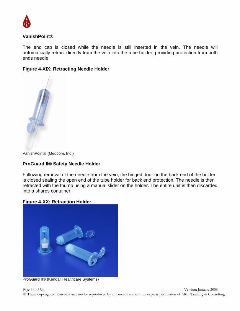
VanishPoint® The end cap is closed while the needle is still inserted in the vein. The needle will automatically retract directly from the vein into the tube holder, providing protection from both ends needle. Figure 4-XIX: Retracting Needle Holder
VanishPoint® (Medcom, Inc.) ProGuard II® Safety Needle Holder Following removal of the needle from the vein, the hinged door on the back end of the holder is closed sealing the open end of the tube holder for back end protection. The needle is then retracted with the thumb using a manual slider on the holder. The entire unit is then discarded into a sharps container. Figure 4-XX: Retraction Holder
ProGuard II® (Kendall Healthcare Systems)
ARO Onsite Training & Consulting Copyright 2008
Page 17 of 34 Version: January 2008© These copyrighted materials may not be reproduced by any means without the express permission of ARO Training & Consulting
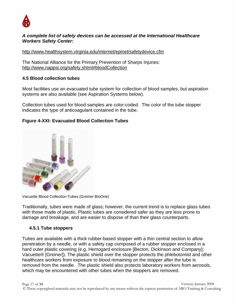
A complete list of safety devices can be accessed at the International Healthcare Workers Safety Center: http://www.healthsystem.virginia.edu/internet/epinet/safetydevice.cfm The National Alliance for the Primary Prevention of Sharps Injuries: http://www.nappsi.org/safety.shtml#bloodCollection 4.5 Blood collection tubes Most facilities use an evacuated tube system for collection of blood samples, but aspiration systems are also available (see Aspiration Systems below). Collection tubes used for blood samples are color-coded. The color of the tube stopper indicates the type of anticoagulant contained in the tube. Figure 4-XXI: Evacuated Blood Collection Tubes
Vacuette Blood Collection Tubes (Greiner-BioOne) Traditionally, tubes were made of glass; however, the current trend is to replace glass tubes with those made of plastic. Plastic tubes are considered safer as they are less prone to damage and breakage, and are easier to dispose of than their glass counterparts.
4.5.1 Tube stoppers Tubes are available with a thick rubber-based stopper with a thin central section to allow penetration by a needle, or with a safety cap composed of a rubber stopper enclosed in a hard outer plastic covering (e.g. Hemogard enclosure [Becton, Dickinson and Company]; Vacuette® [Greiner]). The plastic shield over the stopper protects the phlebotomist and other healthcare workers from exposure to blood remaining on the stopper after the tube is removed from the needle. The plastic shield also protects laboratory workers from aerosols, which may be encountered with other tubes when the stoppers are removed.

ARO Onsite Training & Consulting Copyright 2008
Page 18 of 34 Version: January 2008© These copyrighted materials may not be reproduced by any means without the express permission of ARO Training & Consulting
Figure 4-XXII: Tube Closures
Courtesy and © Becton, Dickinson and Company
Most of the tubes used for serum testing are coated on the inside with silicon to keep blood from sticking to the sides of the tube, and to prevent red blood cell haemolysis. Some tubes contain additives. The term additive refers to any substance that is added to a blood collection tube, and includes anticoagulants, clot activators, and silica gels. Clot activators speed clot production, and barrier gels aid in separation of serum or plasma from the clot following centrifugation. The tube label indicates additives contained in the tube, tube volume and tube expiry date. Anticoagulants prevent blood from clotting so that plasma analysis can be performed – tops are color-coded for quick identification of the specific additives present in the tubes. It is imperative that samples are promptly and thoroughly mixed with the anticoagulant by gently inverting the tube 8 to 10 times upon removal from the needle/tube holder. Specific instructions for each tube suggest inverting the tube anywhere from 5-10 tubes depending on the tube additive. Extra inversion will not affect the sample, whereas insufficient mixing may -incorporating 8 full gentle inversions into your routine will ensure that all of your samples have been adequately mixed. Do not lay the tubes down as you collect them and return to mix them all once the entire collection is complete. If mixing is not thorough, a partial clot may form interfering with test results. Do not shake the tubes as this may result in haemolysis of the red blood cells. Tubes are available in volumes from 2-ml to 15-ml to accommodate various test requirements for adult and paediatric patients.
4.5.2 Evacuated tube system The most common system for collecting blood samples is the evacuated tube system. The evacuated tube system is a closed system in which the patient’s blood goes directly from the vein through the rubber stopper in the tube to the inside of the tube without being exposed to air. Numerous tubes can be collected using a single venipuncture. The evacuated system requires three components: double pointed needle, evacuated sample tube, and plastic
Rubber-stopper
Plastic shield (Hemogard)
ARO Onsite Training & Consulting Copyright 2008
Page 19 of 34 Version: January 2008© These copyrighted materials may not be reproduced by any means without the express permission of ARO Training & Consulting
holder or adapter. All components of the system should be from the same manufacturer to avoid problems with proper needle fit and smooth placement and removal of tubes. Evacuated tubes are used with the evacuated tube system and with the syringe system of blood collection. The tubes produced by Becton, Dickinson and Company are named Vacutainer®; however, the term vacutainer is commonly used to refer to any evacuated tubes. With the evacuated tube, blood can be drawn directly from the vein into the tube without exposing it to air (closed system). The evacuated tube system minimizes accidental contact with blood. With the syringe method, blood is drawn into the barrel of the syringe and then must be transferred into the evacuated tube after collection. Evacuated tubes have a pre-determined vacuum based on the tube volume and the type and amount of additive in the tube. Once the stopper of the tube is punctured with the back-end of the needle in the needle/tube holder, blood is pulled into the tube from the vein by the vacuum. For tubes containing additives, especially anticoagulants, there must be a proper ratio of anticoagulant to blood specimen. The vacuum in the collection tube allows the correct amount of blood to enter the tube. Anticoagulant tubes must be allowed to fill to completion, regardless of whether the anticoagulant is a spray on the inside of the tube, a powder or a liquid. Do not be swayed by co-workers who tell you that you do not have to fill the tubes. The quality and integrity of the sample and the test results may be adversely affected by not filling tubes to completion. A pre-measured vacuum inside the tube allows it to automatically fill with blood when the rubber stopper is penetrated. The tube will not fill with blood if it has lost its vacuum — each tube has a vacuum expiration date guaranteed by the manufacturer and should not be used past this. The tube vacuum can be lost by dropping the tube, opening the tube, advancing the tube too far onto the needle-holder prior to vein puncture or pulling the needle bevel partially out of the skin during the procedure. As mentioned earlier, tubes are available in a number of different sizes ranging from 2 ml to 15 ml. Manufacturers are also beginning to produce shorter draw tubes, which may look the same size as a 5-ml tube, for instance, but are designed to draw in smaller volumes of blood (e.g. 2 ml). These tubes have less vacuum and less additive to maintain recommended ratios of blood to additive for tests performed on that tube. In each case, the tube volume is indicated on the tube. Facilities wishing to reduce the amount of blood drawn from patients may choose to use these small volume or shorter-draw tubes.
4.5.3 Whole blood/plasma tubes anticoagulant tubes In order to obtain whole blood or plasma for testing, an anticoagulant must be contained in the blood collection tube to prevent the blood from clotting. A number of anticoagulants are available depending on the type of testing to be performed.
ARO Onsite Training & Consulting Copyright 2008
Page 20 of 34 Version: January 2008© These copyrighted materials may not be reproduced by any means without the express permission of ARO Training & Consulting
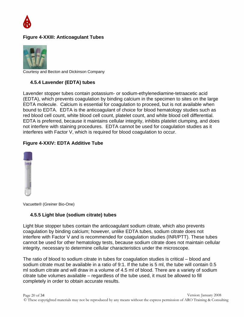
Figure 4-XXIII: Anticoagulant Tubes
Courtesy and Becton and Dickinson Company
4.5.4 Lavender (EDTA) tubes Lavender stopper tubes contain potassium- or sodium-ethylenediamine-tetraacetic acid (EDTA), which prevents coagulation by binding calcium in the specimen to sites on the large EDTA molecule. Calcium is essential for coagulation to proceed, but is not available when bound to EDTA. EDTA is the anticoagulant of choice for blood hematology studies such as red blood cell count, white blood cell count, platelet count, and white blood cell differential. EDTA is preferred, because it maintains cellular integrity, inhibits platelet clumping, and does not interfere with staining procedures. EDTA cannot be used for coagulation studies as it interferes with Factor V, which is required for blood coagulation to occur. Figure 4-XXIV: EDTA Additive Tube
Vacuette® (Greiner Bio-One)
4.5.5 Light blue (sodium citrate) tubes Light blue stopper tubes contain the anticoagulant sodium citrate, which also prevents coagulation by binding calcium; however, unlike EDTA tubes, sodium citrate does not interfere with Factor V and is recommended for coagulation studies (INR/PTT). These tubes cannot be used for other hematology tests, because sodium citrate does not maintain cellular integrity, necessary to determine cellular characteristics under the microscope. The ratio of blood to sodium citrate in tubes for coagulation studies is critical – blood and sodium citrate must be available in a ratio of 9:1. If the tube is 5 ml, the tube will contain 0.5 ml sodium citrate and will draw in a volume of 4.5 ml of blood. There are a variety of sodium citrate tube volumes available – regardless of the tube used, it must be allowed to fill completely in order to obtain accurate results.

ARO Onsite Training & Consulting Copyright 2008
Page 21 of 34 Version: January 2008© These copyrighted materials may not be reproduced by any means without the express permission of ARO Training & Consulting
The 9:1 ratio of blood to sodium citrate is based on the amount of plasma in the specimen; therefore, in patients with polycythemia or other conditions where the hematocrit may be >55%, the amount of anticoagulant must be decreased. In anaemic patients, the amount of anticoagulant must be increased in order to maintain the 9:1 ratio, because of the larger amounts of plasma in these specimens. Figure 4-XXV: Sodium Citrate Additive Tube (1:9 ratio)
Vacuette® (Greiner Bio-One)
4.5.6 Black stopper (sodium citrate) Black stopper tubes contain sodium citrate and can be used for Westergren sedimentation rates (ESR); although many laboratories now perform this test using blood from the lavender topped tube. These tubes differ from the light blue stopper only in that they provide a ratio of blood to anticoagulant of 4:1. Figure 4-XXVI: Sodium Citrate Additive Tube (1:4 Ratio)
Vacuette® (Greiner Bio-One)
4.5.7 Green stopper (heparin) Green stopper tubes contain the anticoagulant heparin linked to ammonium, lithium or sodium salts. Heparin prevents coagulation by inhibiting thrombin, and is the anticoagulant of choice for plasma chemistry testing. Heparin tubes may also be used for STAT chemistry tests when results are required immediately. When serum is used for testing, the laboratory must wait until the blood clot fully forms (~ 30 minutes for silica containing clot activator tubes

ARO Onsite Training & Consulting Copyright 2008
Page 22 of 34 Version: January 2008© These copyrighted materials may not be reproduced by any means without the express permission of ARO Training & Consulting
and ~ 45-60 minutes for non-additive tubes). Ammonia, carboxyhemoglobin, and STAT electrolytes are chemistry tests that may require heparin, depending on the testing laboratory’s collection policies. Light green stopper tubes, called plasma separation tubes (PST), contain lithium heparin and a separation gel. These tubes may be used for potassium testing: heparin prevents release of potassium from platelets, and the gel prevents interference of red blood cell potassium. Figure 4-XXVII: Heparin Additive Tube
Vacuette® (Greiner Bio-One)
4.5.8 Grey stopper (fluoride/oxalate) Grey stopper tubes come with a variety of anticoagulants and additives to preserve glucose. Grey stopper tubes contain glucose preservative (antiglycolytic agent): sodium-fluoride or lithium-iodoacetate. Sodium-fluoride keeps glucose stable for up to 3 days; iodoacetate maintains stability for up to 24 hours. If plasma is required for glucose testing, an anticoagulant must also be present in tubes: potassium oxalate is used which prevents clotting by binding calcium. Tubes cannot be used for other chemistry tests because sodium fluoride interferes with some enzymes. Blood alcohol levels for forensic testing are collected in grey stopper tubes: sodium fluoride inhibits bacterial growth, which could produce alcohol as a metabolic by-product and cause falsely elevated results. Figure 4-XXVIII: Antiglycolytic Additive Tube
Vacuette® (Greiner Bio-One)

ARO Onsite Training & Consulting Copyright 2008
Page 23 of 34 Version: January 2008© These copyrighted materials may not be reproduced by any means without the express permission of ARO Training & Consulting
4.6 Serum tubes
4.6.1 Red stopper Red stopper glass tubes (plain vacuum tubes) do not contain anticoagulants or additives. Blood collected in these tubes clot by the normal coagulation process generally within 60 minutes: serum is yielded when the specimen is centrifuged. These tubes are used for blood bank, but can also be used for chemistry and serology tests. Tubes do not have to be inverted, as there are no additives to mix; however, inversion will not adversely affect the sample. Red stopper plastic tubes contain silica particles that speeds clot formation as in the red/grey stopper tubes. Figure 4-XXIX: Serum Tube
Vacuette® (Greiner Bio-One) 4.6.2 Pink stopper Pink stopper tubes are also available and may be used specifically for blood bank, because they stand out from other specimens. These tubes do not contain anticoagulants or additives.
4.6.3 Gold Hemogard and red/grey stopper Red/grey or mottled rubber stoppers (gold Hemogard®) are found in tubes containing a clot activator and separation gel. Red/grey tubes may be referred to as serum separator tubes (SST). They include substances such as glass or silica particles, and inert clays (celite), which increase surface area for platelet activation, shortening the time for clot formation. Blood will form a clot in red/grey stopper tubes in as few as 20 minutes, reducing the amount of time necessary between sample collection and testing (serum for testing should not be removed from the sample until a clot has fully formed). The gel separator (thixotropic gel) is a synthetic substance that forms a physical barrier between the clot and the serum after the specimen has been centrifuged preventing contamination of serum by cellular elements that may alter test results. Specimens must be allowed to clot completely before they are

ARO Onsite Training & Consulting Copyright 2008
Page 24 of 34 Version: January 2008© These copyrighted materials may not be reproduced by any means without the express permission of ARO Training & Consulting
centrifuged to ensure a solid separation barrier. These tubes are used for most chemistry and immunology tests. They cannot be used for blood bank tests and drug testing. Figure 4-XXX: Gel Separator Serum Tubes
Vacutainer® (Courtesy and © Becton, Dickinson and Company)
4.7.4 Yellow/grey stopper Yellow/grey rubber stopper tubes (orange Hemogard®) contain thrombin as a clot activator, resulting in faster clot formation (5 minutes). These tubes may be used for STAT serum chemistry testing and for patients receiving anticoagulant therapy. 4.7 Specialized blood collection tubes
4.7.1 Dark blue stopper Dark blue stopper tubes are used for toxicology, trace metal, and nutritional testing. These are available with or without anticoagulants (sodium heparin or EDTA). The tubes are chemically clean and the rubber stoppers are guaranteed to contain the lowest possible levels of metal. These tubes must be collected before regular rubber stopper tubes, so that the stopper does not become contaminated with metals and minerals from regular tube stoppers. A discard tube can be collected, as appropriate, depending on the remainder of the tubes to be collected.
4.7.2 Brown stopper Brown stopper tubes (Hemogard®) contain sodium heparin and are used for lead testing. They are certified to contain less than 0.01 g/ml of lead.

Page 25 of 34 Version: January 2008© These copyrighted materials may not be reproduced by any means without the express permission of ARO Training & Consulting
ARO Onsite Training & Consulting Copyright 2008
4.7.3 Yellow stopper
Yellow stopper tubes are available containing two different additives:
1. Acid citrate dextrose (ACD): preserves red blood cells – used for blood bank cellular studies, tissue typing, and some types of molecular testing
2. Sodium polyanetholsulfonate (SPS): inhibits action of complement, phagocytes, and certain antibiotics – used for blood cultures for culture of micro-organisms

Page 26 of 34 Version: January 2008© These copyrighted materials may not be reproduced by any means without the express permission of ARO Training & Consulting
ARO Onsite Training & ConsultingCopyright 2008
Table 4-I: Blood Collection Tubes (Additives, Function and Use) Stopper Colour Additive Mode of Action Comments
Red glass None Blood clots ~ 45 min resulting in serum and clot
May be used for any test requiring serum, especially those affected by presence of gel (e.g. drug levels)
Red plastic Glass/silica Blood clots ~ 20 min resulting in serum and clot
May be used for any test requiring serum, especially those affected by presence of gel (e.g. drug levels)
Gold or red/grey Glass/silica Gel separator
Glass/silica acts as clot activator Gel separates serum from cells following centrifugation
Testing requiring serum –most chemistry and hematology tests
Lavender Ethylenediamine tetra-acetate (EDTA)
Binds calcium Whole blood for hematology tests (whole blood counts, HbA1C, blood smears, etc.)
Light blue Sodium citrate (9:1 ratio) Binds calcium Plasma for coagulation tests (INR/PTT)
Black Sodium citrate (4:1 ratio) Binds calcium Erythrocyte sedimentation rare (ESR)
Green Heparin (sodium, lithium or ammonium heparin)
Anti-thrombin Plasma chemistry tests
Green/grey Lithium heparin Gel separator
Anti-thrombin Gel separates plasma from cells following centrifugation
Tests requiring plasma (e.g. urgent chemistry tests)
Grey Potassium oxalate Sodium fluoride
Binds calcium Glycolytic inhibitor - stabilizes glucose for up to 3 days
Glucose and legal alcohol testing
Dark blue Sodium heparin Anti-thrombin Trace elements, toxicology and nutrient analysis
Brown Sodium heparin Anti-thrombinLead determinations Tube certified to contain < 0.1 µg/ml lead
Stopper Colour Additive Mode of Action Comments
Yellow
Sodium polyanethol-sulfonate (SPS) Acid-citrate dextrose (ACD)
Blood cultures Tissue typing and lymphocyte viability
ARO Onsite Training & Consulting Copyright 2008
Page 27 of 34 Version: January 2008© These copyrighted materials may not be reproduced by any means without the express permission of ARO Training & Consulting
4.8 Aspiration systems
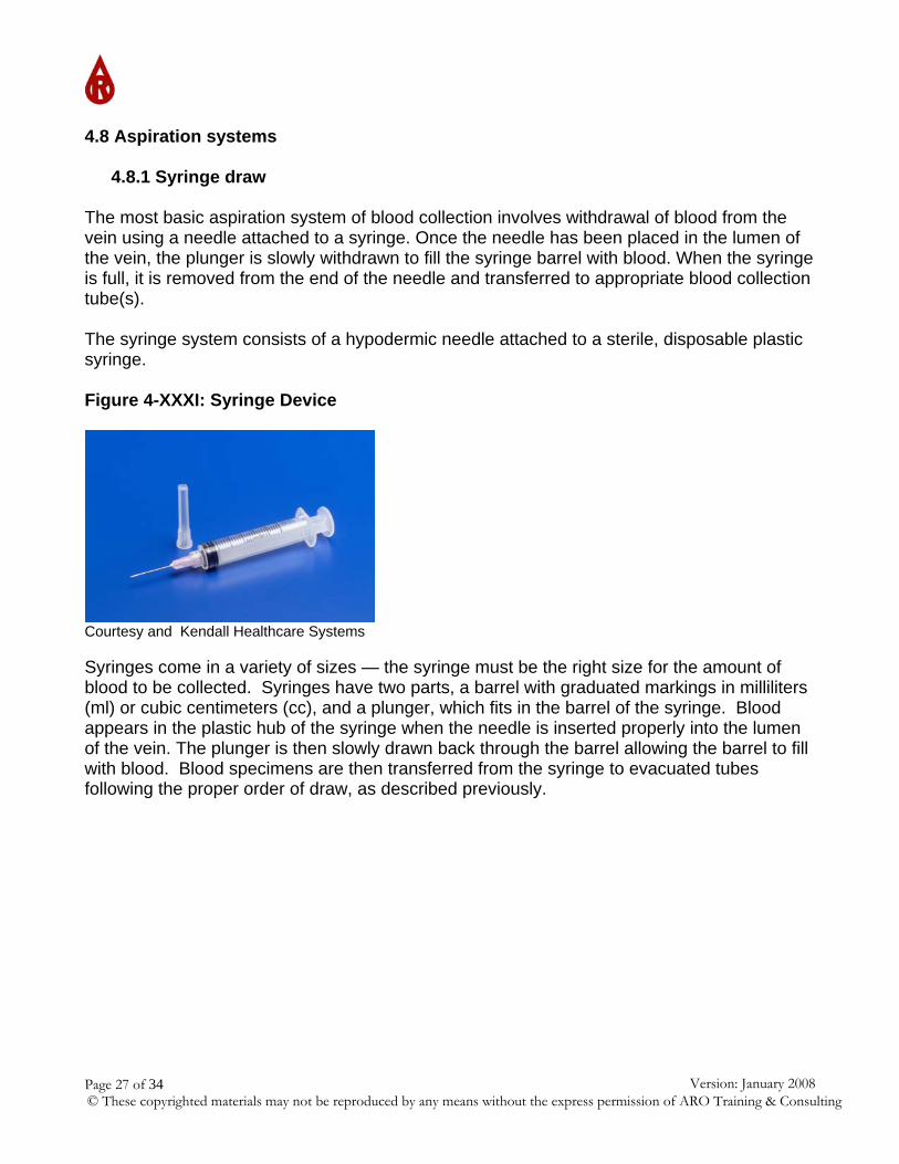
4.8.1 Syringe draw The most basic aspiration system of blood collection involves withdrawal of blood from the vein using a needle attached to a syringe. Once the needle has been placed in the lumen of the vein, the plunger is slowly withdrawn to fill the syringe barrel with blood. When the syringe is full, it is removed from the end of the needle and transferred to appropriate blood collection tube(s). The syringe system consists of a hypodermic needle attached to a sterile, disposable plastic syringe. Figure 4-XXXI: Syringe Device
Courtesy and Kendall Healthcare Systems Syringes come in a variety of sizes — the syringe must be the right size for the amount of blood to be collected. Syringes have two parts, a barrel with graduated markings in milliliters (ml) or cubic centimeters (cc), and a plunger, which fits in the barrel of the syringe. Blood appears in the plastic hub of the syringe when the needle is inserted properly into the lumen of the vein. The plunger is then slowly drawn back through the barrel allowing the barrel to fill with blood. Blood specimens are then transferred from the syringe to evacuated tubes following the proper order of draw, as described previously.

ARO Onsite Training & Consulting Copyright 2008
Page 28 of 34 Version: January 2008© These copyrighted materials may not be reproduced by any means without the express permission of ARO Training & Consulting
Figure 4-XXXII Syringe Sizes
6-ml syringe 12-ml syringe 20-ml syringe Courtesy and © Kendall Healthcare Systems The major advantage to the syringe draw is the ability to control the amount of pressure applied during blood collection. Syringe draw may be necessary when attempting to collect blood from patients whose veins are too fragile to withstand the vacuum pressure associated with evacuated tubes. Pressure can be applied gently to the syringe plunger to draw blood into the syringe without collapsing the vein. Disadvantages associated with syringe use include: • Increased risk of needle-stick injury • Poorer quality samples resulting from delay in transfer of blood to tubes containing
required additives • Increased risk of haemolysis resulting from excessive plunger force, loose fittings of
needle assembly and syringe, and transfer of blood from syringe to tubes Blood draw using needle and syringe should only be performed when absolutely necessary and only by experienced phlebotomists. Use of needle/syringe assembles is associated with a higher rate of needle-stick injury and test results may be affected by delay between transfer of blood to additive containing tubes. One of the risks associated with syringe draws is transfer of blood from the syringe to the appropriate blood collection tubes. Blood transfer should be performed using a blood transfer device rather than by injecting the syringe needle into the tops of the blood collection tubes.
ARO Onsite Training & Consulting Copyright 2008
Page 29 of 34 Version: January 2008© These copyrighted materials may not be reproduced by any means without the express permission of ARO Training & Consulting
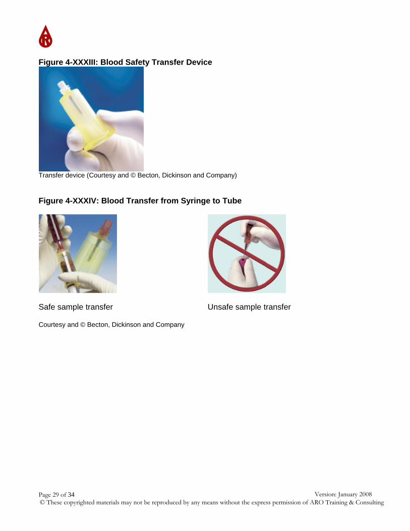
Figure 4-XXXIII: Blood Safety Transfer Device
Transfer device (Courtesy and © Becton, Dickinson and Company) Figure 4-XXXIV: Blood Transfer from Syringe to Tube
Safe sample transfer Unsafe sample transfer Courtesy and © Becton, Dickinson and Company

ARO Onsite Training & Consulting Copyright 2008
Page 30 of 34 Version: January 2008© These copyrighted materials may not be reproduced by any means without the express permission of ARO Training & Consulting
A combination syringe/aspiration tube system is also available from Sarstedt (S-Monovette®). http://www.sarstedt.com/php/main.php The S-Monovette® is an enclosed multiple-sampling blood collection system that utilizes either an aspiration or vacuum method of collection. The aspiration tube system is modeled on blood aspiration using a needle and syringe, except that blood does not have to be transferred to blood collection tubes following withdrawal of blood from the vein. Blood is aspirated into a tube containing a plunger by applying gentle force to the plunger Figure 4-XXXV: Syringe/Aspiration Combination System
S-Monovette® (Courtesy and Sarstedt Company)
4.8.2 Order of draw When multiple tubes are to be drawn, the proper order of draw for blood testing is sterile sample(s), followed by sample(s) for coagulation studies, followed by serum sample(s), then tubes containing anticoagulants. The order of draw recommended by the Clinical and Laboratory Standards Institute (CLSI) is listed below:
1. Blood cultures 2. Light blue stopper, sodium citrate (INR/PTT) tube
(If a light blue-stopper tube is the first or only tube to be drawn and specific factor testing is required (e.g. Factor V assay), a DISCARD tube should be drawn first to eliminate tissue thromboplastin contamination picked up during needle penetration, which may alter test results. A discard tube must also be drawn to avoid a “short draw” when coagulation sample is collected using a winged-infusion set (tubing contains ~ 0.5 ml of air-space reducing the total blood volume by 0.5 ml). Tubes suitable for use as discard tubes are those that do not contain any additives (e.g. plain glass red stopper) or a tube containing the same contents as the coagulation tube (i.e. light-blue sodium-citrate).
3. Serum tubes - red stopper glass tube, red/grey stopper SST tube and/or red stopper plastic tube (contains clot activator).

ARO Onsite Training & Consulting Copyright 2008
Page 31 of 34 Version: January 2008© These copyrighted materials may not be reproduced by any means without the express permission of ARO Training & Consulting
Prior to the most current revision of the CLSI venipuncture document, the recommended order of draw placed the red stopper tube before the light blue sodium citrate tube. With the introduction of plastic blood collection tubes, the order of draw had to be changed to reflect the clot activator that is present in plastic red stopper blood collection tube, precluding their collection before tubes for coagulation studies.
4. Green or green/grey stopper (heparin) tube 5. Lavender stopper, EDTA (CBC) tube 6. Grey stopper, oxalate/fluoride
The purpose of the proper order of draw is to avoid contaminating one tube with carry-over additive from the preceding tube. Certain additives may affect test results. If for any reason the order of draw is inadvertently altered, a discard tube can be collected before collecting the next tube in the recommended order or draw. 4.9 Antiseptics and disinfectants Antiseptics are substances that prevent the growth of bacteria. They are used to prevent sepsis resulting from the presence of microorganisms or their toxic by-products in the bloodstream. They are safe for use on human skin and are used to clean the skin before venipuncture or skin puncture. All skin surfaces contain some bacteria. Bacteria must be washed away before penetrating the skin to reduce the risk of introducing organisms into the bloodstream, which may cause infection. A common antiseptic used for routine blood collection is 70% isopropyl alcohol (isopropanol). Soap and water can also be used for patients who are allergic to isopropanol. Other antiseptics such as povidone-iodine (i.e. Betadine) and 0.5% chlorhexidine gluconate (used on iodine allergic patients) can be used when blood cultures are drawn: skin antisepsis is a very important part of the blood culture collection procedure. Failure to follow proper procedure may result in organisms being introduced into the blood culture bottles. All organisms isolated from these bottles are reported to the patient’s physician: reporting organisms that have actually come from the skin rather than the patient’s blood may result in inappropriate treatment and additional expense. Disinfectants are bactericidal agents that are generally not safe for use on human skin. They are used on surfaces and instruments. The most common disinfectant is household bleach (5.25% sodium hypochlorite): a 1:10 solution will kill the viruses that cause hepatitis and AIDS. It is commonly used to wipe surfaces and clean up blood spills. 4.10 Cotton balls/gauze pads Packaged sterile 2” x 2” or 3” x 3” gauze pads are generally used to hold pressure over the venipuncture site until bleeding has stopped. Cotton or rayon balls may also be used, but tends to stick to the venipuncture site causing it to bleed again on removal.
ARO Onsite Training & Consulting Copyright 2008
Page 32 of 34 Version: January 2008© These copyrighted materials may not be reproduced by any means without the express permission of ARO Training & Consulting
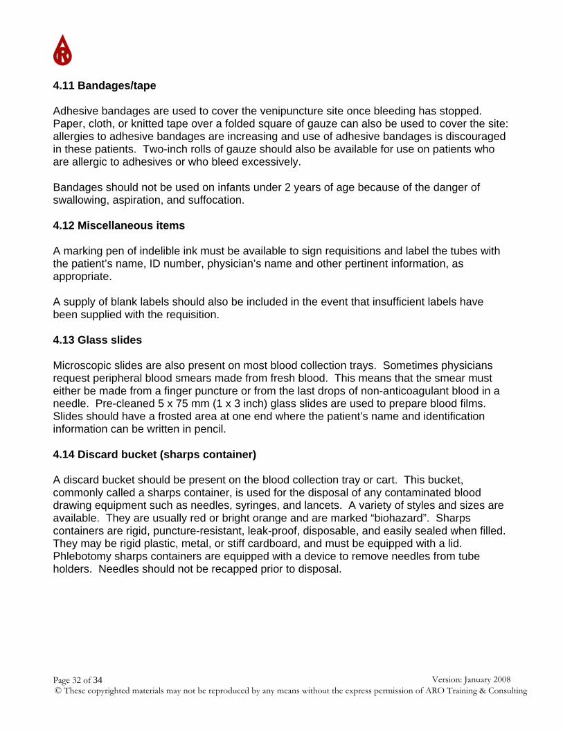
4.11 Bandages/tape Adhesive bandages are used to cover the venipuncture site once bleeding has stopped. Paper, cloth, or knitted tape over a folded square of gauze can also be used to cover the site: allergies to adhesive bandages are increasing and use of adhesive bandages is discouraged in these patients. Two-inch rolls of gauze should also be available for use on patients who are allergic to adhesives or who bleed excessively. Bandages should not be used on infants under 2 years of age because of the danger of swallowing, aspiration, and suffocation. 4.12 Miscellaneous items A marking pen of indelible ink must be available to sign requisitions and label the tubes with the patient’s name, ID number, physician’s name and other pertinent information, as appropriate. A supply of blank labels should also be included in the event that insufficient labels have been supplied with the requisition. 4.13 Glass slides Microscopic slides are also present on most blood collection trays. Sometimes physicians request peripheral blood smears made from fresh blood. This means that the smear must either be made from a finger puncture or from the last drops of non-anticoagulant blood in a needle. Pre-cleaned 5 x 75 mm (1 x 3 inch) glass slides are used to prepare blood films. Slides should have a frosted area at one end where the patient’s name and identification information can be written in pencil. 4.14 Discard bucket (sharps container) A discard bucket should be present on the blood collection tray or cart. This bucket, commonly called a sharps container, is used for the disposal of any contaminated blood drawing equipment such as needles, syringes, and lancets. A variety of styles and sizes are available. They are usually red or bright orange and are marked “biohazard”. Sharps containers are rigid, puncture-resistant, leak-proof, disposable, and easily sealed when filled. They may be rigid plastic, metal, or stiff cardboard, and must be equipped with a lid. Phlebotomy sharps containers are equipped with a device to remove needles from tube holders. Needles should not be recapped prior to disposal.

ARO Onsite Training & Consulting Copyright 2008
Page 33 of 34 Version: January 2008© These copyrighted materials may not be reproduced by any means without the express permission of ARO Training & Consulting
Figure 4-XXXVI: Approved Sharps Containers
© Arbique-Rendell Onsite Training & Consulting 4.15 Warming equipment Equipment for warming the puncture site should be available in case it becomes necessary to dilate the blood vessels. The temperature of warming equipment should not exceed 42°C to prevent damage to the patient’s skin. Warming equipment should also be available to warm blood tubes for tests requiring storage at 370C prior to testing. 4.16 Ice Ice or a refrigerant of some type should be available for transport of specimens that must be chilled during transport to the laboratory.
ARO Onsite Training & Consulting Copyright 2008
Page 34 of 34 Version: January 2008© These copyrighted materials may not be reproduced by any means without the express permission of ARO Training & Consulting
References
1. Becton, and Dickinson Product Catalogue. Retrieved from: http://www.bd.com/vacutainer/products/venous/. Accessed on: 06/12/30.
2. Clinical Laboratory and Standards Institute. Procedure for the Collection of Diagnostic
Blood Specimens by Venipuncture; Approved Standard - Fifth Edition. CLSI; H3-A5; 23(32); 2003.
3. Greiner Bio-One Product Catalogue. Retrieved from: http://www.vacuette.com/.
Accessed on: 06/12/30.
4. ICU Medical Inc. Product Catalogue. Retrieved from: http://www.icumed.com/. Accessed on: 06/12/30.
5. Kendall Healthcare Systems Product Catalogue. Retrieved from:
http://www.kendallhq.com. Accessed on: 06/12/30.
6. Sarstedt Product Catalogue. Retrieved from: http://www.sarstedt.com/php/main.php. Accessed on: 06/12/30.
7. Smiths Medical Product Catalogue. Retrieved from: http://www.smiths-
medical.com/catalog/blood-draw/venipuncture/needle-pro/. Accessed on: 06/12/30.
8. VanishPoint Product Catalogue. Retractable Technologies Inc. Retrieved from: http://www.sarstedt.com/php/main.php. Accessed on: 06/12/30.