veno-arterial extracorporeal membrane oxygenation for
TRANSCRIPT

Chapter 10
Veno-Arterial ExtracorporealMembrane Oxygenation for RefractoryCardiogenic Shock and Cardiac Arrest
Francesco Formica and Giovanni Paolini
Additional information is available at the end of the chapter
http://dx.doi.org/10.5772/54719
1. Introduction
Cardiogenic shock (CS) following acute myocardial infarction (AMI) occurs in 7% to 9% ofpatients affected by AMI with a high mortality rates. Despite all recent advanced treatmentssuch as use of inotropes, vasoconstrictors and intra-aortic balloon pump (IAPB) therapy,revascularization techniques and application of different systems of mechanical circulatorysupport, CS is still the most common cause of hospital mortality ranging between 60%-70%compared to patients with AMI without advanced CS whose hospital mortality is about 10%[1]. Cardiac arrest is a major cause of unexpected death and complicates about 22% of patientswith acute myocardial infarction [2]. Cardiac arrest has a poor prognosis, and despite con‐ventional cardiopulmonary resuscitation (CPR) maneuvers, only a few patients can fullyreturn to a normal lifestyle. The main reasons for very poor outcome and prognosis in CA area lack of return of spontaneous circulation (ROSC), a long time of CPR [3],[4], re-arrest fromhemodynamic instability after ROSC, hypoxic encephalopathy [5], out-of-hospital CA [6-8]. Inboth refractory CS and CA secondary AMI, which are very critical circumstances, Veno-Arterial Extracorporeal Membrane Oxygenation (V-A ECMO) has been proposed and utilizedduring the last decades to obtain rapid resuscitation, stabilization, and subsequent triage to amore permanent treatment strategy.
The aim of this chapter is to describe the more recent indications, techniques and results in theusage of the V-A ECMO in patients with refractory cardiogenic shock and cardiac arrestsecondary to acute myocardial infarction.
© 2013 Formica and Paolini; licensee InTech. This is an open access article distributed under the terms of theCreative Commons Attribution License (http://creativecommons.org/licenses/by/3.0), which permitsunrestricted use, distribution, and reproduction in any medium, provided the original work is properly cited.

2. Definition of V-A ECMO
Extracorporeal membrane oxygenation is essentially a modification of the cardiopulmonarybypass circuit, which is used routinely in cardiac surgery. Blood is removed from the venoussystem, oxygenated by an oxygenator and then returned back to the body by a pump. ECMOprovides both full cardiac and respiratory support. In brief, every ECMO system is basicallya closed tubing loop with the interpolation of a blood pump (centrifugal or roller) and anoxygenator. Two vessel cannulas complete the system. Technically the ECMO system is morecomplex and several configurations have been developed according to the primary etiology.
The veno-arterial ECMO configuration is a tubing loop with a venous arm connected to avenous cannula to allow the venous blood drainage and an arterial arm to return back theoxygenated blood inside the patient’s circulatory system. This mode provides both cardiacand respiratory support and can be achieved by either peripheral or central cannulation(Figure1).
Figure 1. Patient supported with peripheral V-A ECMO
The veno-venous ECMO mode refers to blood, which is drained from the venous system andreturned back to the venous system. This mode only provides respiratory support and isobtained by peripheral cannulation, usually of both femoral veins and jugular vein. ThisECMO configuration will be not discussed in this chapter.
2.1. Definition of cardiogenic shock
Cardiogenic shock is a state of impaired and non-physiologic end-organ perfusion owing to alow cardiac output. Being characterized by hypotension cardiogenic shock is defined mainlyby haemodynamic parameters as follows [9,10]:
a. a systolic blood pressure of less than 90 mmHg for more than 30 min with normovolemia;
b. the need of inotropic drugs to obtain a systolic blood pressure more than 90 mmHg with;
Principles and Practice of Cardiothoracic Surgery274

c. a cardiac index less than 1.8 L/min/m2 without inotropic support and 2.0–2.2 L/min/m2with inotropic and intra-aortic balloon pump (IABP) support;
d. high left ventricular (LV) filling pressures (pulmonary capillary wedge pressure morethan 18 mmHg).
End-organ hypoperfusion may be manifested clinically by:
a. pale, cool, and clammy peripheries;
b. alteration in mental status such delirium, confusion, clouded sensorium, psychomotoragitation;
c. decreased urine output (less than 1 ml/kg/min);
d. pulmonary congestion or edema;
e. tachycardia;
f. hyperlactacidemia (more than 3.mmol/L) as expression of impaired peripheral microcir‐culation;
g. mixed venous saturation of less than 65%.
3. Definition of in-hospital and out-hospital cardiac arrest
Cardiac arrest is a major cause of unexpected death in developed countries with a lowprobability of patient survival. Survival is influenced by several variables common to both in-hospital and out-of-hospital arrest, such as time to recognition of the cardiac arrest, time toinitiation of CPR, rhythm presentation, first defibrillation [2, 11, 12]. In current resuscitationguidelines for in-hospital cardiac arrest (IHCA) patients [13], CPR using ECMO (E-CPR) hasbeen assigned a low-grade recommendation. It is reported that ECMO for out-of-hospitalcardiac arrest (OHCA) has worse outcomes compared with ECMO for IHCA patients [7, 14].In the United States, more than 166,000 patients experience an OHCA annually [15] andapproximately 60% are treated by emergency medical services. OHCA survival to hospitaldischarge range from 0.3% in Detroit [16] to 20.4% in Slovenia [17].Five clinical criteria topredict survival from OHCA [18] have recently been reported. They are: cardiac arrestwitnessed by a bystander, arrest witnessed by emergency medical personnel, provision ofbystander CPR, shockable cardiac rhythm, and return of spontaneous circulation (ROSC) inthe field. These criteria are applicable on IHCA too.
4. Indication and contraindications for v-a ECMO
Patient selection is a crucial point when the physician needs to take the decision to instituteECMO and several considerations must be focused up. Most importantly, it must be consider
Veno-Arterial Extracorporeal Membrane Oxygenation for Refractory Cardiogenic Shock and Cardiac Arresthttp://dx.doi.org/10.5772/54719
275

the likelihood of heart and end organs recovery. If the organs failure is thought to be reversiblewith ECMO, in such situation the device application is to be encouraged. If the likelihood ofrecovery of the heart or other end organs is thought to be very low or even impossible, thenother factors must be taken into account. In such clinical scenario, the decision to instituteECMO should be based on an experienced ECMO team approach, which has to evaluate thepatient’s eligibility for heart transplantation or a definitive mechanical assist device (LVAD)implant as destination therapy.
4.1. Indications for v-a ECMO
The following factors need to be evaluated for the indications [19]:
• Age of patient and body surface area;
• sufficient medical expertise in the field of ECMO;
• possibilities for myocardial revascularization therapy, such as a coronary artery bypassgrafting or coronary angioplasty;
• possibilities for heart transplantation or LVAD implant as destination therapy;
• status of central organs such as kidney, liver, and brain.
4.2. Contraindications for v-a ECMO
Contraindications to the institution of v-a ECMO include [20]:
• disseminated malignancy;
• advanced age;
• graft vs. host disease;
• known severe brain injury;
• unwitnessed cardiac arrest or cardiac arrest of prolonged duration;
• aortic dissection aortic incompetence.
5. Equipment of v-a ECMO circuit
5.1. ECMO circuit
The ECMO circuit is made of PVC tubing and the diameter of lines varies from ¼ inch for aneonate to ½ inch for pediatric and adult patients. The length of the circuit is kept not morethan 2 meters to avoid increasing of resistance within a tube and twisting, but the length shouldbe suitable to allow the movements of the patient by ECMO staff.
Areas of turbulent flow can predispose to clot formation; therefore loop and connectors shouldbe avoided or kept at minimum.
Principles and Practice of Cardiothoracic Surgery276
5.2. ECMO cannulas and cannulation techniques
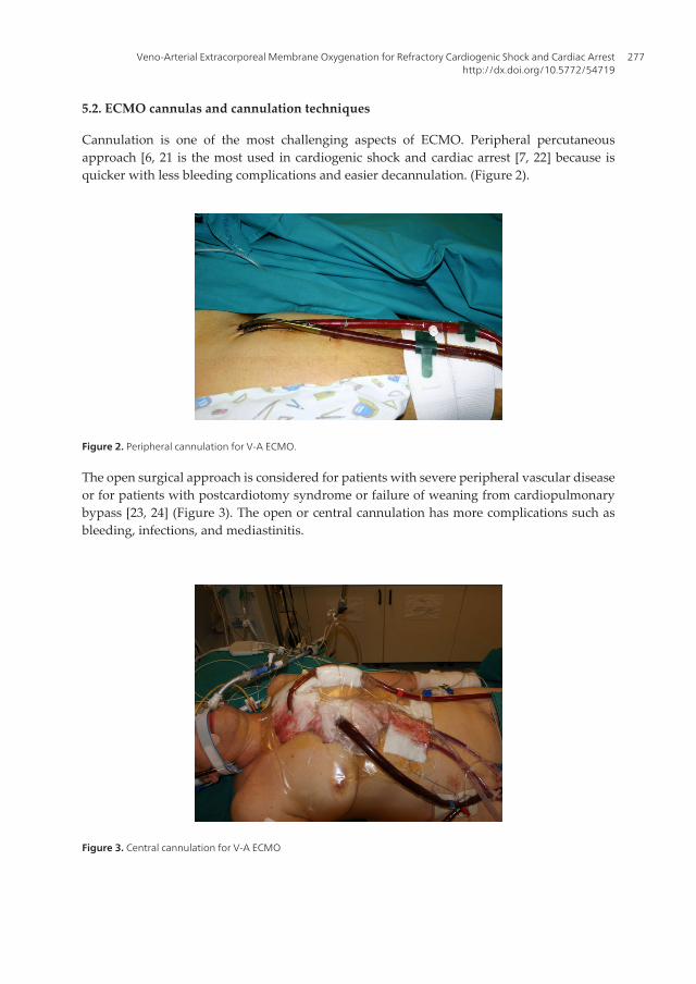
Cannulation is one of the most challenging aspects of ECMO. Peripheral percutaneousapproach [6, 21 is the most used in cardiogenic shock and cardiac arrest [7, 22] because isquicker with less bleeding complications and easier decannulation. (Figure 2).
Figure 2. Peripheral cannulation for V-A ECMO.
The open surgical approach is considered for patients with severe peripheral vascular diseaseor for patients with postcardiotomy syndrome or failure of weaning from cardiopulmonarybypass [23, 24] (Figure 3). The open or central cannulation has more complications such asbleeding, infections, and mediastinitis.
Figure 3. Central cannulation for V-A ECMO
Veno-Arterial Extracorporeal Membrane Oxygenation for Refractory Cardiogenic Shock and Cardiac Arresthttp://dx.doi.org/10.5772/54719
277

Percutaneous cannulas are usually made of polyurethane (Figure 4) and they are inserted usingthe Seldinger technique (Figure 5).
Figure 4. Percutaneous arterial cannulas (right) and percutaneous venous cannulas (left).
Figure 5. Percutaneous cannula insertion by Seldinger technique.
The size of the cannulas depends on the size of the patient; usually the arterial cannula rangesbetween 17 Fr to 21 Fr and the venous cannula ranges between 21 Fr and 25 Fr. Cannulas ofsufficient size are required to support high blood flow with low resistance. Local complica‐tions, particularly at the site of peripheral insertion of VA-ECMO can occur, of which the mostconcerning is leg ischemia. For this reason all attempts the limb perfusion is restored, afternoted the absence of anterior and posterior tibial artery flow, by inserting a 9-Fr catheterdistally to the arterial cannula by means of vascular ultrasound scan as soon as possible afterECMO implantation (Figure 6).
Principles and Practice of Cardiothoracic Surgery278

Figure 6. Distal leg perfusion to restore the blood flow.
Some Authors [25] suggested to insert a catheter for a distal perfusion if the mean pressure ofthe superficial femoral artery is lower than 50 mmHg.
Alternative arterial approach, such as axillary arterial cannulation, has been reported [26](Figure 7).
Figure 7. Cannulation of right axillary artery (tube on the right). Cannula is tunneled below the skin to protect by acci‐dental trauma.
Whatever the type of approach cannulation is considered, it requires always a highly skilledmedical staff, usually a cardiac or vascular surgeon, who are able to undertake this procedureunder often very difficult conditions as the patients are so unstable or even in cardiac arrest.
5.3. Pumps
The pump pushes the blood through the oxygenator and then back to the patient. Themost used pump in adult patients is the centrifugal pump. These pumps consist of a pol‐ycarbonate housing with a one-point sapphire bearing linked to a magnetic field, whichcreate a vortex flow at an adjustable rate (Figure 8). Vortex creates a negative pressure in
Veno-Arterial Extracorporeal Membrane Oxygenation for Refractory Cardiogenic Shock and Cardiac Arresthttp://dx.doi.org/10.5772/54719
279
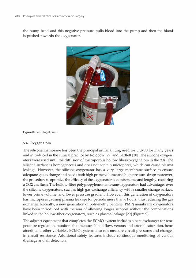
the pump head and this negative pressure pulls blood into the pump and then the bloodis pushed towards the oxygenator.
Figure 8. Centrifugal pump.
5.4. Oxygenators
The silicone membrane has been the principal artificial lung used for ECMO for many yearsand introduced in the clinical practice by Kolobow [27] and Bartlett [28]. The silicone oxygen‐ators were used until the diffusion of microporous hollow fibers oxygenators in the 90s. Thesilicone surface is homogeneous and does not contain micropores, which can cause plasmaleakage. However, the silicone oxygenator has a very large membrane surface to ensureadequate gas exchange and needs both high prime volume and high pressure drop; moreover,the procedure to optimize the efficacy of the oxygenator is cumbersome and lengthy, requiringa CO2 gas flush. The hollow-fiber polypropylene membrane oxygenators had advantages overthe silicone oxygenators, such as high gas exchange efficiency with a smaller change surface,lower prime volume, and lower pressure gradient. However, this generation of oxygenatorshas micropores causing plasma leakage for periods more than 6 hours, thus reducing the gasexchange. Recently, a new generation of poly-methylpentene (PMP) membrane oxygenatorshave been introduced with the aim of allowing longer support without the complicationslinked to the hollow-fiber oxygenators, such as plasma leakage [29] (Figure 9).
The adjunct equipment that completes the ECMO system includes a heat exchanger for tem‐perature regulation, monitors that measure blood flow, venous and arterial saturation, hem‐atocrit, and other variables. ECMO systems also can measure circuit pressures and changesin circuit resistance. Additional safety features include continuous monitoring of venousdrainage and air detection.
Principles and Practice of Cardiothoracic Surgery280

Figure 9. V-A ECMO circuit with polymethilpentene oxygenator (blue case)
Recently a new miniaturized system for V-A ECMO was introduced in the clinical practice.The system has the console directly connected with the oxygenator and the blood pump, whichare integrated to each other. The console has a touch screen where is possible to monitorcontinuously several parameters such has hematocrit, hemoglobin, SVO2, resistance at theinlet and the outlet of oxygenator and the pressure drop (Figure 10).
Figure 10. On the left the miniaturized V-A ECMO system (Cardiohelp by Maquet); on the right the particular of con‐sole.
This system is very useful because of his reduced dimension and weight (the weight of consoleis about 10 Kgs); for this reason this system can be used for transportation of V-A ECMOpatients within the different sites of the hospital or from hospital to other hospital.
Veno-Arterial Extracorporeal Membrane Oxygenation for Refractory Cardiogenic Shock and Cardiac Arresthttp://dx.doi.org/10.5772/54719
281
6. Management of v-a ECMO
Systemic heparinization is obtained with an intravenous bolus of 5,000 UI of heparin, 5 minutesprior to vessels cannulation. ECMO blood flow is calculated to maintain a Cardiac Output(CO) index of 2.5 L/min/m2, an SvO2 of about 70% and a mean blood arterial pressure of 60-70mmHg during the first 24-48 hours. Continuous intravenous heparin is administered in orderto achieve an activate clotting time (ACT) of 160-180 seconds and a prothrombin time value of50-60 seconds. Small doses of inotrope (dobutamine, 5 to 7 μg/Kg/min) are given to maintainthe ventricular ejection, to allow the opening of aortic valve and to prevent the formation ofclots inside the left ventricle (LV). Oxygenator is always connected with a heat exchange tomaintain a body temperature of 36 °C. Those patients who had a cardiac arrest before ECMOimplantation are gradually cooled to 32-34°C during the first 24-36 hours. The assessment ofthe neurologic status is initiated by electroencephalography after body rewarming; serialneurologic evaluations and cerebral computed tomography scan are always considered toassess cerebral hemorrhage, stroke or hypoxic encephalopathy. In those patients with criteriaof irreversible brain damage, ECMO withdrawn is usually considered.
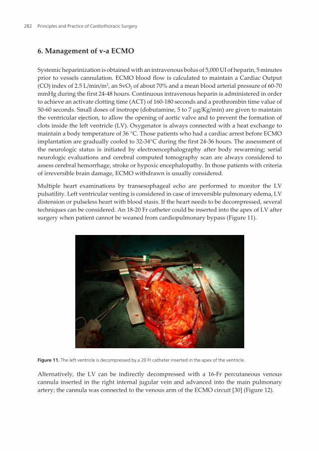
Multiple heart examinations by transesophageal echo are performed to monitor the LVpulsatility. Left ventricular venting is considered in case of irreversible pulmonary edema, LVdistension or pulseless heart with blood stasis. If the heart needs to be decompressed, severaltechniques can be considered. An 18-20 Fr catheter could be inserted into the apex of LV aftersurgery when patient cannot be weaned from cardiopulmonary bypass (Figure 11).
Figure 11. The left ventricle is decompressed by a 20 Fr catheter inserted in the apex of the ventricle.
Alternatively, the LV can be indirectly decompressed with a 16-Fr percutaneous venouscannula inserted in the right internal jugular vein and advanced into the main pulmonaryartery; the cannula was connected to the venous arm of the ECMO circuit [30] (Figure 12).
Principles and Practice of Cardiothoracic Surgery282

Figure 12. The cannula is inserted in the right jugular vein and advanced up to the main arterial pulmonary trunk.
Other techniques of LV decompression have been described such as the use of Pulse-Cath [31],Insertion of a pigtail inside the LV through the aortic valve [32], use of a transeptal atrialcannula [33].
Red blood cells (RBCs) transfusions are given to achieve a hematocrit of 30-32% and plateletsare infused when the platelet count is less than 50,000-60,000/μL. Mechanical ventilation iscontinued throughout the ECMO support with the same management for each patient.Ventilator setting is commonly set at a tidal volume of 8 ml/Kg, 4 breaths/min, positive endexpiratory pressure of 10 cm H20 and a FiO2 of 0.40-0.60.
Intraortic balloon pump (IABP) [24, 34] is employed with the aim to reduce the afterload, toincrease the coronary and cerebral perfusion [35] and to maintain a pulsatile flow.
No attempts to wean off ECMO are usually considered during the first 48 hours. Step bystep is the main strategy for weaning off ECMO using transesophageal echocardiographymonitoring. This consists to reduce the pump flow at 1.0 L/min/m2 for about 40-60 mi‐nutes after having obtained an ACT of 180 seconds. In patients who are supported alsowith IABP, this is set to 1:1 ratio. If systemic pressure, LV contractility, central venouspressure, wedge pressure and SvO2 had not significant changes without the addition ofnew inotropes, then heparin is stopped and ECMO is removed at patient’s bedside or inoperating room within the next few hours.
Transthoracic and transesophageal echocardiography play a very important role in theassessment of LV and RV function and during the delicate phase of weaning from ECMO.Echocardiographic knowledge and facilities are becoming mandatory to start and continue anECMO program with the aim to improve the outcome. Such echocardiographic parameters,such as transmitral E velocity, E/e’ ratio, LV ejection fraction, aortic valve velocity-timeintegral, tissue Doppler lateral Ea, Sa, and parameters derived from Velocity Vector Imaging,
Veno-Arterial Extracorporeal Membrane Oxygenation for Refractory Cardiogenic Shock and Cardiac Arresthttp://dx.doi.org/10.5772/54719
283
including lateral systolic velocity, strain, and strain rate, are now considered important data[36] to drive a safe weaning from ECMO and they should be frequently collected in each ECMOpatient.
7. Results and discussion
Extracorporeal membrane oxygenation is now considered a validate tool to support very illpatients affected by refractory cardiogenic shock to conventional therapy or cardiac arrest [4,7, 37-39] and it is a well-established technology to provide a rapid and full circulatory supportand to reverse the severe hypoperfusion organ injury. The ECMO system has several advan‐tages: a) it can easily be implanted at patient’s bedside, b) it can be initiated through aperipheral percutaneous cannulation, c) it is possible to stabilize the patient in the out-centerhospital [6,22] d) it provides a full cardiopulmonary support, e) it allows to take time fordiagnosis and further decision f) it is a relative low-cost support and g) it is a validate systemfor a “bridge strategy” [40]. However, the use of ECMO for cardiogenic shock has severallimitations. All patients need to be anticoagulated during the ECMO support and complica‐tions such as neurological damage [41], infections [42], limb ischemia [43], bleeding andtransfusion requiring [44] are frequently reported. However, the use of ECMO in patients withacute coronary syndrome complicated by advanced cardiogenic shock or by cardiac arrest isbecoming an increasingly accepted procedure [3, 38, 39, 45].
Although the results about the use of IABP before and during ECMO are not univocal, the useof IABP seems to affect positively the early outcome. Some Authors [23, 24, 34, 46] found thatthe nonuse of IABP was one of the significant predictors of in-hospital death. On the otherside, other Authors [39, 40, 47] could not find significant difference about the use of IABPduring ECMO support. According to these different results, we cannot confirm whether theuse of IABP has a determinant role in the cardiac function improvement. It can be argued thatthe use of IABP during ECMO, through the increase of coronary blood flow as reported byMadershahian [35], could favorite the cardiac recovery in ischemic patients.
Higher lactate levels are an index of severe acidosis and tissue hypoxia. The trends of bloodlactate levels during the first three days of ECMO are considered as independent predictorsof early mortality. Hyperlactatemia (level of blood lactate above 3 mmol/l) during cardiopul‐monary bypass is associated with an increased mortality and morbidity, and appears to berelated primarily to a state of inadequate perfusion [48]. We have already observed [44] that,when blood lactate level is > 3 mmol/l at 48 hours after ECMO initiation, the predictedprobability of mortality is 52%. The earlier ECMO initiation should improve the organperfusion and reduce dramatically the incidence of multi-organ failure. The persistence ofhyperlactacidemia during the first days of ECMO support in nonsurvivors patients, eventhough the flow of the pump during the same period is similar to that of surviving patients,is likely to be referred to the persistent systemic and splanchnic hypoperfusion due to theextent of atherosclerotic disease or other unknown causes.
Principles and Practice of Cardiothoracic Surgery284
Bleeding and transfusion requiring can negatively affect the ECMO course and the earlyoutcome and they are considered important complications during ECMO [23, 24, 47]. It isworthy to point out that lower number of RBCs transfused the number of RBCs units trans‐fused was an independent predictor of in-hospital and late mortality. The need for RBCstransfusion depends not only by the fact that some patients on ECMO have undergone surgery;other factors such as systemic heparinization during ECMO and the use of platelet inhibitorsafter PTCA can cause bleeding and need for transfusions with increased risk of early mortality.Alternative therapy to conventional heparin anticoagulation therapy, such as bivalirudin orfondaparinux, to reduce the risk of bleeding and for the treatment of heparine inducedthrombocitopenya have been recently published [49, 50].
High incidences of central nervous system (CNS) injury meeting the criteria of brain death arereported. These patients usually are withdrawn from ECMO sooner than the rest of the otherpatients. Brain death is frequent in patients who presented with cardiac arrest and received V-A ECMO implantation during cardiopulmonary resuscitation maneuvers. The incidence ofbrain death is ranging between 10% and 40% [6, 7, 39, 51]. Thiagarajan et al.[5] analyzing dataof 297 patients supported by ECPR and extracted from the Extracorporeal Life SupportOrganization (ELSO) Registry reported an incidence of 33% of CNS damage and 21% hadirreversible hypoxic encephalopathy. Other Authors [3, 4] described a very low survival whencardiopulmonary resuscitation (CPR) time is 60 minutes and a survival approaching to 0%when the CPR was more than 90 minutes.
Left ventricular decompression during ECMO support is an important priority in cases inwhich the contractile activity of the heart is inadequate to allow the opening of the aortic valve.In such scenario, the risk of clotting formation inside the left cavities is very high and the clotsmay embolize.
Several techniques to unload the left ventricle such as atrial septostomy [4, 33], direct LV apexcannulation [21], insertion of PulseCath iVAC [31], use of Impella [52, 53], percutaneousinsertion of a pigtail [32] or percutaneous pulmonary truck drainage [30] have been described.One of the most followed strategies is to use as soon as the IABP associated with low dose ofinotrope (dobutamine 5 mcg/min/Kg) with the aim to reduce the systemic resistance, improvethe coronary and cerebral flow and increase the cardiac contractility. Whether the use of IABPis a useful tool to dramatically reduce the afterload mainly in such patients with a peripheralretrograde arterial return [21], and whether the IABP simply increases the coronary blood flow[35], is still debated.
Peripheral percutaneous cannulation represents a big challenging for all ECMO teams. Severalperipheral complications such as retroperitoneal hemorrhage, cannula dislocation, cannula‐tion failure, leg ischemia and leg amputation are described [43, 54]. According to early or latevascular complication following peripheral cannulation, Huang et al. [25] suggested measur‐ing the mean pressure of the superficial femoral artery and they indicate to insert a catheterfor a distal perfusion if the mean pressure is lower than 50 mmHg. It is extremely importantto verify the pulsatility of the anterior and posterior tibial artery by an ultrasound vascularDoppler and to restore the limb perfusion signs of hypoperfusion are observed. In such casean 8-9 F catheter is placed distally to the arterial cannula by means of vascular ultrasound scan.
Veno-Arterial Extracorporeal Membrane Oxygenation for Refractory Cardiogenic Shock and Cardiac Arresthttp://dx.doi.org/10.5772/54719
285

In female patients or in patients with a BSA less than 1.7 m2 or in patients with a severeperipheral vascular disease, the distal catheter is inserted as soon as possible.
Patients with acute coronary syndrome complicated by advanced cardiogenic shock had ahigher survival than patients presented with cardiac arrest. Kim et al. [45] reported an earlysurvival of 59.2% in a group of 27 patients and described a long-term survival of 42.9% at 3years. Bermudez et al. [39] described an early survival of 64% in a group of 33 patients affectedby AMI and advanced CS. The 2-year survival was 48%. Sakamoto et al. [38] reported acumulative early survival of 32.7% in a group of 98 patients affected by refractory CS followingAMI in which 36.7% had CA on arrival. Other early survival rate ranging between 33.3% and56.8% have been reported [37, 55, 56].
8. Future implications
Recently, some Authors have reported early results about the use of IABP in the setting ofcardiogenic shock following acute myocardial infarction and in these articles the IABP seemsto have not robust data to be still considered as the tool of first choice in the treatment ofcardiogenic shock. Seyfart et al [57], in a randomized study of 25 patients with CS, randomlyassigned to IABP (n=13) and percutaneous Impella 2.5 (n=12), reported a superior hemodi‐namic parameter and a significative increasing of cardiac index in patients treated withImpella; the 30 days mortality (46%) was not different in both groups. In a meta-analisyspublished by Sjauw et al. [58] about the use of IABP in the setting of ST-elevation myocardialinfarction complicated by cardiogenic shock, the Authors could not find robust data in favoursof the use of IABP. Different complications such as stroke and bleeding and increasing of 30-days mortality in patients managed with IABP were observed. A very recent article by Thieleand al [59], 600 patients affected by CS following acute myocardial infarction, were randomlyassigned to IABP therapy (n = 300) or conventional therapy (n = 298). The Authors could notfind significant differences in 30-days mortality and in secondary end points or in process-of-care measures, including the time to hemodynamic stabilization, the length of stay in theintensive care unit. No other significant differences with respect to the rates of major bleeding,peripheral ischemic complications and stroke were reported between the two groups. Ho‐wewer all these results have received different criticisms due to small number of patients [57]or a high number of patients with a relatively low mortality risk if treated with conventionaltherapy [59] and therefore these report could be influenced from some confounding factors.
According to these recent results, in the next close future, it can be argued that the use ofECMO could be more encouraged and anticipated in such patient who are in the settingof “pre-shock”, in order to reduce the complications linked to the low cardiac output andto reduce the rate of very late application of ECMO. The current systems are safe andsimple to apply, due to the advance in miniaturized centrifugal pumps and circuits, tothe increased biocompatibility (heparin-coated system), but they are still associated withmajor complications in a relatively high percentage. Big efforts are still needed to im‐prove the current techniques and devices.
Principles and Practice of Cardiothoracic Surgery286

9. Conclusion
The use of V-A ECMO in patients with acute myocardial infarction complicated by refractorycardiogenic shock and or cardiac arrest is widely increasing due to the improving in the earlye mid-term results. The relatively low early survival rate in these very illness patients sup‐ported by ECMO should be considered an encouraging data, because in these patients themortality without the ECMO support is dramatically higher. Bleeding, infections and CNSirreversible damage remain still serious complications and efforts to reduce or prevent themare necessary and strongly recommended to improve the outcome.
Acknowledgements
The Authors wish to thank Dr Giorgia Pavan for her editing and English language support,all the cardiac surgeons and the cardiologist team, the anesthesiologist medical staff, theperfusionist service an the nurse staff of the operating room and the intensive care unit.
Author details
Francesco Formica and Giovanni Paolini
Cardiac Surgery Unit, San Gerardo Hospital, Monza, Department of Surgical Science and In‐terdisciplinary Medicine, University on Milano-Bicocca, Italy
References
[1] Goldberg RJ, Spencer FA, Gore JM, Lessard D, Yarzebski J. Thirty-year trends (1975 to2005) in the magnitude of, management of, and hospital death rates associated withcardiogenic shock in patients with acute myocardial infarction: a population-basedperspective. Circulation 2009;119:1211-9.
[2] Brady WJ, Gurka KK, Mehring B, Peberdy MA, O'Connor RE, American HeartAssociation's Get with the Guidelines (formerly NRCPI. In-hospital cardiac arrest:impact of monitoring and witnessed event on patient survival and neurologic status athospital discharge. Resuscitation 2011;82:845-52.
[3] Chen YS, Chao A, Yu HY, Ko WJ, Wu IH, Chen RJ et al. Analysis and results ofprolonged resuscitation in cardiac arrest patients rescued by extracorporeal membraneoxygenation. J Am Coll Cardiol 2003;41:197-203.
Veno-Arterial Extracorporeal Membrane Oxygenation for Refractory Cardiogenic Shock and Cardiac Arresthttp://dx.doi.org/10.5772/54719
287

[4] Massetti M, Tasle M, Le Page O, Deredec R, Babatasi G, Buklas D et al. Back fromirreversibility: extracorporeal life support for prolonged cardiac arrest. Ann ThoracSurg 2005;79:178-83; discussion 83-4.
[5] Thiagarajan RR, Brogan TV, Scheurer MA, Laussen PC, Rycus PT, Bratton SL. Extrac‐orporeal membrane oxygenation to support cardiopulmonary resuscitation in adults.Ann Thorac Surg 2009;87:778-85.
[6] Beurtheret S, Mordant P, Paoletti X, Marijon E, Celermajer DS, Léger P et al. Emergencycirculatory support in refractory cardiogenic shock patients in remote institutions: apilot study (the cardiac-RESCUE program). Eur Heart J 2012.
[7] Avalli L, Maggioni E, Formica F, Redaelli G, Migliari M, Scanziani M et al. Favourablesurvival of in-hospital compared to out-of-hospital refractory cardiac arrest patientstreated with extracorporeal membrane oxygenation: an Italian tertiary care centreexperience. Resuscitation 2012;83:579-83.
[8] Sasson C, Forman J, Krass D, Macy M, Kellermann AL, McNally BF. A qualitative studyto identify barriers to local implementation of prehospital termination of resuscitationprotocols. Circ Cardiovasc Qual Outcomes 2009;2:361-8.
[9] Cove ME, MacLaren G. Clinical review: mechanical circulatory support for cardiogenicshock complicating acute myocardial infarction. Crit Care 2010;14:235.
[10] Thiele H, Allam B, Chatellier G, Schuler G, Lafont A. Shock in acute myocardialinfarction: the Cape Horn for trials? Eur Heart J 2010;31:1828-35.
[11] Kayser RG, Ornato JP, Peberdy MA, Resuscitation AHANRoC. Cardiac arrest in theEmergency Department: a report from the National Registry of CardiopulmonaryResuscitation. Resuscitation 2008;78:151-60.
[12] Peberdy MA, Kaye W, Ornato JP, Larkin GL, Nadkarni V, Mancini ME et al. Cardio‐pulmonary resuscitation of adults in the hospital: a report of 14720 cardiac arrests fromthe National Registry of Cardiopulmonary Resuscitation. Resuscitation2003;58:297-308.
[13] ECC Committee SbaTFotAHA. 2005 American Heart Association Guidelines forCardiopulmonary Resuscitation and Emergency Cardiovascular Care. Circulation2005;112:IV1-203.
[14] Le Guen M, Nicolas-Robin A, Carreira S, Raux M, Leprince P, Riou B et al. Extracor‐poreal life support following out-of-hospital refractory cardiac arrest. Crit Care2011;15:R29.
[15] Rosamond W, Flegal K, Furie K, Go A, Greenlund K, Haase N et al. Heart disease andstroke statistics--2008 update: a report from the American Heart Association StatisticsCommittee and Stroke Statistics Subcommittee. Circulation 2008;117:e25-146.
[16] Dunne RB, Compton S, Zalenski RJ, Swor R, Welch R, Bock BF. Outcomes from out-of-hospital cardiac arrest in Detroit. Resuscitation 2007;72:59-65.
Principles and Practice of Cardiothoracic Surgery288

[17] Grmec S, Kupnik D. Does the Mainz Emergency Evaluation Scoring (MEES) incombination with capnometry (MEESc) help in the prognosis of outcome fromcardiopulmonary resuscitation in a prehospital setting? Resuscitation 2003;58:89-96.
[18] Morrison LJ, Verbeek PR, Vermeulen MJ, Kiss A, Allan KS, Nesbitt L et al. Derivationand evaluation of a termination of resuscitation clinical prediction rule for advancedlife support providers. Resuscitation 2007;74:266-75.
[19] Beckmann A, Benk C, Beyersdorf F, Haimerl G, Merkle F, Mestres C et al. Positionarticle for the use of extracorporeal life support in adult patients. Eur J CardiothoracSurg 2011;40:676-80.
[20] Marasco SF, Lukas G, McDonald M, McMillan J, Ihle B. Review of ECMO (extracorporeal membrane oxygenation) support in critically ill adult patients. Heart LungCirc 2008;17 Suppl 4:S41-7.
[21] Russo CF, Cannata A, Lanfranconi M, Bruschi G, Milazzo F, Paino R et al. Veno-arterialextracorporeal membrane oxygenation using Levitronix centrifugal pump as bridge todecision for refractory cardiogenic shock. J Thorac Cardiovasc Surg 2010;140:1416-21.
[22] Formica F, Avalli L, Redaelli G, Paolini G. Interhospital stabilization of adult patientswith refractory cardiogenic shock by veno-arterial extracorporeal membrane oxygen‐ation. Int J Cardiol 2011;147:164-5.
[23] Bakhtiary F, Keller H, Dogan S, Dzemali O, Oezaslan F, Meininger D et al. Venoarterialextracorporeal membrane oxygenation for treatment of cardiogenic shock: clinicalexperiences in 45 adult patients. J Thorac Cardiovasc Surg 2008;135:382-8.
[24] Rastan AJ, Dege A, Mohr M, Doll N, Falk V, Walther T et al. Early and late outcomesof 517 consecutive adult patients treated with extracorporeal membrane oxygenationfor refractory postcardiotomy cardiogenic shock. J Thorac Cardiovasc Surg2010;139:302-11, 11.e1.
[25] Huang SC, Yu HY, Ko WJ, Chen YS. Pressure criterion for placement of distal perfusioncatheter to prevent limb ischemia during adult extracorporeal life support. J ThoracCardiovasc Surg 2004;128:776-7.
[26] Javidfar J, Brodie D, Costa J, Miller J, Jurrado J, Lavelle M et al. Subclavian arterycannulation for venoarterial extracorporeal membrane oxygenation. ASAIO J2012;58:494-8.
[27] Kolobow T, Bowman RL. Construction and evaluation of an alveolar membraneartificial heart-lung. Trans Am Soc Artif Intern Organs 1963;9:238-43.
[28] Bartlett RH, Gazzaniga AB, Jefferies MR, Huxtable RF, Haiduc NJ, Fong SW. Extrac‐orporeal membrane oxygenation (ECMO) cardiopulmonary support in infancy. TransAm Soc Artif Intern Organs 1976;22:80-93.
[29] Formica F, Avalli L, Martino A, Maggioni E, Muratore M, Ferro O et al. Extracorporealmembrane oxygenation with a poly-methylpentene oxygenator (Quadrox D). The
Veno-Arterial Extracorporeal Membrane Oxygenation for Refractory Cardiogenic Shock and Cardiac Arresthttp://dx.doi.org/10.5772/54719
289

experience of a single Italian centre in adult patients with refractory cardiogenic shock.ASAIO J 2008;54:89-94.
[30] Avalli L, Maggioni E, Sangalli F, Favini G, Formica F, Fumagalli R. Percutaneous left-heart decompression during extracorporeal membrane oxygenation: an alternative tosurgical and transeptal venting in adult patients. ASAIO J 2011;57:38-40.
[31] Anastasiadis K, Chalvatzoulis O, Antonitsis P, Tossios P, Papakonstantinou C. Leftventricular decompression during peripheral extracorporeal membrane oxygenationsupport with the use of the novel iVAC pulsatile paracorporeal assist device. AnnThorac Surg 2011;92:2257-9.
[32] Barbone A, Malvindi PG, Ferrara P, Tarelli G. Left ventricle unloading by percutaneouspigtail during extracorporeal membrane oxygenation. Interact Cardiovasc Thorac Surg2011;13:293-5.
[33] Aiyagari RM, Rocchini AP, Remenapp RT, Graziano JN. Decompression of the leftatrium during extracorporeal membrane oxygenation using a transseptal cannulaincorporated into the circuit. Crit Care Med 2006;34:2603-6.
[34] Doll N, Kiaii B, Borger M, Bucerius J, Krämer K, Schmitt DV et al. Five-year results of219 consecutive patients treated with extracorporeal membrane oxygenation forrefractory postoperative cardiogenic shock. Ann Thorac Surg 2004;77:151-7; discussion57.
[35] Madershahian N, Liakopoulos OJ, Wippermann J, Salehi-Gilani S, Wittwer T, Choi YHet al. The impact of intraaortic balloon counterpulsation on bypass graft flow in patientswith peripheral ECMO. J Card Surg 2009;24:265-8.
[36] [Firstenberg, 2012, ECMO and ECHO: the evolving role of quantitative echocardiog‐raphy in the management of patients requiring extracorporeal membrane oxygenation
[37] Chen JS, Ko WJ, Yu HY, Lai LP, Huang SC, Chi NH et al. Analysis of the outcome forpatients experiencing myocardial infarction and cardiopulmonary resuscitationrefractory to conventional therapies necessitating extracorporeal life support rescue.Crit Care Med 2006;34:950-7.
[38] Sakamoto S, Taniguchi N, Nakajima S, Takahashi A. Extracorporeal life support forcardiogenic shock or cardiac arrest due to acute coronary syndrome. Ann Thorac Surg2012;94:1-7.
[39] Bermudez CA, Rocha RV, Toyoda Y, Zaldonis D, Sappington PL, Mulukutla S et al.Extracorporeal membrane oxygenation for advanced refractory shock in acute andchronic cardiomyopathy. Ann Thorac Surg 2011;92:2125-31.
[40] Hoefer D, Ruttmann E, Poelzl G, Kilo J, Hoermann C, Margreiter R et al. Outcomeevaluation of the bridge-to-bridge concept in patients with cardiogenic shock. AnnThorac Surg 2006;82:28-33.
Principles and Practice of Cardiothoracic Surgery290
[41] Mateen FJ, Muralidharan R, Shinohara RT, Parisi JE, Schears GJ, Wijdicks EF. Neuro‐logical injury in adults treated with extracorporeal membrane oxygenation. ArchNeurol 2011;68:1543-9.
[42] Pieri M, Greco T, De Bonis M, Maj G, Fumagalli L, Zangrillo A et al. Diagnosis ofinfection in patients undergoing extracorporeal membrane oxygenation: a case-controlstudy. J Thorac Cardiovasc Surg 2012;143:1411-6.
[43] Bisdas T, Beutel G, Warnecke G, Hoeper MM, Kuehn C, Haverich A et al. Vascularcomplications in patients undergoing femoral cannulation for extracorporeal mem‐brane oxygenation support. Ann Thorac Surg 2011;92:626-31.
[44] Formica F, Avalli L, Colagrande L, Ferro O, Greco G, Maggioni E et al. Extracorporealmembrane oxygenation to support adult patients with cardiac failure: predictivefactors of 30-day mortality. Interact Cardiovasc Thorac Surg 2010;10:721-6.
[45] Kim H, Lim SH, Hong J, Hong YS, Lee CJ, Jung JH et al. Efficacy of veno-arterialextracorporeal membrane oxygenation in acute myocardial infarction with cardiogenicshock. Resuscitation 2012;83:971-5.
[46] Smedira NG, Moazami N, Golding CM, McCarthy PM, Apperson-Hansen C, Black‐stone EH et al. Clinical experience with 202 adults receiving extracorporeal membraneoxygenation for cardiac failure: survival at five years. J Thorac Cardiovasc Surg2001;122:92-102.
[47] Wang J, Han J, Jia Y, Zeng W, Shi J, Hou X et al. Early and intermediate results of rescueextracorporeal membrane oxygenation in adult cardiogenic shock. Ann Thorac Surg2009;88:1897-903.
[48] Ranucci M, De Toffol B, Isgrò G, Romitti F, Conti D, Vicentini M. Hyperlactatemiaduring cardiopulmonary bypass: determinants and impact on postoperative outcome.Crit Care 2006;10:R167.
[49] Pappalardo F, Maj G, Scandroglio A, Sampietro F, Zangrillo A, Koster A. Biolineheparin-coated ECMO with bivalirudin anticoagulation in a patient with acute heparin-induced thrombocytopenia: the immune reaction appeared to continue unabated.Perfusion 2009;24:135-7.
[50] Ranucci M, Ballotta A, Kandil H, Isgrò G, Carlucci C, Baryshnikova E et al. Bivalirudin-based versus conventional heparin anticoagulation for postcardiotomy extracorporealmembrane oxygenation. Crit Care 2011;15:R275.
[51] Jaski BE, Ortiz B, Alla KR, Smith SC, Glaser D, Walsh C et al. A 20-year experience withurgent percutaneous cardiopulmonary bypass for salvage of potential survivors ofrefractory cardiovascular collapse. J Thorac Cardiovasc Surg 2010;139:753-7.e1-2.
[52] Beurtheret S, Mordant P, Pavie A, Leprince P. Impella and extracorporeal membraneoxygenation: a demanding combination. ASAIO J 2012;58:291-3.
Veno-Arterial Extracorporeal Membrane Oxygenation for Refractory Cardiogenic Shock and Cardiac Arresthttp://dx.doi.org/10.5772/54719
291
[53] Koeckert MS, Jorde UP, Naka Y, Moses JW, Takayama H. Impella LP 2.5 for leftventricular unloading during venoarterial extracorporeal membrane oxygenationsupport. J Card Surg 2011;26:666-8.
[54] Zimpfer D, Heinisch B, Czerny M, Hoelzenbein T, Taghavi S, Wolner E et al. Latevascular complications after extracorporeal membrane oxygenation support. AnnThorac Surg 2006;81:892-5.
[55] Combes A, Leprince P, Luyt CE, Bonnet N, Trouillet JL, Léger P et al. Outcomes andlong-term quality-of-life of patients supported by extracorporeal membrane oxygena‐tion for refractory cardiogenic shock. Crit Care Med 2008;36:1404-11.
[56] Aissaoui N, Luyt CE, Leprince P, Trouillet JL, Léger P, Pavie A et al. Predictors ofsuccessful extracorporeal membrane oxygenation (ECMO) weaning after assistance forrefractory cardiogenic shock. Intensive Care Med 2011;37:1738-45.
[57] Seyfarth M, Sibbing D, Bauer I, Fröhlich G, Bott-Flugel L, Byrne R, et al. A randomizedclinical trial to evaluate the safety and efficacy of a percutaneous left ventricular assistdevice versus intra-aortic balloon pumping for treatment of cardiogenic shock causedby myocardial infarction. J Am Coll Cardiol 2008;52:1584–8
[58] Sjauw KD, Engstrom AM, Vis MM, van der Schaaf RJ, Baan J, Koch, et al. A systematicreview and meta-analysis of intra-aortic balloon pump therapy in ST-elevationmyocardial infarction: should we change the guidelines? Eu Heart Journal 2009;30:459–468
[59] Thiele H, Zeymer U, Neumann F-J, Ferenc M, Olbrich H-G, M.D., Hausleiter J, et al.Intraaortic Balloon Support for Myocardial Infarction with Cardiogenic Shock. N EnglJ Med 2012;367:1287-96.
Principles and Practice of Cardiothoracic Surgery292