venous thromboembolism in patients receiving multimodality therapy for thoracic malignancies
TRANSCRIPT

Patel et al General Thoracic Surgery
GT
S
Venous thromboembolism in patients receiving multimodalitytherapy for thoracic malignancies
Apurva Patel, MD, Masaki Anraku, MD, Gail E. Darling, MD, Frances A. Shepherd, MD,
Andrew F. Pierre, MD, Thomas K. Waddell, MD, Shaf Keshavjee, MD, and Marc de Perrot, MD, MSc
Objective: The rate of venous thromboembolism in patients undergoing multimodality therapy for lung malig-
nancy and the impact of preoperative venous thromboembolism on postoperative outcome have not been ana-
lyzed systematically.
Methods: We performed a retrospective review of all patients undergoing induction therapy before lung resection
for non–small cell lung cancer and malignant pleural mesothelioma at the University Health Network between
January 1996 and December 2007.
Results: Venous thromboembolism developed in 23 (12.3%) of 186 patients undergoing induction therapy. The
venous thromboembolism was diagnosed during induction therapy in 11 patients. The proportion of pulmonary
embolism was higher during induction therapy (9/11 patients), whereas deep venous thromboses were observed
predominantly postoperatively (7/12 patients) (P¼ .02). The risk of postoperative complications or death was not
increased in patients undergoing surgery despite a preoperative diagnosis of venous thromboembolism. However,
the risk of postoperative pulmonary embolism was higher in patients undergoing surgery without insertion of an
inferior vena cava filter (1/2 patients vs 0/7 after insertion of an inferior vena cava filter, P ¼ .047). The overall
survival was similar between patients with or without venous thromboembolism complications.
Conclusion: This study demonstrates that venous thromboembolism events in patients undergoing multimodality
therapy for lung malignancies is high and deserves careful consideration. Patients with a venous thromboembo-
lism diagnosis during induction therapy may potentially benefit from a temporary inferior vena cava filter before
surgery to limit the risk of recurrent pulmonary embolism. A preoperative diagnosis of venous thromboembolism,
however, does not affect early and late outcomes after surgery and should not be viewed as a negative prognostic
marker.
Venous thromboembolism (VTE) is a common complica-
tion that has been studied extensively in postsurgical pa-
tients because of its impact on morbidity and mortality.1
The rate of postoperative VTE has decreased dramatically
since a large multicenter trial demonstrated that low-dose
heparin (5000 units) administered 2 hours before incision
and then every 8 hours postoperatively significantly reduced
the incidence of VTE.2 However, the impact of induction
therapy on the incidence of preoperative and postoperative
VTE in patients undergoing lung resection for malignancy
has not been reported. This question is of importance consid-
ering that an increasing number of patients with lung cancer
are treated with induction chemotherapy or chemoradiation
therapy for locally advanced disease.3,4 Preoperative VTE
may increase the risk of surgery because of a reduction in
From the Toronto General Hospital and Princess Margaret Hospital, University Health
Network, Toronto, Ontario, Canada.
Received for publication Oct 9, 2008; revisions received Jan 10, 2009; accepted for
publication Feb 9, 2009; available ahead of print April 13, 2009.
Address for reprints: Marc de Perrot, MD, MSc, Division of Thoracic Surgery,
Toronto General Hospital, 9N-961, 200 Elizabeth Street, Toronto, Ontario M5G
2C4, Canada (E-mail: [email protected]).
J Thorac Cardiovasc Surg 2009;138:843-8
0022-5223/$36.00
Copyright � 2009 by The American Association for Thoracic Surgery
doi:10.1016/j.jtcvs.2009.02.028
The Journal of Thoracic and C
the pulmonary vascular bed in patients with other potential
comorbidities and be associated with a higher risk of postop-
erative thrombotic and bleeding events.
The aim of this study is to review the rate of preoperative
and postoperative VTE in patients treated with induction
therapy and to analyze the impact of preoperative VTE on
postoperative outcome. On the basis of our experience and
that of the literature, recommendations for management of
patients presenting with preoperative VTE are presented.
MATERIALS AND METHODSWe performed a retrospective chart review of 186 consecutive patients
undergoing induction therapy for non–small cell lung cancer (NSCLC)
and malignant pleural mesothelioma (MPM) at the University Health Net-
work between January 1996 and December 2007. The study was approved
by the Research Ethics Board of the University Health Network.
All patients with NSCLC presenting with superior sulcus tumors, spinal
involvement, or stage IIIA (N2) disease underwent induction chemoradia-
tion therapy with cisplatin-etoposide concurrently with 45 Gy of radiation,
and all patients with MPM underwent induction chemotherapy with cis-
platin-vinorelbine (n ¼ 26), cisplatin-pemetrexed (n ¼ 24), cisplatin-ralti-
trexed (n ¼ 6), cisplatin-gemcitabine (n ¼ 4), or cisplatin-doxorubicin
(n ¼ 3) followed by extrapleural pneumonectomy (Table 1). Surgery was
usually performed 3 to 6 weeks after completion of induction therapy.
Prophylaxis for VTE during the study period was unfractionated subcu-
taneous heparin 5000 units twice daily starting the day of surgery until hos-
pital discharge. Pneumatic stockings have been used systematically during
ardiovascular Surgery c Volume 138, Number 4 843

General Thoracic Surgery Patel et al
GT
S
Abbreviations and AcronymsIVC ¼ inferior vena cava
MPM ¼ malignant pleural mesothelioma
NSCLC ¼ non–small cell lung cancer
PE ¼ pulmonary embolism
VTE ¼ venous thromboembolism
the perioperative period since 2003. No VTE prophylaxis was used during
induction therapy. No systematic screening for VTE was performed before
or after surgery. The decision to investigate for deep vein thrombosis or pul-
monary embolism (PE) was decided on the basis of clinical suspicion.
PE had to be documented by computed tomography scan or V/Q scan,
and deep vein thrombosis had to be documented by Doppler ultrasonogra-
phy. Patients presenting with sudden death from a suspected PE were also
included in this study. Patients diagnosed with VTE during induction ther-
apy were switched to unfractionated intravenous heparin at the time of sur-
gery, and therapeutic dose of heparin was resumed within 24 to 96 hours
postoperatively. The decision to insert an inferior vena cava (IVC) filter
or not was made by the surgical team on a case-by-case basis. Data were col-
lected from electronic patient records and paper charts. Age, gender, type of
malignancy, type of surgery, timing of VTE, management of VTE, and post-
operative complications were recorded for each patient.
Postoperative deaths included all patients who died within 30 days of
surgery or during the same hospitalization. Postoperative complications
were defined as complications within 30 days of surgery or during the
same hospital stay. Major cardiac and pulmonary complications were de-
fined as grade 3, 4, or 5 complications according to the National Cancer In-
stitute Common Terminology Criteria for Adverse Events 3.0 guidelines.
Results are presented in absolute number and percentage, or median and
range. Categoric variables were compared by chi-square analysis, and nu-
meric variables were compared by Student t test. Cumulative proportion
of VTE from the start of induction therapy was obtained by using the
Kaplan–Meier method. Multivariate analysis was assessed by logistic re-
gression analysis to determine independent predictors of major cardiopul-
844 The Journal of Thoracic and Cardiovascular Su
monary complications and postoperative death. Overall survival was
estimated using the Kaplan–Meier method. Differences in survival were
tested for significance by the log-rank test. Because the characteristics of pa-
tients with VTE complications were different (Table 1), a propensity score
was developed to reduce selection bias in comparing outcomes.5,6 The pro-
pensity score for each patient who underwent surgery was calculated on the
basis of gender, age (continuous variable), type of cancer (NSCLC vs
MPM), and induction treatment (chemotherapy alone vs chemoradiother-
apy). Cox regression modeling was then performed with the calculated
propensity scores and the event of VTE (either pre- or postoperative vs
non-VTE) as variables to determine if VTE was an independent predictor
of outcome. Statview V (Abacus Concept, Berkeley, CA) and STATA 10
(StataCorp LP, College Station, TX) were used for all analyses.
RESULTSVTE developed in 23 (12.3%) of 186 patients undergoing
induction therapy during the 11-year study period. The
cumulative proportion of VTE from the start of induction
therapy is presented in Figure 1. VTE was diagnosed during
induction therapy in 11 patients and postoperatively in 12
patients. The proportion of PE was higher during induction
therapy (9/11 patients), whereas deep vein thromboses were
observed predominantly postoperatively (7/12 patients)
(P ¼ .02). Among patients with NSCLC, VTE was seen
in 8 of 57 patients (14%) with adenocarcinoma compared
with 11 of 66 patients (17%) with other histologic subtypes
of NSCLC (P ¼ .7).
Among the 12 patients with VTE postoperatively, 8 were
diagnosed while in hospital (after admission to the intensive
care unit in 4 patients) and 4 were diagnosed after being dis-
charged from hospital. Postoperative VTE did not develop in
any patient during consolidation chemotherapy. The intro-
duction of pneumatic stockings in 2003 did not reduce the
risk of postoperative VTE (4 postoperative VTEs in 77
TABLE 1. Patient characteristics
All patients
(n ¼ 186)
Patients with VTE complications
(n ¼ 23)
Patients without VTE complications
(n ¼ 163) P value
Age, y .6
Median 60 63 60
Range 21–80 21–75 32–80
Gender .9
Male 122 15 107
Female 64 8 56
Indications for induction therapy .07
MPM 63 4 59
NSCLC 123 19 104
Induction therapy .2
Chemoradiation therapy 119 17 102
Chemotherapy alone 67 6 61
Type of surgery .4
Extrapleural pneumonectomy 48 3 45
Pneumonectomy 20 4 16
Lobectomy 90 13 77
Wedge resection 2 1 1
No surgery 26 2 24
VTE, Venous thromboembolism; MPM, malignant pleural mesothelioma; NSCLC, non–small cell lung cancer.
rgery c October 2009

Patel et al General Thoracic Surgery
GT
S
patients undergoing surgery between 1996 and 2002 and 5
postoperative VTEs in 83 patients between 2003 and 2007
(5.2% vs 6.0%, P ¼ .8).
Among the 11 patients diagnosed with VTE during in-
duction therapy, 9 proceeded to surgery: Five patients
underwent a lobectomy, 2 patients underwent a pneumonec-
tomy, and 2 patients underwent an extrapleural pneumonec-
tomy. The period of time between the diagnosis of VTE and
surgery ranged from 17 to 107 days (median 28 days). In 2
patients, the treatment plan was changed after the diagnosis
of VTE. In 1 patient with large bilateral PE, the radiation
0 1 2 3 4 5 6 7 8 9 10 11 12
0.0
2.5
5.0
7.5
10.0
12.5
15.0
17.5
Months after initiation of induction therapy
VT
E co
mp
licatio
ns (%
)
FIGURE 1. Cumulative proportion of VTE complications after initiation
of induction therapy. Dashed lines represent the 95% confidence interval.
VTE, Venous thromboembolism.
The Journal of Thoracic and C
dose was boosted from 45 to 60 Gy in the event that surgery
was not going to be feasible. In the second patient, a left
pneumonectomy was planned and surgery was delayed by
8 weeks to allow resolution of a large right-sided PE. In
the remaining 7 patients, the protocol of induction therapy
and surgery was completed as anticipated with no delay in
surgery despite the VTE. Seven patients underwent insertion
of an IVC filter immediately before surgery.
Among 160 patients undergoing surgery, major cardio-
pulmonary complications developed in 42 (26%) and 10
(6.3%) died postoperatively (Table 2). The risk of major car-
diopulmonary complications was significantly higher in men
and in patients aged more than 60 years (Table 3). Patients
undergoing pneumonectomy were also at greater risk of ma-
jor cardiopulmonary complications and death than patients
TABLE 2. Major postoperative cardiopulmonary complications*
Complications No. of patients (%)
Major cardiopulmonary complications 42 (26%)
Pulmonary complications
Respiratory failure/Pneumonia 21
Empyema 6
Bronchopleural fistula 3
Pulmonary emboli 3
Cardiac complications
Atrial fibrillation 19
Myocardial infarction 1
Cardiac herniation 1
*Some patients had>1 complication.
TABLE 3. Risk factors for major cardiopulmonary complications
Complications P value
Factors
All patients
undergoing surgery (n ¼ 160) Yes (n ¼ 42) No (n ¼ 118) Univariate analysis Multivariate analysis
Type of resection .06 .06
Pneumonectomy or EPP 68 23 45
Lobectomy or wedge 92 19 73
Side of surgery .6 .6
Right 89 22 67
Left 71 20 51
VTE during induction therapy .2 .2
Yes 9 4 5
No 151 38 113
Age>60 y .04 .04
Yes 74 25 49
No 86 17 69
Gender .04 .04
Male 101 32 69
Female 59 10 49
Indication for induction therapy .6 .3
MPM 48 14 34
NSCLC 112 28 84
Type of induction therapy .3 .6
Chemoradiation therapy 108 27 81
Chemotherapy 52 15 37
EPP, Extrapleural pneumonectomy; VTE, venous thromboembolism; MPM, malignant pleural mesothelioma; NSCLC, non–small cell lung cancer.
ardiovascular Surgery c Volume 138, Number 4 845

General Thoracic Surgery Patel et al
GT
S
TABLE 4. Risk factors for postoperative deaths
Postoperative death P value
Factors
All patients
undergoing surgery (n ¼ 160) Yes (n ¼ 10) No (n ¼ 150) Univariate analysis Multivariate analysis
Type of resection .01 .03
Pneumonectomy or EPP 68 8 60
Lobectomy or wedge 92 2 90
Side of surgery .3 .4
Right 89 7 82
Left 71 3 68
VTE during induction therapy .4 .9
Yes 9 0 9
No 151 10 141
Age>60 y .8 .8
Yes 74 5 69
No 86 5 81
Gender .6 .6
Male 101 7 94
Female 59 3 56
Indication for induction therapy .5 .8
MPM 48 4 44
NSCLC 112 6 106
Type of induction therapy .2 .2
Chemoradiation therapy 108 5 103
Chemotherapy 52 5 47
EPP, Extrapleural pneumonectomy; VTE, venous thromboembolism; MPM, malignant pleural mesothelioma; NSCLC, non–small cell lung cancer.
undergoing lobectomy (Tables 3 and 4). A preoperative di-
agnosis of VTE, however, did not increase the risk of post-
operative cardiopulmonary complications or death (Tables 3
and 4). Major positive complications developed in 3 of the
9 patients (33%) with a preoperative diagnosis of VTE:
recurrent PE (n ¼ 1), hemothorax (n ¼ 1), and pneumonia
(n ¼ 1). Among patients with a diagnosis of VTE made
during induction therapy, the risk of postoperative PE was
higher in those undergoing surgery without insertion of an
IVC filter (PE developed in 1/2 patients without IVC filter
vs 0/7 patients with an IVC filter, P ¼ .047).
After a median follow-up of 1.5 years after surgery, 11 of
21 patients (52%) with a history of VTE and 80 of 139 pa-
tients (58%) without VTE have died (P ¼ .7). The overall
survival was similar between patients with or without VTE
complications (Figure 2). After adjustment for the propen-
sity score, the hazard ratio of VTE was 1.004 (95% confi-
dence interval, 0.53–1.89; P ¼ .99), confirming that
preoperative or postoperative VTE did not affect patients’
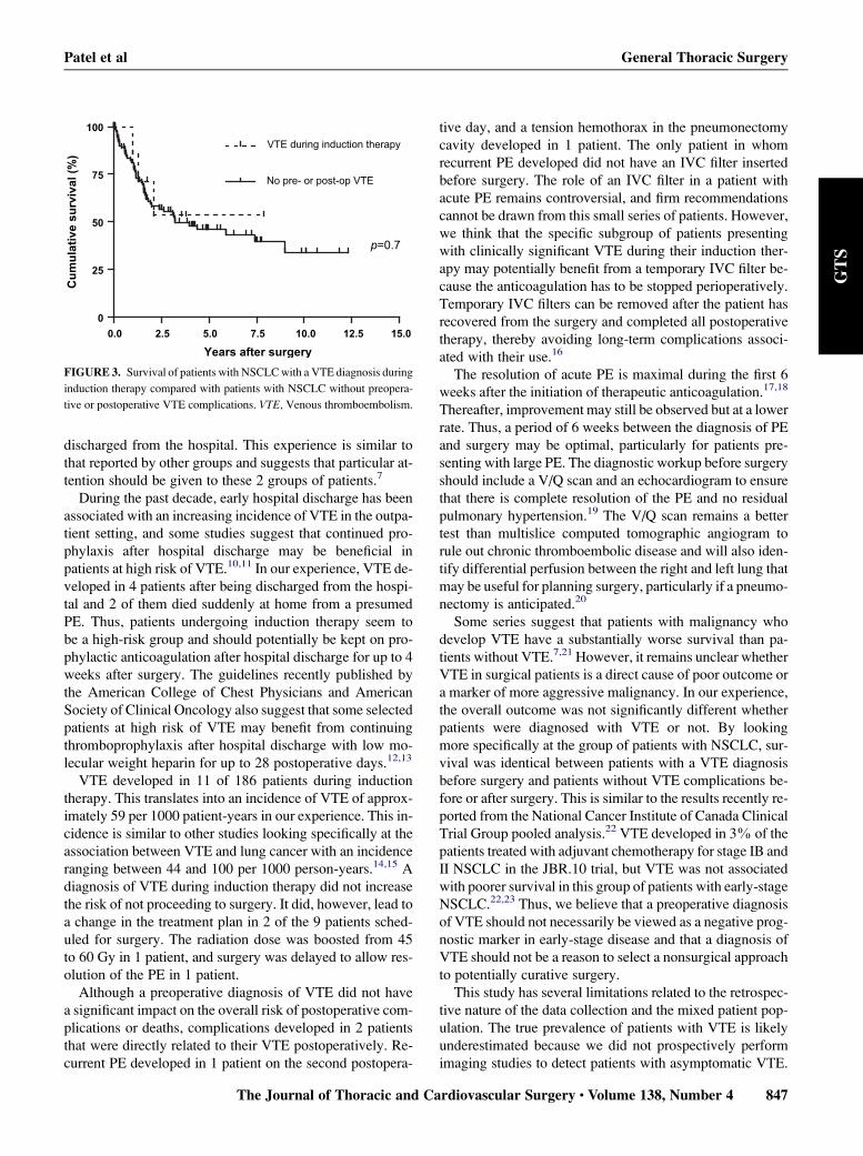
survival after surgery. Among patients with a preoperative
diagnosis of VTE, 5 are alive without recurrence 2.5 to 8.5
years after surgery (median 2.1 years), and the 5-year sur-
vival reached 54% in patients with NSCLC (Figure 3).
DISCUSSIONThis study demonstrates that the rate of VTE in patients
undergoing induction therapy for lung malignancy is high,
846 The Journal of Thoracic and Cardiovascular Su
ranging between 6.3% in patients undergoing induction
chemotherapy for MPM and 15.4% in patients undergoing
induction chemoradiation therapy for NSCLC. The number
of VTE events was higher than in other series of lung resec-
tion for malignancy and relates to the fact that approximately
half of the VTE complications occurred during induction
therapy before surgery.7-9
The rate of postoperative VTE in our series ranged be-
tween 7% and 8%. The majority of postoperative VTE oc-
curred in patients admitted to the intensive care unit for
major postoperative complications or in patients already
0.0 2.5 5.0 7.5 10.0 12.5 15.0
0
25
50
75
100
All VTE complications
No pre- or post-op VTE
p=0.8
Years after surgery
Cu
mu
lative su
rvival (%
)
FIGURE 2. Overall survival of patients with or without VTE complica-
tions. VTE, Venous thromboembolism.
rgery c October 2009
Patel et al General Thoracic Surgery
GT
S
discharged from the hospital. This experience is similar to
that reported by other groups and suggests that particular at-
tention should be given to these 2 groups of patients.7
During the past decade, early hospital discharge has been
associated with an increasing incidence of VTE in the outpa-
tient setting, and some studies suggest that continued pro-
phylaxis after hospital discharge may be beneficial in
patients at high risk of VTE.10,11 In our experience, VTE de-
veloped in 4 patients after being discharged from the hospi-
tal and 2 of them died suddenly at home from a presumed
PE. Thus, patients undergoing induction therapy seem to
be a high-risk group and should potentially be kept on pro-
phylactic anticoagulation after hospital discharge for up to 4
weeks after surgery. The guidelines recently published by
the American College of Chest Physicians and American
Society of Clinical Oncology also suggest that some selected
patients at high risk of VTE may benefit from continuing
thromboprophylaxis after hospital discharge with low mo-
lecular weight heparin for up to 28 postoperative days.12,13
VTE developed in 11 of 186 patients during induction
therapy. This translates into an incidence of VTE of approx-
imately 59 per 1000 patient-years in our experience. This in-
cidence is similar to other studies looking specifically at the
association between VTE and lung cancer with an incidence
ranging between 44 and 100 per 1000 person-years.14,15 A
diagnosis of VTE during induction therapy did not increase
the risk of not proceeding to surgery. It did, however, lead to
a change in the treatment plan in 2 of the 9 patients sched-
uled for surgery. The radiation dose was boosted from 45
to 60 Gy in 1 patient, and surgery was delayed to allow res-
olution of the PE in 1 patient.
Although a preoperative diagnosis of VTE did not have
a significant impact on the overall risk of postoperative com-
plications or deaths, complications developed in 2 patients
that were directly related to their VTE postoperatively. Re-
current PE developed in 1 patient on the second postopera-
0.0 2.5 5.0 7.5 10.0 12.5 15.0
0
25
50
75
100
No pre- or post-op VTE
VTE during induction therapy
p=0.7
Years after surgery
Cu
mu
lative su
rvival (%
)
FIGURE 3. Survival of patients with NSCLC with a VTE diagnosis during
induction therapy compared with patients with NSCLC without preopera-
tive or postoperative VTE complications. VTE, Venous thromboembolism.
The Journal of Thoracic and C
tive day, and a tension hemothorax in the pneumonectomy
cavity developed in 1 patient. The only patient in whom
recurrent PE developed did not have an IVC filter inserted
before surgery. The role of an IVC filter in a patient with
acute PE remains controversial, and firm recommendations
cannot be drawn from this small series of patients. However,
we think that the specific subgroup of patients presenting
with clinically significant VTE during their induction ther-
apy may potentially benefit from a temporary IVC filter be-
cause the anticoagulation has to be stopped perioperatively.
Temporary IVC filters can be removed after the patient has
recovered from the surgery and completed all postoperative
therapy, thereby avoiding long-term complications associ-
ated with their use.16
The resolution of acute PE is maximal during the first 6
weeks after the initiation of therapeutic anticoagulation.17,18
Thereafter, improvement may still be observed but at a lower
rate. Thus, a period of 6 weeks between the diagnosis of PE
and surgery may be optimal, particularly for patients pre-
senting with large PE. The diagnostic workup before surgery
should include a V/Q scan and an echocardiogram to ensure
that there is complete resolution of the PE and no residual
pulmonary hypertension.19 The V/Q scan remains a better
test than multislice computed tomographic angiogram to
rule out chronic thromboembolic disease and will also iden-
tify differential perfusion between the right and left lung that
may be useful for planning surgery, particularly if a pneumo-
nectomy is anticipated.20
Some series suggest that patients with malignancy who
develop VTE have a substantially worse survival than pa-
tients without VTE.7,21 However, it remains unclear whether
VTE in surgical patients is a direct cause of poor outcome or
a marker of more aggressive malignancy. In our experience,
the overall outcome was not significantly different whether
patients were diagnosed with VTE or not. By looking
more specifically at the group of patients with NSCLC, sur-
vival was identical between patients with a VTE diagnosis
before surgery and patients without VTE complications be-
fore or after surgery. This is similar to the results recently re-
ported from the National Cancer Institute of Canada Clinical
Trial Group pooled analysis.22 VTE developed in 3% of the
patients treated with adjuvant chemotherapy for stage IB and
II NSCLC in the JBR.10 trial, but VTE was not associated
with poorer survival in this group of patients with early-stage
NSCLC.22,23 Thus, we believe that a preoperative diagnosis
of VTE should not necessarily be viewed as a negative prog-
nostic marker in early-stage disease and that a diagnosis of
VTE should not be a reason to select a nonsurgical approach
to potentially curative surgery.
This study has several limitations related to the retrospec-
tive nature of the data collection and the mixed patient pop-
ulation. The true prevalence of patients with VTE is likely
underestimated because we did not prospectively perform
imaging studies to detect patients with asymptomatic VTE.
ardiovascular Surgery c Volume 138, Number 4 847

General Thoracic Surgery Patel et al
GT
S
The type of induction therapy is also variable between
institutions, and, although unlikely, induction therapy with
platinum and taxane may have a different impact on the
risk of VTE than we observed in our series.
CONCLUSIONSThis large study of patients undergoing induction therapy
for lung malignancy demonstrates that the rate of VTE dur-
ing induction therapy is high and deserves careful consider-
ation. Patients with a VTE diagnosis during induction
therapy may require a change in their treatment plan and
are at risk of recurrent VTE and bleeding postoperatively.
The insertion of a temporary ICV filter at the time of surgery
in patients with a preoperative diagnosis of VTE remains
controversial but may be beneficial in this subgroup of pa-
tients. The immediate postoperative outcome and long-
term survival of patients with a preoperative diagnosis of
VTE are similar to those of other patients without any
VTE complications. Thus, we believe that a preoperative di-
agnosis of VTE should not be viewed as a negative prognos-
tic marker and should not be a reason to select a nonsurgical
approach to potentially curative surgery.
We thank Dr Luis Garrido-Olivares for assistance with the statis-
tical analysis.
References1. Zurawska U, Parasuraman S, Goldhaber SZ. Prevention of pulmonary embolism
in general surgery patients. Circulation. 2007;115:302-7.
2. Kakkar VV, Corrigan TP, Fossard DP. Prevention of fatal postoperative pulmo-
nary embolism by low doses of heparin. Lancet. 1975;2:45-51.
3. Fischer S, Darling G, Pierre AF, Sun A, Leighl N, Waddell TK, et al. Induction
chemoradiation therapy followed by surgical resection for non-small cell lung
cancer (NSCLC) invading the thoracic inlet. Eur J Cardiothorac Surg. 2008;
33:1129-34.
4. Uy KL, Darling G, Xu W, Yi QL, De Perrot M, Pierre AF, et al. Improved results
of induction chemoradiation before surgical intervention for selected patients with
stage IIIA-N2 non-small cell lung cancer. J Thorac Cardiovasc Surg. 2007;134:
188-93.
5. Blackstone EH. Comparing apples and oranges. J Thorac Cardiovasc Surg. 2002;
123:8-15.
6. Rubin DB. The design versus the analysis of observational studies for causal ef-
fects: parallels with the design of randomized trials. Statist Med. 2007;26:20-36.
7. Mason DP, Quader MA, Blackstone EH, Rajeswaran J, DeCamp MM,
Murthy SC, et al. Thromboembolism after pneumonectomy for malignancy: an in-
dependent marker of poor outcome. J Thorac Cardiovasc Surg. 2006;131:711-8.
848 The Journal of Thoracic and Cardiovascular Su
8. Sugarbaker DJ, Jaklitsch MT, Bueno R, Richards W, Lukanich J, Mentzer SJ,
et al. Prevention, early detection, and management of complications after 328 con-
secutive extrapleural pneumonectomies. J Thorac Cardiovasc Surg. 2004;128:
138-46.
9. Dentali F, Malato A, Ageno W, Imperatori A, Cajozzo M, Rotolo N, et al.
Incidence of venous thromboembolism in patients undergoing thoracotomy
for lung cancer. J Thorac Cardiovasc Surg. 2008;135:705-6.
10. Bergqvist D, Agnelli G, Cohen AT, Eldor A, Nilsson PE, Le Moigne-Amrani A,
et al. Duration of prophylaxis against venous thromboembolism with enoxaparin
after surgery for cancer. N Engl J Med. 2002;346:975-80.
11. Kakkar AK, Brenner B, Dahl OE, Eriksson BI, Mouret P, Muntz J, et al. Extended
duration rivaroxaban versus short-term enoxaparin for the prevention of venous
thromboembolism after total hip arthroplasty: a double-blind, randomised con-
trolled trial. Lancet. 2008;372:31-9.
12. Geerts WH, Bergqvist D, Pineo GF, Heit JA, Samama CM, Lassen MR, et al.
Prevention of venous thromboembolism: American College of Chest Physicians
Evidence-Based Clinical Practice Guidelines (8th Edition). Chest. 2008;133(6
Suppl):381S-453S.
13. Lyman GH, Khorana AA, Falanga A, Clarke-Pearson D, Flowers C, Jahanzeb M,
et al. American Society of Clinical Oncology guideline: recommendations for ve-
nous thromboembolism prophylaxis and treatment in patients with cancer. J Clin
Oncol. 2007;25:5490-505.
14. Blom JW, Vanderschoot JP, Oostindier MJ, Osanto S, van der Meer FJ,
Rosendaal FR. Incidence of venous thrombosis in a large cohort of 66,329 can-
cer patients: results of a record linkage study. J Thromb Haemost. 2006;4:
529-35.
15. Chew HK, Davies AM, Wun T, Harvey D, Zhou H, White RH. The incidence of
venous thromboembolism among patients with primary lung cancer. J Thromb
Haemost. 2008;6:601-8.
16. Berczi V, Bottomley JR, Thomas SM, Taneja S, Gaines PA, Cleveland TJ. Long-
term retrievability of IVC filters: should we abandon permanent devices? Cardi-
ovasc Intervent Radiol. 2007;30:820-7.
17. Ribeiro A, Lindmarker P, Johnsson H, Juhlin-Dannfelt A, Jorfeldt L. Pulmonary
embolism: one-year follow-up with echocardiography Doppler and five-year sur-
vival analysis. Circulation. 1999;99:1325-30.
18. Ribeiro A, Lindmarker P, Johnsson H, Juhlin-Dannfelt A, Jorfeldt L. Pulmo-
nary embolism: a follow-up study of the relation between the degree of right
ventricle overload and the extent of perfusion defects. J Intern Med. 1999;245:
601-10.
19. de Perrot M, Fadel E, McRae K, Tan K, Slinger P, Paul N, et al. Evaluation of per-
sistent pulmonary hypertension after acute pulmonary embolism. Chest. 2007;
132:780-5.
20. Tunariu N, Gibbs SJ, Win Z, Gin-Sing W, Graham A, Gishen P, et al. Ventilation-
perfusion scintigraphy is more sensitive than multidetector CTPA in detecting
chronic thromboembolic pulmonary disease as a treatable cause of pulmonary
hypertension. J Nucl Med. 2007;48:680-4.
21. Sorensen HT, Mellemkjaer L, Olsen JH, Baron JA. Prognosis of cancers associ-
ated with venous thromboembolism. N Engl J Med. 2000;343:1846-50.
22. Shepherd F, Hasan B, Hicks L, Cheung M, Leighl N, Winton T, et al. Venous
thromboembolism (VTE) and non-small cell lung cancer: a pooled analysis of
National Cancer Institute of Canada Clinical Trials Group (NCIC CTG) trials.
Eur J Cancer. 2007;4(Suppl):361, (Abst 6516).
23. Winton T, Livingston R, Johnson D, Rigas J, Johnston M, Butts C, et al. Vinor-
elbine plus cisplatin vs. observation in resected non-small-cell lung cancer. N Engl
J Med. 2005;352:2589-97.
rgery c October 2009