venous thromboembolism (vte)– what you need to know patrick rendon, md thursday school july 2015
TRANSCRIPT

Venous Thromboembolism (VTE)– What You Need to Know
Patrick Rendon, MDThursday School July 2015


For another time…
•Arterial thrombi
•HIT
•VTE prophylaxis
•Caveats to treating superficial vein thrombosis

Objectives
•Be able to determine the pre-test probability for a VTE
•Describe the approach to a patient with suspected VTE
•State at least three methods of managing VTE

Road Map
• Case 1 - Diagnosis • Tips • From now on don’t say…
• Case 2 - Triage• Tips • From now on don’t say…
• Case 3 - Treatment• Tips • From now on don’t say…

DIAGNOSISDVT

It’s 6 pm in the evening. You are tired and trying to wrap up the call day. Your medical student is admitting a patient with you:It is a 66-year-old male veteran who presented with shortness of breath. He has not had chest pain, cough, or other localizing symptoms. He fractured his left leg in an automobile accident 7 weeks ago, and his short-leg cast was removed 2 weeks ago.On physical examination, temperature is 37.8 °C (100.0 °F), pulse rate is 102/min, and respiration rate is 20/min O2S 94% on 2L nasal cannula (89% RA). The chest is clear and the legs are not swollen or tender. The remainder of the examination is unremarkable.Labs: troponin, leukocytes and CXR count are normal. EKG: sinus tachycardiaThe student wants to know from you which of the following is the most appropriate next step in evaluation? And WHY?
A. CT angiographyB. Pulmonary angiogramC. VQ scanD. Serum D-dimer measurementE. Tell the student to go “look it up.” You’re too tired to think.


Answer A. CT angiography

How I think about VTE
• Approximately 300,000 persons die annually in the US from acute PE…so it’s a big problem
• The greatest risk factor for having a VTE is prior VTE!
• Most PEs arise from proximal DVTs (at or above the popliteal fossae) and travel to the pulmonary arteries
• Provoked or unprovoked?
• Use Virchow’s triad!


History & Physical for PE
•Symptoms• Dyspnea (73-85%)• Pleuritic chest pain (66-85%)
•Signs• Tachypnea (92%)• Crackles (51%)
PIOPED II

Labs & Studies for PE
• ABG• Hypoxemia • Widened alveolar-arterial gradient
• EKG: • For right heart strain = large clot burden• RAD, RBBB, S1Q3T3 • Most common: sinus tachycardia
• TTE• RV dysfunction: RV dilatation/hypokinesis• Elevated PA systolic pressure
• CXR: • Rules out other conditions

How do we evaluate for VTE?
PRE-TEST PROBABILITY

http://www.uptodate.com/contents/calculator-pulmonary-embolism-wells-score?source=see_link&utdPopup=true

VQ scan is 2nd line

Diagnosing VTE
• Vein anatomy is important for DVT
• If you find a DVT, why look for PE?
• Order a VQ scan (sensitivity 98%, specificity 10%) if CTA is:
• Contraindicated• Inconclusive• Negative but discordant with a high clinical suspicion


Wells Score ≤ 0
Wells Score ≥ 3

What about unprovoked VTE?
• Whether VTE is unprovoked vs provoked = greater prognostic information regarding recurrence risk
• Results of thrombophilia testing do not really influence treatment duration• FVL heterozygosity modestly increases (1.5 fold) recurrence risk
• Potent thrombophilias (e.g. ATIII deficiency) are rarely identified
• NEJM 2015 (and others) - Screening for Occult Cancer in Unprovoked VTE• Not cost effective to perform abdominal CT looking for neoplasm• Age appropriate screening with basic bloodwork is appropriate

• Anti-thrombin III Deficiency
• Protein C Clottable (“activity”)
• Protein S Activity
• Factor V Leiden mutation
• Factor II (prothrombin) mutation
• APAS “panel”
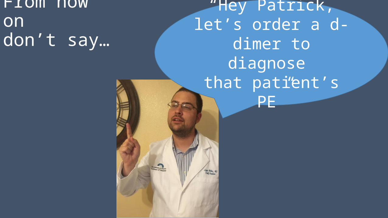
From now on don’t say… “Hey Patrick, let’s order
a d-dimer to diagnose that patient’s PE”

But do say…“Hey team, I would wait on the the full hypercoag workup or aggressive testing
until this VTE has been anticoagulated for 3-6 months or if we have a specific worry
about something; such as hemoptysis in a patient who smokes for example. In that
scenario we could order a CT chest for an unexplained VTE”
Thoughtful look

Triage and
Disposition DVT
PE

You are on the medicine consult rotation. The Ortho resident calls you to tell you about a 63-year-old woman who just developed severe shortness of breath that awoke her from sleep. The Ortho resident tells you that the patient underwent elective right hip replacement for severe osteoarthritis 5 days ago. She has no history of coronary artery disease. She has received SQ enoxaparin for VTE prophylaxis.
Blood pressure is 78/50 mm Hg, pulse 130/min, RR 34/min, and O2S is 70% on RA. JVD is present. Lungs are clear, and there are no murmurs. Her extremities are cold and clammy.
An EKG shows sinus tach. CXR is ordered and preparations are made for intubation.
Which of the following is the most appropriate next step in management?
A. Cardiac enzymes and D-dimer levels
B. CT angiogram of the chest
C. Emergent bedside TTE
D. Ventilation/perfusion (V/Q) scan
E. Call Paul Andre, he’s in the MICU 951-1816
REAL PHONE NUMBER

Answer C. Emergent bedside TTE• Key point: Perform bedside TTE to confirm RV overload if CTA or VQ
scan is unavailable and/or the patient is critically ill

Anticoagulate & call the fellow – thrombolytics?
Approach to
suspected PE
Anticoagulate & call the fellow – thrombolytics?

Disposition Tip: To admit or not to admit?The PESI Score –
• DVTs can be treated outpatient unless there are social, insurance, comorbid conditions, or concurrent PE
• Patients with PE and hypoxemia should be admitted
• Base disposition on severity• Use The Pulmonary Embolism Severity Index (PESI) score
• Determines the severity, mortality and outcome of patients with new PE

Case 1: 66 M with shortness of breath, temp 37.8 °C (100.0 °F), pulse of 102/min, and RR 20/min O2S 94% on 2L nasal cannula (89% RA) is diagnosed with a right segmental PE via CT angiogram.
What is his mortality and should he be admitted?
High and Yes
Class I and II patients may possibly be treated as outpatients in the right clinical setting.
Our patient is class IV

From now on do think… “Why DOES the ER want this
patient admitted? It’s probably because this patient
with PE has concurrent history of heart failure,
cancer, and COPD.”
QI work
Fantasy football
stats

TREATMENT
Bilateral DVTs Ouch.

A 65-year-old female veteran is evaluated at UNM Hospital for a 1-day history of right leg swelling. Three days ago, she underwent nephrectomy for renal cell carcinoma. Her only medications are unfractionated heparin, 5000 units subcutaneously twice daily, and lisinopril.BP is 130/75 mm Hg, pulse is 85/min, and RR is 20/min. Wt is 80 kg. The RLE calf is swollen, warm, and tender to palpation. The remainder of the examination is normal.
Laboratory studies:
Hematocrit 29%
Platelet count 275,000/µL (275 × 109/L)
Creatinine 2.1 mg/dL (194.5 µmol/L)
Estimated glomerular filtration rate 29 mL/min/1.73 m2

Venous duplex ultrasonography shows a right lower extremity femoral and popliteal vein DVT. There is no hydronephrosis or surgical site hematoma on an ultrasound.In addition to cessation of subcutaneous unfractionated heparin, which of the following is the most appropriate treatment?
A. Adjusted-dose, intravenous unfractionated heparinB. Enoxaparin, 80 mg twice dailyC. WarfarinD. RivaroxabanE. Wait, did you say veteran? Check amion. This patient should appropriately be transferred.

A 65-year-old female veteran is evaluated at UNM Hospital for a 1-day history of right leg swelling. Three days ago, she underwent nephrectomy for renal cell carcinoma. Her only medications are unfractionated heparin, 5000 units subcutaneously twice daily, and lisinopril.BP is 130/75 mm Hg, pulse is 85/min, and RR is 20/min. Wt is 80 kg. The RLE calf is swollen, warm, and tender to palpation. The remainder of the examination is normal.
Venous duplex ultrasonography shows a right lower extremity femoral and popliteal vein DVT. There is no hydronephrosis or surgical site hematoma on an ultrasound.In addition to cessation of subcutaneous unfractionated heparin, which of the following is the most appropriate treatment?
A. Adjusted-dose, IV unfractionated heparinB. Enoxaparin, 80 mg twice dailyC. WarfarinD. RivaroxabanE. Wait, did you say veteran? Check amion. This patient should appropriately be transferred.
Laboratory studies:Hematocrit 29%Platelet count 275,000/µLCreatinine 2.1 mg/dLEstimated glomerular filtration rate 29 mL/min/1.73 m2


Answer A. Adjusted-dose, IV unfractionated heparin
• Use IV unfractionated heparin for DVT in patients with recent surgery and CKD
• Enoxaparin and Fondaprinux are contraindicated with estimated GFR <30 mL/min/1.73 m2

Treatment for Acute VTE• Enoxaparin (Lovenox, LMWH)
• First-line therapy • 50% lower incidence of recurrent VTE than warfarin in patients with advanced cancer
• Unfractionated heparin (IV or sub Q) and Enoxaparin• Include an overlap of several days with a stable, therapeutic INR • Can monitor the PTT and Xa levels
• Fondaparinux
• Dabigatran, Rivaroxaban, Apixaban• Long half-lives• No antidote• Inability to determine the anticoagulant effect

Warfarin Treatment for Acute VTE
• Inhibits vitamin K oxide reductase affecting the synthesis of factors II, VII, IX, and X and proteins C and S
• Begin Warfarin with Heparin/LMWH/Fonda• Factor II and X levels require at least 5 days to decline
• Should overlap for at least 5 days and until an INR of 2 or more is achieved

From now ondon’t say… “Hey team, let’s put
that patient on a heparin drip”
My desk Do say…Enoxaparin
is first line


Special
Circumstances

Thrombolytics
Current risk-benefit studies do not generally support the use of thrombolytics except for hemodynamic instability
Thrombolytics are recommended in patients with RV dysfunction causing hemodynamic compromise
CALL YOUR FELLOW BEFORE GIVING
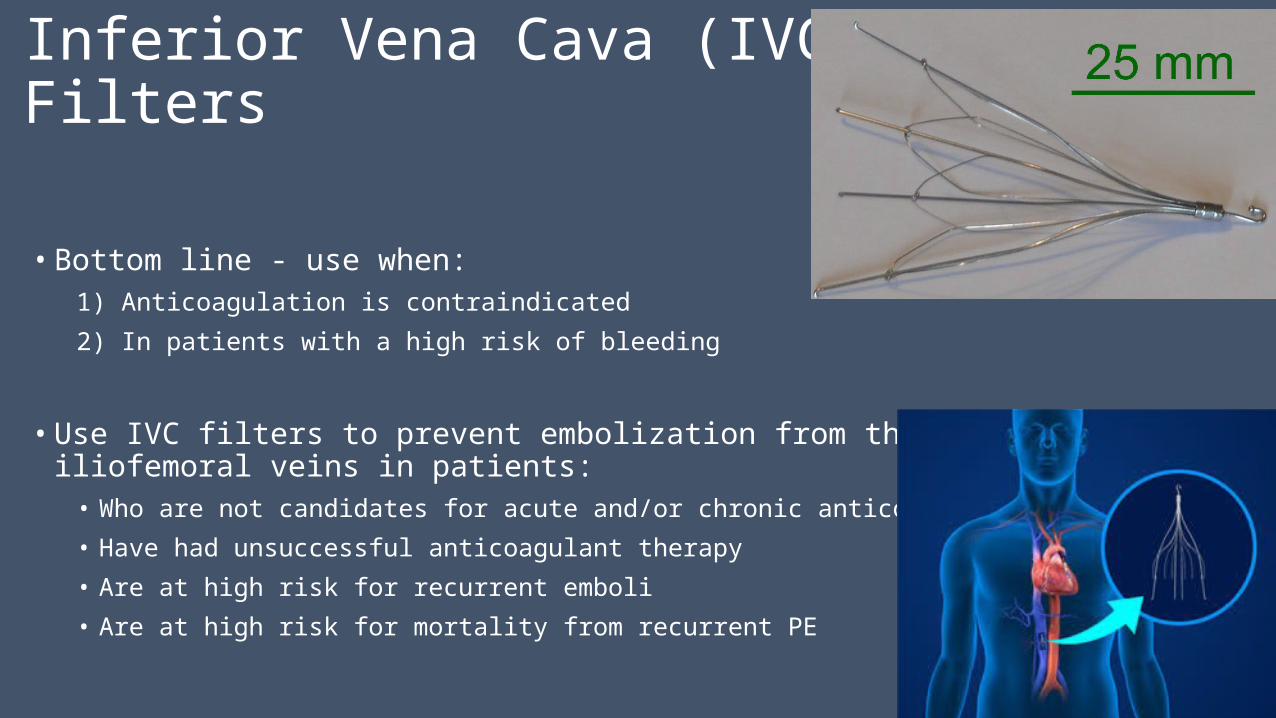
Inferior Vena Cava (IVC) Filters
• Bottom line - use when:1) Anticoagulation is contraindicated
2) In patients with a high risk of bleeding
• Use IVC filters to prevent embolization from the iliofemoral veins in patients:• Who are not candidates for acute and/or chronic anticoagulation
• Have had unsuccessful anticoagulant therapy
• Are at high risk for recurrent emboli
• Are at high risk for mortality from recurrent PE

Superficial Venous Thrombophlebitis
• Treat SVT (thrombophlebitis) with compression stockings and NSAIDs
• Consider anticoagulation because DVT or symptomatic PE can develop

Incidental and small subsegmental PE• Unclear significance
• A single subsegmental PE probably does not have the same clinical significance as other larger PEs
• Most experts anticoagulate
“Clot cupcake”

Reversal for life-threatening bleeding• Warfarin
• 10 mg IV vitamin K• Fresh frozen plasma (FFP)
• Heparin• Protamine
• LMWH• Protamine has 60% reversal
• Fondaparinux• Not technically reversible• FFP
• Dabigatran, Rivaroxaban• Not technically reversible (yet)
• Prothrombin complex concentrates (PCCs)
• Requires pharmacy approval• Can cause thrombi

Consultation
• UNMH Anticoagulation Pharmacy 505-264-6970
• VA: Ask your team pharmacist

Thank you to the Chiefs

Questions?
• Great Resources for you
• Annals of Internal Medicine DVT “In the Clinic” article
• ACP Smart Medicine
• Cleveland Clinic: Venous thromboembolism What to do after anticoagulation is started