ventral aspect of the visual form pathway is not critical
TRANSCRIPT

Ventral aspect of the visual form pathway is not criticalfor the perception of biological motionSharon Gilaie-Dotana,b,1, Ayse Pinar Sayginc, Lauren J. Lorenzid, Geraint Reesa,b, and Marlene Behrmannd,e
aInstitute of Cognitive Neuroscience and bWellcome Trust Centre for Neuroimaging, Institute of Neurology, University College London, London, WC1N 3AR,United Kingdom; cDepartment of Cognitive Science and Neurosciences Program, University of California, San Diego, CA 92093-0515; and dDepartment ofPsychology and eCenter for the Neural Basis of Cognition, Carnegie Mellon University, Pittsburgh, PA 15213
Edited by Michael E. Goldberg, Columbia University College of Physicians and Surgeons and the New York State Psychiatric Institute, New York, NY, andapproved December 12, 2014 (received for review August 6, 2014)
Identifying the movements of those around us is fundamental formany daily activities, such as recognizing actions, detecting preda-tors, and interacting with others socially. A key question concernsthe neurobiological substrates underlying biological motion percep-tion. Although the ventral “form” visual cortex is standardly acti-vated by biologically moving stimuli, whether these activations arefunctionally critical for biological motion perception or are epiphe-nomenal remains unknown. To address this question, we examinedwhether focal damage to regions of the ventral visual cortex, result-ing in significant deficits in form perception, adversely affects bio-logical motion perception. Six patients with damage to the ventralcortex were tested with sensitive point-light display paradigms. Allpatients were able to recognize unmasked point-light displays andtheir perceptual thresholds were not significantly different fromthose of three different control groups, one of which comprisedbrain-damaged patients with spared ventral cortex (n > 50). Impor-tantly, these six patients performed significantly better than patientswith damage to regions critical for biological motion perception. Toassess the necessary contribution of different regions in the ventralpathway to biological motion perception, we complement the be-havioral findings with a fine-grained comparison between the lesionlocation and extent, and the cortical regions standardly implicated inbiological motion processing. This analysis revealed that the ventralaspects of the form pathway (e.g., fusiform regions, ventral extras-triate body area) are not critical for biological motion perception.Wehypothesize that the role of these ventral regions is to provide en-hanced multiview/posture representations of the moving personrather than to represent biological motion perception per se.
ventral stream | visual form agnosia | action perception | EBA |point-light displays
Perception of the movements of other peoples’ bodies is fun-damental to our daily interactions (e.g., motor learning,
social interactions, anticipating actions of others), and is sufficientlyrobust so as to succeed even under suboptimal conditions [e.g.,poor illumination and even partial occlusion (1–6)]. A clear dem-onstration of the resilience of this ability is the ease with whichpeople recognize biological motion from point-light displays(PLDs) that consist of only a small set of moving points that markjoints on the body (7), (Fig. 1A, Left). These stimuli appear to naïveobservers as a set of incoherent dots when static, but evoke a vividpercept of a moving person when in motion. Observers are able toinfer movement information, such as the motion or direction of thefigure, in these impoverished PLDs even under conditions ofmasking, added noise (8–11), or night driving (3, 4, 12).Examination of the neural correlates of the perception of body
movement reveals a widespread cortical network (13). Becausebiological motion perception, in natural vision or in PLDs, in-volves both form and motion perception (14), unsurprisingly,cortical regions associated with both form and motion perceptionare activated. It is unclear, however, whether all of these brainareas contribute causally to the perception of biological motion.Neuropsychological studies in patients and transcranial magneticstimulation (TMS) studies in normal observers have identified
several motion-sensitive areas as critical for biological motionperception, including the posterior superior temporal sulcus(pSTS) and ventral premotor cortex (vPMC) (11, 15–17), giventhat a sustained or transient lesion to these regions impairsbiological motion perception. However, whether form-sensitiveregions in the ventral “form” visual pathway [for example, theextrastriate body area (EBA) (18–20) in the lateral occipitalcortex], which are consistently activated in response to bi-ological motion in neuroimaging studies, play a critical role inbiological motion perception remains unknown.PLDs constitute ideal stimuli with which to explore whether the
engagement of the ventral visual cortex is necessary for biologicalmotion perception, as these displays permit the presentation ofrecognizable body movements while dissociating them from“classic ventral” form cues, such as contour, surface, shape, tex-ture, and color. As such, PLDs are thought to depict dynamic bodyand action information solely via motion cues. To the extent thatthe ventral form pathway is involved in PLD perception, thiscannot be attributed to processing classic ventral form cues. Evenin the absence of classic form cues, however, moving PLDs conveycoarse form information of the dynamic body, and investigatingthe structure of the articulated body that can be retrieved from thecoherent movement of the dots has been a central driving moti-vation in biological motion research (e.g., refs. 21–27). This ap-proach has been true since the pioneering work of Johansson (7),and therefore, unsurprisingly, these displays have often beentermed biological structure-from-motion or form-from-motion. Inlight of the above, whether the processes supporting biologicalmotion perception—in the absence of classic form cues—critically
Significance
Perceiving the movements of people around us is critical for manydaily skills (from detecting threats to social interactions) andinvolves both form and motion perception. Even though the“form” visual pathway is standardly activated by biological mo-tion stimuli, it is unknown whether this pathway’s integrity iscritical for the perception of biological motion. Here, we examinedwhether damage to different aspects of the form pathway affectsbiological motion perception. Individuals with lesions to the ven-tral aspects of this pathway evinced normal biological motionperception despite their impairments in form perception. Ourcounterintuitive findings indicate that biological motion can beperceived and processed normally even when the ability to per-ceive the form or the actor executing the movements is impaired.
Author contributions: S.G.-D., A.P.S., G.R., and M.B. designed research; S.G.-D., A.P.S., andL.J.L. performed research; A.P.S. contributed new reagents/analytic tools; S.G.-D. andA.P.S. analyzed data; and S.G.-D., A.P.S., G.R., and M.B. wrote the paper.
The authors declare no conflict of interest.
This article is a PNAS Direct Submission.
Freely available online through the PNAS open access option.1To whom correspondence should be addressed. Email: [email protected].
This article contains supporting information online at www.pnas.org/lookup/suppl/doi:10.1073/pnas.1414974112/-/DCSupplemental.
www.pnas.org/cgi/doi/10.1073/pnas.1414974112 PNAS Early Edition | 1 of 10
NEU
ROSC
IENCE
PNASPL
US

depend on the form computations of the form visual pathway stillremains unknown (20, 28–30).One way to address this issue is to study whether damage to the
ventral “form” visual pathway affects the perception of biologicalmotion. The predictions are straightforward: if ventral stream in-tegrity and ventral form representations are necessary for theperception of biological motion, then individuals with form per-ception deficits following damage to the ventral visual cortex [in-cluding damage to specific areas implicated in biological motionprocessing or areas implicated in body form perception (e.g., theEBA) (18–20)], should be impaired at perceiving biological motion.To examine this hypothesis, we tested a group of six patients with
form perception deficits following a circumscribed lesion to theventral visual cortex in adulthood (Table 1). Using PLDs andparadigms that are successful in detecting biological motion per-ceptual deficits following brain damage (11, 17), we measured thepatients’ recognition and perception of biological motion in twodifferent experiments. We compared each patient’s performance tothat of three different control groups: (i) a brain-damaged controlgroup of 54 patients, whose cortical lesions fell outside of theventral visual cortex; (ii) a group of healthy age-matched controls(patient-specific); and (iii) a group of 13 young control participants.The importance of the brain-damaged control group is twofold.First, comparing the ventral patients to patients with nonventrallesions allowed us to determine whether biological motion per-ception, if affected, is specifically a consequence of a ventral lesionor of brain damage, more generally. Second, because a subset ofpatients in the nonventral control group have lesions to brain areasknown to significantly impair biological motion perception (pSTSand vPMC), we were able to compare directly the perceptualthresholds of the ventral patients with those of individuals withidentified deficits in biological motion following damage to thepSTS or vPMC. Finally, given that the ventral cortex constitutesa large swath of the cortex and that patients’ lesions were notidentical, we assessed the brain–behavior correspondences furtherby examining, at a finer grain, which—if any—affected subareasimpair biological motion perception. To do so, for each patient wecarefully delineated the lesion, assessed the magnitude of thedamage, and situated the lesion relative to regions in the ventral,lateral, and middle temporal cortex that are standardly associatedwith biological motion processing, including body parts and visualmotion-sensitive regions (e.g., refs. 13, 14, 20, 31–33).To anticipate our findings, we show that biological motion
recognition and perceptual thresholds of the ventral patientsconsistently fall within the normal range of all three controlgroups. Thus, our results indicate that the perception of biologicalmotion (i) does not depend on the integrity of the ventral aspectsof the form visual pathway or on the integrity of the ventral por-tion of the EBA (ventral to the human middle temporal V5complex, MT+/V5), and (ii) can be dissociated from form per-ception. We conclude, therefore, that computations that sufficefor the perception of biological motion are mediated by mecha-nisms independent of the form ventral cortex, and that suchcomputations may be based on motion cues that representmovement kinematics rather than on form information per se.
ResultsExperiment 1. Recognition and perceptual thresholds for biological motion.At the start of Exp. 1, participants were presented with unmaskedPLDs (Fig. 1A, Left) and were asked to describe what they per-ceived. For these unmasked PLDs, all six ventral patients (as wellas almost all individuals in three groups of controls) were able toname the movements effortlessly and immediately, even withoutprior knowledge of or training on PLDs. This observation isconsistent with previous work showing that patients are generallyable to recognize unmasked PLDs of biological motion (34–39).Only two of the control patients with brain-damage outside of the
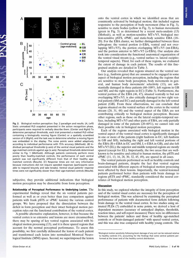
Fig. 1. Biological motion perception: Exp. 1 paradigm and results. (A, Left)Static snapshots from the unmasked PLDs presented in the action recog-nition phase; participants were required to verbally describe the display.(Right) To determine perceptual thresholds, two masked PLDs embedded innoise points were presented simultaneously, one on the right and one onthe left, one containing a biological movement (here on the left), and theother a spatially scrambled version of that movement. The task was todetermine which side contained the moving human figure. The noisepoints were added adaptively according to individual performance(Methods). (B) Individual perceptual thresholds (y axis) of the ventral visualpatients and the three control groups against age (x axis). Perceptualthresholds represent the number of noise points that can be toleratedwhile performing at 82% accuracy (more noise points correspond to betterperformance). Each ventral patient was not significantly different from anyof the control groups, and performed similarly to or better than the braindamaged controls with spared ventral cortex (11) (Table 2). (C) Ventral vi-sual patients’ performance was significantly better than that of brain-damaged controls (11) whose lesions invaded regions critical for biologicalmotion perception (pSTS, vPMC). (D) Response times (y axis) as recordedduring the experiment plotted against age (x axis). Response times arenot very informative as instructions did not require speeded responses(participants were able to respond leisurely and take breaks). Responsetimes of the brain-damaged control group were not available. Ventralvisual patients’ response times (apart from EC) (Results) were not signifi-cantly slower than those of their age-matched controls.
2 of 10 | www.pnas.org/cgi/doi/10.1073/pnas.1414974112 Gilaie-Dotan et al.

ventral cortex (from the “brain-damaged control group” in thepresent study) were unable to recognize unmasked PLDs (11).After this recognition phase, perceptual thresholds for bi-
ological motion (number of noise points at which performance is82% accurate; see Fig. 1A, Right, for illustration) were measuredfor all participants (Fig. 1B, and detailed in Table 2). Consistentwith previous results showing that the perceptual thresholds ofbrain-damaged patients for biological motion are significantlylower than those of healthy age-matched controls (11), four ofthe ventral visual patients’ (CR, SM, EC, and SH) perceptualthresholds were at the lower end of their matched controls’distribution (control group 1; light diamonds in Fig. 1B), but notstatistically different (see Table 2 for statistical details). More-over, the perceptual threshold of each ventral patient was also
within the norm of the younger control group [control group 2;all jt(12)js < 1.44, all Ps > 0.17 (40); Fig. 1B, light gray circles].We then compared the thresholds of the ventral patients and
those of 54 patients with unilateral nonventral brain damage[control group 3 (11); dark circles in Fig. 1B]. If the integrity ofthe ventral visual cortex is critical for biological motion per-ception, then the performance of the ventral visual patientsshould be significantly poorer than that of patients with braindamage elsewhere. In contrast, the ventral patients’ thresholdswere trending to be significantly better than their brain-damagedcontrols [Wilcoxon nonparametric rank-sum test: ventral patients(median = 13.11, n = 6) vs. brain damaged controls (median = 9.82,n = 54): U = 258, P = 0.06]. In addition, in an individual caseanalysis, each of the ventral visual patients’ performance was betterthan the average performance of the right only (n = 11), left only
Table 1. Summary of the visual perceptual functions and impairments of the six ventral patients
Function/Impairment EL GB SH CR SM EC
Lesioned hemisphere Left Left Left Right (+ left) Right RightAge (sex) 61 (F) 70 (F) 69 (M) 31 (M) 37 (M) 48 (F)Time from injury 15 y 3 y 6 y 15 y 19 y 8 y
Visual acuity Correctedto normal
Correctedto normal
Correctedto normal
Normal Normal Normal
Accommodation/convergencedeficit
Noneapparent
or reported
Noneapparent
or reported
Noneapparent
or reported
Noneapparent orreported
Noneapparent
or reported
Noneapparent
or reportedVisual field Upper right
quadrantanopiaUpper right
quadrantanopiaRight homonymous
hemianopia(largely resolved)
Full field Full field Full field
Object perception Mild impairment(1–2 SDs)
Mild impairment(1–2 SDs)
Mild impairment(1–2 SDs)
Agnosic(3 SDs)
Agnosic(3 SDs)
Object recognitiondifficulties(screening)
Face perception Mild impairment(1–2 SDs)
Mild impairment(1–2 SDs)
Mild impairment(1–2 SDs)
Prosopagnosic(3 SDs)
Prosopagnosic(3 SDs)
Facerecognitiondifficulties(screening)
Word perception Pure alexic(3 SDs)
Pure alexic(3 SDs)
Pure alexic(3 SDs)
Mild impairment(1-2 SDs)
Mild impairment(1-2 SDs)
Unknown
Motion perception –
basic (detection)Normal Normal Unknown Impaired
(very slow motion)Impaired(very slowmotion)
Impaired(very slowmotion)
Motion perception –
basic (coherence)Normal Normal Unknown Impaired
(very fast motion)Impaired (medium
to veryfast motion)
Unknown
Motion perception –
structure (SFM)Normal Normal Unknown Impaired Impaired Impaired
Motion perception –
biological unmaskedPLDs (Exp. 1)
Normal Normal Normal Normal Normal Normal
Motion perception –
biological perceptualthresholds (Exp. 1)
Normal Normal Normal Normal Normal Normal
Motion perception –
biological unmaskedPLDs (Exp. 2)
Normal Normal Normal Normal Normal Normal
Motion perception –
biological perceptualthresholds (Exp. 2)
Normal Normal Normal Normal Normal Normal
Most of these data have been reported earlier [EL (61, 70–75), GB (61, 75), SH (70, 75), CR (61, 65–67, 70), SM (61, 64–69), EC (61)]. The data from this studyare presented in the four bottom rows. We summarize the patients’ abilities by noting the number of SDs each score deviates from the controls’ mean. Visualimpairments are denoted in bold.
Gilaie-Dotan et al. PNAS Early Edition | 3 of 10
NEU
ROSC
IENCE
PNASPL
US

(n = 43), or combined right and left hemisphere brain-damagedcontrol patients (see Table 2 for full details). All of thesecomparisons indicate that the six ventral patients performedwell within the range of other (nonventral) brain-damaged pa-tients, thereby ruling out a specific role for the ventral cortex inbiological motion perception.The data from the control brain-damaged patients were taken
from a previous study (11) that revealed that lesions to the leftpSTS (L-pSTS) or to left vPMC (L-vPMC) had the greatest ad-verse effect on biological motion perception. The function andstructure of these regions are associated with biological motionperception (11, 15, 16, 20, 41–45) and their role in biologicalmotion perception has been confirmed in several TMS studies (15,16). In light of this finding, these data permit a stringent com-parison between the performance of our ventral patients and thatof the brain-damaged patients with lesions to L-pSTS or L-vPMC(the two “critical” lesion groups). As shown in Fig. 1C, the ventralvisual patient group performed significantly better (had higherperceptual thresholds, meaning they could tolerate more noisepoints) than both of the critical lesion groups [Wilcoxon non-parametric rank-sum test: ventral patients (median = 13.11, n = 6)vs. lesioned L-pSTS (median = 7.1, n = 9): U = 73, P = 0.0016;ventral patients vs. lesioned L-vPMC (median = 7.6, n = 10): U =77, P = 0.003]. Furthermore, in single-case comparisons [eachventral patient vs. the critical control groups (40)], four of theventral visual patients performed significantly better than thecritical control groups (Table 2). These results indicate thatdamage to the ventral visual cortex, unlike damage to the pSTS orvPMC, does not impair biological motion perception.Response times for biological motion. The results thus far indicate thatbiological motion perception does not rely on ventral stream in-tegrity. To confirm this finding and ensure that the results werenot a product of a speed-accuracy trade-off, we examined reactiontimes even though participants were informed that speededresponses were not required and participants were allowed tospeak and take breaks during the experiment (Fig. 1D). Theresponses of the patients were not significantly slower than theirage-matched controls [Wilcoxon nonparametric rank-sum test:ventral patients (median = 7.78 s, n = 6) vs. age-matched controls(median = 5.21 s, n = 33): U = 159, P = 0.134, z = 1.5]. Thisfinding also held true for five ventral patients under single-casecomparisons of patient vs. age-matched control group [two-tailed,jtjs < 0.5, Ps > 0.63 (40)]. EC was significantly slower than her age-matched controls [t(11) = 4.74, P = 0.0008]; however, this is almostcertainly a result of the fact that she spoke during the experiment.These results confirm that reaction times were within the normal
range for the ventral patients and that these normal perceptualthresholds did not result from elongated response times.
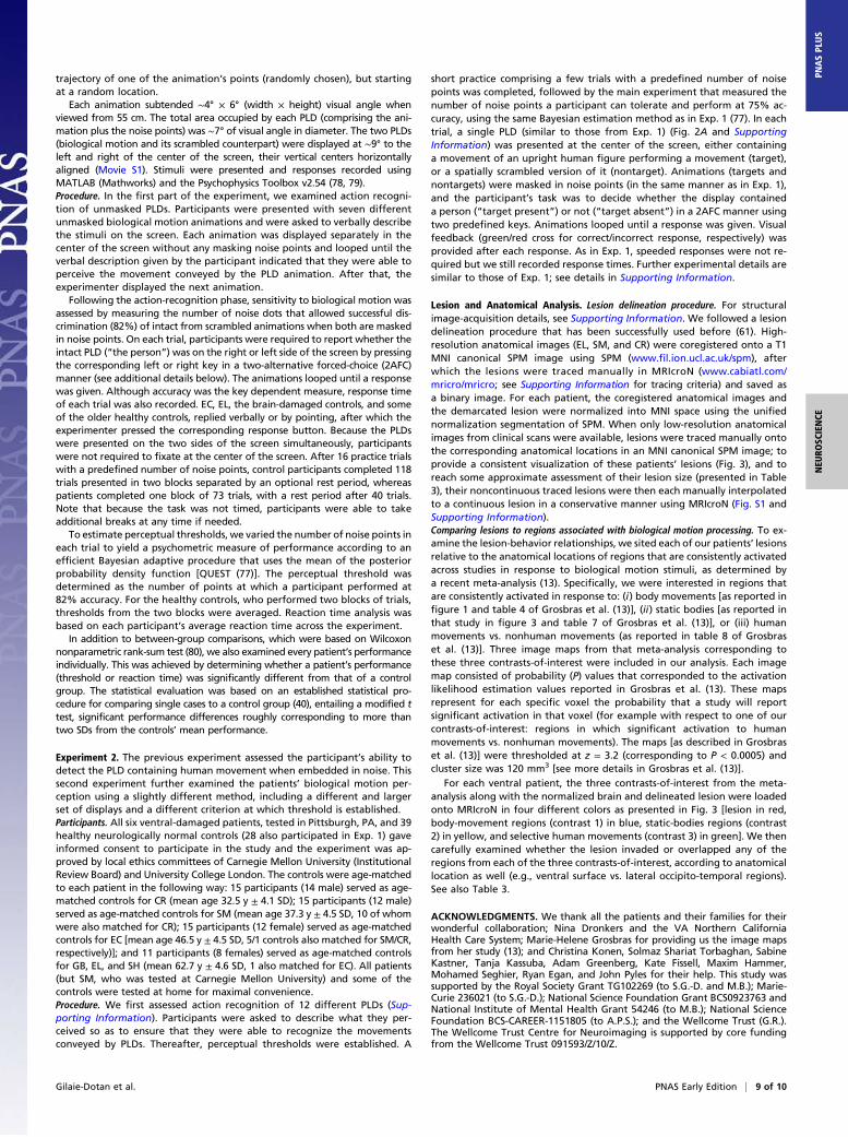
Experiment 2. Recognition and perceptual thresholds for biologicalmotion under a different paradigm. To provide additional supportfor the findings from Exp. 1, we further examined the perceptualthresholds of the ventral patients using a modified biological motionexperimental paradigm. This task included a larger set of biologicalmotion animations, different presentation and task requirements,and provided feedback. In this experiment, each trial consisted ofone centrally displayed PLD (Fig. 2A) and observers determinedwhether there was a moving human figure embedded in the display(compare Fig. 2A, Center and Right; see Methods). During theaction-recognition phase (Fig. 2A, Left), all ventral patients and theircontrols effortlessly reported the actions present in the PLDs.Moreover, the ventral patients’ perceptual thresholds fell within thenormal range of their age-matched controls (Fig. 2B and Table S1)[CR: t(14) = −1.42, P > 0.17; SM: t(14) = −0.88, P > 0.39; EC: t(14) =−0.76, P > 0.45; EL: t(10) = 0.30, P > 0.77; GB: t(10) = 1.56, P > 0.15;SH: t(10) = −0.65, P > 0.53); the performance of EL, GB, and SHalso fell in the normal range of a bigger control group (n = 14), aged60.2 ± 6.45 (SD): jt(13)js < 0.5, Ps > 0.6.] Of great interest, theperceptual thresholds established here for each of the patients (andthe relative rank ordering of the patients) were very similar to thoseobtained in Exp. 1, reflecting the reliability and consistency ofthese measures.Response times for biological motion under a different paradigm. Theanalysis of the reaction times of the patients versus the age-matched controls revealed no significant group differences (Fig.2C) [all patients but EL: jtjs < 0.79, Ps >0.45, EL: t(10) = 1.51,P > 0.16], again confirming that the patients performed withinthe normal range.
Subjective Reports About Biological Motion Perception. As a con-verging source of evidence, we obtained self-reports from thepatients and controls in response to questions such as whether,on the basis of gait, they were able to recognize individuals anddiscriminate the age and sex of an individual and, for thepatients, whether these abilities have changed postinjury. Allpatients, as well as controls, reported that they were able tocomprehend movement patterns and actions even when theywere unable to recognize the person doing it. Subjects alsoreported that they were able to discriminate sex and age basedon gait, and that they could easily recognize atypical gait (e.g.,limping). None of the patients reported that their abilitieschanged following their brain injury. Although these reports are
Table 2. Exp. 1: Biological motion perceptual thresholds of patients and controls
Patient Threshold
Versus healthy age-matched controls(control group 1)
Versus brain-damaged controls (control group 3)
All (n = 54)LH damageonly (n = 43)
RH damageonly (n = 11)
With “critical”pSTS lesion
(n = 9)
With “critical”vPMC lesion(n = 10)
Mean threshold ± SD t P t P t P t P t P t P
EL 18.38 19.15 ± 8.74 −0.086 0.933 1.47 0.15* 1.5 0.14* 1.29 0.22* 4.187 0.002* 3.66 0.003*GB 18.73 19.15 ± 8.74 −0.047 0.963 1.54 0.13* 1.57 0.12* 1.35 0.20* 4.319 0.002* 3.78 0.003*SH 11.33 19.15 ± 8.74 −0.866 0.401 0.12 0.90 0.09 0.93 0.22 0.83 1.528 0.082* 1.27 0.117*CR 13.97 28.66 ± 7.91 −1.8 0.09 0.63 0.53 0.62 0.53 0.62 0.54 2.52 0.018* 2.17 0.03*SM 12.25 25.06 ± 7.54 −1.64 0.12 0.3 0.77 0.27 0.78 0.36 0.72 1.875 0.049* 1.58 0.07*EC 9.57 20.08 ± 7.87 −1.29 0.22 −0.21 0.83 −0.26 0.79 −0.047 0.96 0.864 0.206* 0.676 0.26*
Thresholds indicate the number of noise points masking the stimuli while performance is at 82% accuracy (Methods). Statistical values (t, P) representsingle-case vs. control group comparisons (40). Ventral visual patients’ perceptual thresholds for biological motion were not significantly different from thoseof three control groups: healthy age-matched (group 1), brain-damaged (group 3), and younger controls (group 2; see Results). Importantly, the group ofventral patients performed significantly better than the group of patients with lesions to pSTS or vPMC (Results).*At the upper end of the controls’ distribution (i.e., performing better than the average).
4 of 10 | www.pnas.org/cgi/doi/10.1073/pnas.1414974112 Gilaie-Dotan et al.
subjective, they provide additional indications that biologicalmotion perception may be dissociable from form perception.
Relationship of Perceptual Performance to Underlying Lesion. Theexperimental findings reveal that the six ventral patients per-formed as well as or even better than (see comparison againstpatients with frank pSTS or vPMC lesions) the various controlgroups. We have proposed that the dissociation between thedeficit in form perception and their intact biological motion per-ception rules out the functional contribution of the ventral cortex.A possible alternative explanation, however, is that because the
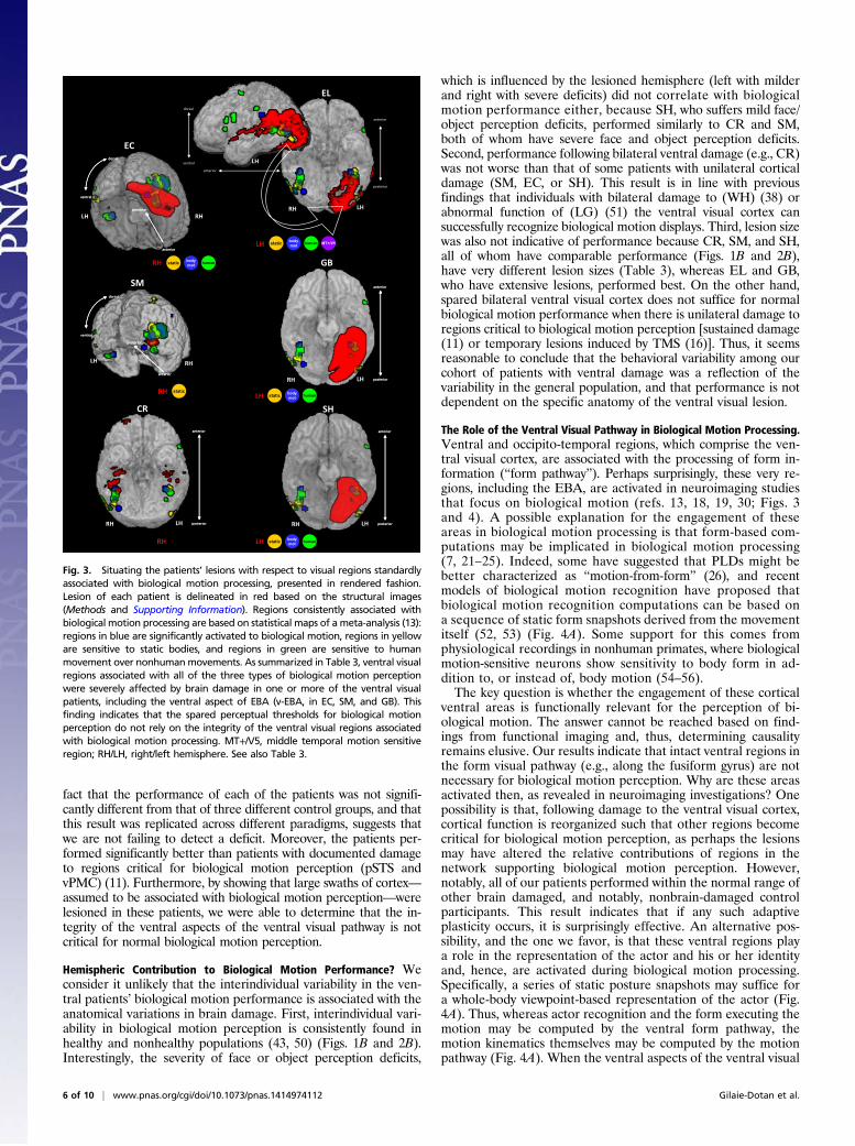
ventral cortex is so extensive and lesions are more circumscribed,there may be sparing of key ventral regions associated with bi-ological motion processing (13), and it is these spared regions thataccount for the normal perceptual performance. To assess thispossibility, we first carefully delineated the lesion of each patientand transformed each lesion into normalized Montreal Neuro-logical Institute (MNI) space. Second, we superimposed the lesion
onto the ventral cortex in which we identified areas that areconsistently activated by biological motion; this included regionsresponsive to the perception of body movement (blue in Fig. 3),sensitive to static bodies (yellow in Fig. 3), to human movements(green in Fig. 3) as determined by a recent meta-analysis (13)(Methods), as well as motion-sensitive MT+/V5, biological mo-tion-sensitive pSTS, vPMC, and static-body sensitive EBA (18–20). For the EBA cluster, we further distinguished between threesubregions: the ventral portion (v-EBA, ventral and not over-lapping MT+/V5), the portion overlapping MT+/V5 (mt-EBA),and the portion anterior to MT+/V5 (a-EBA). Our analysis alsotook into consideration the functional anatomical organization ofthe ventral visual stream (e.g., ventral surface vs. lateral occipito-temporal aspects). Third, for each of these regions, we evaluatedthe extent of damage in each patient. The results of this fine-grained analysis are detailed in Table 3.Our analysis revealed that regions situated on the ventral sur-
face (e.g., fusiform gyrus) that are assumed to be engaged in someaspect of biological motion perception, including the regions thatare sensitive to static body perception, body movements in gen-eral, and human body movements’ selectivity (13), are sub-stantially damaged in three patients (60–100%, left regions in GBand SH, and the right regions in EC) (Table 3). Furthermore, theventral portion of the EBA (46, 47), situated ventrally to but notoverlapping MT+/V5, is also critically damaged in two right ven-tral patients (SM and EC) and partially damaged in the left ventralpatient (GB). From these observations, we can conclude thatregions situated on the ventral surface of the cortex in the ventralstream (20, 31, 48) and the ventral aspect of the EBA are notcontributing critically to biological motion perception. Becauseother regions, such as those on the lateral occipito-temporal sur-face, including MT+/V5 and other parts of EBA, are only partiallydamaged in some of the patients (20–60%), we cannot rule outtheir possible role in biological motion perception.Each of the regions associated with biological motion in the
ventral aspect of the ventral visual cortex is significantly damagedin one or more of the patients (right fusiform in EC, left fusiformin GB and SH), including the right and left ventral portions ofthe EBA (R-v-EBA in EC and SM, L-v-EBA in GB), and also leftMT+/V5 (EL); the superior and middle temporal regions are mostlyspared (except for EL). Importantly, the two areas that are well-known to be sensitive and critical to human motion, the pSTS andvPMC (11, 13, 16, 28, 30, 32, 45, 49), are spared in all cases.The ventral patients performed as well as healthy controls and
brain-damaged patients, despite the fact that ventral regionsassociated with different aspects of biological motion processingwere affected by their lesions. Importantly, the ventral-lesionedpatients performed better than patients with brain damage toregions pSTS and vPMC, standardly considered the neural cor-relates of biological motion perception.
DiscussionIn this study, we explored whether the integrity of form perceptionand of the ventral visual cortex are necessary for the perception ofbiological motion. This was achieved by examining the perceptualperformance of patients with documented form deficits followingbrain damage to the ventral visual cortex. In two studies using an-imated PLDs (7) embedded in noise points, we derived a host ofdependent measures (accuracy of unmasked displays, thresholds,reaction times, and self-report measures) There were no differencesbetween the patients’ indices and those of healthy age-matchedcontrols or of brain-damaged patients* with spared ventral cortex,and this was also true compared with healthy young controls. The
Fig. 2. Biological motion perception: Exp. 2 paradigm and results. (A, Left)Static unmasked PLD snapshot presented in the action recognition phase,participants were required to verbally describe them. (Center and Right) Todetermine perceptual thresholds, each trial presented a masked PLD eithercontaining a biologically moving figure (Center) or a spatially scrambledversion of it (Right), and the task was to determine whether a moving figurewas embedded in the display. The noise points were added adaptivelyaccording to individual performance with 75% accuracy (Methods). (B) In-dividual perceptual thresholds (y axis) of the ventral visual patients and theage-matched controls against age (x axis). Perceptual thresholds determinedsimilar to Fig. 1 (Methods). Although patients commonly perform morepoorly than healthy-matched controls, the performance of each ventralpatient was not significantly different from that of their healthy age-matched controls (Results). (C) Response times are not very informativebecause instructions did not require speeded responses (participants wereable to respond leisurely and take breaks). Ventral visual patients’ responsetimes were not significantly slower than their age-matched controls (Results).
*Biological motion sensitivity following brain damage of any sort can be reduced relativeto healthy controls (11), accounting for the findings that some ventral patients per-formed at the lower end of the healthy control distribution.
Gilaie-Dotan et al. PNAS Early Edition | 5 of 10
NEU
ROSC
IENCE
PNASPL
US
fact that the performance of each of the patients was not signifi-cantly different from that of three different control groups, and thatthis result was replicated across different paradigms, suggests thatwe are not failing to detect a deficit. Moreover, the patients per-formed significantly better than patients with documented damageto regions critical for biological motion perception (pSTS andvPMC) (11). Furthermore, by showing that large swaths of cortex—assumed to be associated with biological motion perception—werelesioned in these patients, we were able to determine that the in-tegrity of the ventral aspects of the ventral visual pathway is notcritical for normal biological motion perception.
Hemispheric Contribution to Biological Motion Performance? Weconsider it unlikely that the interindividual variability in the ven-tral patients’ biological motion performance is associated with theanatomical variations in brain damage. First, interindividual vari-ability in biological motion perception is consistently found inhealthy and nonhealthy populations (43, 50) (Figs. 1B and 2B).Interestingly, the severity of face or object perception deficits,
which is influenced by the lesioned hemisphere (left with milderand right with severe deficits) did not correlate with biologicalmotion performance either, because SH, who suffers mild face/object perception deficits, performed similarly to CR and SM,both of whom have severe face and object perception deficits.Second, performance following bilateral ventral damage (e.g., CR)was not worse than that of some patients with unilateral corticaldamage (SM, EC, or SH). This result is in line with previousfindings that individuals with bilateral damage to (WH) (38) orabnormal function of (LG) (51) the ventral visual cortex cansuccessfully recognize biological motion displays. Third, lesion sizewas also not indicative of performance because CR, SM, and SH,all of whom have comparable performance (Figs. 1B and 2B),have very different lesion sizes (Table 3), whereas EL and GB,who have extensive lesions, performed best. On the other hand,spared bilateral ventral visual cortex does not suffice for normalbiological motion performance when there is unilateral damage toregions critical to biological motion perception [sustained damage(11) or temporary lesions induced by TMS (16)]. Thus, it seemsreasonable to conclude that the behavioral variability among ourcohort of patients with ventral damage was a reflection of thevariability in the general population, and that performance is notdependent on the specific anatomy of the ventral visual lesion.
The Role of the Ventral Visual Pathway in Biological Motion Processing.Ventral and occipito-temporal regions, which comprise the ven-tral visual cortex, are associated with the processing of form in-formation (“form pathway”). Perhaps surprisingly, these very re-gions, including the EBA, are activated in neuroimaging studiesthat focus on biological motion (refs. 13, 18, 19, 30; Figs. 3and 4). A possible explanation for the engagement of theseareas in biological motion processing is that form-based com-putations may be implicated in biological motion processing(7, 21–25). Indeed, some have suggested that PLDs might bebetter characterized as “motion-from-form” (26), and recentmodels of biological motion recognition have proposed thatbiological motion recognition computations can be based ona sequence of static form snapshots derived from the movementitself (52, 53) (Fig. 4A). Some support for this comes fromphysiological recordings in nonhuman primates, where biologicalmotion-sensitive neurons show sensitivity to body form in ad-dition to, or instead of, body motion (54–56).The key question is whether the engagement of these cortical
ventral areas is functionally relevant for the perception of bi-ological motion. The answer cannot be reached based on find-ings from functional imaging and, thus, determining causalityremains elusive. Our results indicate that intact ventral regions inthe form visual pathway (e.g., along the fusiform gyrus) are notnecessary for biological motion perception. Why are these areasactivated then, as revealed in neuroimaging investigations? Onepossibility is that, following damage to the ventral visual cortex,cortical function is reorganized such that other regions becomecritical for biological motion perception, as perhaps the lesionsmay have altered the relative contributions of regions in thenetwork supporting biological motion perception. However,notably, all of our patients performed within the normal range ofother brain damaged, and notably, nonbrain-damaged controlparticipants. This result indicates that if any such adaptiveplasticity occurs, it is surprisingly effective. An alternative pos-sibility, and the one we favor, is that these ventral regions playa role in the representation of the actor and his or her identityand, hence, are activated during biological motion processing.Specifically, a series of static posture snapshots may suffice fora whole-body viewpoint-based representation of the actor (Fig.4A). Thus, whereas actor recognition and the form executing themotion may be computed by the ventral form pathway, themotion kinematics themselves may be computed by the motionpathway (Fig. 4A). When the ventral aspects of the ventral visual
SM
RHLH
posterior
anterior
dorsal
ventral
posterior
anterior
dorsal
ventral
CR
RH LH posterior
anterior
posterior
anterior
GB
RH LH posterior
anterior
posterior
anterior
EC
RHLH
dorsal
ventral
posterior
anterior
dorsal
ventral
posterior
anterior
static bodymot humanRH
SH
RH LH posterior
anterior
posterior
anterior
staticRH
RH LH
posterior
anterior
LHposterioranterior
dorsal
ventral
MT+/V5LH
static bodymot humanLH
static bodymot human
RH
static bodymot humanLH
EL
Fig. 3. Situating the patients’ lesions with respect to visual regions standardlyassociated with biological motion processing, presented in rendered fashion.Lesion of each patient is delineated in red based on the structural images(Methods and Supporting Information). Regions consistently associated withbiological motion processing are based on statistical maps of ameta-analysis (13):regions in blue are significantly activated to biological motion, regions in yelloware sensitive to static bodies, and regions in green are sensitive to humanmovement over nonhumanmovements. As summarized in Table 3, ventral visualregions associated with all of the three types of biological motion perceptionwere severely affected by brain damage in one or more of the ventral visualpatients, including the ventral aspect of EBA (v-EBA, in EC, SM, and GB). Thisfinding indicates that the spared perceptual thresholds for biological motionperception do not rely on the integrity of the ventral visual regions associatedwith biological motion processing. MT+/V5, middle temporal motion sensitiveregion; RH/LH, right/left hemisphere. See also Table 3.
6 of 10 | www.pnas.org/cgi/doi/10.1073/pnas.1414974112 Gilaie-Dotan et al.

cortex are damaged (an example is conveyed in Fig. 4A by red-colored markings based on our ventral visual patients), snapshotsleading to actor recognition might be disrupted. However, becausethe computations of kinematics mediated by the motion pathwayare not significantly affected, movement perception is unaffected.Fig. 4B shows the prediction of this model for PLDs to illustrate ourcurrent findings. In addition to the supporting evidence from ourpatients, as well as from studies of individuals with developmentalagnosia (51, 57), form-based and motion-based processing of bodymotion can be dissociated among healthy controls (50).
Parallel Processing Routes Supporting Biological Motion Perception?The notion that biological motion perception might be computedin more than one way is also compatible with findings froma series of neuropsychological case studies. For example, LM,the “motion blind” patient with lesioned bilateral MT+/V5 (36),and AF, with severe damage to the dorsal cortex (35), are bothable to recognize unmasked PLDs above chance. In addition,patients with brain damage or abnormal vision, such as patientMM (37), who recovered from long-term visual deprivation, orpatient JW, who has widespread occipital damage followinghypoxia (58), are both able to successfully recognize unmaskedPLDs. Finally, the above-chance biological motion performanceof patients with lesions that appear to invade early visual areas(35, 59) and are very different from the lesions of the patientsexamined in the present study, also seems to suggest multipleprocessing routes supporting biological motion perception.
Although all of these findings are consistent with an account inwhich biological motion perception may be achieved via multiplepathways, there is still some selectivity to the processing, andthere are indications that biological motion perception is in-dependent from other lower-level motion perception. For ex-ample, performance in biological motion and motion-coherencetasks are not correlated, as revealed in studies of patients (11),following congenital cataracts (60), or in healthy controls (43).Indeed, all of our ventral patients performed normally in thebiological motion tasks but some have basic motion perceptiondeficits [SM, CR, and EC are impaired in motion coherence andmotion detection tasks (61)]. Similarly, patients AF and LMperformed poorly on early motion tasks despite above-chanceperformance on biological motion (35, 36).If biological motion recognition can be achieved via multiple
pathways, this duplication might reflect the importance of thisprocess to a multiplicity of abilities, such as social communica-tion, motor learning, and theory of mind. Whether these path-ways achieve movement recognition independently remains to beresolved. What is certain, though, is that the integrity of theventral aspects of the ventral visual stream is not, in and of itself,critical for the normal perception of biological motion.
ConclusionsWe have shown that biological motion perception can be achieveddespite damage to the ventral aspect of the form visual pathway(e.g., fusiform body area, v-EBA). Although regions such as the
Table 3. Summary of patients’ lesions
The lesion size is based on the results of the lesion delineating procedure (see also Supporting Information). Because of the low spatial coverage of GB’s,SH’s, and EC’s clinical scans, their lesion size reflects a spatial interpolation across the lesion locations in the clinical images that were available. The assessmentof the overlap of the lesion with regions associated with biological motion processing is predominantly based on a recent meta-analysis (13) (see text fordetails), as demonstrated in Fig. 3. Overlap with MT+/V5 is based on SM’s functional localization, and on MT+/V5 reported location for the other patients (81)(Methods). Overlap scale relates to foci reported in the meta-analysis with z ≥ 3.2 score (equivalent to P ≤ 0.0005). Overlap notations: No, none; Hardly, ∼<10%; Mildly, ∼10–20%; Partially, ∼20–60%; Mostly, >60–70%; All, 100%. Regions that are significantly damaged are indicated in bold and shaded grayaccording to the extent of damage (Mostly to All in dark gray, Partially in medium gray, and Mildly in light gray). EBA partitions: v-EBA, aspect of EBA ventralto MT/V5; mt-EBA, aspect of EBA overlapping MT/V5; a-EBA, aspect of EBA anterior to MT/V5 (46, 47).*GB’s, CR’s, and EC’s lesion sizes were approximated on an MNI template brain and therefore provided in MNI normalized space units (mm3), which might bean overestimation relative to native space volume (82).
Gilaie-Dotan et al. PNAS Early Edition | 7 of 10
NEU
ROSC
IENCE
PNASPL
US

pSTS and vPMC are critical for biological motion perception (11,15, 16, 41, 62, 63), we speculate that the ventral regions of the formvisual pathway are critical for recognizing the person performing themovement, but not for recognizing the motion being performed.
MethodsPatients with Ventral Visual Lesions. Six premorbidly normal right-handedindividuals who sustained brain damage to the right (n = 2), left (n = 3), orbilateral (n = 1) ventral visual cortex participated in the study. Followinga lesion sustained in adulthood (except for CR, who was aged 16 y), allindividuals reported visual perceptual problems and have well-establishedform-processing deficits. Table 1 summarizes the key demographics, neuro-psychological descriptions, and detailed visual performance (including visualmotion perception) of each patient; further details are available in SupportingInformation and in earlier publications [SM (61, 64–70), CR (61, 65–67, 70),EL (61, 70–75), GB (61, 75), SH (70, 75), and EC (61)].
Experiment 1. In this experiment, we used unmasked PLDs to assess recog-nition of biological motion and then measured perceptual thresholds usingPLDs masked in noise points. To assess recognition of biological motion,unmasked PLD animations of actions (see below) were presented and par-ticipants were required to verbally describe the stimuli without having prior
knowledge of what these would be. Each PLD animation looped until a co-herent verbal description was given, after which the experimenter presentedthe next animation. Tomeasure perceptual thresholds, on each trial two PLDswere presented simultaneously on the right and left sides of the screen, onecontaining a moving upright human figure performing one of seven actions(see Stimuli below) (Fig. 1A, Supporting Information, and Movies S1–S3) (11,51, 76), and the other a spatially scrambled version of the same action. Theside of the biological motion animation was randomly determined on eachtrial. Participants were instructed to identify which of the two displayscontained the animation of the human movement [but did not have toidentify the movement (i.e., jogging or walking), except in the action recog-nition phase; see below]. Both animations (intact and scrambled movements)were embedded in a number of noise points adaptively determined accordingto the participant’s performance (77). The task became more difficult as thenumber of noise points increased. Perceptual thresholds were determinedbased on the number of noise points with which a participant could performat a predefined level of accuracy (82%). Stimuli and further procedures arefully described in Supporting Information and elsewhere (11, 51).Participants. All six ventral-lesion patients, tested in Pittsburgh, PA, and allhealthy control participants (tested in Pittsburgh or in London) gave writteninformed consent to participate in the study and the experiments wereapproved by local ethics committees (Institutional Review Board, CarnegieMellon University and University College London). All patients (except SM,who was tested at Carnegie Mellon University) and the older controls weretested at home for maximal convenience.
Procedures regarding the data collection from the nonventral brain-damaged patient control group (control group 3, see below) are providedelsewhere (11). Informed consent was obtained from these patients at thetime of testing in accordance with guidelines of the University of California,San Diego and VA Northern California Health Care System Human ResearchProtections Programs. The findings from these patients have been publishedpreviously (11) and we simply adopted the de-identified data to serve as anadditional benchmark against which to compare the ventral patients’performance.
Healthy controls. All healthy control participants had normal or corrected-to-normal vision, no history of neurological disorders, and were right-handed.
Control group 1 was the first neurologically normal control group thatparticipated in this study and included 42 healthy adults, age-matched to thepatients: 16 male control participants served as age-matched controls for CR(mean age 32.0 y ± 2.9 SD); 15 males served as age-matched controls for SM(mean age 35.2 y ± 3.3 SD, 11 of whom were also matched for CR); 13females and 1 male (matched for SM as well) served as age-matched controlsfor EC (mean age 48.0 y ± 3.8 SD); and 14 females and 1 male served as age-matched controls for GB, EL, and SH (aged 50–70 y, mean 59.2 ± 6.1 SD, ofwhom 5 females were matched for EC as well).
Control group 2 was the second neurologically normal control group andincluded 13 healthy young controls (aged 20.4 y ± 1.1 SD).
Brain-damaged controls. Control group 3 included 54 right-handed, brain-damaged patients (13 females, 41 males, aged 36.9–84.9 y) with focal, uni-lateral lesions (43 in the left hemisphere, 11 in the right hemisphere). Fromthe 60 patients who had completed Exp. 1 in an earlier study (11), we se-lected for this control group only those for whom we could definitivelydetermine that their ventral visual cortex was not affected by their lesion (asascertained and confirmed by the lesion boundaries) as determined fromcomputerized lesion reconstructions of the brain. The time between testingand patients’ cerebrovascular accident ranged from 6 mo to 22 y (mean of6.5 y). Patients with diagnosed or suspected vision or hearing loss, dementia,head trauma, tumors, multiple infarcts, or prior psychiatric or neurologicalabnormalities were excluded from the sample. Motor and languageimpairments ranged from very mild to severe in the sample, but all patientswere able to understand and carry out the task. None of the patients pre-sented with spatial neglect or other attentional disorders.Stimuli. Briefly, biological motion PLD animations made of 12 white points ona black background depicting one of seven actions—walking, jogging,overarm throwing, underarm throwing (bowling), stepping up, high kickinginto the air, and lower kicking—and lasting 0.8 s were presented and loopeduntil a response was given.
For the perceptual threshold assessment, a matched spatially scrambledversion was created for each animation so that the local motion of each pointwas preserved, without the global form (11, 51, 76).
In each trial of the perceptual threshold assessment, additional movingnoise points were randomly superimposed on both PLDs (the biologicalmotion and its scrambled counterpart) (9). The motion trajectory of eachnoise point that was added to the animations was equivalent to a motion
Localorientationdetectors
Invariant bardetectors
Snapshotneurons
V1 V1/V4??
Localmotion
detectors
Local OFdetectors
OF pattern neurons
V1/V2, MT+/V5 MT+/V5, KO pSTS
Snapshots foractor recogni�on
kinematics forac�on recognition
pSTS andvPMC
Human motionpatternneurons
Formpathway
Motionpathway
XITS, FBA,v-EBA?
?
Localorientationdetectors
Invariant bar detectors
Snapshotneurons
V1 V1/V4 ITS, FBA,v-EBA?X
Localmotion
detectors
Local OFdetectors
OF patternneurons
V1/V2, MT+/V5 MT+/V5, KO pSTS
Snapshots foractor recogni�on
kinematics foraction recogni�on
pSTS andvPMC
Human motionpatternneurons
Formpathway
Motionpathway
XA
B
Fig. 4. Adaptation of the model for biological motion recognition based onGiese and Poggio (52). (A) The original model by Giese and Poggio with pro-posed distinctions: the form pathway’s main role involves snapshots for actorrecognition, and the motion pathway’s main role involves kinematic patternsfor human movement recognition. Brick color indicates how brain damage tothe ventral cortex predominantly affects the form processing pathway that isinvolved in snapshot creation, thereby impairing actor but not action recog-nition. (B) Adaptation of the model to PLDs. Information flow in the case ofPLDs resembles that of a damaged ventral visual cortex (A), because the in-formation processed by the form pathway is insufficient, leading to abnormalactor recognition. At the same time, the information processed by the motionpathway is not significantly affected, so that themovement can be recognized.Based on our results, we speculate that the perception of human movementcan be achieved based on motion kinematics alone. FBA, fusiform body area;ITS, inferior temporal sulcus; KO, kinetic occipital; MT+/V5, middle temporalmotion-sensitive region; OF, optic flow; V1/V2/V4, visual retinotopic regions; v-EBA, ventral aspect of extrastriate body area.
8 of 10 | www.pnas.org/cgi/doi/10.1073/pnas.1414974112 Gilaie-Dotan et al.
trajectory of one of the animation’s points (randomly chosen), but startingat a random location.
Each animation subtended ∼4° × 6° (width × height) visual angle whenviewed from 55 cm. The total area occupied by each PLD (comprising the ani-mation plus the noise points) was ∼7° of visual angle in diameter. The two PLDs(biological motion and its scrambled counterpart) were displayed at ∼9° to theleft and right of the center of the screen, their vertical centers horizontallyaligned (Movie S1). Stimuli were presented and responses recorded usingMATLAB (Mathworks) and the Psychophysics Toolbox v2.54 (78, 79).Procedure. In the first part of the experiment, we examined action recogni-tion of unmasked PLDs. Participants were presented with seven differentunmasked biological motion animations and were asked to verbally describethe stimuli on the screen. Each animation was displayed separately in thecenter of the screen without any masking noise points and looped until theverbal description given by the participant indicated that they were able toperceive the movement conveyed by the PLD animation. After that, theexperimenter displayed the next animation.
Following the action-recognition phase, sensitivity to biological motion wasassessed by measuring the number of noise dots that allowed successful dis-crimination (82%) of intact from scrambled animations when both are maskedin noise points. On each trial, participants were required to report whether theintact PLD (“the person”) was on the right or left side of the screen by pressingthe corresponding left or right key in a two-alternative forced-choice (2AFC)manner (see additional details below). The animations looped until a responsewas given. Although accuracy was the key dependent measure, response timeof each trial was also recorded. EC, EL, the brain-damaged controls, and someof the older healthy controls, replied verbally or by pointing, after which theexperimenter pressed the corresponding response button. Because the PLDswere presented on the two sides of the screen simultaneously, participantswere not required to fixate at the center of the screen. After 16 practice trialswith a predefined number of noise points, control participants completed 118trials presented in two blocks separated by an optional rest period, whereaspatients completed one block of 73 trials, with a rest period after 40 trials.Note that because the task was not timed, participants were able to takeadditional breaks at any time if needed.
To estimate perceptual thresholds, we varied the number of noise points ineach trial to yield a psychometric measure of performance according to anefficient Bayesian adaptive procedure that uses the mean of the posteriorprobability density function [QUEST (77)]. The perceptual threshold wasdetermined as the number of points at which a participant performed at82% accuracy. For the healthy controls, who performed two blocks of trials,thresholds from the two blocks were averaged. Reaction time analysis wasbased on each participant’s average reaction time across the experiment.
In addition to between-group comparisons, which were based on Wilcoxonnonparametric rank-sum test (80), we also examined every patient’s performanceindividually. This was achieved by determining whether a patient’s performance(threshold or reaction time) was significantly different from that of a controlgroup. The statistical evaluation was based on an established statistical pro-cedure for comparing single cases to a control group (40), entailing a modified ttest, significant performance differences roughly corresponding to more thantwo SDs from the controls’ mean performance.
Experiment 2. The previous experiment assessed the participant’s ability todetect the PLD containing human movement when embedded in noise. Thissecond experiment further examined the patients’ biological motion per-ception using a slightly different method, including a different and largerset of displays and a different criterion at which threshold is established.Participants. All six ventral-damaged patients, tested in Pittsburgh, PA, and 39healthy neurologically normal controls (28 also participated in Exp. 1) gaveinformed consent to participate in the study and the experiment was ap-proved by local ethics committees of Carnegie Mellon University (InstitutionalReview Board) and University College London. The controls were age-matchedto each patient in the following way: 15 participants (14 male) served as age-matched controls for CR (mean age 32.5 y ± 4.1 SD); 15 participants (12 male)served as age-matched controls for SM (mean age 37.3 y ± 4.5 SD, 10 of whomwere also matched for CR); 15 participants (12 female) served as age-matchedcontrols for EC [mean age 46.5 y ± 4.5 SD, 5/1 controls also matched for SM/CR,respectively)]; and 11 participants (8 females) served as age-matched controlsfor GB, EL, and SH (mean 62.7 y ± 4.6 SD, 1 also matched for EC). All patients(but SM, who was tested at Carnegie Mellon University) and some of thecontrols were tested at home for maximal convenience.Procedure. We first assessed action recognition of 12 different PLDs (Sup-porting Information). Participants were asked to describe what they per-ceived so as to ensure that they were able to recognize the movementsconveyed by PLDs. Thereafter, perceptual thresholds were established. A
short practice comprising a few trials with a predefined number of noisepoints was completed, followed by the main experiment that measured thenumber of noise points a participant can tolerate and perform at 75% ac-curacy, using the same Bayesian estimation method as in Exp. 1 (77). In eachtrial, a single PLD (similar to those from Exp. 1) (Fig. 2A and SupportingInformation) was presented at the center of the screen, either containinga movement of an upright human figure performing a movement (target),or a spatially scrambled version of it (nontarget). Animations (targets andnontargets) were masked in noise points (in the same manner as in Exp. 1),and the participant’s task was to decide whether the display containeda person (“target present”) or not (“target absent”) in a 2AFC manner usingtwo predefined keys. Animations looped until a response was given. Visualfeedback (green/red cross for correct/incorrect response, respectively) wasprovided after each response. As in Exp. 1, speeded responses were not re-quired but we still recorded response times. Further experimental details aresimilar to those of Exp. 1; see details in Supporting Information.
Lesion and Anatomical Analysis. Lesion delineation procedure. For structuralimage-acquisition details, see Supporting Information. We followed a lesiondelineation procedure that has been successfully used before (61). High-resolution anatomical images (EL, SM, and CR) were coregistered onto a T1MNI canonical SPM image using SPM (www.fil.ion.ucl.ac.uk/spm), afterwhich the lesions were traced manually in MRIcroN (www.cabiatl.com/mricro/mricro; see Supporting Information for tracing criteria) and saved asa binary image. For each patient, the coregistered anatomical images andthe demarcated lesion were normalized into MNI space using the unifiednormalization segmentation of SPM. When only low-resolution anatomicalimages from clinical scans were available, lesions were traced manually ontothe corresponding anatomical locations in an MNI canonical SPM image; toprovide a consistent visualization of these patients’ lesions (Fig. 3), and toreach some approximate assessment of their lesion size (presented in Table3), their noncontinuous traced lesions were then each manually interpolatedto a continuous lesion in a conservative manner using MRIcroN (Fig. S1 andSupporting Information).Comparing lesions to regions associated with biological motion processing. To ex-amine the lesion-behavior relationships, we sited each of our patients’ lesionsrelative to the anatomical locations of regions that are consistently activatedacross studies in response to biological motion stimuli, as determined bya recent meta-analysis (13). Specifically, we were interested in regions thatare consistently activated in response to: (i) body movements [as reported infigure 1 and table 4 of Grosbras et al. (13)], (ii) static bodies [as reported inthat study in figure 3 and table 7 of Grosbras et al. (13)], or (iii) humanmovements vs. nonhuman movements (as reported in table 8 of Grosbraset al. (13)]. Three image maps from that meta-analysis corresponding tothese three contrasts-of-interest were included in our analysis. Each imagemap consisted of probability (P) values that corresponded to the activationlikelihood estimation values reported in Grosbras et al. (13). These mapsrepresent for each specific voxel the probability that a study will reportsignificant activation in that voxel (for example with respect to one of ourcontrasts-of-interest: regions in which significant activation to humanmovements vs. nonhuman movements). The maps [as described in Grosbraset al. (13)] were thresholded at z = 3.2 (corresponding to P < 0.0005) andcluster size was 120 mm3 [see more details in Grosbras et al. (13)].
For each ventral patient, the three contrasts-of-interest from the meta-analysis along with the normalized brain and delineated lesion were loadedonto MRIcroN in four different colors as presented in Fig. 3 [lesion in red,body-movement regions (contrast 1) in blue, static-bodies regions (contrast2) in yellow, and selective human movements (contrast 3) in green]. We thencarefully examined whether the lesion invaded or overlapped any of theregions from each of the three contrasts-of-interest, according to anatomicallocation as well (e.g., ventral surface vs. lateral occipito-temporal regions).See also Table 3.
ACKNOWLEDGMENTS. We thank all the patients and their families for theirwonderful collaboration; Nina Dronkers and the VA Northern CaliforniaHealth Care System; Marie-Helene Grosbras for providing us the image mapsfrom her study (13); and Christina Konen, Solmaz Shariat Torbaghan, SabineKastner, Tanja Kassuba, Adam Greenberg, Kate Fissell, Maxim Hammer,Mohamed Seghier, Ryan Egan, and John Pyles for their help. This study wassupported by the Royal Society Grant TG102269 (to S.G.-D. and M.B.); Marie-Curie 236021 (to S.G.-D.); National Science Foundation Grant BCS0923763 andNational Institute of Mental Health Grant 54246 (to M.B.); National ScienceFoundation BCS-CAREER-1151805 (to A.P.S.); and the Wellcome Trust (G.R.).The Wellcome Trust Centre for Neuroimaging is supported by core fundingfrom the Wellcome Trust 091593/Z/10/Z.
Gilaie-Dotan et al. PNAS Early Edition | 9 of 10
NEU
ROSC
IENCE
PNASPL
US

1. Billino J, Bremmer F, Gegenfurtner KR (2008) Motion processing at low light levels:Differential effects on the perception of specific motion types. J Vis 8(3):1–10.
2. Wood JM, et al. (2011) Using biological motion to enhance the conspicuity of road-way workers. Accid Anal Prev 43(3):1036–1041.
3. Tyrrell RA, et al. (2009) Seeing pedestrians at night: Visual clutter does not mask bi-ological motion. Accid Anal Prev 41(3):506–512.
4. Balk SA, Tyrrell RA, Brooks JO, Carpenter TL (2008) Highlighting human form and motioninformation enhances the conspicuity of pedestrians at night. Perception 37(8):1276–1284.
5. Owens DA, Antonoff RJ, Francis EL (1994) Biological motion and nighttime pedestrianconspicuity. Hum Factors 36(4):718–732.
6. Neri P, Morrone MC, Burr DC (1998) Seeing biological motion. Nature 395(6705):894–896.7. Johansson G (1973) Visual perception of biological motion and a model for its anal-
ysis. Percept Psychophys 14(2):201–211.8. Pinto J, Shiffrar M (1999) Subconfigurations of the human form in the perception of
biological motion displays. Acta Psychol (Amst) 102(2-3):293–318.9. Bertenthal B, Pinto J (1994) Global processing of biological motion. Psychol Sci 5(4):
221–225.10. Pavlova M, Staudt M, Sokolov A, Birbaumer N, Krägeloh-Mann I (2003) Perception
and production of biological movement in patients with early periventricular brainlesions. Brain 126(Pt 3):692–701.
11. Saygin AP (2007) Superior temporal and premotor brain areas necessary for biologicalmotion perception. Brain 130(Pt 9):2452–2461.
12. Wood JM, Tyrrell RA, Carberry TP (2005) Limitations in drivers’ ability to recognizepedestrians at night. Hum Factors 47(3):644–653.
13. Grosbras MH, Beaton S, Eickhoff SB (2012) Brain regions involved in human movementperception: A quantitative voxel-based meta-analysis. Hum Brain Mapp 33(2):431–454.
14. Kourtzi Z, Krekelberg B, van Wezel RJ (2008) Linking form and motion in the primatebrain. Trends Cogn Sci 12(6):230–236.
15. Grossman ED, Battelli L, Pascual-Leone A (2005) Repetitive TMS over posterior STSdisrupts perception of biological motion. Vision Res 45(22):2847–2853.
16. vanKemenadeBM,MuggletonN,WalshV,SayginAP(2012)EffectsofTMSoverpremotorandsuperior temporal cortices on biological motion perception. J Cogn Neurosci 24(4):896–904.
17. Vaina LM, Gross CG (2004) Perceptual deficits in patients with impaired recognition ofbiological motion after temporal lobe lesions. Proc Natl Acad Sci USA 101(48):16947–16951.
18. Downing PE, Jiang Y, Shuman M, Kanwisher N (2001) A cortical area selective forvisual processing of the human body. Science 293(5539):2470–2473.
19. Peelen MV, Downing PE (2007) The neural basis of visual body perception. Nat RevNeurosci 8(8):636–648.
20. Jastorff J, Orban GA (2009) Human functional magnetic resonance imaging revealsseparation and integration of shape and motion cues in biological motion processing.J Neurosci 29(22):7315–7329.
21. Thompson JC, Clarke M, Stewart T, Puce A (2005) Configural processing of biologicalmotion in human superior temporal sulcus. J Neurosci 25(39):9059–9066.
22. Beintema JA, Lappe M (2002) Perception of biological motion without local imagemotion. Proc Natl Acad Sci USA 99(8):5661–5663.
23. Lu H (2010) Structural processing in biological motion perception. J Vis 10(12):13.24. Reid R, Brooks A, Blair D, van der Zwan R (2009) Snap! Recognising implicit actions in
static point-light displays. Perception 38(4):613–616.25. Thirkettle M, Scott-Samuel NE, Benton CP (2010) Form overshadows ‘opponent mo-
tion’ information in processing of biological motion from point light walker stimuli.Vision Res 50(1):118–126.
26. Lange J, Georg K, Lappe M (2006) Visual perception of biological motion by form: Atemplate-matching analysis. J Vis 6(8):836–849.
27. Troje NF (2008) Biological motion perception. The Senses: A Comprehensive Refer-ence, Vol 1, eds Basbaum A, et al. (Elsevier, Oxford), pp 231–238.
28. Vaina LM, Solomon J, Chowdhury S, Sinha P, Belliveau JW (2001) Functional neuroanatomyof biological motion perception in humans. Proc Natl Acad Sci USA 98(20):11656–11661.
29. Beauchamp MS, Lee KE, Haxby JV, Martin A (2002) Parallel visual motion processingstreams for manipulable objects and human movements. Neuron 34(1):149–159.
30. Grossman ED, Blake R (2002) Brain areas active during visual perception of biologicalmotion. Neuron 35(6):1167–1175.
31. Peelen MV, Wiggett AJ, Downing PE (2006) Patterns of fMRI activity dissociate overlappingfunctional brain areas that respond to biological motion. Neuron 49(6):815–822.
32. Pyles JA, Garcia JO, Hoffman DD, Grossman ED (2007) Visual perception and neuralcorrelates of novel ‘biological motion’. Vision Res 47(21):2786–2797.
33. Grossman ED, Jardine NL, Pyles JA (2011) fMR-adaptation reveals invariant coding ofbiological motion on human STS. Front Hum Neurosci 5:12.
34. Schenk T, Zihl J (1997) Visual motion perception after brain damage: II. Deficits inform-from-motion perception. Neuropsychologia 35(9):1299–1310.
35. Vaina LM, Lemay M, Bienfang DC, Choi AY, Nakayama K (1990) Intact “biologicalmotion” and “structure from motion” perception in a patient with impaired motionmechanisms: A case study. Vis Neurosci 5(4):353–369.
36. McLeod P, Dittrich W, Driver J, Perrett D, Zihl J (1996) Preserved and impaired de-tection of structure from motion by a “motion blind” patient. Vis Cogn 3(4):363–391.
37. Fine I, et al. (2003) Long-term deprivation affects visual perception and cortex. NatNeurosci 6(9):915–916.
38. Huberle E, Rupek P, Lappe M, Karnath HO (2012) Perception of biological motion invisual agnosia. Front Behav Neurosci 6:56.
39. Pavlova M, et al. (2005) Recruitment of periventricular parietal regions in processingcluttered point-light biological motion. Cereb Cortex 15(5):594–601.
40. Crawford JR, Howell DC (1998) Comparing an individual’s test score against normsderived from small samples. J Clin Exp Neuropsychol 12(4):482–486.
41. Saygin AP, Wilson SM, Hagler DJ, Jr, Bates E, Sereno MI (2004) Point-light biologicalmotion perception activates human premotor cortex. J Neurosci 24(27):6181–6188.
42. Sereno MI, Saygin AP, Hagler DJ, Jr (2003) Retinotopy in parietal and temporal cortex.Neuroimage 19:S1523.
43. Gilaie-Dotan S, Kanai R, Bahrami B, Rees G, Saygin AP (2013) Neuroanatomical cor-relates of biological motion detection. Neuropsychologia 51(3):457–463.
44. Grossman E, et al. (2000) Brain areas involved in perception of biological motion.J Cogn Neurosci 12(5):711–720.
45. Peuskens H, Vanrie J, Verfaillie K, Orban GA (2005) Specificity of regions processingbiological motion. Eur J Neurosci 21(10):2864–2875.
46. Weiner KS, Grill-Spector K (2011) Not one extrastriate body area: Using anatomicallandmarks, hMT+, and visual field maps to parcellate limb-selective activations inhuman lateral occipitotemporal cortex. Neuroimage 56(4):2183–2199.
47. Ferri S, Kolster H, Jastorff J, Orban GA (2013) The overlap of the EBA and the MT/V5cluster. Neuroimage 66:412–425.
48. Schwarzlose RF, Baker CI, Kanwisher N (2005) Separate face and body selectivity onthe fusiform gyrus. J Neurosci 25(47):11055–11059.
49. Bonda E, PetridesM,Ostry D, EvansA (1996) Specific involvement of humanparietal systemsand the amygdala in the perception of biological motion. J Neurosci 16(11):3737–3744.
50. Miller LE, Saygin AP (2013) Individual differences in the perception of biologicalmotion: Links to social cognition and motor imagery. Cognition 128(2):140–148.
51. Gilaie-Dotan S, Bentin S, Harel M, Rees G, Saygin AP (2011) Normal form from biologicalmotion despite impaired ventral stream function. Neuropsychologia 49(5):1033–1043.
52. Giese MA, Poggio T (2003) Neural mechanisms for the recognition of biologicalmovements. Nat Rev Neurosci 4(3):179–192.
53. Lange J, Lappe M (2006) A model of biological motion perception from configuralform cues. J Neurosci 26(11):2894–2906.
54. Vangeneugden J, et al. (2011) Distinct mechanisms for coding of visual actions inmacaque temporal cortex. J Neurosci 31(2):385–401.
55. Jellema T, Perrett DI (2003) Cells in monkey STS responsive to articulated body motions andconsequent static posture: a case of implied motion? Neuropsychologia 41(13):1728–1737.
56. Singer JM, Sheinberg DL (2010) Temporal cortex neurons encode articulated actionsas slow sequences of integrated poses. J Neurosci 30(8):3133–3145.
57. Gilaie-Dotan S, Perry A, Bonneh Y, Malach R, Bentin S (2009) Seeing with profoundlydeactivated mid-level visual areas: Non-hierarchical functioning in the human visualcortex. Cereb Cortex 19(7):1687–1703.
58. Rosenthal O, Behrmann M (2006) Acquiring long-term representations of visualclasses following extensive extrastriate damage. Neuropsychologia 44(5):799–815.
59. Huberle E, Rupek P, Lappe M, Karnath HO (2009) Perception of global gestalt bytemporal integration in simultanagnosia. Eur J Neurosci 29(1):197–204.
60. Hadad BS, Maurer D, Lewis TL (2012) Sparing of sensitivity to biological motion butnot of global motion after early visual deprivation. Dev Sci 15(4):474–481.
61. Gilaie-Dotan S, et al. (2013) The role of human ventral visual cortex in motion per-ception. Brain 136(Pt 9):2784–2798.
62. Pelphrey KA, et al. (2003) Brain activity evoked by the perception of human walking:Controlling for meaningful coherent motion. J Neurosci 23(17):6819–6825.
63. Tai YF, Scherfler C, Brooks DJ, Sawamoto N, Castiello U (2004) The human premotorcortex is ‘mirror’ only for biological actions. Curr Biol 14(2):117–120.
64. Behrmann M, Kimchi R (2003) What does visual agnosia tell us about perceptualorganization and its relationship to object perception? J Exp Psychol Hum PerceptPerform 29(1):19–42.
65. Marotta JJ, Genovese CR, Behrmann M (2001) A functional MRI study of face rec-ognition in patients with prosopagnosia. Neuroreport 12(8):1581–1587.
66. Gauthier I, Behrmann M, Tarr MJ (1999) Can face recognition really be dissociatedfrom object recognition? J Cogn Neurosci 11(4):349–370.
67. Behrmann M, Williams P (2007) Impairments in part-whole representations of objectsin two cases of integrative visual agnosia. Cogn Neuropsychol 24(7):701–730.
68. Nishimura M, Doyle J, Humphreys K, Behrmann M (2010) Probing the face-space ofindividuals with prosopagnosia. Neuropsychologia 48(6):1828–1841.
69. Konen CS, Behrmann M, Nishimura M, Kastner S (2011) The functional neuroanatomyof object agnosia: A case study. Neuron 71(1):49–60.
70. Behrmann M, Plaut DC (2014) Bilateral hemispheric processing of words and faces:Evidence from word impairments in prosopagnosia and face impairments in purealexia. Cereb Cortex 24(4):1102–1118.
71. McKeeff TJ, BehrmannM (2004) Pure alexia and covert reading: Evidence from Strooptasks. Cogn Neuropsychol 21(2):443–458.
72. Mycroft RH, Behrmann M, Kay J (2009) Visuoperceptual deficits in letter-by-letterreading? Neuropsychologia 47(7):1733–1744.
73. Montant M, Behrmann M (2001) Phonological activation in pure alexia. Cogn Neu-ropsychol 18(8):697–727.
74. Behrmann M, Nelson J, Sekuler EB (1998) Visual complexity in letter-by-letter reading:“Pure” alexia is not pure. Neuropsychologia 36(11):1115–1132.
75. Habekost T, Petersen A, Behrmann M, Starrfelt R (2014) From word superiority toword inferiority: Visual processing of letters and words in pure alexia. Cogn Neuro-psychol 31(5-6):413–436.
76. Ahlström V, Blake R, Ahlström U (1997) Perception of biological motion. Perception26(12):1539–1548.
77. Watson AB, Pelli DG (1983) QUEST: A Bayesian adaptive psychometric method. Per-cept Psychophys 33(2):113–120.
78. Brainard DH (1997) The Psychophysics Toolbox. Spat Vis 10(4):433–436.79. Pelli DG (1997) The VideoToolbox software for visual psychophysics: Transforming
numbers into movies. Spat Vis 10(4):437–442.80. Wilcoxon F (1945) Individual comparisons by ranking methods. Biom Bull 1(6):80–83.81. Kolster H, Peeters R, Orban GA (2010) The retinotopic organization of the human
middle temporal area MT/V5 and its cortical neighbors. J Neurosci 30(29):9801–9820.82. Allen JS, et al. (2008) Effects of spatial transformation on regional brain volume
estimates. Neuroimage 42(2):535–547.
10 of 10 | www.pnas.org/cgi/doi/10.1073/pnas.1414974112 Gilaie-Dotan et al.