vertebral column
TRANSCRIPT

Dr.Poonam KalavadiyaMPT (Cardio Pulmonary)
Vertibral Colum 1
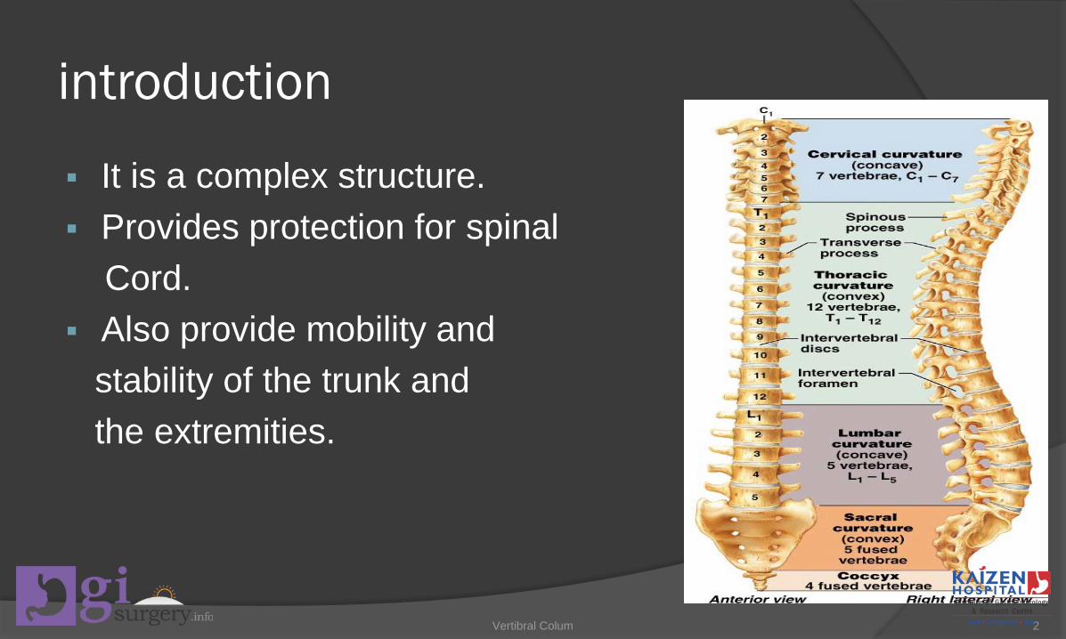
introduction
It is a complex structure. Provides protection for spinal
Cord. Also provide mobility and
stability of the trunk and the extremities.
Vertibral Colum 2

structure
Vertebral column composed of 33 vertebrae and 23 intervertebral disks.
And divided in to five regions.
Vertibral Colum 3
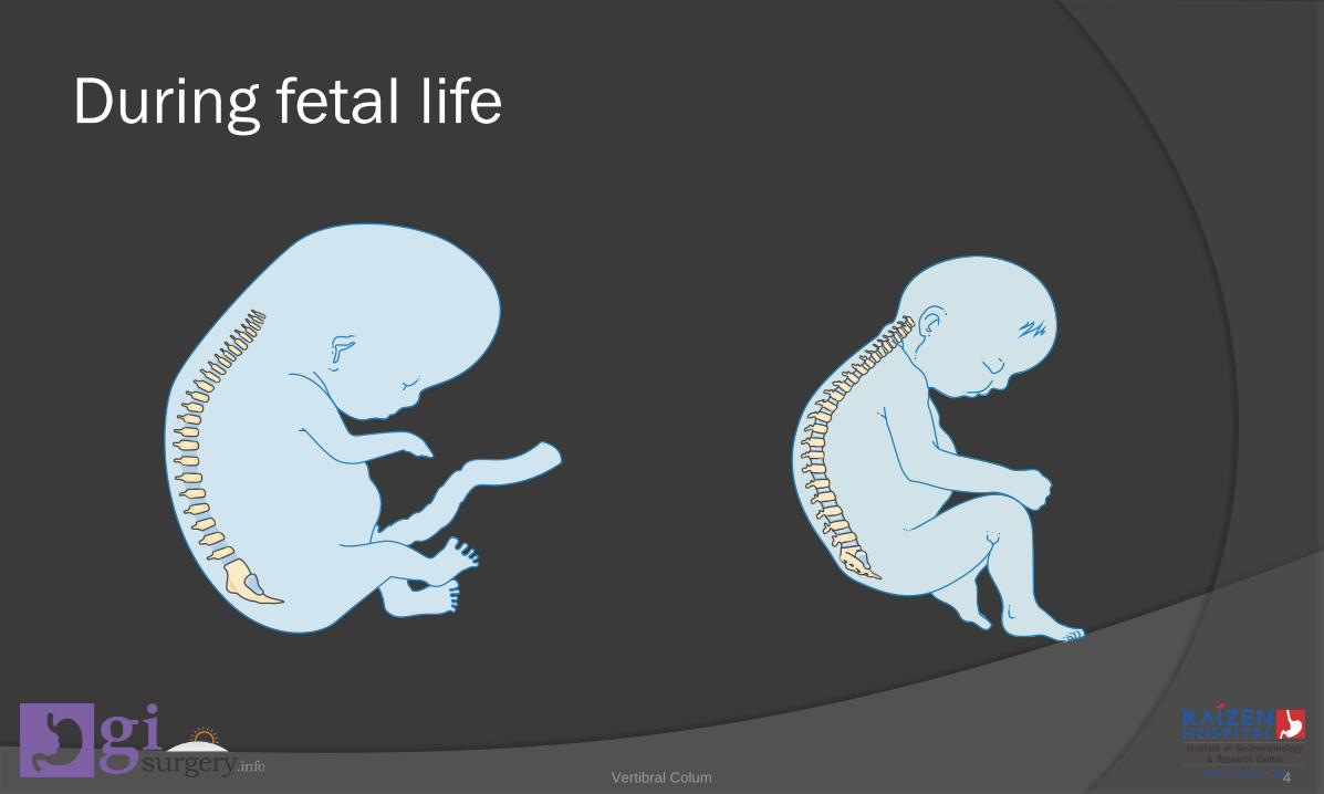
During fetal life
Vertibral Colum 4

The two curves (thoracic and sacral) that retain the original posterior convexity throughout life are called primary curves or kyphotic curves.
And the two curves (cervical and lumbar) that show a reversal of the original posterior convexity are called secondary or lordotic curves.
Vertibral Colum 5

The secondary or lordotic curves develop as a result of the accommodation of the skeleton to the upright posture.
Vertibral Colum 6

ADVANTAGE OF CURVES
A curved vertebral column provides significant advantage over a straight rod in that it is able to resist much higher compressive loads.
According to kapandji, a spinal column with the normal lumbar, thoracic, and cervical curves has a 10-fold ability to resist axial compression in comparison with a straight rod.
Vertibral Colum 7

The mobile segment
A smallest functional unit in a spine. One mobile segment=two adjacent vertebrae, the
intervening intervertebral disk and all the soft tissue around.
Vertibral Colum 8

A typical vertebra
There are two major parts1)anterior - vertebral body2)posterior - neural arch
Vertibral Colum 9
1)Vertebral body
Is designed to be the weight-bearing structure of the spinal column.
It is not a solid block of bone but a shell of a cortical bone surrounding by a cancellous cavity.
The cortical shell is reinforced by trabeculae in the cancellous bone, which provide resistance to compressive forces.
Vertibral Colum 10

Structure of the Typical Vertebra Vertebral body – ant Vertebral arch – pos – shape
of horse shoe - articular processes divide the arch in to two parts Ant – the pedicels, pos –laminae.
Spinous process – attached to the midline post
Vertebral arch – attached to the vertebral through pedicels
Vertibral Colum 11

Structure of the Typical VertebraPedicel Connections between the
post. elements & vertebral bodies
Transmit tension & bending forces from the post. elements to the vertebral bodies
Size is bigger in lumbar vertebrae
Vertibral Colum 12
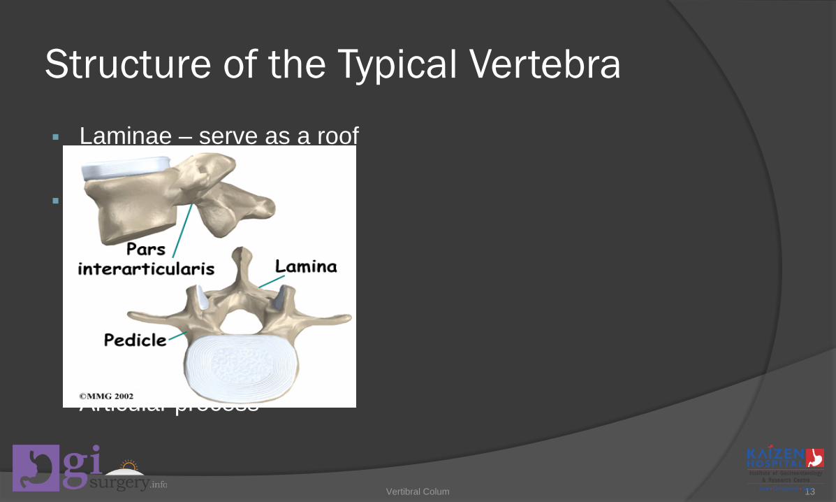
Structure of the Typical Vertebra Laminae – serve as a roof
– protect the spinal cord Transmit forces from the
vertically placed laminae to the horizontal oriented pedicel, then to vertebral body by means of PARS INTERARTIULARIS –between sup. & inf. Articular process
Vertibral Colum 13

INTERVERTEBRAL DISCTwo principle functions
1.To separate two vertebral bodies
2.To transmit load from one vertebral to the next
Vertibral Colum 14

INTERVERTEBRAL DISC
Disc thickness varies with disc position in the vertebral column
Lumbar region – 9mm Thoracic region – 5mm Cervical region – 3mm The greater the ratio – greater the mobility
Vertibral Colum 15
INTERVERTEBRAL DISC
The ratio of disc thickness to the height of the vertebral body
Cervical column – 2/5 Lumbar column – 1/3 Thoracic column – 1/5
Vertibral Colum 16
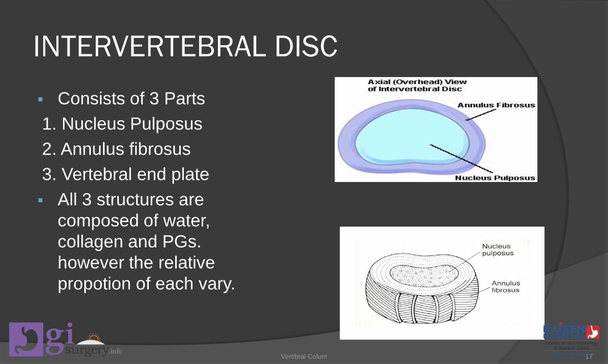
INTERVERTEBRAL DISC Consists of 3 Parts1. Nucleus Pulposus2. Annulus fibrosus3. Vertebral end plate All 3 structures are
composed of water, collagen and PGs. however the relative propotion of each vary.
Vertibral Colum 17

Nucleus Pulposus Has more water 70% - 90% and PGs & remainder 15% consists
of collagen, elastin, proteolytic enzymes PG are macro-molecules Attract and retain water Hydrophilic gel–like matter Resists compression
Amount of water Activity related Varies throughout the day
Vertibral Colum 18
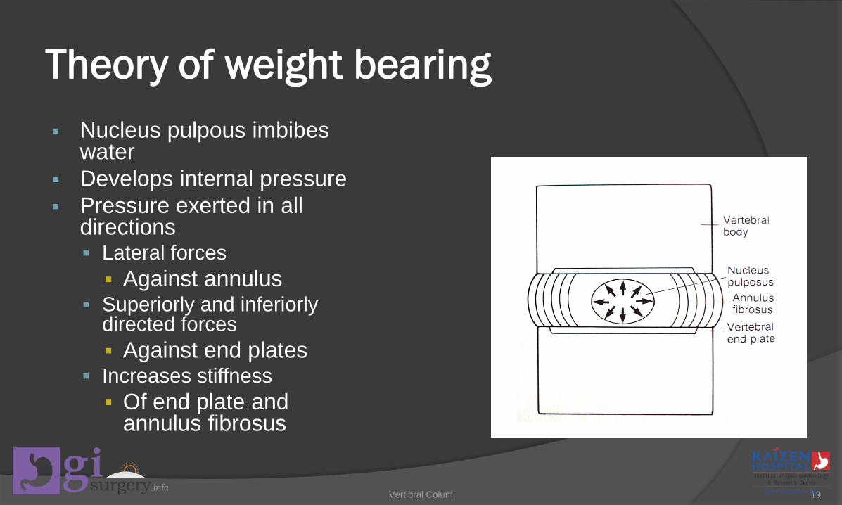
Theory of weight bearing Nucleus pulpous imbibes
water Develops internal pressure Pressure exerted in all
directions Lateral forces Against annulus
Superiorly and inferiorly directed forces Against end plates
Increases stiffness Of end plate and
annulus fibrosus
Vertibral Colum 19

Anulus Fibrosus Strong radial tire–like
structure Series of lamellae Concentric sheets of
collagen fibers Connected to end plates Orientated at various
angles Under compression Become horizontal
Encloses nucleus pulposus
Vertibral Colum 20

Vertebral End Plates Layers of cartilage 0.6 –
1mm thick cover the vertebral bodies
It cover the entire nucleus pulposus but not the anulusfibrosus
It consists of both hyaline & fibrocartilage
The vertebral end plate is strongly attached to the vertebral body, which is why it is considered to be a component of the disk rather than the vertebral body.
Vertibral Colum 21

Disk innervation
Disks are innervated in the outer one third to one half of the fibers of the anulus fibrosus.
Cervical and lumbar – vertebral and sinuvertebralnerves.
Vertibral Colum 22
Disc Nutrition
Avascular structure of the human body Nutrients for the disc found within the tiny capillary
beds of the metaphyseal arteries that are in the subchondral bone, just above the vertebral plates
It supply the outer surface of the anulus fibrosus Remaining of the disc receives its nutrition through
diffusion process
Vertibral Colum 23

Articulations
Two types1. Cartilaginous – between the vertebral bodies –
also called as INTERBODY JOINTS2. Diarthrodial joints or synovial – between the
zygapophyseal facets located on the superior articular process of one vertebra & zygapophysealfacets located on the inferior articular process of an adjacent vertebra
Vertibral Colum 24

Movements at the Interbody joints
Gliding – Frontal Plane Distraction & Compression – Vertically Anterior – Posterior translation – Sagittal plane Rotation – Side to side rotation - Frontal plane Rotation – transverse plane Anterior – posterior tilting – sagittal plane
Vertibral Colum 25

Vertibral Colum 26
Zygapophyseal articulations
They are diartrodial joints and have regional variations in structure.
These accessory structures appear to be of several types, but most are classified as either adipose tissue pads or fibro adipose meniscoids.
The structures are most likely involved in protecting articular surfaces that are exposed during flexion and extension of vertebral column.
Vertibral Colum 27