vertical bundles of the white matter fibers in the pons revisited: … · 2019-02-02 · vertical...
TRANSCRIPT

Vol:.(1234567890)
Anatomical Science International (2019) 94:180–183https://doi.org/10.1007/s12565-018-0465-7
1 3
ORIGINAL ARTICLE
Vertical bundles of the white matter fibers in the pons revisited: preliminary study utilizing the Klingler technique
Michał Grzegorczyk1,3 · Cristobal Krasucki1 · Bogdan Ciszek1,2
Received: 31 March 2018 / Accepted: 16 October 2018 / Published online: 28 October 2018 © The Author(s) 2018
AbstractThe inner structure of the pons contains several layers of transverse and vertical fibers and many nuclei. The vertical bun-dles are described as fibers of the corticospinal tract, corticonuclear tract, frontopontine tract and parieto-temporopontine tract organized in three layers. The aim of this study was to investigate the structure of the vertical bundles in the ventral pons using the modified Klingler method. Ten brain stem specimens were investigated. Specimens were fixed in 10% formalin, frozen for 24 h to separate nerve fibers by ice crystals, and then unfrozen again in 10% formalin solution. Afterwards, the specimens were dissected using a sharpened spatula. Results point to the existence of three main layers of vertical bundles and a small, constant, and superficial fourth fascicle that is yet to be described in the literature. We propose the name fasciculus longitudinalis superficialis (superficial longitudinal fascicle) for this group of vertical fibers of the pons.
Keywords Pons · Corticospinal tract · Anatomy · Klingler technique · Brain stem
Introduction
The inner structure of the pons is very complex, given that this compact structure contains many crossing neural tracts as well as many nuclei. Frontopontine (FPT), corticospi-nal (CST), parieto-temporopontine (PTT), corticonuclear (CNT), and cerebellopontine (CrPT) tracts are found in the ventral pons. These tracts are constructed from vertical (FPT, CST, CNT, and PTT) and transverse (CrPT) nerve fibers which cross each other in an inconstant manner (Clara 1959; De Simone et al. 2005; Fernández-Gil et al. 2010; Oh et al. 2003; Standring et al. 2008). The CST is located in the ventrolateral part of the pons. It is arranged in bun-dles of vertical fibers separated by transverse fibers and pontine nuclei. It is important to note that CST fascicles maintain somatotopic order along their course through the pons (Fernández-Gil et al. 2010; Hong et al. 2010; Hua et al. 2012; Jang 2011; Prats-Galino et al. 2012). Knowledge of
the precise anatomical localization of a lesion affecting the pons is crucial to the neurological diagnosis, but imaging a small and compact structure such as the pons or some other part of the brain stem remains problematic due to its com-plexity (Fernández-Gil et al. 2010; Jang 2011; Skadorwa et al. 2009). Pathologies that may require neurosurgical interventions, such as tumors or cavernous hemangiomas, should be localized in relation to the CST to avoid serious postoperative complications. However, even with modern tractography, this can be a difficult task (Kwon et al. 2011). It is therefore essential to localize the individual neural tracts in the pons.
Aim
The aim of this study was to investigate the spatial arrange-ment of the vertical nerve fibers in the pons.
Materials and methods
Ten human brain stem specimens of both sexes (five women, five men) aged 21–90 years were investigated using the mod-ified Klingler method. The specimens, fixed in 10% forma-lin, were placed in a freezer at a temperature of – 20 °C
* Michał Grzegorczyk
1 Department of Descriptive and Clinical Anatomy, Medical University of Warsaw, Warsaw, Poland
2 Department of Neurosurgery, Boganowicz Children Hospital, Warsaw, Poland
3 Warsaw, Poland
181Vertical bundles of the white matter fibers in the pons revisited: preliminary study utilizing…
1 3
for 24 h. The frozen specimens were then placed in 10% formalin solution again. After 24 h, the specimens were dis-sected in layers using a sharpened spatula and an anatomical needle under a surgical microscope with a magnification of up to 40×. After the removal of each nerve fiber layer, pho-tographic documentation was carried out using a Fujifilm Finepix S1000FD camera.
Results
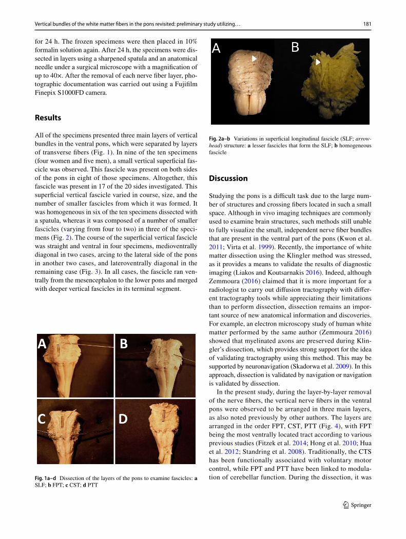
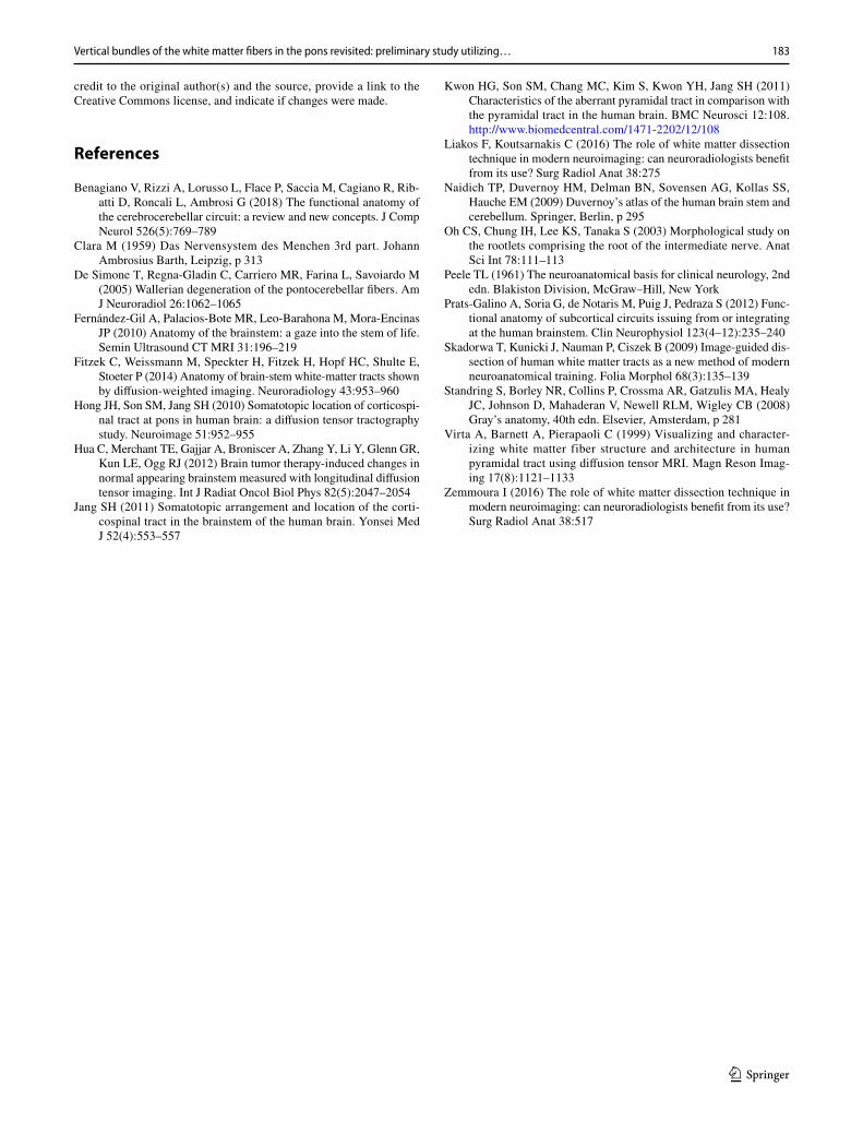
All of the specimens presented three main layers of vertical bundles in the ventral pons, which were separated by layers of transverse fibers (Fig. 1). In nine of the ten specimens (four women and five men), a small vertical superficial fas-cicle was observed. This fascicle was present on both sides of the pons in eight of those specimens. Altogether, this fascicle was present in 17 of the 20 sides investigated. This superficial vertical fascicle varied in course, size, and the number of smaller fascicles from which it was formed. It was homogeneous in six of the ten specimens dissected with a spatula, whereas it was composed of a number of smaller fascicles (varying from four to two) in three of the speci-mens (Fig. 2). The course of the superficial vertical fascicle was straight and ventral in four specimens, medioventrally diagonal in two cases, arcing to the lateral side of the pons in another two cases, and lateroventrally diagonal in the remaining case (Fig. 3). In all cases, the fascicle ran ven-trally from the mesencephalon to the lower pons and merged with deeper vertical fascicles in its terminal segment.
Discussion
Studying the pons is a difficult task due to the large num-ber of structures and crossing fibers located in such a small space. Although in vivo imaging techniques are commonly used to examine brain structures, such methods still unable to fully visualize the small, independent nerve fiber bundles that are present in the ventral part of the pons (Kwon et al. 2011; Virta et al. 1999). Recently, the importance of white matter dissection using the Klingler method was stressed, as it provides a means to validate the results of diagnostic imaging (Liakos and Koutsarnakis 2016). Indeed, although Zemmoura (2016) claimed that it is more important for a radiologist to carry out diffusion tractography with differ-ent tractography tools while appreciating their limitations than to perform dissection, dissection remains an impor-tant source of new anatomical information and discoveries. For example, an electron microscopy study of human white matter performed by the same author (Zemmoura 2016) showed that myelinated axons are preserved during Klin-gler’s dissection, which provides strong support for the idea of validating tractography using this method. This may be supported by neuronavigation (Skadorwa et al. 2009). In this approach, dissection is validated by navigation or navigation is validated by dissection.
In the present study, during the layer-by-layer removal of the nerve fibers, the vertical nerve fibers in the ventral pons were observed to be arranged in three main layers, as also noted previously by other authors. The layers are arranged in the order FPT, CST, PTT (Fig. 4), with FPT being the most ventrally located tract according to various previous studies (Fitzek et al. 2014; Hong et al. 2010; Hua et al. 2012; Standring et al. 2008). Traditionally, the CTS has been functionally associated with voluntary motor control, while FPT and PTT have been linked to modula-tion of cerebellar function. During the dissection, it was Fig. 1a–d Dissection of the layers of the pons to examine fascicles: a
SLF; b FPT; c CST; d PTT
Fig. 2a–b Variations in superficial longitudinal fascicle (SLF; arrow-head) structure: a lesser fascicles that form the SLF; b homogeneous fascicle

182 M. Grzegorczyk et al.
1 3
clear that the fascicles were connected by minor groups of fibers and so do not constitute separate entities. However, it was difficult to identify the spatial arrangement of these connections using the Klingler method. Serial transverse cross-sections with 3D reconstruction are necessary to visualize the internal structure of the vertical fascicles in the pons. An as-yet undescribed element in the superfi-cial layer of the pons was discovered in the present work: a consistently observed fascicle (9/10 specimens, 17/20 sides) of vertical fibers located ventrally to the main group of vertical bundles, between superficial transverse fibers. For more than 50 years, this fascicle has been visualized in some neuroanatomical atlases and textbooks without any explanation or comment (Naidich et al. 2009; Peele 1961), whereas in other important academic publications, ranging from the studies of Max Clara to contemporary editions of Gray’s anatomy, it has not been visualized at all (Clara 1959; Standring et al. 2008). We therefore propose a name for this fascicle—fasciculus longitudinalis super-ficialis (superficial longitudinal fascicle; SLF). The func-tional role of this fascicle remains unclear at this stage of
our investigations. This structure should be measured and traced upward to the mesencephalon and the diencephalon, and then downward to the medulla. More detailed studies should show whether it is an independent fascicle of verti-cal fibers or a portion of frontopontine fibers that become separated from the main fascicle along its pontine course. Localization of SLF part in upper pons corresponds to the localization of fascicle fibers in medial1/5 width of base of the pons (four specimens) or the middle part of the cerebral peduncle. This suggests that it originates in the motor and premotor cortex, a suggestion strengthened by the localization of the SLF in the medial and interme-diate group of pontine fibers as described by Benagiano et al., thus making it a part of the cerebrocerebellar circuit (Benagiano et al. 2018). It may also resemble the aberrant pyramidal tract described by Kwon et al. (2011). Observa-tion of its course, especially the ventral direction of its ter-minal segment, suggests the latter two options. However, the consistent presentation of this structure may indicate that it has a specific function. Our observations are rather incidental, as new studies of the structure of the brain stem using classical anatomical dissection are rare nowadays (Liakos and Koutsarnakis 2016; Skadorwa et al. 2009). While Klingler’s technique is archaic, the arrangement of the vertical fascicles of the pons is invisible to standard MRI, meaning that the discovery of such minute structures as the superficial longitudinal fascicle using MRI is cur-rently highly improbable. In conclusion, we can state that the vertical fibers of the ventral pons are arranged in four fascicles: the superficial, frontopontine, corticospinal, and parieto-temporopontine fibers.
Compliance with ethical standards
Conflict of interest The authors declare they have no conflict of inter-est.
Open Access This article is distributed under the terms of the Crea-tive Commons Attribution 4.0 International License (http://creat iveco mmons .org/licen ses/by/4.0/), which permits unrestricted use, distribu-tion, and reproduction in any medium, provided you give appropriate
Fig. 3a–d Variations in the course of the superficial longitudinal fascicle (arrow): a medioventral, b arcing, c straight, d lateroventral
Fig. 4 Visualization of the transverse section of the pons at the level of the medial cerebellar peduncle. SLF superficial longitudinal fas-cicle, FPT frontopontine tract, CST corticospinal tract, PTT parieto-temporopontine tract
183Vertical bundles of the white matter fibers in the pons revisited: preliminary study utilizing…
1 3
credit to the original author(s) and the source, provide a link to the Creative Commons license, and indicate if changes were made.
References
Benagiano V, Rizzi A, Lorusso L, Flace P, Saccia M, Cagiano R, Rib-atti D, Roncali L, Ambrosi G (2018) The functional anatomy of the cerebrocerebellar circuit: a review and new concepts. J Comp Neurol 526(5):769–789
Clara M (1959) Das Nervensystem des Menchen 3rd part. Johann Ambrosius Barth, Leipzig, p 313
De Simone T, Regna-Gladin C, Carriero MR, Farina L, Savoiardo M (2005) Wallerian degeneration of the pontocerebellar fibers. Am J Neuroradiol 26:1062–1065
Fernández-Gil A, Palacios-Bote MR, Leo-Barahona M, Mora-Encinas JP (2010) Anatomy of the brainstem: a gaze into the stem of life. Semin Ultrasound CT MRI 31:196–219
Fitzek C, Weissmann M, Speckter H, Fitzek H, Hopf HC, Shulte E, Stoeter P (2014) Anatomy of brain-stem white-matter tracts shown by diffusion-weighted imaging. Neuroradiology 43:953–960
Hong JH, Son SM, Jang SH (2010) Somatotopic location of corticospi-nal tract at pons in human brain: a diffusion tensor tractography study. Neuroimage 51:952–955
Hua C, Merchant TE, Gajjar A, Broniscer A, Zhang Y, Li Y, Glenn GR, Kun LE, Ogg RJ (2012) Brain tumor therapy-induced changes in normal appearing brainstem measured with longitudinal diffusion tensor imaging. Int J Radiat Oncol Biol Phys 82(5):2047–2054
Jang SH (2011) Somatotopic arrangement and location of the corti-cospinal tract in the brainstem of the human brain. Yonsei Med J 52(4):553–557
Kwon HG, Son SM, Chang MC, Kim S, Kwon YH, Jang SH (2011) Characteristics of the aberrant pyramidal tract in comparison with the pyramidal tract in the human brain. BMC Neurosci 12:108. http://www.biome dcent ral.com/1471-2202/12/108
Liakos F, Koutsarnakis C (2016) The role of white matter dissection technique in modern neuroimaging: can neuroradiologists benefit from its use? Surg Radiol Anat 38:275
Naidich TP, Duvernoy HM, Delman BN, Sovensen AG, Kollas SS, Hauche EM (2009) Duvernoy’s atlas of the human brain stem and cerebellum. Springer, Berlin, p 295
Oh CS, Chung IH, Lee KS, Tanaka S (2003) Morphological study on the rootlets comprising the root of the intermediate nerve. Anat Sci Int 78:111–113
Peele TL (1961) The neuroanatomical basis for clinical neurology, 2nd edn. Blakiston Division, McGraw–Hill, New York
Prats-Galino A, Soria G, de Notaris M, Puig J, Pedraza S (2012) Func-tional anatomy of subcortical circuits issuing from or integrating at the human brainstem. Clin Neurophysiol 123(4–12):235–240
Skadorwa T, Kunicki J, Nauman P, Ciszek B (2009) Image-guided dis-section of human white matter tracts as a new method of modern neuroanatomical training. Folia Morphol 68(3):135–139
Standring S, Borley NR, Collins P, Crossma AR, Gatzulis MA, Healy JC, Johnson D, Mahaderan V, Newell RLM, Wigley CB (2008) Gray’s anatomy, 40th edn. Elsevier, Amsterdam, p 281
Virta A, Barnett A, Pierapaoli C (1999) Visualizing and character-izing white matter fiber structure and architecture in human pyramidal tract using diffusion tensor MRI. Magn Reson Imag-ing 17(8):1121–1133
Zemmoura I (2016) The role of white matter dissection technique in modern neuroimaging: can neuroradiologists benefit from its use? Surg Radiol Anat 38:517