vestibular and visual dysfunction after … and visual dysfunction after concussion ... calcium...
TRANSCRIPT
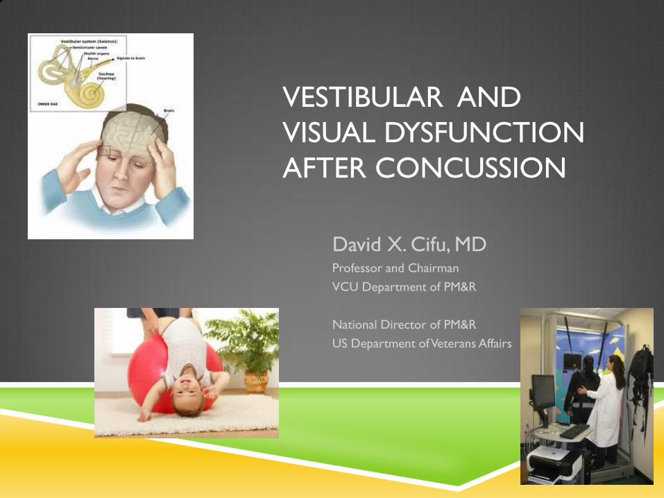
VESTIBULAR AND
VISUAL DYSFUNCTION
AFTER CONCUSSION
David X. Cifu, MD Professor and Chairman
VCU Department of PM&R
National Director of PM&R
US Department of Veterans Affairs

GOALS
Define commonly used terms related to post-concussive vestibular and visual deficits
Outline the incidence and prevalence of post-concussive vestibular and visual deficits
Discuss the anatomic and pathologic correlates of post-concussive vestibular and visual deficits
Develop the framework for interventions for post-concussive vestibular and visual deficits

DEFINITIONS: CONCUSSION
Concussion is a complex pathophysiologic process induced by traumatic forces secondary to direct or indirect impulsive forces to the head that disrupts the function of the brain.
Concussion is defined as an alteration or loss of consciousness for up to 30 minutes with associated loss of memory surrounding the event (post-traumatic amnesia) for up to 24 hours.
Transient (<24 hours) neurologic sequelae may also be present, including numbness, dizziness, cognitive deficits, discoordination, and alterations in special senses.
This disturbance of brain function is typically associated with normal structural neuroimaging findings.
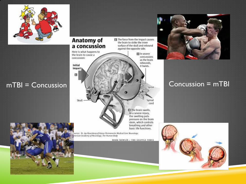
mTBI = Concussion

DEFINITIONS:
POTENTIALLY CONCUSSIVE EVENT
In addition to traditional concussive injury, there is an increasing
“awareness” of potentially concussive events (PCE).
A PCE is defined as an impulsive force to the head of sufficient intensity
that results in acute or chronic symptoms in some individuals, but
remain asymptomatic (“subclinical”) in others - no demonstrable
neurologic or symptomatic effect.
The term PCE is often applied to all traumatic events that either could
have caused a concussion (with altered/lost consciousness) but did not
and those that did produce a concussion.

DEFINITIONS:
POST-CONCUSSIVE SYMPTOMS
Persistent physical, cognitive, emotional, and/or sleep-related symptoms
occur in more than half of concussions, but usually resolve in 1-4 weeks.
Symptoms presenting in the first 1-2 weeks after a concussion are
commonly ascribed to the concussion.
Continued symptoms after 3 months may be labeled as Post-Concussive
Syndrome and may occur in up to 30% of injuries.

DEFINITIONS: POSTCONCUSSIVE SYMPTOMS
Ongoing symptoms are either a prolonged version of the concussion pathophysiology or a manifestation of other processes, such as cervical injury, migraine headaches, depression, chronic pain, vestibular dysfunction, visual dysfunction, or some combination of conditions.
The pathophysiology of ongoing symptoms from the original concussion injury may reflect multiple causes: anatomic, neurometabolic, and physiologic.
Neurometabolic Cascade
Release of neurotransmitters- Glutamate
Massive neuron firing. Creates large cellular demand.
Calcium influx blocking oxygen preventing cellular respiration
K+ efflux cause vasoconstriction
This prevents fuel, glucose, from getting to the cells.
This leads to cellular death and dysfunction

RISK FACTORS FOR
PERSISTENT CONCUSSIVE SYMPTOMS
Presenting with four or more symptoms was associated with double the risk for concussive symptoms ≥1 week for both football and non-football players.
History of prior concussion was associated with double the risk for concussive symptoms ≥1 week in football players only.
Several symptoms were associated with concussive symptoms ≥1 week in all athletes: drowsiness, nausea and concentration difficulties.
Sensitivity to light and noise was associated with concussive symptoms ≥1 week in non-football players only.
Amnesia was associated with concussive symptoms ≥1 week in males, but not females.
Loss of consciousness was not significant. Chrisman SP, et al. Brain Inj. 2013
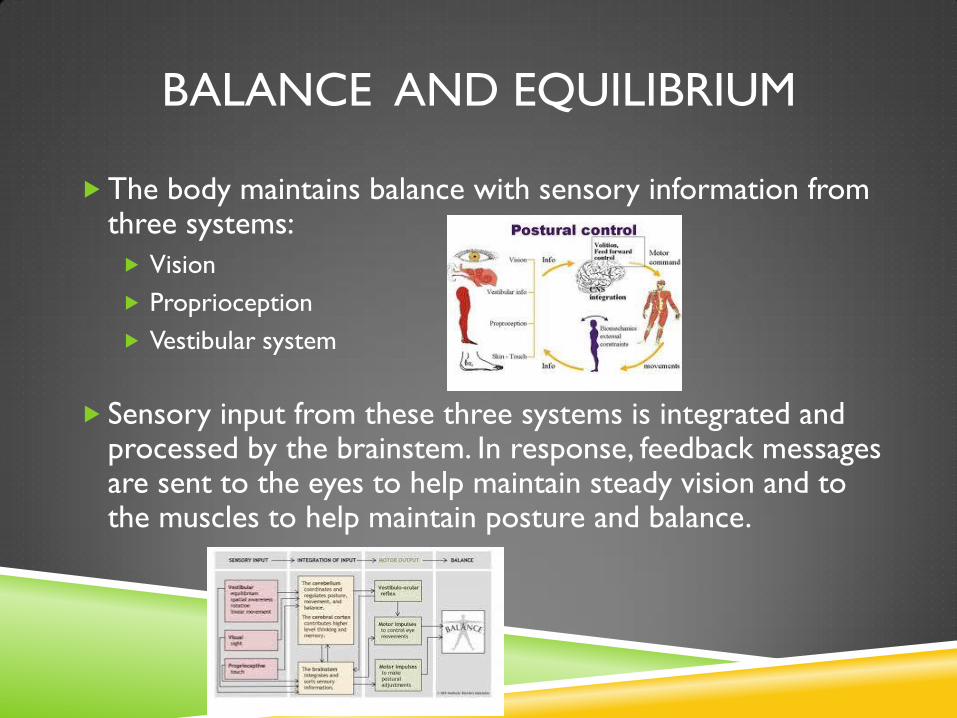
BALANCE AND EQUILIBRIUM
The body maintains balance with sensory information from three systems:
Vision
Proprioception
Vestibular system
Sensory input from these three systems is integrated and processed by the brainstem. In response, feedback messages are sent to the eyes to help maintain steady vision and to the muscles to help maintain posture and balance.

DEFINITIONS: IMBALANCE
Dizziness, vertigo and disequilibrium are common terms used to
describe vestibular dysfunction as well as other difficulties.
They are all symptoms that can result from a peripheral vestibular
disorder (a dysfunction of the balance organs of the inner ear) or
central vestibular disorder (a dysfunction of one or more parts of the
central nervous system that help process balance and spatial
information).
Although these three symptoms can be linked by a common cause, they
have different meanings, and describing them accurately can mean the
difference between a successful diagnosis and one that is missed.

DEFINITIONS: IMBALANCE
Disequilibrium means unsteadiness, imbalance, or loss of equilibrium
that is often accompanied by spatial disorientation.
Dizziness describes a sensation of lightheadedness, faintness, or
unsteadiness. Dizziness does not involve a rotational component.
Vertigo has a rotational, spinning component, and is the perception of
movement, either of the self or surrounding objects.

DEFINITIONS: VISUAL DEFICITS
Visual field defects
Versional - Disturbances of gaze stabilization
Saccades - Quick, simultaneous movements of both eyes in the
same direction.
Fixation - maintaining the gaze in a constant direction
Smooth Pursuit - tracking by the eyes of a slowly moving object at a
steady coordinated velocity
Vergence – Disturbances of focus
Blurred vision/Accommodation
Diplopia/Ocular motility

DEFINITIONS: VISUAL/AUDITORY “PAIN”
Photophobia/Photosensitivity – Light sensitivity
Hyperacusis – Sound sensitivity
Tinnitus – Perception of sound when no sound is present
Unclear peripheral and/or central neurologic etiologies.

DEFINITIONS: VISUAL AND
AUDITORY PERCEPTUAL DEFICITS
Central Auditory Processing Deficit (CAPD) – Inaccurate
perception, interpretation or understanding of auditory stimuli,
due to cortical injury of Primary Auditory Cortex.
Bilaterally at upper sides of the Temporal Lobes on the superior temporal plane,
within the lateral fissure and comprising parts of Heschl’s gyrus and the superior
temporal gyrus, including planum polare and planum temporale (Brodmann areas 41,
42 and, and partially 22)
Central Visual Perceptual Deficits - Inaccurate perception,
interpretation or understanding of visual stimuli, due to cortical
injury of Primary Visual Cortex.
Visual cortex includes areas of occipital, temporal and parietal lobes, as well as areas
of the limbic cortex. The left cortex plays a major role in recognizing the meaning of
common objects.

EPIDEMIOLOGY: CONCUSSIONS
The CDC estimates that there are 3.5 million civilian concussions in the United
States annually.
Up to 30% of individuals with concussion will continue to be symptomatic at 3 months (i.e., post-
concussion syndrome) and 5% or more will demonstrate abnormalities on testing or by symptom
report at one year post-injury.
The DoD/VA estimates that 16% of all service members who served in the
OEF/OIF conflicts experienced at least 1 concussion.
9% of all OEF/OIF Veterans seen for care have persistent symptoms related to
these concussions.

EPIDEMIOLOGY: LONG TERM EFFECTS
The long term effects of a subclinical PCE’s, single
concussion, multiple concussions (whether separated by a
short or long period of time) are not know, nor is the
impact of persistent post-concussive symptoms.
Recent reports suggest that a subset of individuals who
sustain concussion are at an elevated risk to develop
degenerative neurologic disorders (i.e., dementia,
Parkinson’s disease, behavioral dysfunction).

EPIDEMIOLOGY: IMBALANCE
35% adults aged 40 years or older in the United States—approximately
69 million Americans—have experienced some form of vestibular
dysfunction.
4% (8 million) of American adults report a chronic problem with
balance, while an additional 1.1% (2.4 million) report a chronic problem
with dizziness alone.
Agrawal Y, Carey JP, Della Santina CC, Schubert MC, Minor LB. Disorders of balance and vestibular
function in US adults. Arch Intern Med. 2009;169(10): 938-944.
National Institute on Deafness and Other Communication Disorders (NIDCD). Strategic Plan (FY
2006-2008). Available at: www.nidcd.nih.gov/StaticResources/about/plans/strategic/strategic06-08.pdf.
Accessed May 20, 2010.

EPIDEMIOLOGY: IMBALANCE
Eighty percent of people aged 65 years and older have experienced
dizziness, and BPPV, the most common vestibular disorder, is the cause
of approximately 50% of dizziness in older people.
Overall, vertigo from a vestibular problem accounts for a third of all
dizziness and vertigo symptoms reported to health care professionals.
Ator GA. Vertigo—Evaluation and Treatment in the Elderly.
Fife TD, Iverson DJ, Lempert T, Furman JM, Baloh RW, Tusa RJ, Hain TC, Herdman S, Morrow MJ,
Gronseth GS. Practice parameter: therapies for benign paroxysmal positional vertigo (an evidence-
based review): report of the Quality Standards Subcommittee of the American Academy of Neurology.
Neurol. 2008;70:2067–2074.
Neuhauser HK, Radtke A, von Brevern M et al. Burden of dizziness and vertigo in the community. Arch
Intern Med. 2008;168(19):2118–2124.

EPIDEMIOLOGY:
POST-CONCUSSIVE IMBALANCE
Nearly one-quarter of the patients with acute concussion present with dizziness. Reports of 80% after blast injury.
Causes include;
Inner ear disorders
Benign Paroxysmal Positional Vertigo
Labyrinthine concussion
Perilymphatic fistula
CNS Disorders
Post-traumatic migraine
Brainstem concussion
Autonomic dysregulation (Orthostatic Hypotension)
Occulomotor abnormalities
Seizures
Psychological disorders
Musculoskeletal disorders

EPIDEMIOLOGY: VISUAL DISTURBANCES
Sensorimotor vision symptoms after mTBI are reported in frequencies
ranging from 10 to 85%, depending upon the nature of the vision deficit
and the criteria used in the study .
Subtle visual deficits are often overlooked, but may have subtle to
profound impacts on basic (reading, walking) to advanced (driving,
sports, working) functional tasks.
Lew HL, Garvert DW, Pogoda TK, et al. Auditory and visual impairments in patients with blast-
related traumatic brain injury: Effect of dual sensory impairment on Functional Independence
Measure. J. Rehabil. Res. Dev. 46(6), 819–826 (2009).
Kapoor N, Ciuffreda KJ. Vision Disturbances Following Traumatic Brain Injury. Curr. Treat.
Options Neurol. 4(4), 271–280 (2002).

EPIDEMIOLOGY: VISUAL DEFICITS
Visual field defects (35% of all TBI, rare with mTBI)
Versional - Disturbances of gaze stabilization (40-80% mTBI)
Saccades - Quick, simultaneous movements of both eyes in the
same direction.
Fixation - maintaining the gaze in a constant direction (rare with
mTBI)
Smooth Pursuit - tracking by the eyes of a slowly moving object at a
steady coordinated velocity
Vergence – Disturbances of focus
Blurred vision/Accommodation (10-40% of mTBI)
Diplopia/Ocular motility (40-56% mTBI)
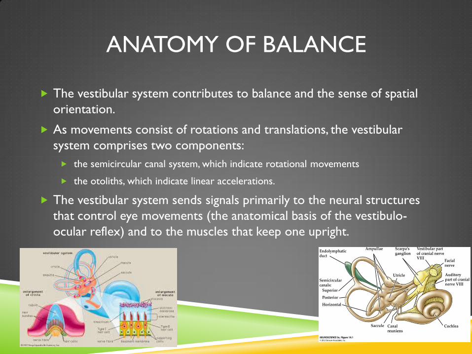
ANATOMY OF BALANCE
The vestibular system contributes to balance and the sense of spatial
orientation.
As movements consist of rotations and translations, the vestibular
system comprises two components:
the semicircular canal system, which indicate rotational movements
the otoliths, which indicate linear accelerations.
The vestibular system sends signals primarily to the neural structures
that control eye movements (the anatomical basis of the vestibulo-
ocular reflex) and to the muscles that keep one upright.


SEMICIRCULAR CANALS
The vestibular system contains three semicircular canals in each labyrinth.
They are approximately orthogonal (right angles) to each other, and are called the
horizontal (or lateral), the anterior semicircular canal (or superior) and the
posterior (or inferior) semicircular canal. Anterior and posterior canals may be
collectively called vertical semicircular canals.
Movement of fluid within the horizontal semicircular canal corresponds to rotation
of the head around a vertical axis (i.e. the neck), as when doing a pirouette.
The anterior and posterior semicircular canals detect rotations of the head in the
sagittal plane (as when nodding), and in the frontal plane, as when cartwheeling. Both
anterior and posterior canals are orientated at approximately 45° between frontal
and sagittal planes.

OTOLITHIC ORGAN
While the semicircular canals respond to rotations, the otolithic organs sense linear accelerations.
Humans have two of these organs on each side, one called utricle, the other saccule. Otoconia crystals (otoliths) float within the organs.
Most of the utricular signals elicit eye movements, while the majority of the saccular signals projects to muscles that control our posture.
The otoconia crystals in the otoconia layer rest on a viscous gel layer, and are heavier than their surroundings. Therefore they get displaced during linear acceleration, which in turn deflects the ciliary bundles of the hair cells and thus produces a sensory signal.

CENTRAL PROCESSING

CENTRAL PROCESSING
Signals from the semicircular canals and otolithic organs project
via Cranial Nerve VIII to the 4 main vestibular nuclei in the
brainstem
The vestibular nuclei on either sides of the brain stem exchange
signals regarding movement and body position. These signals are
sent down the following projection pathways.
Cerebellum: Signals sent to the cerebellum are relayed back as
muscle movements of the head, eyes, and posture. The VOR is also
modulated here.
Nuclei of Nerves III, IV, and VI through MLF: Signals sent to these
nerves cause the vestibular-ocular reflex. They allow for the eyes to
fix on a moving object while staying in focus.

CENTRAL PROCESSING
The vestibular nuclei on either sides of the brain stem exchange
signals regarding movement and body position. These signals are
sent down the following projection pathways.
Reticular Formation: Signals sent to the reticular formation signal
the new posture the body has taken on and how to adjust
circulation and breathing due to body position.
Spinal Cord: Signals sent via medial; and lateral vestibulospinal tracts
to the spinal cord allow quick reflex reactions to both the limbs and
trunk to regain balance.
Thalamus: Signals sent to the thalamus allow for head and body
motor control as well as being conscious of body position.

CENTRAL PROCESSING
Autonomic: Signals sent to through efferent projections onto the solitary nucleus of cranial nerves IX and X coordinate vestibulo-sympathetic reflexes that modulate autonomic changes in blood circulation, heart rate and respiratory rate.
Projections from the Vestibular Nuclei also go to areas of the cortex, however their role is unclear
Persistent dizziness and vertigo with acute injury, disease or dysfunction of the vestibular system may therefore be due to;
Permanent damage and hypofunction/dysfunction of the vestibular system
Dysfunction of the VIII cranial nerve
Dysfunction of the visual or peripheral sensory system
Dysfunction of the central structures
Recalibration of the cerebellum (? or cortex)

ANATOMY OF VISION
Light passes through the refractive components (i.e., the cornea,
aqueous chamber, crystalline lens, and vitreous) to reach the retina.
The photoreceptors at the retina transmit signals at the level of the
optic nerve via retinal ganglion cell (RGC).
The RGCs maintain long axons that comprise the optic nerve, leaving
the retina and proceeding to the optic chiasm.
At the chiasm, the visual information separates into right and left hemi-
fields of visual space; this divided information travels post-chiasm as
optic tracts to the lateral geniculate nucleus (LGN), ultimately becoming
optic radiations, post-LGN.

CENTRAL PROCESSING
The vast majority of the axons continue onwards to the
occipital cortex (V1).
At V1, the neural signals for the primary visual pathway
begin the transformation into a high-resolution, neural
image with appropriate form, color vision, and contrast, in
addition to maintaining peripheral vision integrity.
Parallel processing begins at this level with a ventral
pathway and dorsal pathway.
The ventral visual pathway is predominantly for object identification, with
its axons eventually reaching the posterior-inferior temporal lobe.
The dorsal visual pathway is predominantly for spatial representation and
visually-guided action, with its axons eventually reaching the parietal lobe.

STABILIZING VISUAL AND
VESTIBULAR REFLEXES The vestibulo-ocular reflex (VOR) is a reflex eye movement that
stabilizes images on the retina during head movement by producing an
eye movement in the direction opposite to head movement, thus
preserving the image on the center of the visual field. For example,
when the head moves to the right, the eyes move to the left, and vice
versa.
Since slight head movements are present all the time, the VOR is very
important for stabilizing vision: patients whose VOR is impaired find it
difficult to read, because they cannot stabilize the eyes during small head
tremors. The VOR reflex does not depend on visual input and works
even in total darkness or when the eyes are closed.

STABILIZING VISUAL AND
VESTIBULAR REFLEXES
The optokinetic reflex (OKR) responds to visual motion
stimulation. It is observed when one follows a moving
object with their eyes. Once that object moves out of their
field of vision, their eyes move back to the original position
when first seeing the object.
The cervico-ocular reflex (COR) is a rotational eye reflex
elicited by neck musculature and cervical spine
proprioception that works in conjunction with the VOR and
OKR.
The vestibulocolic reflex (VCR) is involved in head
stabilization in space.

CENTRAL VS. PERIPHERAL IMBALANCE
CENTRAL PERIPHERAL
ONSET GRADUAL SUDDEN (THOUGH
SOME LATENCY)
FREQUENCY INTERMITTANT CONSTANT
SEVERITY MILD/MODERATE SEVERE
POSITIONAL YES VARIABLE
NAUSEA/VOMITING NO YES
FATIGUEABLE NO YES
VISUAL SUPRESSION NO YES
PHOTOPHOBIA YES NO
PHONOPHOBIA YES NO
TINNITUS YES NO
HEARING LOSS NO NO

PERIPHERAL VESTIBULAR DISORDERS
BPPV
Perilymph Fistula (PLF)
Labyrinthine Concussion
Post-Traumatic Endolymphatic Hydrops
Meniere’s-like presentation
Vestibular Hypofunction/Dysfunction
Cervical vertigo
Eighth nerve complex

BPPV: ETIOLOGY
BPPV is the most common vestibular disorder
2.4% of all people will experience it at some point in their lifetimes.
BPPV accounts for at least 20% of diagnoses made by physicians who
specialize in dizziness and vestibular disorders, and is the cause of
approximately 50% of dizziness in older people.
The most common cause of BPPV in people under age 50 is head injury
and is presumably a result of concussive force that displaces the
otoconia.

BPPV: DEFINITION
Sudden, intermittent and intense episodes of vertigo lasting for seconds
and triggered by certain movements of the head.
BPPV consists of brief, intermittent episodes of nystagmus,
lightheadedness and/or vertigo without hearing loss; the episodes are
provoked by head movement in the same planes as the semi-circular
canals, dislodging displaced otoconia.
>80% time, it is the posterior canal that is affected. In certain head
positions, these particles shift and create a fluid wave which displaces
the cupula of the canal affected, which leads to dizziness, vertigo and
nystagmus.

BPPV: PATHOLOGY
BPPV occurs as a result of otoconia, tiny crystals of calcium carbonate that are a normal part of the inner ear’s anatomy, detaching from the otolithic membrane in the utricle and collecting in one of the semicircular canals.
When the head is still, gravity causes the otoconia to clump and settle.
When the head moves, the otoconia shift. This stimulates the cupula to send false signals to the brain, producing vertigo and triggering nystagmus (involuntary eye movements).

BPPV: TESTING
BPPV is diagnosed based on medical history, physical examination, the
results of vestibular and auditory (hearing) tests, and lab work to rule
out other diagnoses.
Vestibular tests include the Dix-Hallpike maneuver and the Supine Roll
test.
These tests allow a physician to observe the nystagmus elicited in
response to a change in head position.
The problematic semicircular canal can be identified based on the
characteristics of the observed nystagmus.

BPPV: TESTING
Frenzel goggles, especially of the type using a TV camera, are sometimes
used as a diagnostic aid in order to magnify and illuminate nystagmus.
If electronystagmography (ENG) is employed to observe nystagmus with
position changes, it is important that the equipment used is capable of
measuring vertical eye movements.
Magnetic resonance imaging scan (MRI) may be used to rule out other
problems such as a stroke or brain tumor, but such scans are not helpful
in diagnosing BPPV.
Auditory tests may help to pinpoint a specific cause of BPPV, such as
Ménière’s disease or labyrinthitis.
PERILYMPH FISTULA
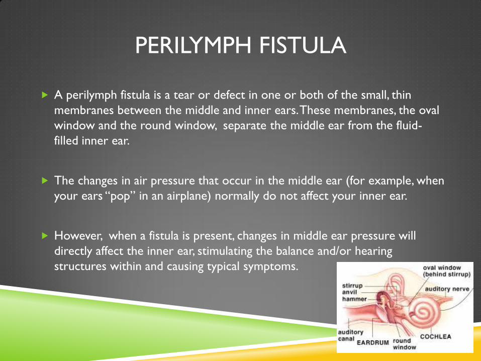
A perilymph fistula is a tear or defect in one or both of the small, thin
membranes between the middle and inner ears. These membranes, the oval
window and the round window, separate the middle ear from the fluid-
filled inner ear.
The changes in air pressure that occur in the middle ear (for example, when
your ears “pop” in an airplane) normally do not affect your inner ear.
However, when a fistula is present, changes in middle ear pressure will
directly affect the inner ear, stimulating the balance and/or hearing
structures within and causing typical symptoms.

PERILYMPH FISTULA
The symptoms of perilymph fistula may include;
Dizziness
Vertigo
Imbalance
Nausea
Vomiting.
Ringing or fullness in the ears
Hearing loss
Most people with fistulas find that their symptoms get worse with changes in altitude (elevators, airplanes, travel over mountain passes), air pressure (weather changes), and with exertion/activity.

PERILYMPH FISTULA
If symptoms are severe and have not responded to conservative
treatment (bed rest), or if progressive hearing loss has occurred, surgical
repair of the fistulas may be required. This procedure involves placing a
graft over the fistula defect in the oval and/or round window.
Persons with fistulas should avoid lifting, straining, bending over, or any
activity that would increase head pressure, since all of these will worsen
symptoms and prevent the fistula from healing. It is also important to
avoid air pressure changes as these changes will tend to worsen
symptoms.

LABYRINTHINE CONCUSSION
Posttraumatic vertigo that resolves spontaneously over time, after other diagnoses have been excluded, is known as labyrinthine concussion.
Typically presents with abrupt hearing loss and persistent vertigo following head trauma in the absence of a temporal bone fracture.
Symptoms generally can improve over a few days and sensations of persistent vertigo will eventually transition to movement-induced vertigo.
The pathophysiology of labyrinthine concussion is not well-defined or distinguished from BPPV.
POST-TRAUMATIC ENDOLYMPHATIC HYDROPS
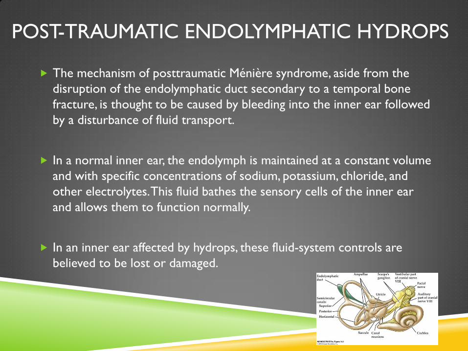
The mechanism of posttraumatic Ménière syndrome, aside from the
disruption of the endolymphatic duct secondary to a temporal bone
fracture, is thought to be caused by bleeding into the inner ear followed
by a disturbance of fluid transport.
In a normal inner ear, the endolymph is maintained at a constant volume
and with specific concentrations of sodium, potassium, chloride, and
other electrolytes. This fluid bathes the sensory cells of the inner ear
and allows them to function normally.
In an inner ear affected by hydrops, these fluid-system controls are
believed to be lost or damaged.

POST-TRAUMATIC ENDOLYMPHATIC HYDROPS
This may cause the volume and concentration of the endolymph to fluctuate in response to changes in the body’s circulatory fluids and electrolytes.
Symptoms typical of hydrops include pressure or fullness in the ears (aural fullness), tinnitus (ringing or other noise in the ears), hearing loss, dizziness, and imbalance.
Compared with primary (viral/idiopathic), secondary (post-traumatic) Meniere’s disease tends to have continuous symptoms, that are less violent in nature and less likely to cause long-term damage to hearing and balance.

VESTIBULAR HYPOFUNCTION/DYSFUNCTION
Vestibular hypofunction (VH) is a general term used to describe inability
of vestibular system to adequately detect and convey signals to
brainstem.

EIGHTH NERVE COMPLEX
The eighth nerve complex is at risk for injury, even in cases of mild
trauma, because of the shearing effect on the root entry zone of the
nerve to the brainstem.
Also known as brainstem concussion.
This mechanism has been demonstrated in experimental models and in
autopsy reports.
VISUAL FIELD DEFICITS
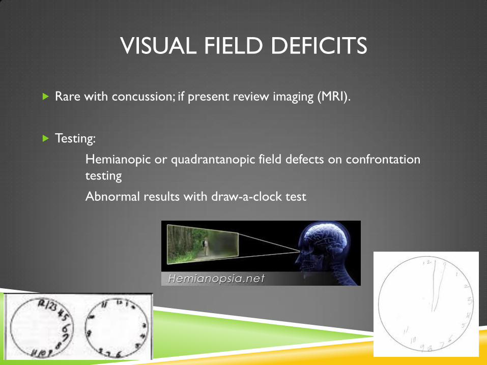
Rare with concussion; if present review imaging (MRI).
Testing:
Hemianopic or quadrantanopic field defects on confrontation
testing
Abnormal results with draw-a-clock test

VISUAL ACUITY DEFICITS – BLURRED VISION
Deficit of accommodation – maintaining clear image or focus on an
object as its distance varies. Unclear relationship to TBI, but seems
related to difficulty with vergence.
Seen in 10-40% of TBI
Blurred vision
constant or intermittent
evident when changing viewing distances (i.e., near-far and/or far-near blur)
Eyestrain, brow-aches, eye fatigue evident after brief periods of
sustained near vision work
Dizziness, nausea, or motion sickness during or following a vision-based
task

VISUAL ACUITY DEFICITS – DIPLOPIA
Deficit of vergence – Disjunctive changes in eye position as one attends
to objects at varying distances in the visual field. Seen in 40-56% of TBI
Findings
Double vision
Eliminated with occlusion
Constant or intermittent
At any viewing distance
More evident in one position of gaze than another
Eyestrain, eye fatigue, closing or squinting one eye after a brief vision-related task
Avoidance of prolonged vision-related tasks
Dizziness, nausea, and motion sickness during or following vision-based tasks

VISUAL ACUITY DEFICITS – VERSIONAL
Disturbances of gaze stabilization (40-80% mTBI).
Findings:
Reading-related difficulty
Slower reading speed
Loss of place while reading/ skipping lines
The print appears to “float”/ “swim”
Avoidance of prolonged vision-related tasks
Difficulty shifting to/ tracking objects
Dizziness/nausea/motion sickness during or following vision-based
tasks

VISUAL ACUITY DEFICITS – VERSIONAL
Saccades - Quick, simultaneous movements of both eyes in
the same direction.
noticeable undershooting or overshooting of the target on saccadic testing
Fixation - maintaining the gaze in a constant direction.
nystagmus on fixation assessment
Smooth Pursuit - tracking by the eyes of a slowly moving
object at a steady coordinated velocity.
restriction of eye movements on pursuit testing

VISUAL-VESTIBULAR DYSFUNCTION:
MANAGEMENT STRATEGIES
VISUAL-VESTIBULAR REHABILITATION
Limited strict scientific evidence in concussed populations demonstrating
efficacy or identifying specifics of visual and vestibular rehabilitation
programs. Extensive literature for geriatric imbalance.
No role for medications other than acute sedation/nausea management.
Typically recommended if persistent symptoms after 2-4 weeks.
Focuses on specific deficits with sensory integration of vestibular, visual,
proprioceptive, touch/pressure and hearing systems.

VISUAL-VESTIBULAR REHABILITATION
Types of Sensory Reintegration:
Oculomotor training
Eye-Head coordination
Balance training
Visual motion sensitivity training
Neuromuscular control
Body mechanics and posture

SUMMARY
The majority of balance deficits seen acutely after mTBI are related to injury of the peripheral vestibular system. Early return to upright mobility is crucial.
The majority of visual deficits seen acutely after mTBI are related to central coordination of gaze version and vergence.
Vestibular rehabilitation focuses on reintegrating the labyrinthian organs with the proprioceptive, visual, and muscular systems.
Visual rehabilitation focuses on progressively optimizing the brain’s coordination and perception of eye movements.