vestibulo-ocular reflex responses to a multichannel vestibular ... · vestibulo-ocular reflex...
TRANSCRIPT
Vestibulo-Ocular Reflex Responses to a MultichannelVestibular Prosthesis Incorporating a 3D CoordinateTransformation for Correction of Misalignment
GENE Y. FRIDMAN,1 NATAN S. DAVIDOVICS,1,2 CHENKAI DAI,1 AMERICO A. MIGLIACCIO,1,2
AND CHARLES C. DELLA SANTINA1,2
1Vestibular NeuroEngineering Laboratory, Department of Otolaryngology - Head & Neck Surgery, Johns Hopkins School ofMedicine, Ross Bldg Rm 830, 720 Rutland Ave., Baltimore, 11 MD 21205, USA2Department of Biomedical Engineering, Johns Hopkins School of Medicine, Baltimore, MD 21205, USA
Received: 25 June 2009; Accepted: 17 January 2010; Online publication: 23 February 2010
ABSTRACT
There is no effective treatment available for individ-uals unable to compensate for bilateral profound lossof vestibular sensation, which causes chronic disequi-librium and blurs vision by disrupting vestibulo-ocularreflexes that normally stabilize the eyes during headmovement. Previous work suggests that a multichan-nel vestibular prosthesis can emulate normal semi-circular canals by electrically stimulating vestibularnerve branches to encode head movements detectedby mutually orthogonal gyroscopes affixed to theskull. Until now, that approach has been limited bycurrent spread resulting in distortion of the vestibularnerve activation pattern and consequent inability toaccurately encode head movements throughout thefull 3-dimensional (3D) range normally transduced bythe labyrinths. We report that the electrically evoked3D angular vestibulo-ocular reflex exhibits vectorsuperposition and linearity to a sufficient degree thata multichannel vestibular prosthesis incorporating aprecompensatory 3D coordinate transformation tocorrect misalignment can accurately emulate semi-circular canals for head rotations throughout therange of 3D axes normally transduced by a healthylabyrinth.
Keywords: electrical, stimulation, vestibular,prosthesis, VOR, gyroscopes, implant, labyrinth,vestibulo-ocular reflex
INTRODUCTION
Bilateral loss of vestibular sensation can occur as theresult of ototoxic drug exposure, ischemia, infection,genetic abnormality, or trauma to the inner ears.Because input from the vestibular labyrinths normallymediates perception of movement and reflexes thatstabilize posture and visual gaze, loss of this inputcauses chronic disequilibrium, postural instability, andvisual blur due to illusory movement of the worldduring head motion (Grunbauer et al. 1998; Minor1998; Gillespie and Minor 1999). A multichannelneuro-electronic prosthesis that senses rotation ofthe head in three dimensions and delivers corre-sponding stimuli to the ampullary branches of thevestibular nerve should restore sensation of headrotation, which in turn should help to stabilize gazeand at least partially restore perception of headmovement. Restoring function to just one labyrinthis a reasonable goal for such a device because theadaptive capacity of vestibulocerebellar circuits issufficient to ensure that individuals with a single,normal labyrinth (e.g., after unilateral injury orinfection) typically compensate well enough to sufferlittle o disability in activities of daily life (Curthoys andHalmagyi 1995; Black et al. 1996).
Correspondence to: Charles C. Della Santina & VestibularNeuroEngineeringLaboratory &Johns Hopkins School of Medicine &Ross Bldg Rm 830, 720Rutland Ave., Baltimore, MD 21205, USA. Telephone: +1-410-5027909;fax: +1-410-6148610; email: [email protected]
JARO 11: 367–381 (2010)DOI: 10.1007/s10162-010-0208-5D 2010 Association for Research in Otolaryngology
367
JAROJournal of the Association for Research in Otolaryngology

Normally, the eyes rotate opposite the direction ofhead rotation in order to stabilize images on theretinae. This compensatory eye rotation is driven bythe angular vestibulo-ocular reflex (aVOR), for whichsensory input is provided by three mutually orthogo-nal semicircular canals (SCC) in each inner ear’svestibular labyrinth. Discharge rates of vestibularafferent fibers in each SCC’s ampullary nerve aremodulated by the component of head angular velocityabout that SCC’s axis (Ewald 1892; Wilson and Melvill1979; Carey and Della Santina 2005; Della Santina etal. 2005b). The resulting eye movements can bemeasured precisely in 3D, giving insight into relativeexcitation of the ampullary nerves driving the reflex(Robinson and Murphy 1932; Robinson 1963; Aw etal. 1996a, b; Migliaccio et al. 2005).
Classic experiments by Cohen, Suzuki, et al.(Cohen et al. 1963, 1964; Cohen and Suzuki 1963a,b; Suzuki et al. 1964, 1969; and Suzuki and Cohen1964) demonstrated that pulses of electrical stimula-tion delivered to the ampullary nerves via wireelectrodes elicit an aVOR about axes of rotationsimilar to those elicited by the hydrodynamic excita-tion of the individual canals (Ewald 1892). Merfeld,Gong, Lewis, et al. (Gong and Merfeld 2000, 2002;Lewis et al. 2001, 2002; Merfeld et al. 2006, 2007; andGong et al. 2008) described a single-channel prosthe-sis based on the idea of sensing rotational motionusing a gyroscope and stimulating the appropriatebranch of the vestibular nerve. Della Santina et al.(Della Santina et al. 2005b, 2006, 2007; Chiang et al.2009; and Davidovics et al. 2009) extended thatapproach to 3D using a multichannel prosthesis thatcontains three mutually orthogonal gyroscopes tosense head rotational velocity and deliver electricalstimulus via multiple electrodes to the appropriatenerves. Tests of this device in chinchillas renderedbilaterally vestibular-deficient via gentamicin treat-ment revealed partial restoration of the 3D aVOR.However, current spread within implanted labyrinthslimited the ability to selectively stimulate each ampul-lary nerve without incurring spurious excitation ofadjacent branches of the vestibular nerve. Increasingstimulus current amplitude initially increased aVORmagnitude without changing the aVOR axis, but athigher currents, the eye rotation axis deviated fromthe ideal as spurious stimulation of other vestibularnerve branches altered the head rotation axis “per-ceived” by brainstem neurons mediating the aVOR.
In theory, one should be able to overcome theeffects of current spread from an electrode targetingone ampullary nerve by adjusting the input deliveredvia other electrodes targeting two other ampullarynerves in the same (or contralateral) labyrinth. Forexample, if current intended for the horizontalampullary nerve spuriously excites the anterior and
posterior ampullary nerves, then head rotation purelyabout the horizontal SCC axis might be encoded bymodulating not only the horizontal electrode inputbut instead by simultaneously modulating stimuli on allthree electrodes to represent a horizontal head rota-tion via vector summation. If linearity and vectorsuperposition hold, this procedure amounts to asimple linear transformation between two different3D coordinate systems, analogous to thematrix algebratransformations popularized by Robinson (1982) foranalysis of the 3D aVOR. One should be able tocharacterize this transformation by delivering a set ofstimuli s*i (with each s*i being a 3-vector representing atriplet of stimulus intensities delivered via threeelectrodes targeting the three ampullary nerves) andmeasuring the corresponding responses r*i (with eachr*i being a 3-vector representing the axis and speed ofobserved eye movement responses) for a set of Nvirtual head movements spanning the range of headmovement axes and rotational velocities normallyencoded by the labyrinth. A single 3×3 matrix M canthen be found using least-squares techniques suchthat
r*i � M s*i for i ¼ 1:::N : ð1ÞOnce M has been established, the appropriate
pattern of electrode activation a* the prosthesis shoulddeliver to the three ampullary nerve electrodes duringa head rotation eliciting gyroscope signals is
ð2Þ
This procedure would require that M is computedduring an occasional “fitting” session, analogous tofitting required for patients with cochlear implants.For this procedure to work properly, the axes of eyemovement responses to individual stimulation of eachthe three ampullary nerves must be linearly inde-pendent with respect to each other. Otherwise, theinverse of M cannot exist and the matrix pseudoin-verse calculation will fail. In this context, linearindependence means that the 3D axis of aVORresponses to stimuli delivered via any one electrodealone cannot be in the plane defined by the axes ofresponses to the other two electrodes. The accuracywith which linear precompensation can correct forcurrent spread also depends on the extent to whichthe electrically evoked aVOR response is linear andexhibits vector summation.
Observations by Cohen et al. (1964) during elec-trical stimulation of ampullary nerves alone and incombination demonstrated that the aVOR at leastqualitatively exhibits vector summation. Recently,Gong et al. (2008) showed that responses to pros-thetic stimuli delivered to electrodes implanted in
368 FRIDMAN ET AL.: VOR Responses to a Multichannel Vestibular Prosthesis - Misalignment Correction
coplanar canals on opposite sides of the head showevidence of linear summation. However, significantfrequency- and amplitude-dependent nonlinearitiesare evident in aVOR responses of individuals with asingle intact labyrinth (who most closely represent thefunctional goal of a vestibular prosthesis implantedunilaterally in a bilaterally vestibular-deficient individ-ual; Carey et al. 2002a; Weber et al. 2008, 2009). The3D aVOR may therefore be more complex than astatic vector summation of different ampullary nerveinputs. Furthermore, nonlinearities could arise due tothe dynamics of action potential initiation in ampul-lary nerve fibers spuriously excited or inhibited bycurrent spread. Therefore, linear behavior of theelectrically evoked 3D aVOR cannot be assumed.
In the present study, we assessed the linearity of theelectrically evoked aVOR and then examined whetherincorporating a linear precompensation encodingscheme in a multichannel vestibular prosthesis ena-bles implanted animals to accurately sense andrespond to sinusoidal head rotation about any speci-fied 3D axis at any specified velocity within the rangetypical of normal behavior.
METHODS
Subjects
Adult wild-type 450–650 g chinchillas were used for allexperiments, which were performed in accordancewith a protocol approved by Johns Hopkins UniversityAnimal Care and Use Committee.
Surgical procedures
Surgical procedures were similar to those describedpreviously in detail (Della Santina et al. 2007). Undergeneral anesthesia (isoflurane, 3–5%), a phenolicpost was positioned on the skull in the midline,perpendicular to the mean horizontal semicircularcanal plane and embedded in dental cement (ESPEProTemp II, 3M Corp., Minneapolis, MN), forming arigid connection to the skull. The post served torestrain the animal during aVOR testing. Electrodeswere fashioned from pairs of Teflon-insulated 75-µmPt-Ir wire (Cooner Wire, Chatsworth, CA) for each ofthe canal electrodes and 125-µm Pt-Ir wire forreference electrodes.
To provide reference data on the aVOR of ananimal with a single normal labyrinth (against whichto compare responses of bilaterally vestibular-deficientanimals treated with unilateral prosthetic stimula-tion), three otherwise normal chinchillas were treatedunilaterally via intratympanic injection of ∼0.5 mLgentamicin (26.7 mg/mL buffered to pH 7.4 withsodium bicarbonate) using a protocol described
previously (Hirvonen et al. 2005). Head posts wereplaced subsequently as described above.
Electrodes were positioned in the left labyrinth forall four implanted chinchillas. The electrode for theanterior canal ampullary nerve stimulation was posi-tioned via a fenestration in the internal auditory canalexposing the ampullary nerve just medial of theanterior canal ampulla. The electrode for horizontalcanal stimulation was positioned via a ∼100-µmfenestration on the anterolateral wall of the horizon-tal ampulla. The electrode intended for the posteriorcanal stimulation was inserted into a fenestration inthe thin segment of the posterior canal and advancedtoward the ampulla until resistance was encountered.
Two reference electrodes were implanted to pro-vide a choice of two possible returns for the monop-olar stimulation delivered via any of the stimulatingelectrodes. A distant return was inserted into neckmusculature, and a near return was inserted into thecommon crus via a fenestration in the superior aspectof the anterior SCC. The distant return was strippedof approximately 5 mm of insulation, and the nearreturn was stripped of approximately 1 mm ofinsulation. For each stimulating electrode, we selectedthe reference electrode that yielded the highestvelocity aVOR response with lower axis misalignment.
At the time of electrode implantation, the semi-circular canals of the right labyrinth were pluggedwith fascia and bone chips via fenestrations in theirthin segments. In order to assess the impact ofelectrode placement alone, we did not plug the canalsin the left labyrinth. Instead, the two animals (ch206and ch207) that had residual aVOR after the surgerywere treated with intratympanic gentamicin on theleft side, which ablated all residual sensitivity to headrotation without prosthetic input in each case. (Intra-tympanic gentamicin kills type I vestibular hair cellsand renders type II hair cells insensitive to endolymphmotion (Della Santina et al. 2005c; Hirvonen et al.2005; Lyford-Pike et al. 2007)).
Eye movement measurement and analysis
Real-time, binocular 3D video-oculography (3D VOG)was used for recording eye movements in response tohead movement or electrical stimulation. The methodwe employed is analogous to that described in detailpreviously (Migliaccio et al. 2005; Della Santina et al.2007), except for the use of faster cameras yielding ahigher sample rate. The system was installed on a dualcore 2-GHz 2-GB RAM PC running Windows XP.Firewire cameras (Dragonfly Express, Point GreyResearch, Richmond, BC, Canada) retrofitted with25.0-mm focal length, f/2.0 microvideo lenses wereused to acquire 500×400 pixel, 8-bit grayscale imagesat 180 Hz for each eye. An array of three fluorescent
FRIDMAN ET AL.: VOR Responses to a Multichannel Vestibular Prosthesis - Misalignment Correction 369
yellow 0.5×0.5-mm squares, separated by 0.5 mm andarranged in a 45° right triangle on a black film, wasplaced on the topically anesthetized cornea of eacheye using a small amount of veterinary tissue glue(e.g., VetBond, 3M Corp) after application of propar-acaine and saline eye drops. The glue forms aconformal coat over the saline film on the corneaup to the limbus, making a translucent film thatremains on the eye and passes under the lids duringeye movement. A set of ultraviolet light-emittingdiodes (UV LEDs) illuminated each marker, and aUV cut filter on each camera maximized contrast ofthe markers versus reflections of the LEDs on theconjunctiva. LabVIEW software (National Instru-ments, Austin, TX) was used to control camerasettings, threshold the image, correct lens distortion,and determine the center of each marker using acenter of mass algorithm. Each eye’s center ofrotation was aligned by centering a pattern of cornealreflections from LEDs attached to the camera atpoints equidistant from the lens axis.
Eye movement data were analyzed using customsoftware incorporating 3D rotational kinematics.Recordings from each eye indicating horizontal,vertical, and torsional eye rotations in eye coordinateswere converted to 3D rotational position in rotationvector form in a right-hand-rule canal-referencedcoordinate system. Velocity vectors were calculatedfrom the corresponding rotation vectors, then filteredusing a running spline interpolation filter (LabVIEW“Cubic Spline Fit’ module with balance parameter0.99995) applied to data linearly interpolated on a1-kHz time base. For eye responses to sinusoidalstimuli, each of the three eye movement components[horizontal, LARP (about the mean axis of the left-anterior/right-posterior SCCs), and RALP (right-anterior/left-posterior)] were separately averagedcycle by cycle for cycles free of saccades and blinks.LARP and RALP axes were approximated as being 45°off the midline and in the mean plane of thehorizontal SCCs.
Motion stimulation
Whole body rotations about mean SCC plane axeswere used for testing aVOR responses to naturalstimuli. Each animal was rigidly mounted in a gimbalbolted to a servo-controlled motor positioned so thatthe center of the animal’s skull was aligned with therotator’s Earth-vertical axis. The gimbal was reor-iented to bring each axis of interest coincident withthe rotator’s axis for each of three axes: horizontal,LARP (left ear down nose up) and RALP (right eardown nose up). The horizontal aVOR was tested withthe gimbal in its reference position in which theanimal’s horizontal semicircular canal axes were
approximately Earth-vertical. LabVIEW-based softwarecontrolled the motor. Sinusoids included rotations at2 Hz at peak velocity 50°/s. Stimulus duration wassufficient to include at least six cycles at eachfrequency, and only cycles not corrupted by blinksor other artifacts were analyzed.
Electrical stimulation
The complete dataset for each individual animal wasobtained in a single session lasting approximately 3 h.In all experiments, angular velocity of real or emu-lated head rotation about a semicircular canal’s axiswas used to modulate the pulse frequency of charge-balanced, symmetric, biphasic current pulses deliv-ered to that canal’s ampullary nerve. Pulse rate wasmodulated between 0 and 400 pps at 2 Hz along atwo-segment, piecewise-linear operating characteristicrunning linearly from 0 pps at −300°/s (inhibitoryhead rotation for the left labyrinth) to 60 pps (headstationary) then linearly from 60 to 400 pps at +300°/s(excitatory head rotation for the left labyrinth). Wechose the 60-pps baseline stimulation rate based onpreliminary experiments (Davidovics et al. 2009) thatrevealed much larger responses for lower baselinerates than for the supranormal baseline rates we(Della Santina et al. 2007) and others (Lewis et al.2001, 2002; Merfeld et al. 2006) have used previously.This rate approximates the mean resting spike rate ofvestibular afferent fibers in normal chinchillas (Bairdet al. 1988; Hullar et al. 2005) and is about 50%higher than the mean rate in labyrinths treated withintratympanic gentamicin (Della Santina et al. 2005c;Hirvonen et al. 2005).
The current amplitude of biphasic current pulsesdelivered at 350 µs/phase and 1-Hz modulationbetween 30 and 230 pps (half-maximal modulation)was slowly increased until signs of facial nervestimulation were observed or an obvious shift in theeye response axis away from the target axis wasdetected, then lowered until these gross effectsabated, typically resulting in a response amplitude of25–50°/s (amplitude of the best-fit single-frequencywhole cycle sinusoid fit via single-frequency Fouriertransform). The current amplitude and the returnelectrode used for monopolar stimulation delivered tothe electrodes implanted in each canal are listed inTable 1. Charge per phase was kept constant for eachanimal once it was set during the preliminarystimulation session.
Stimulation current amplitude for each electrodewas then downloaded to the multichannel vestibularprosthesis, which has been described in detail else-where (Della Santina et al. 2007). Pulse frequency-modulated biphasic pulse trains on the three electrodeswere presented concurrently, but asynchronously,
370 FRIDMAN ET AL.: VOR Responses to a Multichannel Vestibular Prosthesis - Misalignment Correction
ensuring no overlap in pulse presentations. In thecase of a timing conflict between the channels, pulseswere queued up and presented in first-in/first-outorder so that only one electrode was active at anymoment.
To assay eye movement responses during a largevariety of prosthetically encoded rotation angles in aprecise and repeatable manner and to ensure thatresponses measured were solely due to prostheticinput, all subsequent experiments were performedwith the head stationary. In place of the gyroscopeinputs that are normally sampled by the prosthesiscentral processor, we substituted three analog signalsgenerated by a USB-3103DAQ data acquisition card(Measurement Computing, Norton, MA) controlledby a PC running custom software written in LabVIEW.
Assessment of linearity of electrically evokedaVOR
To assess the degree of linearity for responses tostimuli targeting a given ampullary nerve, we meas-ured eye rotation velocities during 2-Hz sinusoidalmodulation of the pulse rate at various stimulusintensities (SI) about baseline on that ampullarynerve’s electrode, while pulse rates on electrodestargeting the other two ampullary nerves were keptconstant at 60 pps. Changes in pulse rate encode headangular velocity, so SI is analogous to the amplitude ofa sinusoidal head velocity. Down-modulating pulserate from 60 toward 0 pps effectively encodes headrotation in the inhibitory direction, while up-modulat-ing from 60 toward 400 pps encodes excitatory headrotation about the axis of the stimulated ampullarynerve’s SCC. Using the piecewise linear mapping ofthe gyro-signal to pulse rate, SI of 0% delivered asteady 60 pps. SI of 50% modulated between 30 and230 pps so that for half of the sinusoidal cycle, thepulse rate changed from 60 to 30 pps and back, and
for the second half of the cycle, the pulse rate changedfrom 60 to 230 pps and back. SI of 100%modulated thepulse rate from 60 to 0 pps and back during theinhibitory half of the cycle and from 60 to 400 ppsduring the excitatory half of the cycle. Eye responseswere recorded during sinusoidal modulation stimulipresented with SI of 20%, 40%, 60%, 80%, and 100%,in random order on each of the three electrodes.
Finding parameters for precompensation matrix
To determine the linear precompensation matrix fora given animal, we first tuned pulse current asdescribed above, typically resulting in response veloc-ities of approximately 25–50°/s (amplitude of best-fitwhole cycle sinusoid) for 50% SI on each of the threeelectrode channels activated individually. We thenpresented a series of stimulus combinations on eachof the three active electrodes, with each tripletrepresenting an axis of head rotation in a 3D spacedefined by the mean horizontal, LARP, and RALPsemicircular canal axes. The stimulation vector com-prised three parameters, each indicating the SI oneach of the individual electrodes after precompensa-tory remapping. Sixty-five evenly spaced stimulusvectors spanning the space of possible rotation axesin 3D were initially presented at corresponding ratiosof 50% SI about the 60-pps baseline on eachelectrode, typically producing aVOR response velocityamplitudes in the range 25–50°/s. Using the com-plete set of aVOR velocity vectors in response to allcommanded stimulus vectors, we then computed anoptimal mapping matrix M for each animal using QRdecomposition (Matlab, Mathworks, Cambridge, MA).This matrix described the least squares optimal linear3D remapping relationship between the set of stim-ulus vectors and corresponding response vectors. Theinverse of this matrix was then used for subsequentexperiments to compute SI commands for eachelectrode given an arbitrary desired aVOR rotationaxis and velocity. A similar approach was independ-ently described in a recently awarded patent thatincluded claims to the use of linear algebraic coor-dinate transformations in the setting of prostheticvestibular stimulation (Merfeld 2008).
Preliminary experiments revealed that using SI otherthan 50% produced matrices which were similar to theone derived from using the SI of 50%. For consistency,we used 50% SI to derive the optimal mappingmatrixMfor each of the animals used in these experiments.
Assessing aVOR responses to precompensatedstimuli
To assay the response to precompensated prostheticstimuli for a given desired head rotation axis, a ten-
TABLE 1
Stimulation current levels (μA/phase) and reference electrodeused for each active electrode in each animal
Animal ID
Stimulation level (μA/phase) and reference electrode
LA electrode LH electrode LP electrode
ch205 120 (CC) 120 (M) 70 (M)ch206 350 (CC) 225 (M) 70 (M)ch207 140 (CC) 130 (CC) 170 (CC)ch208 35 (CC) 55 (M) 120 (CC)
Amplitude and reference electrodes were determined for stimulatingelectrode and for each animal at the beginning of the recording session andkept constant for the duration of that experiment
LA, LH, and LP active electrode’s target ampullary nerve (left anterior,horizontal, and posterior, respectively), M muscle reference electrode, CCcommon crus reference
FRIDMAN ET AL.: VOR Responses to a Multichannel Vestibular Prosthesis - Misalignment Correction 371

cycle burst of 2-Hz sinusoidal pulse frequency modu-lation was presented with the relative SI on each ofthe three active electrode channels set according tothe appropriate output of Eq. 2. This was repeated inrandom order for 65 axes chosen to span a hemi-sphere at 22.5° intervals. For each axis tested,stimulation was presented at different SI, correspond-ing to 12.5, 25, 37.5, 50, 75, and 100°/s desired peakeye rotation response at 2 Hz. Assuming a normalchinchilla aVOR gain of ∼0.4, these correspondapproximately to head rotations at peak velocity 32,62, 94, 125, 188, and 250°/s. An analogous set ofresponses was measured on each animal using fre-quencies of 0.2, 0.5, 2, and 5 Hz at 25°/s desired peakeye rotation velocity. The aVOR responses about eachof the three spatial components (horizontal, LARP,and RALP) were measured in terms of the amplitudesof the best-fit whole cycle sinusoid at the samefrequency as the stimulus. Measured aVOR responseswere compared to the expected eye rotations, definedas being about the same axis as the intended virtualhead rotation and with speed equal to that predictedfor a normal chinchilla undergoing a true headrotation about the specified axis. We assumed a meanaVOR gain and phase of 0.5° and 0°, respectively, fornormal chinchillas at 2 Hz (Della Santina et al. 2007).
Statistical methods
Linearity of single-channel responses was quantifiedusing the Pearson correlation coefficient for a leastmean square error linear fit. To determine whethermisalignment and error in eye velocity were signifi-cantly different between the uncompensated andprecompensated paradigms, we used a multivariategeneral linear model constructed and analyzed usingSPSS 16.0 software (SPSS Inc., Chicago IL).
RESULTS
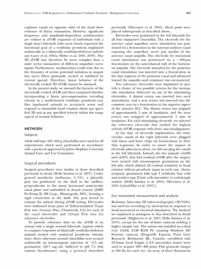
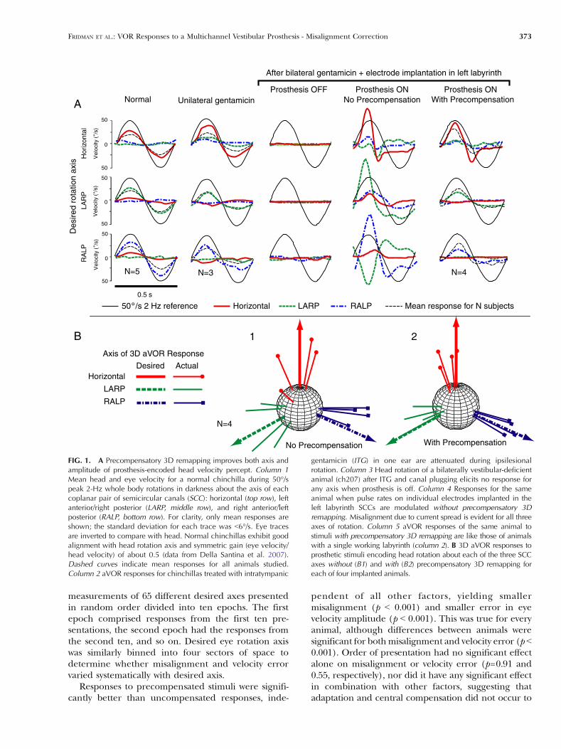
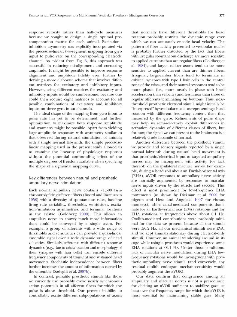
When a normal chinchilla is rotated sinusoidally at2 Hz and 50°/s peak about the mean axis of eachcoplanar pair of semicircular canals, normal aVORresponses are only partially compensatory for thehead rotation, with the resulting eye movementsbeing opposite the direction of head rotation but atonly about half the head velocity (Fig. 1A, column 1).After unilateral gentamicin treatment, responses tosinusoidal head rotation become asymmetric, with anear-normal aVOR response during rotation towardthe normal ear and attenuated response duringipsilesional rotation (Fig. 1A, column 2). After canalplugging, electrode insertion, and gentamicin treat-ment resulting in bilateral vestibular deficiency, noaVOR response is observed for head rotation in either
direction (Fig. 1A, column 3). When the same animal(ch207) is electrically stimulated at 50% SI onindividual SCCs’ electrodes, current spread to non-targeted branches of the vestibular nerve is evident inthat the aVOR response axis does not align well withthe intended axis (especially for that RALP case ofFig. 1A, column 4). Precompensatory adjustment ofthe stimuli attenuated the undesired response com-ponents and normalized both the axis and amplitudeof the response (Fig. 1A, column 5). Asymmetry wasobserved in the responses for all four animals,indicating that unilateral prosthetic stimulationrestores a bilaterally vestibular-deficient animal to astate similar that of an animal with a single normallabyrinth (i.e., Fig. 1A, column 2).
Similar improvement in the axis alignment wasevident for all four experimental animals when wecompared observed and actual axes of aVOR responsefor modulation of each SCC’s electrode individually(Fig. 1B).
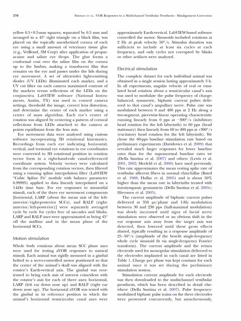
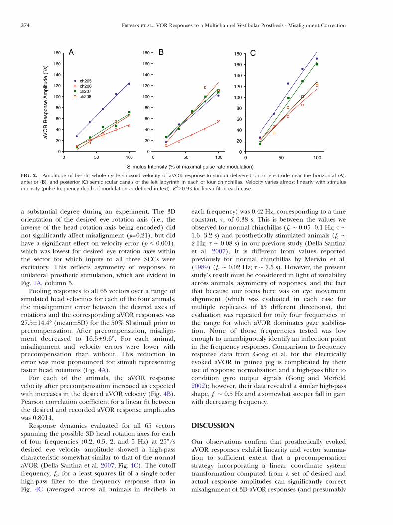
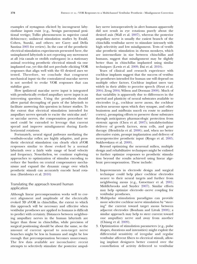
To assess whether the relationship between single-channel SCC stimulation intensity and aVORresponse velocity is sufficiently linear to justify use ofa linear precompensation scheme, we assayed aVORresponse amplitude for each animal as a function ofSI amplitude (Fig. 2). The Pearson correlationcoefficient for a linear fit between the SI amplitudesand the corresponding aVOR response amplitudeswas 90.93 in all cases.
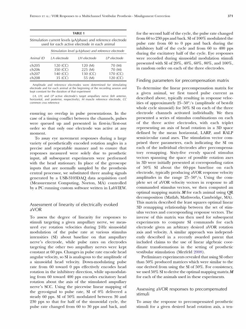
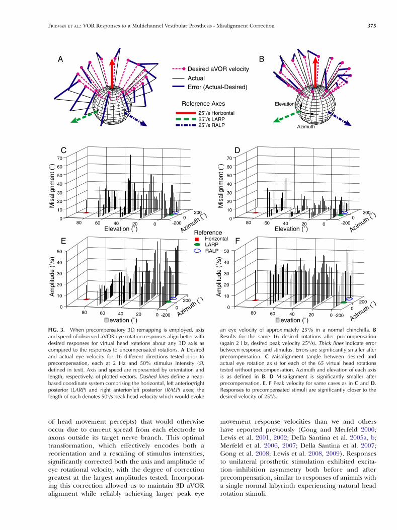
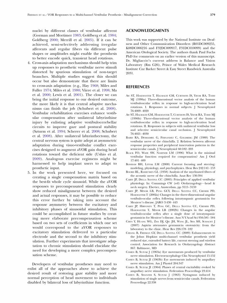
Observed aVOR responses matched desired ampli-tude and axis better when precompensation was usedto correct for axis misalignment than did responses tostimuli presented without precompensation. Figure 3compares desired and actual aVOR responses for 16stimulus vectors without (panel A) and with precom-pensation (right panel B). In the first case, stimuliwithout precompensation were presented at 50% SI.In the second case, precompensation was used tocorrect for axis misalignment and to specify desired25°/s peak eye velocities. Misalignment is clearlyreduced by precompensation. This reduction ofmisalignment is also apparent in (Fig. 3C, D), whichshows misalignment without and with precompensa-tion for all 65 head rotation axes emulated during theexperiment. Precompensation also reduced errors inthe speed of eye movement responses (Fig. 3E, F).
To discern whether animal tested, order of stim-ulus presentation, axis of desired response, and/orcombinations of these factors significantly affectedmisalignment and amplitude error of responses, thesefactors were included in the multivariate generallinear model as independent variables. Order ofpresentation was defined by binning data acquiredwithin each of two test sessions (either uncompen-sated at 50% SI and 2 Hz or precompensated at 25°/sdesired velocity and 2 Hz), each of which comprised
372 FRIDMAN ET AL.: VOR Responses to a Multichannel Vestibular Prosthesis - Misalignment Correction
measurements of 65 different desired axes presentedin random order divided into ten epochs. The firstepoch comprised responses from the first ten pre-sentations, the second epoch had the responses fromthe second ten, and so on. Desired eye rotation axiswas similarly binned into four sectors of space todetermine whether misalignment and velocity errorvaried systematically with desired axis.
Responses to precompensated stimuli were signifi-cantly better than uncompensated responses, inde-
pendent of all other factors, yielding smallermisalignment (p G 0.001) and smaller error in eyevelocity amplitude (p G 0.001). This was true for everyanimal, although differences between animals weresignificant for bothmisalignment and velocity error (p G0.001). Order of presentation had no significant effectalone on misalignment or velocity error (p=0.91 and0.55, respectively), nor did it have any significant effectin combination with other factors, suggesting thatadaptation and central compensation did not occur to
RA
LP
A
s 5.0
LAR
P
0
05
05
Vel
ocity
(/s
)
0
05
05
Vel
ocity
(/s
)
Hor
izon
tal
0
05
05
Vel
ocity
(/s
)No Precompensation With PrecompensationNormal Unilateral gentamicin
Prosthesis OFF
After bilateral gentamicin + electrode implantation in left labyrinth
Prosthesis ON Prosthesis ON
N=3N=5 N=4
Des
ired
rota
tion
axis
Mean response for N subjects50 /s 2 Hz reference Horizontal LARP RALP
BAxis of 3D aVOR Response
Horizontal
LARP
RALP
Desired Actual
No Precompensation With Precompensation
N=4
1 2
FIG. 1. A Precompensatory 3D remapping improves both axis andamplitude of prosthesis-encoded head velocity percept. Column 1Mean head and eye velocity for a normal chinchilla during 50°/speak 2-Hz whole body rotations in darkness about the axis of eachcoplanar pair of semicircular canals (SCC): horizontal (top row), leftanterior/right posterior (LARP, middle row), and right anterior/leftposterior (RALP, bottom row). For clarity, only mean responses areshown; the standard deviation for each trace was G6°/s. Eye tracesare inverted to compare with head. Normal chinchillas exhibit goodalignment with head rotation axis and symmetric gain (eye velocity/head velocity) of about 0.5 (data from Della Santina et al. 2007).Dashed curves indicate mean responses for all animals studied.Column 2 aVOR responses for chinchillas treated with intratympanic
gentamicin (ITG) in one ear are attenuated during ipsilesionalrotation. Column 3 Head rotation of a bilaterally vestibular-deficientanimal (ch207) after ITG and canal plugging elicits no response forany axis when prosthesis is off. Column 4 Responses for the sameanimal when pulse rates on individual electrodes implanted in theleft labyrinth SCCs are modulated without precompensatory 3Dremapping. Misalignment due to current spread is evident for all threeaxes of rotation. Column 5 aVOR responses of the same animal tostimuli with precompensatory 3D remapping are like those of animalswith a single working labyrinth (column 2). B 3D aVOR responses toprosthetic stimuli encoding head rotation about each of the three SCCaxes without (B1) and with (B2) precompensatory 3D remapping foreach of four implanted animals.
FRIDMAN ET AL.: VOR Responses to a Multichannel Vestibular Prosthesis - Misalignment Correction 373
a substantial degree during an experiment. The 3Dorientation of the desired eye rotation axis (i.e., theinverse of the head rotation axis being encoded) didnot significantly affect misalignment (p=0.21), but didhave a significant effect on velocity error (p G 0.001),which was lowest for desired eye rotation axes withinthe sector for which inputs to all three SCCs wereexcitatory. This reflects asymmetry of responses tounilateral prosthetic stimulation, which are evident inFig. 1A, column 5.
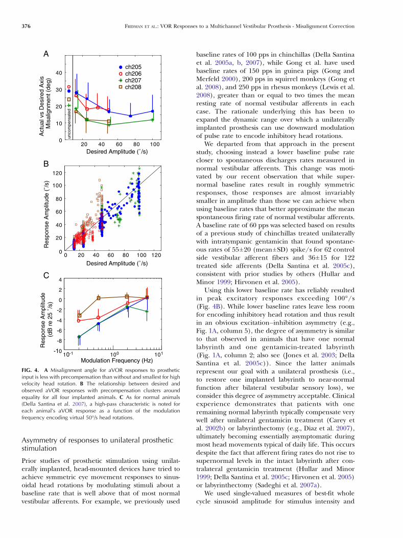
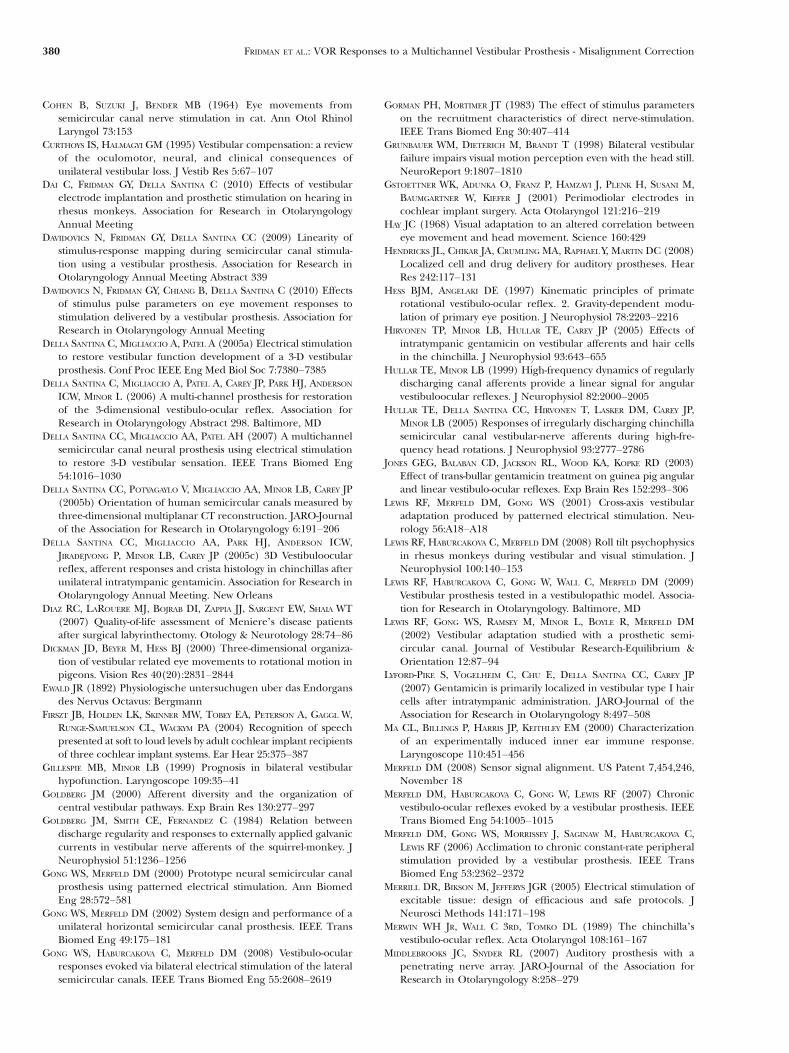
Pooling responses to all 65 vectors over a range ofsimulated head velocities for each of the four animals,the misalignment error between the desired axes ofrotations and the corresponding aVOR responses was27.5±14.4° (mean±SD) for the 50% SI stimuli prior toprecompensation. After precompensation, misalign-ment decreased to 16.5±9.6°. For each animal,misalignment and velocity errors were lower withprecompensation than without. This reduction inerror was most pronounced for stimuli representingfaster head rotations (Fig. 4A).
For each of the animals, the aVOR responsevelocity after precompensation increased as expectedwith increases in the desired aVOR velocity (Fig. 4B).Pearson correlation coefficient for a linear fit betweenthe desired and recorded aVOR response amplitudeswas 0.8014.
Response dynamics evaluated for all 65 vectorsspanning the possible 3D head rotation axes for eachof four frequencies (0.2, 0.5, 2, and 5 Hz) at 25°/sdesired eye velocity amplitude showed a high-passcharacteristic somewhat similar to that of the normalaVOR (Della Santina et al. 2007; Fig. 4C). The cutofffrequency, fc, for a least squares fit of a single-orderhigh-pass filter to the frequency response data inFig. 4C (averaged across all animals in decibels at
each frequency) was 0.42 Hz, corresponding to a timeconstant, τ, of 0.38 s. This is between the values weobserved for normal chinchillas (fc ∼ 0.05–0.1 Hz; τ ∼1.6–3.2 s) and prosthetically stimulated animals (fc ∼2 Hz; τ ∼ 0.08 s) in our previous study (Della Santinaet al. 2007). It is different from values reportedpreviously for normal chinchillas by Merwin et al.(1989) (fc ∼ 0.02 Hz; τ ∼ 7.5 s). However, the presentstudy’s result must be considered in light of variabilityacross animals, asymmetry of responses, and the factthat because our focus here was on eye movementalignment (which was evaluated in each case formultiple replicates of 65 different directions), theevaluation was repeated for only four frequencies inthe range for which aVOR dominates gaze stabiliza-tion. None of those frequencies tested was lowenough to unambiguously identify an inflection pointin the frequency responses. Comparison to frequencyresponse data from Gong et al. for the electricallyevoked aVOR in guinea pig is complicated by theiruse of response normalization and a high-pass filter tocondition gyro output signals (Gong and Merfeld2002); however, their data revealed a similar high-passshape, fc ∼ 0.5 Hz and a somewhat steeper fall in gainwith decreasing frequency.
DISCUSSION
Our observations confirm that prosthetically evokedaVOR responses exhibit linearity and vector summa-tion to sufficient extent that a precompensationstrategy incorporating a linear coordinate systemtransformation computed from a set of desired andactual response amplitudes can significantly correctmisalignment of 3D aVOR responses (and presumably
502hc602hc702hc802hc
aVO
R R
espo
nse
Am
plitu
de (
˚/s)
0
20
40
60
80
100
120
140
160
180
0
20
40
60
80
100
120
140
160
180
0
20
40
60
80
100
120
140
160
180
0 50 1000 50 1000 50 100
A B C
Stimulus Intensity (% of maximal pulse rate modulation)
FIG. 2. Amplitude of best-fit whole cycle sinusoid velocity of aVOR response to stimuli delivered on an electrode near the horizontal (A),anterior (B), and posterior (C) semicircular canals of the left labyrinth in each of four chinchillas. Velocity varies almost linearly with stimulusintensity (pulse frequency depth of modulation as defined in text). R290.93 for linear fit in each case.
374 FRIDMAN ET AL.: VOR Responses to a Multichannel Vestibular Prosthesis - Misalignment Correction
of head movement percepts) that would otherwiseoccur due to current spread from each electrode toaxons outside its target nerve branch. This optimaltransformation, which effectively encodes both areorientation and a rescaling of stimulus intensities,significantly corrected both the axis and amplitude ofeye rotational velocity, with the degree of correctiongreatest at the largest amplitudes tested. Incorporat-ing this correction allowed us to maintain 3D aVORalignment while reliably achieving larger peak eye
movement response velocities than we and othershave reported previously (Gong and Merfeld 2000;Lewis et al. 2001, 2002; Della Santina et al. 2005a, b;Merfeld et al. 2006, 2007; Della Santina et al. 2007;Gong et al. 2008; Lewis et al. 2008, 2009). Responsesto unilateral prosthetic stimulation exhibited excita-tion–inhibition asymmetry both before and afterprecompensation, similar to responses of animals witha single normal labyrinth experiencing natural headrotation stimuli.
latnoziroHPRALPLAR
)˚( noitavelE Azimuth
(˚)002-
0002
0020406080
01
02
03
04
05
06
07
Mis
alig
nmen
t (˚)
)˚( noitavelE Azimuth
(˚)002-
0002
0020406080
01
02
03
04
05
06
07
002-
0
002
0020406080
01
02
03
04
05
002-
0
002
0020406080
01
02
03
04
05
Mis
alig
nmen
t (˚)
Am
plitu
de (
˚/s)
Am
plitu
de (
˚/s)
)˚( noitavelE)˚( noitavelE Azimuth
(˚)
Azimuth
(˚)
C D
E FReference
htumizA
A
latnoziroH s/˚52PRAL s/˚25PLAR s/˚25
Error (Actual-Desired)
Desired aVOR velocity
Actual
Reference Axes
B
Elevation
FIG. 3. When precompensatory 3D remapping is employed, axisand speed of observed aVOR eye rotation responses align better withdesired responses for virtual head rotations about any 3D axis ascompared to the responses to uncompensated rotations. A Desiredand actual eye velocity for 16 different directions tested prior toprecompensation, each at 2 Hz and 50% stimulus intensity (SI,defined in text). Axis and speed are represented by orientation andlength, respectively, of plotted vectors. Dashed lines define a head-based coordinate system comprising the horizontal, left anterior/rightposterior (LARP) and right anterior/left posterior (RALP) axes; thelength of each denotes 50°/s peak head velocity which would evoke
an eye velocity of approximately 25°/s in a normal chinchilla. BResults for the same 16 desired rotations after precompensation(again 2 Hz, desired peak velocity 25°/s). Thick lines indicate errorbetween response and stimulus. Errors are significantly smaller afterprecompensation. C Misalignment (angle between desired andactual eye rotation axis) for each of the 65 virtual head rotationstested without precompensation. Azimuth and elevation of each axisis as defined in B. D Misalignment is significantly smaller afterprecompensation. E, F Peak velocity for same cases as in C and D.Responses to precompensated stimuli are significantly closer to thedesired velocity of 25°/s.
FRIDMAN ET AL.: VOR Responses to a Multichannel Vestibular Prosthesis - Misalignment Correction 375
Asymmetry of responses to unilateral prostheticstimulation
Prior studies of prosthetic stimulation using unilat-erally implanted, head-mounted devices have tried toachieve symmetric eye movement responses to sinus-oidal head rotations by modulating stimuli about abaseline rate that is well above that of most normalvestibular afferents. For example, we previously used
baseline rates of 100 pps in chinchillas (Della Santinaet al. 2005a, b, 2007), while Gong et al. have usedbaseline rates of 150 pps in guinea pigs (Gong andMerfeld 2000), 200 pps in squirrel monkeys (Gong etal. 2008), and 250 pps in rhesus monkeys (Lewis et al.2008), greater than or equal to two times the meanresting rate of normal vestibular afferents in eachcase. The rationale underlying this has been toexpand the dynamic range over which a unilaterallyimplanted prosthesis can use downward modulationof pulse rate to encode inhibitory head rotations.
We departed from that approach in the presentstudy, choosing instead a lower baseline pulse ratecloser to spontaneous discharges rates measured innormal vestibular afferents. This change was moti-vated by our recent observation that while super-normal baseline rates result in roughly symmetricresponses, those responses are almost invariablysmaller in amplitude than those we can achieve whenusing baseline rates that better approximate the meanspontaneous firing rate of normal vestibular afferents.A baseline rate of 60 pps was selected based on resultsof a previous study of chinchillas treated unilaterallywith intratympanic gentamicin that found spontane-ous rates of 55±20 (mean±SD) spike/s for 62 controlside vestibular afferent fibers and 36±15 for 122treated side afferents (Della Santina et al. 2005c),consistent with prior studies by others (Hullar andMinor 1999; Hirvonen et al. 2005).
Using this lower baseline rate has reliably resultedin peak excitatory responses exceeding 100°/s(Fig. 4B). While lower baseline rates leave less roomfor encoding inhibitory head rotation and thus resultin an obvious excitation–inhibition asymmetry (e.g.,Fig. 1A, column 5), the degree of asymmetry is similarto that observed in animals that have one normallabyrinth and one gentamicin-treated labyrinth(Fig. 1A, column 2; also see (Jones et al. 2003; DellaSantina et al. 2005c)). Since the latter animalsrepresent our goal with a unilateral prosthesis (i.e.,to restore one implanted labyrinth to near-normalfunction after bilateral vestibular sensory loss), weconsider this degree of asymmetry acceptable. Clinicalexperience demonstrates that patients with oneremaining normal labyrinth typically compensate verywell after unilateral gentamicin treatment (Carey etal. 2002b) or labyrinthectomy (e.g., Diaz et al. 2007),ultimately becoming essentially asymptomatic duringmost head movements typical of daily life. This occursdespite the fact that afferent firing rates do not rise tosupernormal levels in the intact labyrinth after con-tralateral gentamicin treatment (Hullar and Minor1999; Della Santina et al. 2005c; Hirvonen et al. 2005)or labyrinthectomy (Sadeghi et al. 2007a).
We used single-valued measures of best-fit wholecycle sinusoid amplitude for stimulus intensity and
01 1- 01 0 01 101-
8-
6-
4-
2-
0
2
4
)zH( ycneuqerF noitaludoM
Res
pons
e A
mpl
itude
(d
B r
e 25
˚/s
)A
B
C
0
02
04
06
08
001
021
)s/˚( edutilpmA deriseD
Res
pons
e A
mpl
itude
(˚/
s)
0 02 04 06 08 001 021
02 04 06 08 001
01
02
03
04
)s/˚( edutilpmA deriseD
Act
ual v
s D
esire
d A
xis
Mis
alig
nmen
t (de
g)
502hc602hc702hc802hc
unco
mpe
nsat
ed
0
FIG. 4. A Misalignment angle for aVOR responses to prostheticinput is less with precompensation than without and smallest for highvelocity head rotation. B The relationship between desired andobserved aVOR responses with precompensation clusters aroundequality for all four implanted animals. C As for normal animals(Della Santina et al. 2007), a high-pass characteristic is noted foreach animal’s aVOR response as a function of the modulationfrequency encoding virtual 50°/s head rotations.
376 FRIDMAN ET AL.: VOR Responses to a Multichannel Vestibular Prosthesis - Misalignment Correction
response velocity rather than half-cycle measuresbecause we sought to design a single optimal pre-compensation matrix for each animal. Excitation–inhibition asymmetry was explicitly incorporated viathe piecewise-linear, two-segment mapping from gyroinput to pulse rate on the corresponding electrodechannel. As evident from Fig. 3, this approach wassuccessful in reducing misalignment and correctingamplitude. It might be possible to improve responsealignment and amplitude fidelity even further bydevising a more elaborate scheme that involves differ-ent matrices for excitatory and inhibitory inputs.However, using different matrices for excitatory andinhibitory inputs would be cumbersome, because onecould then require eight matrices to account for allpossible combinations of excitatory and inhibitoryinputs on three gyro input channels.
The ideal shape of the mapping from gyro input topulse rate has yet to be determined, and furtheroptimization to maximize both response amplitudeand symmetry might be possible. Apart from yieldinglarge-amplitude responses with asymmetry similar tothat observed during natural stimulation of animalswith a single normal labyrinth, the simple piecewise-linear mapping used in the present study allowed usto examine the linearity of physiologic responseswithout the potential confounding effect of themultiple degrees of freedom available when specifyingthe shape of a sigmoidal mapping curve.
Key differences between natural and prostheticampullary nerve stimulation
Each normal ampullary nerve contains ∼1,500 asyn-chronously firing afferent fibers (Boord and Rasmussen1958) with a diversity of spontaneous rates, baselinefiring rate variability, thresholds, sensitivities, excita-tion/inhibition asymmetries, and terminal locationsin the cristae (Goldberg 2000). This allows anampullary nerve to convey much more informationthan could be conveyed by a single axon. Forexample, a group of afferents with a wide range ofthresholds and sensitivities can provide a quasi-linearensemble signal over a wide dynamic range of headvelocities. Similarly, afferents with different responsedynamics (e.g., due to crista location andmorphology oftheir synapses with hair cells) can encode differentfrequency components of transient and sustained headmovements. Stochastic independence between fibersfurther increases the amount of information carried bythe ensemble (Sadeghi et al. 2007b).
In contrast, pulsatile prosthetic stimuli like thosewe currently use probably evoke nearly synchronousaction potentials in all afferent fibers for which thepulse is above threshold. Our present inability tocontrollably excite different subpopulations of axons
that normally have different thresholds for headrotation probably restricts the dynamic range overwhich we can accurately encode head velocity. Thepattern of fiber activity presented to vestibular nucleiis probably further distorted by the fact that fiberswith irregular spontaneous discharge are more sensitiveto applied currents than are regular fibers (Goldberg etal. 1984), and larger caliber axons tend to be moresensitive to applied current than are thinner fibers.Irregular, large-caliber fibers tend to terminate incalyceal synapses with type I hair cells in the centralzone of the crista, and their natural responses tend to bemore phasic (i.e., more nearly in phase with headacceleration than velocity) and less linear than those ofregular afferents terminating on boutons. Thus, near-threshold prosthetic electrical stimuli might initially be“interpreted” by vestibular nuclei as representing a headrotation with different frequency content than thatmeasured by the gyros. Refinements of pulse shapemay help us semi-selectively exploit differences inactivation dynamics of different classes of fibers, butfor now, the signal we can present to the brainstem is arelatively crude facsimile of normal.
Another difference between the prosthetic stimuliwe provide and sensory signals reported by a singlenormal labyrinth during natural head movement isthat prosthetic/electrical input to targeted ampullarynerves may be incongruent with activity (or lackthereof) on the ipsilateral macular nerves. For exam-ple, during a head roll about an Earth-horizontal axis(EHA), aVOR responses to ampullary nerve activityare normally augmented by responses to macularnerve inputs driven by the utricle and saccule. Thiseffect is most prominent for low-frequency EHAmovements (as shown by Dickman et al. 2000 forpigeon and Hess and Angelaki 1997 for rhesusmonkeys), while canal-mediated components domi-nate for all Earth-vertical axis (EVA) rotations and forEHA rotations at frequencies above about 0.1 Hz.Otolith-mediated contributions were probably mini-mal for the data we present, because all our stimuliwere ≥0.2 Hz, all our mechanical stimuli were EVA,and we kept animals stationary during electrical-onlystimuli. However, an animal wandering around in itscage while using a prosthesis would experience someEHA rotations at G0.1 Hz. Under those conditions,lack of macular nerve modulation during EHA low-frequency rotations would be incongruent with pros-thetic ampullary nerve stimuli (and conversely, anyresidual otolith endorgan mechanosensitivity wouldprobably augment the aVOR).
Our data confirm that congruence among allampullary and macular nerves is not a prerequisitefor eliciting an aVOR sufficient to stabilize gaze, atleast over the frequency range for which the aVOR ismost essential for maintaining stable gaze. Many
FRIDMAN ET AL.: VOR Responses to a Multichannel Vestibular Prosthesis - Misalignment Correction 377
examples of nystagmus elicited by incongruent laby-rinthine inputs exist (e.g., benign paroxysmal posi-tional vertigo; Tullio phenomenon in superior canaldehiscence; unilateral stimulation studies by Ewald,Cohen, Suzuki, and others; see Carey and DellaSantina 2005 for review). In the case of the prostheticelectrical stimulation experiments presented here, theunimplanted labyrinth is not reporting any movementat all (via canals or otolith endorgans) in a stationaryanimal receiving prosthetic electrical stimuli via oneimplanted ear, yet this did not preclude eliciting VORresponses that align well with the 65 desired axes wetested. Therefore, we conclude that congruentmechanical input via the contralateral macular nervesis not needed to evoke VOR responses that canstabilize gaze.
How ipsilateral macular nerve input is integratedwith prosthetically evoked ampullary nerve input is aninteresting question. Using the prosthesis shouldallow partial decoupling of parts of the labyrinth tofacilitate answering this question in future studies. Tothe extent that electrical stimulation intended forampullary nerves spreads to excite the utricular and/or saccular nerves, the compensation procedure weused could be extended to leverage that currentspread and improve misalignment during Earth-horizontal rotations.
Fortunately, neural signal pathways mediating thevestibulo-ocular reflex are highly adaptive, and pros-thetic electrical stimulation can clearly elicit aVORresponses similar to those evoked by a normallabyrinth over a fairly wide range of head velocityand frequency. Nonetheless, we continue exploringapproaches to optimization of stimulus encoding toreduce the burden on central compensatory mecha-nisms and expand the dynamic range over whichprosthetic stimuli can accurately encode head rota-tion (Davidovics et al. 2010)
Translating the approach toward humanapplication
Although linear precompensation works well to cor-rect alignment and amplitude of the electricallyevoked 3D aVOR in chinchillas, the extent to whichthis approach will be necessary and effective whenvestibular prostheses are applied to humans is difficultto predict with certainty. Distances between neighbor-ing ampullary nerves in the human labyrinth arelarger than those in chinchillas, while precision ofsurgical positioning should be about the same, so theamount of current spread to non-target nervebranches might be less in humans and might be lowenough that precompensation becomes unnecessary.The few data available are inconclusive: recentattempts to selectively stimulate the posterior ampul-
lary nerve intraoperatively in alert humans apparentlydid not result in eye rotations purely about thedesired axis (Wall et al. 2007), whereas the posteriorampullary nerve is usually the easiest branch of thechinchilla vestibular nerve to stimulate intensely withhigh selectivity and low misalignment. Tests of vestib-ular prosthetic stimulation in rhesus monkeys, whichare intermediate in size between chinchillas andhumans, suggest that misalignment may be slightlybetter than in chinchillas implanted using similartechniques (Lewis et al. 2009; Dai et al. 2010).
Years of clinical and research experience withcochlear implants suggest that the success of vestibu-lar prostheses intended for human use will depend onmultiple other factors. Cochlear implant users varywidely in their ability to perceive speech (Firszt et al.2004; Zeng 2004; Wilson and Dorman 2008). Much ofthat variability is apparently due to differences in thesurvival and plasticity of neural substrates available toelectrodes (e.g., cochlear nerve axons, the cochlearnucleus neurons upon which they synapse, and otherbrainstem and midbrain nuceli en route to auditorycortex), prompting efforts to preserve those substratesthrough anticipatory pharmacologic protection fromototoxic agents (Chen et al. 2007); intralabyrinthinedelivery of growth factors, stem cells, and genetherapy (Hendricks et al. 2008); and, when no betteralternative exists, prompt implantation and delivery ofneuroprotective prosthetic input (Ryugo et al. 2005;Stakhovskaya et al. 2008).
Beyond optimizing the neuronal milieu, multipledesign and rehabilitative techniques might be enlistedto further optimize responses to prosthetic stimula-tion beyond the results achieved using linear alge-braic precompensation. These include:
1. Improvements in electrode design and surgicaltechnique could help place cochlear electrodesnearer to their neural targets and further fromneighboring axons (e.g., Gstoettner et al. 2001;Middlebrooks and Snyder 2007). Similar effortsmay help optimize electrode–nerve coupling forvestibular prostheses.
2. Multipolar stimulation paradigms can providemore selective cochlear nerve stimulation by “steer-ing” the current toward target axons betweenadjacent electrodes (Bonham and Litvak 2008); asimilar approach may help to steer current towardone ampullary nerve and away from another(Chiang et al. 2009).
3. Optimization of stimulation parameters (e.g., pulseshapes, durations and intensities) might exploit thedifferential sensitivity of irregular and regularvestibular afferent fibers to electrical stimuli, allow-ing implant designers better control over theconstellation of activity delivered to vestibular
378 FRIDMAN ET AL.: VOR Responses to a Multichannel Vestibular Prosthesis - Misalignment Correction
nuclei by different classes of vestibular afferent(Gorman and Mortimer 1983; Goldberg et al. 1984;Goldberg 2000; Merrill et al. 2005). If it can beachieved, semi-selectively addressing irregularafferents and regular fibers via different pulseshapes or amplitudes might enable the prosthesisto better encode quick, transient head rotations.
4. Cross-axis adaptation mechanisms should help trimup responses to prosthetic vestibular nerve stimulidistorted by spurious stimulation of non-targetbranches. Multiple studies suggest this shouldoccur but also demonstrate that there are limitsto cross-axis adaptation (e.g., Hay 1968; Miles andFuller 1974; Miles et al. 1980; Viirre et al. 1998; Maet al. 2000; Lewis et al. 2001). The closer we canbring the initial response to our desired outcome,the more likely it is that central adaptive mecha-nisms can finish the job (Schubert et al. 2008).Vestibular rehabilitation exercises enhance vestib-ular compensation after unilateral labyrinthineinjury by enlisting adaptive vestibulocerebellarcircuits to improve gaze and postural stability(Szturm et al. 1994; Scherer et al. 2008; Schubertet al. 2008). After unilateral labyrinthectomy, thecentral nervous system is quite adept at asymmetricadaptation during visuo-vestibular conflict exer-cises designed to augment aVOR gain during headrotations toward the deficient side (Ushio et al.2009). Analogous exercise regimens might beharnessed to help implant users to adapt toprosthetic input.
5. In the work presented here, we focused oncreating a single compensation matrix based onthe best-fit whole cycle sinusoid. While the aVORresponses to precompensated stimulation clearlyshow reduced misalignment between the desiredand actual responses, it may be possible to reducethis error further by taking into account theresponse asymmetry between the excitatory andinhibitory phases of sinusoidal stimulation. Thiscould be accomplished in future studies by creat-ing more elaborate precompensation schemebased on two sets of coefficients in which one setwould correspond to the aVOR responses toexcitatory stimulation delivered to a particularelectrode and the second to the inhibitory stim-ulation. Further experiments that investigate adap-tation to chronic stimulation should elucidate theneed for developing a more complex precompen-sation scheme.
Developers of vestibular prostheses may need toenlist all of the approaches above to achieve thedesired result of restoring gaze stability and morenormal perception of head movement to individualsdisabled by bilateral loss of labyrinthine function.
ACKNOWLEDGMENTS
This work was supported by the National Institute on Deaf-ness and Other Communication Disorders (R01DC009255,K08DC006216 and F32DC009917, F31DC010099) and theAmerican Otological Society. The authors thank Paul FuchsPhD for comments on an earlier version of this manuscript.Dr. Migliaccio’s current address is Balance and VisionLaboratory (Rm G26), Prince of Wales Medical ResearchInstitute Cnr Barker Street & Easy Street Randwick Australia2031.
REFERENCES
AW ST, HASLWANTER T, HALMAGYI GM, CURTHOYS IS, YAVOR RA, TODD
MJ (1996a) Three-dimensional vector analysis of the humanvestibuloocular reflex in response to high-acceleration headrotations. I. Responses in normal subjects. J Neurophysiol76:4009–4020
AW ST, HALMAGYI GM, HASLWANTER T, CURTHOYS IS, YAVOR RA, TODD MJ(1996b) Three-dimensional vector analysis of the humanvestibuloocular reflex in response to high-acceleration headrotations. II. Responses in subjects with unilateral vestibular lossand selective semicircular canal occlusion. J Neurophysiol76:4021–4030
BAIRD RA, DESMADRYL G, FERNANDEZ C, GOLDBERG JM (1988) Thevestibular nerve of the chinchilla. 2. Relation between afferentresponse properties and peripheral innervation patterns in thesemicircular canals. J Neurophysiol 60:182–203
BLACK FO, WADE SW, NASHNER LM (1996) What is the minimalvestibular function required for compensation? Am J Otol17:401–409
BONHAM BH, LITVAK LM (2008) Current focusing and steering:modeling, physiology, and psychophysics. Hear Res 242:141–153
BOORD RL, RASMUSSEN GL (1958) Analysis of the myelinated fibers ofthe acoustic nerve of the chinchilla. Anat Rec 130:394
CAREY JP, DELLA SANTINA CC (2005) Principles of applied vestibularphysiology. In: Cummings CW (ed) Otolaryngology—head &neck surgery. Elsevier, Amsterdam, pp 3115–3159
CAREY JP, MINOR LB, PENG GCY, DELLA SANTINA CC, CREMER PD,HASLWANTER T (2002a) Changes in the three-dimensional angularvestibulo-ocular reflex following intratympanic gentamicin forMeniere’s disease. JARO 3:430–443
CAREY JP, HIRVONEN T, PENG GC, DELLA SANTINA CC, CREMER PD,HASLWANTER T, MINOR LB (2002b) Changes in the angularvestibulo-ocular reflex after a single dose of intratympanicgentamicin for Meniere’s disease. Ann N YAcad Sci 956:581–584
CHEN Y, HUANG WG, ZHA DJ, QIU JH, WANG JL, SHA SH, SCHACHT J(2007) Aspirin attenuates gentamicin ototoxicity: from thelaboratory to the clinic. Hear Res 226:178–182
CHIANG B, FRIDMAN GY, DELLA SANTINA CC (2009) Enhancements tothe Johns Hopkins multi-channel vestibular prosthesis yieldreduced size, extended battery life, current steering and wirelesscontrol. Association for Research in Otolaryngology Abstract867. Baltimore, MD
COHEN B, SUZUKI J (1963a) Eye movements produced by vestibularnerve stimulation. Electroencephalogr Clin Neurophysiol 15:152
COHEN B, SUZUKI JI (1963b) Eye movements induced by ampullarynerve stimulation. Am J Physiol 204:347
COHEN B, SUZUKI JI (1964) Phases of central excitability evoked byampullary nerve stimulation. Federation Proceedings 23:414
COHEN B, SHANZER S, SUZUKI J (1963) Nystagmus induced bystimulation of single nerves from semicircular canals. FederationProceedings 22:338
FRIDMAN ET AL.: VOR Responses to a Multichannel Vestibular Prosthesis - Misalignment Correction 379
COHEN B, SUZUKI J, BENDER MB (1964) Eye movements fromsemicircular canal nerve stimulation in cat. Ann Otol RhinolLaryngol 73:153
CURTHOYS IS, HALMAGYI GM (1995) Vestibular compensation: a reviewof the oculomotor, neural, and clinical consequences ofunilateral vestibular loss. J Vestib Res 5:67–107
DAI C, FRIDMAN GY, DELLA SANTINA C (2010) Effects of vestibularelectrode implantation and prosthetic stimulation on hearing inrhesus monkeys. Association for Research in OtolaryngologyAnnual Meeting
DAVIDOVICS N, FRIDMAN GY, DELLA SANTINA CC (2009) Linearity ofstimulus-response mapping during semicircular canal stimula-tion using a vestibular prosthesis. Association for Research inOtolaryngology Annual Meeting Abstract 339
DAVIDOVICS N, FRIDMAN GY, CHIANG B, DELLA SANTINA C (2010) Effectsof stimulus pulse parameters on eye movement responses tostimulation delivered by a vestibular prosthesis. Association forResearch in Otolaryngology Annual Meeting
DELLA SANTINA C, MIGLIACCIO A, PATEL A (2005a) Electrical stimulationto restore vestibular function development of a 3-D vestibularprosthesis. Conf Proc IEEE Eng Med Biol Soc 7:7380–7385
DELLA SANTINA C, MIGLIACCIO A, PATEL A, CAREY JP, PARK HJ, ANDERSON
ICW, MINOR L (2006) A multi-channel prosthesis for restorationof the 3-dimensional vestibulo-ocular reflex. Association forResearch in Otolaryngology Abstract 298. Baltimore, MD
DELLA SANTINA CC, MIGLIACCIO AA, PATEL AH (2007) A multichannelsemicircular canal neural prosthesis using electrical stimulationto restore 3-D vestibular sensation. IEEE Trans Biomed Eng54:1016–1030
DELLA SANTINA CC, POTYAGAYLO V, MIGLIACCIO AA, MINOR LB, CAREY JP(2005b) Orientation of human semicircular canals measured bythree-dimensional multiplanar CT reconstruction. JARO-Journalof the Association for Research in Otolaryngology 6:191–206
DELLA SANTINA CC, MIGLIACCIO AA, PARK HJ, ANDERSON ICW,JIRADEJVONG P, MINOR LB, CAREY JP (2005c) 3D Vestibuloocularreflex, afferent responses and crista histology in chinchillas afterunilateral intratympanic gentamicin. Association for Research inOtolaryngology Annual Meeting. New Orleans
DIAZ RC, LAROUERE MJ, BOJRAB DI, ZAPPIA JJ, SARGENT EW, SHAIA WT(2007) Quality-of-life assessment of Meniere’s disease patientsafter surgical labyrinthectomy. Otology & Neurotology 28:74–86
DICKMAN JD, BEYER M, HESS BJ (2000) Three-dimensional organiza-tion of vestibular related eye movements to rotational motion inpigeons. Vision Res 40(20):2831–2844
EWALD JR (1892) Physiologische untersuchugen uber das Endorgansdes Nervus Octavus: Bergmann
FIRSZT JB, HOLDEN LK, SKINNER MW, TOBEY EA, PETERSON A, GAGGL W,RUNGE-SAMUELSON CL, WACKYM PA (2004) Recognition of speechpresented at soft to loud levels by adult cochlear implant recipientsof three cochlear implant systems. Ear Hear 25:375–387
GILLESPIE MB, MINOR LB (1999) Prognosis in bilateral vestibularhypofunction. Laryngoscope 109:35–41
GOLDBERG JM (2000) Afferent diversity and the organization ofcentral vestibular pathways. Exp Brain Res 130:277–297
GOLDBERG JM, SMITH CE, FERNANDEZ C (1984) Relation betweendischarge regularity and responses to externally applied galvaniccurrents in vestibular nerve afferents of the squirrel-monkey. JNeurophysiol 51:1236–1256
GONG WS, MERFELD DM (2000) Prototype neural semicircular canalprosthesis using patterned electrical stimulation. Ann BiomedEng 28:572–581
GONG WS, MERFELD DM (2002) System design and performance of aunilateral horizontal semicircular canal prosthesis. IEEE TransBiomed Eng 49:175–181
GONG WS, HABURCAKOVA C, MERFELD DM (2008) Vestibulo-ocularresponses evoked via bilateral electrical stimulation of the lateralsemicircular canals. IEEE Trans Biomed Eng 55:2608–2619
GORMAN PH, MORTIMER JT (1983) The effect of stimulus parameterson the recruitment characteristics of direct nerve-stimulation.IEEE Trans Biomed Eng 30:407–414
GRUNBAUER WM, DIETERICH M, BRANDT T (1998) Bilateral vestibularfailure impairs visual motion perception even with the head still.NeuroReport 9:1807–1810
GSTOETTNER WK, ADUNKA O, FRANZ P, HAMZAVI J, PLENK H, SUSANI M,BAUMGARTNER W, KIEFER J (2001) Perimodiolar electrodes incochlear implant surgery. Acta Otolaryngol 121:216–219
HAY JC (1968) Visual adaptation to an altered correlation betweeneye movement and head movement. Science 160:429
HENDRICKS JL, CHIKAR JA, CRUMLING MA, RAPHAEL Y, MARTIN DC (2008)Localized cell and drug delivery for auditory prostheses. HearRes 242:117–131
HESS BJM, ANGELAKI DE (1997) Kinematic principles of primaterotational vestibulo-ocular reflex. 2. Gravity-dependent modu-lation of primary eye position. J Neurophysiol 78:2203–2216
HIRVONEN TP, MINOR LB, HULLAR TE, CAREY JP (2005) Effects ofintratympanic gentamicin on vestibular afferents and hair cellsin the chinchilla. J Neurophysiol 93:643–655
HULLAR TE, MINOR LB (1999) High-frequency dynamics of regularlydischarging canal afferents provide a linear signal for angularvestibuloocular reflexes. J Neurophysiol 82:2000–2005
HULLAR TE, DELLA SANTINA CC, HIRVONEN T, LASKER DM, CAREY JP,MINOR LB (2005) Responses of irregularly discharging chinchillasemicircular canal vestibular-nerve afferents during high-fre-quency head rotations. J Neurophysiol 93:2777–2786
JONES GEG, BALABAN CD, JACKSON RL, WOOD KA, KOPKE RD (2003)Effect of trans-bullar gentamicin treatment on guinea pig angularand linear vestibulo-ocular reflexes. Exp Brain Res 152:293–306
LEWIS RF, MERFELD DM, GONG WS (2001) Cross-axis vestibularadaptation produced by patterned electrical stimulation. Neu-rology 56:A18–A18
LEWIS RF, HABURCAKOVA C, MERFELD DM (2008) Roll tilt psychophysicsin rhesus monkeys during vestibular and visual stimulation. JNeurophysiol 100:140–153
LEWIS RF, HABURCAKOVA C, GONG W, WALL C, MERFELD DM (2009)Vestibular prosthesis tested in a vestibulopathic model. Associa-tion for Research in Otolaryngology. Baltimore, MD
LEWIS RF, GONG WS, RAMSEY M, MINOR L, BOYLE R, MERFELD DM(2002) Vestibular adaptation studied with a prosthetic semi-circular canal. Journal of Vestibular Research-Equilibrium &Orientation 12:87–94
LYFORD-PIKE S, VOGELHEIM C, CHU E, DELLA SANTINA CC, CAREY JP(2007) Gentamicin is primarily localized in vestibular type I haircells after intratympanic administration. JARO-Journal of theAssociation for Research in Otolaryngology 8:497–508
MA CL, BILLINGS P, HARRIS JP, KEITHLEY EM (2000) Characterizationof an experimentally induced inner ear immune response.Laryngoscope 110:451–456
MERFELD DM (2008) Sensor signal alignment. US Patent 7,454,246,November 18
MERFELD DM, HABURCAKOVA C, GONG W, LEWIS RF (2007) Chronicvestibulo-ocular reflexes evoked by a vestibular prosthesis. IEEETrans Biomed Eng 54:1005–1015
MERFELD DM, GONG WS, MORRISSEY J, SAGINAW M, HABURCAKOVA C,LEWIS RF (2006) Acclimation to chronic constant-rate peripheralstimulation provided by a vestibular prosthesis. IEEE TransBiomed Eng 53:2362–2372
MERRILL DR, BIKSON M, JEFFERYS JGR (2005) Electrical stimulation ofexcitable tissue: design of efficacious and safe protocols. JNeurosci Methods 141:171–198
MERWIN WH JR, WALL C 3RD, TOMKO DL (1989) The chinchilla’svestibulo-ocular reflex. Acta Otolaryngol 108:161–167
MIDDLEBROOKS JC, SNYDER RL (2007) Auditory prosthesis with apenetrating nerve array. JARO-Journal of the Association forResearch in Otolaryngology 8:258–279
380 FRIDMAN ET AL.: VOR Responses to a Multichannel Vestibular Prosthesis - Misalignment Correction
MIGLIACCIO AA, MACDOUGALL HG, MINOR LB, DELLA SANTINA CC(2005) Inexpensive system for real-time 3-dimensional video-oculography using a fluorescent marker array. J NeurosciMethods 143:141–150
MILES FA, FULLER JH (1974) Adaptive plasticity in vestibulo-ocularresponses of rhesus-monkey. Brain Res 80:512–516
MILES FA, FULLER JH, BRAITMAN DJ, DOW BM (1980) Long-termadaptive-changes in primate vestibuloocular reflex. 3. Electro-physiological observations in flocculus of normal monkeys. JNeurophysiol 43:1437–1476
MINOR LB (1998) Gentamicin-induced bilateral vestibular hypo-function. JAMA-Journal of the American Medical Association279:541–544
ROBINSON DA (1963) A method of measuring eye movement using ascleral search coil in a magnetic field. IEEE Trans Biomed EngBM10:137
ROBINSON DA (1982) The use of matrices in analyzing the 3-dimensional behavior of the vestibulo-ocular reflex. Biol Cybern46:53–66
ROBINSON FP, MURPHY PG (1932) The validity of measuring eyemovements by direct observation. Science 76:171–172
RYUGO DK, KRETZMER EA, NIPARKO JK (2005) Restoration of auditorynerve synapses in cats by cochlear implants. Science 310:1490–1492
SADEGHI SG, MINOR LB, CULLEN KE (2007a) Response of vestibular-nerve afferents to active and passive rotations under normalconditions and after unilateral labyrinthectomy. J Neurophysiol97:1503–1514
SADEGHI SG, CHACRON MJ, TAYLOR MC, CULLEN KE (2007b) Neuralvariability, detection thresholds, and information transmission inthe vestibular system. J Neurosci 27:771–781
SCHERER M, MIGLIACCIO AA, SCHUBERT MC (2008) Effect of vestibularrehabilitation on passive dynamic visual acuity. Journal ofVestibular Research-Equilibrium & Orientation 18:147–157
SCHUBERT MC, DELLA SANTINA CC, SHELHAMER M (2008) Incrementalangular vestibulo-ocular reflex adaptation to active head rota-tion. Exp Brain Res 191:435–446
STAKHOVSKAYA O, HRADEK GT, SNYDER RL, LEAKE PA (2008) Effects ofage at onset of deafness and electrical stimulation on thedeveloping cochlear nucleus in cats. Hear Res 243:69–77
SUZUKI J, COHEN B, BENDER MB (1964) Compensatory eye movementsinduced by vertical semicircular canal stimulation. Exp Neurol9:137
SUZUKI JI, COHEN B (1964) Head eye body + limb movements fromsemicircular canal nerves. Exp Neurol 10:393
SUZUKI JI, GOTO K, TOKUMASU K, COHEN B (1969) Implantation ofelectrodes near individual vestibular nerve branches in mam-mals. Ann Otol Rhinol Laryngol 78:815
SZTURM T, IRELAND DJ, LESSING-TURNER M (1994) Comparison ofdifferent exercise programs in the rehabilitation of patients withchronic peripheral vestibular dysfunction. J Vestib Res 4:461–479
USHIO M, LASKER D, MINOR L (2009) Unidirectional rotations inmacaques produce asymmetric changes in gain of the horizontalVOR before and after unilateral labyrinthectomy (Abstract 760).Society for Neuroscience Annual Meeting 2009. Chicago, IL
VIIRRE E, DRAPER M, GAILEY C, MILLER D, FURNESS T (1998) Adaptationof the VOR in patients with low VOR gains. J Vestib Res 8:331–334
WALL C, KOS MI, GUYOT JP (2007) Eye movements in response toelectric stimulation of the human posterior ampullary nerve.Ann Otol Rhinol Laryngol 116:369–374
WEBER KP, AW ST, TODD MJ, MCGARVIE LA, CURTHOYS IS, HALMAGYI
GM (2008) Head impulse test in unilateral vestibular loss:vestibulo-ocular reflex and catch-up saccades. Neurology70:454–463
WEBER KP, AW ST, TODD MJ, MCGARVIE LA, CURTHOYS IS, HALMAGYI GM(2009) Horizontal head impulse test detects gentamicin vestibu-lotoxicity. Neurology 72:1417–1424
WILSON BS, DORMAN MF (2008) Cochlear implants: a remarkablepast and a brilliant future. Hear Res 242:3–21
WILSON VJ, MELVILL J (1979) Mammalian vestibular physiology.Plenum, New York
ZENG FG (2004) Trends in cochlear implants. Trends in Amplifica-tion 8:1–34
FRIDMAN ET AL.: VOR Responses to a Multichannel Vestibular Prosthesis - Misalignment Correction 381