view - libreria universo
TRANSCRIPT
Hallmarks of Metastasis
M. Roshni Ray and David M. Jablons
Abstract Metastasis is rarely due to accidental sloughing off of cancerous cells
from a nonmalignant tumor and colonizing elsewhere; on the contrary, it is an
active process requiring genetic and/or epigenetic mechanisms leading to the
formation of a cell capable of responding to certain chemotactic signals that
direct motility, interacting with other cells to be co-translocated, implanting in
foreign locations, avoiding immune response, being refractory to growth inhi-
bitory signals, and proliferating independently of growth factors for sustained
cell division. The complexity of these processes necessitates a detailed under-
standing of the molecular and cellular mechanisms behind each of these steps.
In this chapter, we will discuss hallmarks of metastatic process, along with
theories proposed, genes involved, techniques to monitor, and therapeutic
implications.
Introduction
Cancer is a general term describing hundreds of diseases in which cells aggres-
sively proliferate without regard for normal growth limits of the original tissue
or organ site and then invade surrounding and adjoining tissues. Most cancers
are diseases of the epithelial tissue [1], where in late stages the cancerous cells
invade the mesoderm and the endodermal layers. Metastasis, the subsequent
spread of these invasive cells throughout the body to other organs, accounts for
90% of human cancer deaths [2]. Interestingly, 5-year survival of stage IV
patients is a dismal 3%. By contrast, 5-year survival of early-stage cancers is
49% [3]. Thus, in addition to early detection efforts, understanding themechan-
isms of metastasis and halting its course are imperative to the treatment of
cancer.
D.M. Jablons (*)Thoracic Oncology Program, Comprehensive Cancer Center, University of California,San Francisco, CA, USAe-mail: [email protected]
V. Keshamouni et al. (eds.), Lung Cancer Metastasis,DOI 10.1007/978-1-4419-0772-1_2, Ó Springer ScienceþBusiness Media, LLC 2009
29
Traditionally, metastasis has been characterized as a late-stage phenomenonin cancer. Pathologic staging describes first the size and local invasion of aprimary tumor, then metastasis to lymph nodes, and finally distal metastases.Gene expression profiling studies [4] suggest that metastatic potential is intrin-sic to all tumor cells and that metastatic spread could be an early event intumorigenesis. However, there is some evidence for the existence of a smallpopulation of cells within a tumor, which exclusively are capable of metastasis –the so-called cancer stem cells.
Regardless of which cells are capable of metastasizing, metastasis is anextremely complex process and, thankfully, highly inefficient as only a smallfraction of tumor cells are actually able to fully metastasize. The processinvolves migration of a cancerous cell out of the original location, overcomingbarriers to implantation in a foreign location, subsequently dividing uncon-trolled, and/or metastasizing further. Whereas in most cancers the cell cyclecheckpoint arrest is overcome by at least one transformative event early incancer development in the traditional model of metastasis, a second genetic orepigenetic event is usually necessary for transition of a non-metastatic tumor tometastatic. Metastasis is rarely due to accidental sloughing off of cancerouscells from a nonmalignant tumor and colonizing elsewhere; on the contrary, it isan active process requiring genetic and/or epigenetic mechanisms leading to theformation of a cell capable of responding to certain chemotactic signals thatdirect motility, interacting with other cells to be co-translocated, implanting inforeign locations, avoiding immune response, being refractory to growth inhi-bitory signals, and proliferating independently of growth factors for sustainedcell division. The complexity of these processes necessitates a detailed under-standing of the molecular and cellular mechanisms behind each of these steps.
The Metastatic Process
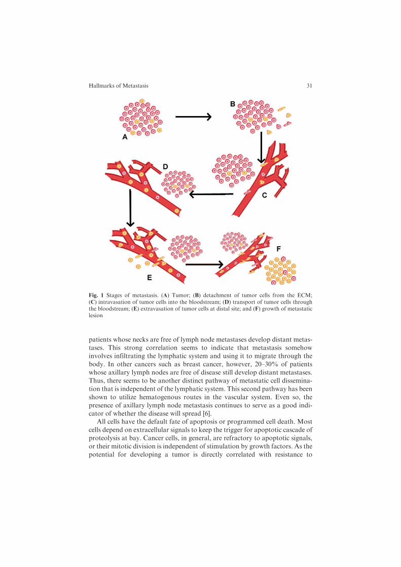
Cancer metastasis involves several interrelated steps, each of which can be ratelimiting in that failure to achieve any state can shut down the entire metastaticprocess. Moreover, only certain cells within a heterogeneous tumor populationare capable of achieving these steps. Metastasis consists of (1) detachment ofepithelial cells from the extracellular matrix (ECM), (2) survival within thebloodstream, and (3) growth at the metastatic site (Fig. 1).
As metastasis tends to be the lethal aspect of cancer, dissecting its biologicalbasis is of utmost importance in pinpointing therapeutic targets to prevent andcure it. The existence of lymph node metastases in a cancer is a strong indicatorof survival in patients as well as a prognosticator of whether other distalmetastases will develop. In some cancers, lymph node metastases are betterindicators of distant metastasis than in other cancers. In head and neck cancer,for example, the correlation is strong – the presence of lymph node metastasesin the neck halves the survival rate in patients [5]. Moreover, only 7% of
30 M.R. Ray and D.M. Jablons
patients whose necks are free of lymph node metastases develop distant metas-
tases. This strong correlation seems to indicate that metastasis somehow
involves infiltrating the lymphatic system and using it to migrate through the
body. In other cancers such as breast cancer, however, 20–30% of patients
whose axillary lymph nodes are free of disease still develop distant metastases.
Thus, there seems to be another distinct pathway of metastatic cell dissemina-
tion that is independent of the lymphatic system. This second pathway has been
shown to utilize hematogenous routes in the vascular system. Even so, the
presence of axillary lymph node metastasis continues to serve as a good indi-
cator of whether the disease will spread [6].All cells have the default fate of apoptosis or programmed cell death. Most
cells depend on extracellular signals to keep the trigger for apoptotic cascade of
proteolysis at bay. Cancer cells, in general, are refractory to apoptotic signals,
or their mitotic division is independent of stimulation by growth factors. As the
potential for developing a tumor is directly correlated with resistance to
Fig. 1 Stages of metastasis. (A) Tumor; (B) detachment of tumor cells from the ECM;(C) intravasation of tumor cells into the bloodstream; (D) transport of tumor cells throughthe bloodstream; (E) extravasation of tumor cells at distal site; and (F) growth of metastaticlesion
Hallmarks of Metastasis 31
apoptosis, so is resistance to apoptosis correlated with the metastatic potential
of a tumor. The reason behind this correlation is somewhat unclear. It is
possible that certain rare cells having the ability to undergo mitosis indepen-
dently of growth factors preferentially become metastatic; alternatively, the
same molecular events that give rise to metastatic transformation of a tumor
cell cause it to be resistant to apoptosis. Therefore, anoikis (cell death by
disruption of cell adhesion and cell–ECM interactions) and amorphosis (cell
death by loss of cytoskeletal structure) are vital to preventing metastasis.
Normally anoikis and amorphosis are triggered by detachment of usually
adherent cells from the ECM and through disruption of the actin cytoskeleton
[7], which is consistent with the general observation that specific cell–cell and/or
cell–matrix contact and ligand-mediated signaling are necessary to keep the
apoptotic cascade from being activated. Abrogation of the need for signaling
through contact for suppressing apoptosis might, therefore, lead to both
immortalization and cell detachment. Alternatively, the two processes might
be unrelated.
Theories of Metastasis
It is currently unclear whether any given cell within a tumor once transformed
into the metastatic stage can migrate and form a secondary tumor or whether a
special group of cells within a solid tumor, cancer stem cells, a rare tumor cell
type with indefinite self-renewal capability, is the only cell type capable of
migrating and colonizing secondary tissues and organs. In the traditional
model of cancer metastasis, every malignant tumor cell supposedly possesses
metastatic potential. A normal cell accumulates random mutations eventually
leading to cancer, and these neoplastic cells continue to accrue mutations until
some become metastatic by chance. Nevertheless, small populations of cells in
many malignant tumors, including acute myeloblastic leukemia (AML) [8],
glioblastoma [9], small cell lung cancer [10], non-small cell lung cancer [11],
malignantmelanoma [12], and breast cancer [13], display properties reminiscent
of stem cells, the cell group that indefinitely retains the property of self-renewal
by mitosis [14]. It is conceivable that migration of these cells could in principle
lead to metastasis and successful colonization at a distant site, and this may
explain why successful primary metastasis is a relatively rare event. Strong
evidence implicating these ‘‘cancer stem cells’’ (see Chapter 3) in metastasis is
provided by the observation of overlap between the genes and signaling path-
ways necessary for normal stem cell motility and those for metastatic cancer
cells [15]. Invasive metastasis appears, for a number of cancer types, to be a
property of a subpopulation of tumor cells which appear to have stem cell-like
properties. Since differentiated cells rarely reenter the somatic stem cell state,
progeny of previously differentiated cells should rarely metastasize.
32 M.R. Ray and D.M. Jablons
Dissemination from the Primary Tumor
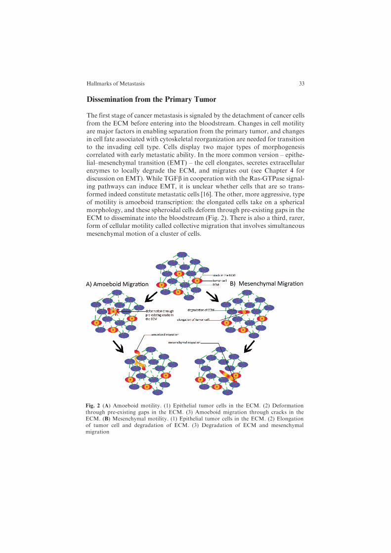
The first stage of cancer metastasis is signaled by the detachment of cancer cellsfrom the ECM before entering into the bloodstream. Changes in cell motilityare major factors in enabling separation from the primary tumor, and changesin cell fate associated with cytoskeletal reorganization are needed for transitionto the invading cell type. Cells display two major types of morphogenesiscorrelated with early metastatic ability. In the more common version – epithe-lial–mesenchymal transition (EMT) – the cell elongates, secretes extracellularenzymes to locally degrade the ECM, and migrates out (see Chapter 4 fordiscussion on EMT). While TGFb in cooperation with the Ras-GTPase signal-ing pathways can induce EMT, it is unclear whether cells that are so trans-formed indeed constitute metastatic cells [16]. The other, more aggressive, typeof motility is amoeboid transcription: the elongated cells take on a sphericalmorphology, and these spheroidal cells deform through pre-existing gaps in theECM to disseminate into the bloodstream (Fig. 2). There is also a third, rarer,form of cellular motility called collective migration that involves simultaneousmesenchymal motion of a cluster of cells.
Fig. 2 (A) Amoeboid motility. (1) Epithelial tumor cells in the ECM. (2) Deformationthrough pre-existing gaps in the ECM. (3) Amoeboid migration through cracks in theECM. (B) Mesenchymal motility. (1) Epithelial tumor cells in the ECM. (2) Elongationof tumor cell and degradation of ECM. (3) Degradation of ECM and mesenchymalmigration
Hallmarks of Metastasis 33
Intra- and intercellular signaling mechanisms enable these morphogenetic
processes, and cell–cell cooperation is likely involved in controlling swarm-like
behavior of rare metastasizing cells. Cells, however, appear to be capable of
switching between different forms of motility, which renders therapeutics that
target genes or proteins associated with distinct aspects of mobility somewhat
refractory [17]. There is, nonetheless, evidence suggesting that certain transcrip-
tional regulators may control entire sets of motility genes. For example, AP-1
transcription factor activity is correlated with expression of cell motility genes
[18]. Twist, Six-1, and BRMS1, all transcriptional regulators, have also been
implicated. Other genes involved in cell–cell signaling such as ErbB1, encoding
epidermal growth factor receptor (EGFR), are implicated in cancer cell motility
but not necessarily in growth of the primary tumor. Genes or proteins that are
specifically implicated in cell motility in metastatic transformation are potential
drug targets.The significance of EMT lies in that disseminating metastatic cells must be
able to survive without normal matrix components and evade anoikis. This
survival is important in metastasis because intra/extravasating tumor cells
either do not adhere to a matrix at all or encounter foreign matrices along the
way [19]. Overexpression of BCL2 increases the metastatic potential of breast
cancer epithelial cells by inhibiting matrix-degradation-induced apoptosis but
does not affect primary tumor growth or cell motility [20, 21].The developmental signaling pathways Wnt, Notch, and Hedgehog have
also been linked to EMT [19]. Wnt signaling is of particular interest as it has
been associated with collective migration and is aberrantly activated in lung
cancers [22].With regard tometastasis, loss of signaling byWnt1, the first of the
Wnt proteins to be discovered, has been shown to reduce the size of lung
metastases in mice [23].As stated earlier, most motile cells normally move mesenchymally (Fig. 2).
After tumor cells undergo EMT, they migrate by polarizing and extending
pseudopodia-like projections (lamellipods) on their anterior ends, binding spe-
cific cell surface or extracellular matrix ligands, pushing themselves forward
through actin-based contractions of the cell body, and then releasing the
adhesive bonds at the rear. Adhesion to the ECM substrate is mediated through
interactions of beta-integrins, a major group of cell surface receptor ligands.
Subsequently, signaling by the integrins, as well as integrins themselves, coop-
erate with and recruit cell surface proteases (such as matrix metalloproteinases,
MMPs) to locally degrade the ECM. MMPs break down collagen in the ECM
(collagenolysis). Mesenchymal motion is proteolytic and path generating: gaps
in the ECM through which the cell ultimately passes are created by the cell itself
[24]. Actin filaments are the dominant structural component of lamellipods [25].
H-, N-, and K-Ras are small GTPases that promote mesenchymal lamellipod
extension by regulating PtdIns(3,4,5)P3 levels. Cdc42 and Rac1 are also small
GTPases that promote formation of actin-rich protrusions. Unfortunately for
patients, near-total inhibition of cell surface proteases by protease inhibitor
34 M.R. Ray and D.M. Jablons
treatment induces conversion of mesenchymal cells to spherical morphologyand virtually no change in migration rates [24].
Amoeboid motion – where spherically shaped cells deform and slip throughpre-existing cracks in the ECM – is protease independent and path finding (incontrast to the proteolytic path-generating nature of mesenchymal locomotion)(Fig. 2). RhoA (a small GTPase) activates the ROCK protein, which phosphor-ylates MLC2, a myosin light chain protein, which in turn activates a signalingcascade implicated in the development of spheroid structure and induction ofcellular locomotion. The Smurf1 protein, an ubiquitin ligase, is responsible fortargeting RhoA for degradation. When Smurf1 is activated, RhoA activity isdepleted due to ubiquitin-mediated proteolysis, possibly involving the protea-some, and cells form lamellipods that aid in mesenchymal cell movement. Onthe other hand, when Smurf1 activity is downregulated, the RhoA cascade isactivated and induces amoeboid cell invasion, which is actually more aggressivethan mesenchymal motion [26].
The signaling cascades leading to different types of motion ultimately influ-ence cytoskeletal elements to reorganize and the molecular motors to generateforce that leads to cellular motion. Altered MLC organization is related to theamoeboid tumor cells’ ability to generate sufficient mechanical force to deformthe extracellular mesh of collagen fibers and to enable the cell to push throughthe ECM [27]. ROCK regulates MLC phosphorylation, and inhibition ofROCK (but not of MMPs) reduces in vivo cancer cell motility. The proteinezrin is localized in the direction of cell movement in amoeboid cells [28]. Ezrinprovides a functional link between the plasma membrane and the cortical actincytoskeleton of the cell. Forced ezrin expression induces a highly metastaticstate in certain poorly metastatic tumor cell lines [27]. Combined blockade ofextracellular proteases andROCKprevents tumor cells from switching betweentypes of motility and also blocks cell invasion.
In a study to identify a gene expression signature associated with the pro-pensity for metastasis, invasive breast cancer tumor cells were collected in vitroby virtue of their chemotactic ability (migration toward a source of EGF), andtheir mRNA expression levels were assayed in relation to their less invasivecounterparts [17]. Genes associated with motility were most strikingly differen-tially regulated in invasive cells compared to those in non-invasive cells. Forexample, cofilin, Arp2/3 complex, and capping protein, all involved in lamelli-pod protrusion, extension, and tail retraction, were coordinately upregulated inthe invasive cells. Genes encoding Rho and ROCK were significantly upregu-lated. Upregulation of cofilin, Arp2/3, and capping protein results in increasedprotrusion velocities of up to 10-fold higher than those in cells with lower levelsof expression of these proteins. By contrast, the ZBP1 gene is strongly down-regulated in invasive breast carcinoma cells. ZBP1 binds to beta-actin mRNAand localizes the mRNA to the leading edge of cells. Beta-actin is the mostcommon form of actin which is polymerized as filaments within the lamellipodand is acted on by cofilin, capping protein, and the Arp2/3 pathways. The site ofsub-cellular localization of the ZBP1 protein likely determines, by controlling
Hallmarks of Metastasis 35
localization of the beta-actin mRNA (thus its site of translation), the site atwhich these pathways converge. Downregulation of ZBP1-mediated beta-actinmRNA targeting, which is associated with inhibition of lamellipod formation inmesenchymal cells, results in the formation of highly invasive amoeboid cellsand increased chemotaxis [4].
Vascular Transport of Metastatic Cells
Even though both mesenchymal and amoeboid cells can migrate toward andintravasate into blood vessels, amoeboid cells are better suited to survive withinthe vascular system. A mesenchymal to amoeboid transformation is importantduring entry into blood vessels because elongated mesenchymal cells tend toshatter, or undergo amorphosis, in response to the force of blood flow(hemodynamic shearing). The spheroid morphology characteristic of amoeboidcells can better withstand high shear stress and hence these cells survive better inthe bloodstream [29]. Thus, solitary cancer cells in circulation are sensitive toapoptosis, particularly to that induced by mechanical stress and immune-mediated destruction. Potentially metastatic cells that have entered thebloodstream are destroyed either by mechanical stress or, supposedly, byimmune-directed cell death.
Once disseminated into the bloodstream, tumor cells are able to circulatethroughout the body. Oftentimes cancer patients have significant quantities ofthese cells both in blood and in bone marrow long after removal of the primarytumor and before any sign of metastasis occurs [30]. Are each of these dormantcells capable of producing their own clonal metastases or do only a fractionpossess the pluripotency to form the seed of metastatic malignancy? To answerthis question, a distinction between the tumor cells circulating in peripheralblood (circulating tumor cells – CTCs) as opposed to those aggregated in thebone marrow (disseminated tumor cells – DTCs) must be established.
DTCs have been documented in the bonemarrow for most types of epithelialcancers [30]. While a number of studies have revealed correlation between thepresence of DTCs and postoperative metastatic relapse, viable use of DTCs asprognosticators of recurrence has yet to be unequivocally demonstrated. Apoint of interest is that presence of DTCs is associated not only with bonemetastases but also with distal tumor development in lung, brain, and liver;thus, it is likely that DTCs accumulating in the bone marrow eventually reenterthe vasculature to travel throughout the body [31]. Incidentally, the processesby which tumor cells disseminate appear to vary by cancer. In early-stage breastcancer, DTCs are heterogeneous and do not possess the same changes as thetumor; however, in late stages the DTC genotypes are largely homogeneous.This observation seems to indicate that dissemination of DTCs is an early eventafter which the cells accumulate other mutations, some of which eventuallyoverwhelmingly favor metastasis. In prostate cancer, however, CTCs appear
36 M.R. Ray and D.M. Jablons
genotypically homogeneous and similar to cells in the primary tumor. Thisobservation suggests that as the tumor develops, cells with metastatic potentialeventually arise directly from the primary tumor and spread forth throughoutthe body [30].
At present, only limited data exist correlatingDTCswith concurrentCTCs, andthe significance of CTCs in peripheral blood is as yet unclear [30]. In most cancers,patients appear to present with a higher fraction of DTCs than CTCs, suggestingthat bone marrow may provide better conditions for tumor cell homing andsurvival [30]. Another hypothesis of note is the speculation as to whether surgeryitself can dislodge tumor cells from the primarymalignancy, thereby allowing thesecells to become CTCs. Bone marrow possibly forms a pre-metastatic niche andthat it offers a site for dormancy is evidenced by the presence ofDTCs in colorectalcarcinomas, a cancer in which bone metastases are rare [31].
It is usually assumed that invasive tumor cells entering blood vessels andforeign tissues will be recognized and targeted by the immune system. Thomasand Burnet [32, 33] suggested that immunosurveillance was responsible for thetargeted elimination of cancerous cells, particularly in that immune response isoften associated with advanced carcinogenesis (for discussion on immunesur-veilance and tumor progression see Chapter 6). Indeed, immune surveillancemay contribute to dormancy in DTCs. The nature of dormancy is variable inthat in some cases it is characterized by a balance between apoptosis andproliferation whereas in other cases it describes either non- or slowly proliferat-ing cells. It has been shown inmurine models that upon depletion of CD4+ andCD8+ leukocytes, progressive growth is initiated in previously dormant tumorcells [30]. In lung cancer, increased presence of CD8+ cytotoxic lymphocyteshas been observed, but this escalation in immune response does not appear tocorrelate with outcome. Tumor cells appear to evade the immune systembecause, as native cells, they are poorly immunogenic. Moreover, tumor cellsalso downregulate antigens by interfering with antigen-presenting cells andsecrete cytokines that may aid in both immune tolerance and suppression.Tumor cells are often also resistant to cytotoxic T-lymphocytes as evidencedby the cells’ failure to undergo apoptosis upon attack. Nonetheless, reduction ofmalignant tumor has been observed alongside bacterial infection, leading tohope of an anti-tumor vaccine created from dead bacteria. Although no con-clusively successful anti-tumor vaccines have yet been reported, a number ofsuch vaccines have entered clinical trials [34].
All this being said, our understanding of the role immunosurveillance playsin maintaining dormancy is murky, at best. In most cases, tumor developmentappears to be similar in normal and in immunocompromised animal models,and any systematic correlation between immune deficiency and human cancerhas yet to be demonstrated. Thus, it is unlikely that immune surveillance offersmuch protection against anything but pathogen-associated cancers [35].
What then of the heightened immune cell activity at primary tumor andmetastatic sites? Macrophages have been observed to produce matrix metallo-proteinases that promote tumor cell mobility by degrading the ECM. These
Hallmarks of Metastasis 37
tumor-associated macrophages (TAMs) express EGF and promote EGFR-dependent cell invasion (for more detailed discussion of TAMs and otherimmune cells see Chapter 11). TAMs tend to aggregate along blood vesselsand tumor margins and create an EGF gradient responsible for directingchemotaxis. This gradient likely promotes intra- and extravasation. In fact,prior to extravasation, leukocytes are recruited when tumor cells attach toblood vessel walls, and the leukocytes are thought to extravasate ahead of thecancer cells, in essence by ushering them out [36].
Proliferative Ability at Site of Metastasis
Metastasis has been directly correlated, on a single animal basis, with the bloodburden of tumor cells. A high concentration of CTCs in the bloodstream isrelated to a higher likelihood of metastasis [17]. Nevertheless, most cells dierapidly after extravasation [37, 38], which is likely why DTCs seem to correlatebetter with prognosis than CTCs. In as far back as 1889, Stephen Paget positedthe ‘‘seed and soil’’ hypothesis [39] – that metastatic ability was dependent uponcross-communication between specific tumorigenic cells (seeds) and the distalorgan microenvironment (soil). The more tumor ‘‘seeds’’ that are available indistal organs, the more likely it is that some will take root and metastasize. Theactual site of metastasis is, incidentally, not directly linked to blood profusionthrough that specific organ. This is because certain markers at the secondarytumor site genetically predispose tumor cells to attach and develop into micro-metastases – the importance of "soil."
Organs with dense capillary beds (bone, liver, and lung) are common meta-static sites probably because CTCs aremechanically arrested in small vessels [7].That being said, certain cancers are predisposed tometastasize to certain organsand there is no systematic correlation between blood burden and metastasis onan organ level. Instead, organs with high metastatic potential in a specificinstance tend to exhibit predisposition toward angiogenesis. For example,cells of these tissues express high levels of growth factors such as VEGF,HGF, FGF, and EGF. These growth factors stimulate macrophages to produceMMPs that locally degrade the ECM and allow tumor cells to take root andproliferate. CD44 has been implicated in tumor cell adhesion and is importantfor endowing the expressing cells with the ability to form micrometastases atdistal site. Disruption of the CD44–ECM interaction tends to induce apoptosisand prevent metastasis [40].
Without a blood supply, tumor cells adhered at a secondary site constitutefoci of dormant micrometastases, and although they may remain dormant foryears, they undergo rapid proliferation once angiogenesis occurs. While dor-mant, these cells are largely quiescent or, in some cases, proliferate at extremelyslow rates. Once growth factor-induced angiogenesis takes place, however, thecells begin to rapidly divide. Blood supply provides oxygen, growth factors, andnutrients vital to the proliferation of metastatic cells. Angiogenesis is triggered
38 M.R. Ray and D.M. Jablons
when angiogenic inducers (mainly growth factors) are favored over inhibitors(for further discussion on angiogenic diversity see Chapters 7 and 8). Theinhibitors tend to be ECM proteins or protein fragments such as thrombos-pondin and endostatin [31]. Inhibition of angiogenesis appears to stymie meta-static spread and has thus received much attention of late with regard totargeted therapeutics.
Genes Involved in Metastasis
Are metastatic cancer cells genetically different from non-metastatic cancercells? The genetic variability usually associated with most solid tumor cellsprovides a window into the mechanism of metastatic spread. The spectrum ofgenetic variability among secondary tumors of diverse locations resemblesmoreclosely those present in cells of the primary tumors in the same individual thaneither is to the same cancer type from different individuals [41]. These results areconsistent with the idea that metastatic cells are clonally derived from primarytumor cells but do not necessarily signify that metastatic tumors, or tumors ingeneral from different individuals, are genetically heterogeneous. On the con-trary, the question of whether the same or similar genetic or epigenetic changesare necessary for all metastatic cells remains open.
Given the complexity of themetastatic process, genetic mechanisms behind itare likely to be complex. The motivations for studying genes involved inmetastasis are 2-fold: understanding the biological basis of the process andfinding a molecular signature of metastasis for better diagnosis, prognosis, andtherapeutic intervention to restrict it. Both motivations are well served bystudies that aim to identify the predominant genetic factors correlated withmetastasis. A groundbreaking step in understanding the genetic basis of metas-tasis was taken by Ramaswamy et al. [42] when the authors measured genome-wide gene expression profiles of 12 samples of confirmed metastatic adenocar-cinomas of diverse origins (breast, lung, prostate, uterine, and ovarian cancers)and compared them with those obtained from 64 confirmed non-metastasizingcancers of the same types. The comparison yielded a best descriptor transcriptset of 128 genes, of which 64 were overexpressed and 64 were underexpressed inthe metastatic cancer samples. The descriptor gene set did not provide anyobvious set of genes with related function. In fact, some genes that are under-expressed are unexpected (e.g.,MLC2) and others that are overexpressed are ofunknown significance for metastasis (e.g., glucose phosphate isomerase). Fromthis larger gene set, the authors derived a core gene expression signature with arefined set of 17 metastasis markers (8 overexpressed and 9 underexpressed).These 17 genes performed well as predictors of metastasis on other unrelatedtumors with or without metastasis. This refined core set of genes also includedseveral genes whose expression signatures defy simple logical expectations: e.g.,actin gamma 2, myosin heavy chain 11, and myosin light chain kinase genesare underexpressed, whereas lamin B and type 1 collagens a1 and a2 are
Hallmarks of Metastasis 39

overexpressed. Thus, despite the obvious utility of this core set of gene expres-sion signature markers for more accurate prognosis, a biological understandingof their basis was not forthcoming.
The results of Ramaswamy et al. [42] can in principle be interpreted to meanthat a majority of cells in solid tumors, which carry a signature set of geneexpression values defining potential for metastasis, are able to metastasize. Thisconclusion could be drawn because a minority contribution of rare metastaticcells to the overall mRNA levels would have gone undetected in their experi-ments and could give credence to the traditional theory of metastatic progres-sion. Alternatively, it might also mean that a small population of cancer stemcells in these tumors are actually capable of metastasis, yet by cell division, theygive rise to two cell types: one along the linear stem cell line that maintains aconstant cell number and the another that proliferates to differentiate into non-stem cell character but retains the epigenetic signature of the original metastaticstem cells. The reason for not detecting this core gene expression signature innon-metastatic tumors might just be that the stem cell populations in thesetumors are below a critical number or that there is a reversal of epigeneticsignatures among some of their progeny.
A biologically insightful understanding of metastasis has come from identi-fying genes that suppress tumor metastasis using several different in vivometastasis assays [43]. Metastasis suppressor genes, or MSGs, are a specialclass of genes that are turned off in metastatic cells but, when re-expressed,inhibit metastasis without affecting tumorigenicity. At least 12 suppressor geneshave been identified to date, beginning with the discovery of NM23 in 1988 [44].Other MSGs include NME1 – a member of the nucleoside diphosphate kinasefamily of proteins implicated in cell cycle regulation, KISS1 – a regulator ofmetalloproteases and a ligand of a G-protein-coupled receptor [45], a mitogen-activated protein kinase gene (MKK4), and BRMS1 which functions in gapjunctions and reduces motility. Each of these genes provides interesting anchorto the spectrum of events thought to be responsible for distinct cellular stages ofmetastasis. In lung cancer, the invasion suppressor CRMP1 (collapsinresponse-mediator protein 1) has been identified as an invasion suppressor,but its efficacy as anMSG has only been demonstrated in vitro and thus has yetto be validated as a true MSG [44]. More work is needed in this direction tounderstand the detailed molecular pathways that integrate functions of MSGsin gene regulatory and signaling networks.
Techniques for Monitoring Metastasis
Because only a small fraction of malignant cells eventually metastasize, andthese are difficult to identify early in a heterogeneous tumor cell population,studying metastasis has proven difficult until only recently. With the advent ofrefined optical imaging techniques, on both a whole-body and microscopic
40 M.R. Ray and D.M. Jablons
scale, and by the identification of cellular and molecular markers to define themetastatic stage, a better understanding of metastasis is now possible. Whole-body imaging allows non-invasive study of the tumorigenic and metastaticprocesses and their development within a single organism. Microscopic techni-ques allow morphological analysis on a cellular and sub-cellular level [46].Currently, most in vivo techniques for studying metastasis are usable only inanimal models.
Goodale et al. developed a flow cytometry method to quantify CTCs in miceand further adapted this technique along with laser scanning cytometry meth-ods to study both bone marrow and lymph node dissemination of tumor cells[47]. Essentially, mice were injected with metastatic human breast cancer cellsand at progressive time points, the animals were sacrificed for harvest ofperipheral blood, lymph nodes, and bone marrow. These samples were thenfluorescently labeled and studied using cytometric techniques. Unfortunately,since this method requires sacrifice of the animal model, it is not translatable tohuman research.
Multiphoton confocal microscopy has also been used to study metastaticcells in vivo by tagging these cells with green fluorescent protein and trackingtheir motion but again, this technology is not approved for use in clinic patients[46]. Sipkins and colleagues have used dynamic intravital confocal imaging todemonstrate unique regions within bone marrow to which metastatic leukemiacells may home [48]. A group at the University of Pennsylvania employed GFPtagging to study apoptosis in potentially metastatic melanomas and found thatpropensity to apoptose after arrest in pulmonary vasculature was a distinguish-ing factor between metastatic and non-metastatic cells [49].
Most human models of metastasis involve ex vivo analysis of cells purifiedfrom either blood or surgically resected samples [46]. Such methodology, how-ever, limits the insight gained regarding the initial process of metastasis awayfrom the original tumor. In terms of monitoring metastases in patients, PETand CT scans are used to regularly check cancer patients for new lesions andpathologists use traditional observation and staining methods to determinewhether these lesions are new primary cancers or secondary or tertiarymetastases.
Therapeutics and Future Directions
Since, as mentioned earlier, metastasis accounts for nearly 90% of cancer-related deaths, recognizing and preemptively treating carcinomas with highmetastatic potential is vital to reducing disease mortality. Identifying andtargeting the so-called cancer stem cells is a key step in such early treatment.However, to do so requires definite identification of such cancer stem cells aswell as therapeutic regimens that demonstrably target only cancer stem cells butnot normal stem cells [50].
Hallmarks of Metastasis 41
Apoptosis resistance has been shown to be a key feature both in tumorigen-
esis and inmetastatic spread but the direct correlation withmetastasis has yet to
be illuminated due to limited models [7]. Moreover, a cell’s intrinsic survival
properties likely play a role in its ability to survive in a distal microenvironment,
and these properties must be elucidated to better target highly metastatic cells.Total blood and bone marrow burdens of disseminated cells seem to play a
role in the likelihood of cancer recurrence, so early detection of these dissemi-
nated tumor cells could perhaps determine whether patients should undergo
systemic therapies adjuvant to surgical resection. Although all such therapies
do target disease relapse, there is currently little selection in place to determine
which patients are at greater statistical relapse than others, leading to toxic and
unpleasant overtreatment of patients [51]. For example, currently less than 25%
of breast cancer patients lacking overt lymph node metastases suffer from
relapse within 10 years after operation, but greater than 90% receive che-
motherapy [52]. In non-small cell lung cancer (NSCLC), patients presenting
with early-stage disease generally forgo adjuvant therapy post-resection, but
some of these patients, those who suffer relapse within 5 years of operation,
need to be selectively identified for therapies complementing surgery [53].Inhibition of cellular motility is also an important target, particularly in
managing early-stage disease [17]. As early detection becomes more common-
place, targeted therapies limiting dissemination of cancers that have not yet
undergone micrometastases become more important. Unfortunately, evaluat-
ing the effectiveness of therapies based on limiting invasiveness of tumor cells
has proven difficult because cellular motility cannot be assessed in patients and
histological analysis has thus far proven unreliable. In fact, this difficulty in
assessing the efficacy of motility-targeting therapeutics was a likely cause for
the failure of clinical trials using MMP inhibitors [54].A major area of current study on therapeutic directions focuses upon the
targeting of angiogenesis. Without its own blood supply, a distal micrometas-
tasis is unable to continue proliferation. Angiogenesis appears to be closely
linked to the presence of a variety of growth factors and their receptors, in
particular basic fibroblast growth factor (bFGF), vascular endothelial growth
factor (VEGF) [55], and epidermal growth factor receptor (EGFR) [56]. These
factors have been shown to be of importance for tumor growth and invasive-
ness. A majority of lifetime non-smoking NSCLC patients, females of Cauca-
sian and Asian descent in particular, present with an EGFRmutation treatable
by the targeted small molecule EGFR inhibitor erlotinib (Tarceva) [57]. In
recent years, various solid tumors have been treated with a reasonable degree
of success by targeted monoclonal antibodies (MAbs) against EGFR and
VEGF. Such therapies include cetuximab, panitumumab, and bevacizumab
[56]. The two former antibodies target EGFR whereas bevacizumab is a huma-
nized IgG1-type MAb directed against soluble VEGF. Bevacizumab, in parti-
cular, has become part of the standard first-line chemotherapy regimen for
NSCLC patients [57].
42 M.R. Ray and D.M. Jablons
In conclusion, the metastatic cascade is grossly implicated in the lethality ofcancer, lung cancer included, and dissecting the process is essential to treatingthe disease. Medical scientists are faced with a number of key questions inmetastasis to tackle: Is metastatic spread a capability intrinsic to all cells or toonly a select few cancer stem cells? Which are the genes responsible for EMT?What genes control all subtypes of cellular locomotion? Are cell detachmentfrom the ECM and the bypass of apoptosis separate or linked processes? Howdo DTCs and CTCs relate to each other, and what are their prognostic andmechanistic roles with regard to metastasis? What role does immunosurveil-lance play in the spread of cancer? What factors cause metastatic cells to‘‘home’’ in on specific organs? How do dormant disseminated cancer cellsbegin to rapidly proliferate? By what biology do metastatic prognosticatorgenes actually aid and abet metastasis? How canMSGs be better characterized,and how can they be reactivated in later stage cancers? Although the processesleading up to cellular metastasis are still poorly understood, a great deal hasbeen learned in recent years. As the chain of events leading to metastasis isbetter elucidated, our ability to medically target various parts of the process willin turn be enhanced.
Acknowledgments The authors would like to thank Dr. Animesh Ray of the Keck GraduateInstitute, Claremont, CA, for invaluable help and assistance in preparing this manuscript.
References
1. Cairns, J. Mutation selection and the natural history of cancer. Nature 255: 187–200, 1975.2. Weigelt, B., Peterse, J.L., and van ‘t Veer, L.J. Breast cancer metastasis: markers and
models. Nature Rev Cancer 5: 591–602, 2005.3. Jemal, A., Siegel, R., Ward, E., Hao, Y., Xu, J., Murray, T., and Thun, M.J. Cancer
statistics. Ca Cancer J Clin 58: 71–96, 2008.4. Wang, W., Goswami, S., Lapidus, K., Wells, A.L., Wyckoff, J.B., Sahai, E., Singer, R.H.,
Segall, J.E., and Condeelis, J.S. Identification and testing of gene expression signature ofinvasive carcinoma cells within primary mammary tumors. Cancer Res 64: 8585–8594,2004.
5. Leemans, C.R., Tiwari, R., Nauta, J.J., van der Waal, I., and Snow, G.B. Regional lymphnode involvement and its significance in the development of distant metastases in head andneck carcinoma. Cancer Res 71: 452–456, 1993.
6. Braun, S., Pantel, K., Mueller, P., Janni, W., Hepp, F., Kentenich, C.R.M., Gastroph, S.,Wischnik, A., Dimpfl, T., Kindermann, G., Riethmueller, G., and Schlimok, G. Cytoker-atin-positive bonemarrowmicrometastases an survival of breast cancer patients with stageI-III disease. N Engl J Med 342: 525–533, 2000.
7. Mehlen, P. and Puisieux, A. Metastasis: a question of life or death. Nature Rev Cancer 6:449–458, 2006.
8. Lapidot, T., Sirard, C., Vormoor, J., Murdoch, B., Hoang, T., Caceres-Cortes, J., Minden,M., Paterson, B., Caliguri, M.A., and Dick, J.E. A cell initiating human acute myeloidleukemia after transplantation into SCID mice. Nature 17: 645–648, 1994.
9. Singh, S.K., Clarke, I.D., Terasaki, M., Bonn, V.E., Hawkins, C., Squire, J., andDirks, P.B.Identification of a cancer stem cell in human brain tumours. Cancer Res 63: 5821–5828,2003.
Hallmarks of Metastasis 43
10. Krystal, G.W., Hines, S.J., and Organ, C.P. Autocrine growth of small cell lungcancer mediated by coexpression of c-kit and stem cell factor. Cancer Res 56:370–376, 1996.
11. Kim, C.F., Jackson, E.L., Woolfenden, A.E., Lawrence, S., Babar, I., Vogel, S., Crowley,D., Bronson, R.T., and Jacks, T. Identification of bronchioalveolar stem cells in normallung and lung cancer. Cell 121: 823–835, 2005.
12. Klein, W.M., Wu, B.P., Zhao, S., Wu, H., Klein-Szanto, A.J.P., and Tahan, S.R.Increased expression of stem cell markers in malignant melanoma. Modern Pathol 20:102–107, 2007.
13. Al-Hajj, M., Wicha, M.S., Benito-Hernandez, A., Morrison, S.J., and Clarke, M.F.Prospective identification of tumorigenic breast cancer cells. Proc Natl Acad Sci USA100: 3983–3988, 2003.
14. Pardal, R., Clarke, M.F., and Morrison, S.J. Applying the principles of stem cell biologyto cancer. Nature Rev Cancer 3: 895–902, 2003.
15. Kucia, M., Reca, R., Miekus, K., Wanzeck, J., Wojakowski, W., Janowska-Wieczorek,A., Ratajczak, J., and Ratajczak, M.Z. Trafficking of normal stem cells and metastasis ofcancer stem cells involve similar mechanisms: pivotal role of the SDF-1-CXCR4 axis.Stem Cells 23: 879–894, 2005.
16. Oft, M., Akhurst, R.J., and Balmain, A. Metastasis is driven by sequential elevation ofH-ras and Smad2 levels. Nature Cell Biol. 4: 487–494, 2002.
17. Sahai, E. Mechanisms of cancer cell invasion. Curr Opin Gen Dev 15: 87–96, 2005.18. Ozanne, B.W., McGarry, L., Spence, H.J., Johnston, I., Winnie, J., Meagher, L., and
Stapleton, G. Transcriptional regulation of cell invasion: AP-1 regulation of a multigenicinvasion programme. Eur J Cancer 36: 1640–1648, 2000.
19. Eccles, S.A. and Welch, D.R. Metastasis: recent discoveries and novel treatment strate-gies. Lancet 369: 1742–1757, 2007.
20. Martin, S.S. and Leder, P. HumanMCF10Amammary epithelial cells undergo apoptosisfollowing actin depolymerization that is independent of attachment and rescued by Bcl-2.Mol. Cell Biol 21: 6529–6536, 2001.
21. Pinkas, J., Martin, S. S., and Leder, P. Bcl-2-mediated cell survival promotes metastasisof EpH4 bMEKDD mammary epithelial cells. Mol Cancer Res 2: 551–556, 2004.
22. Mazieres, J., He, B., You, L., Xu, Z., and Jablons, D.M. Wnt signaling in lung cancer.Cancer Lett 222: 1–10, 2005.
23. You, L., Kim, J., He, B., Xu, Z., McCormick, F., and Jablons, D.M. Wnt-1 signal as apotential cancer therapeutic target. Drug News Perspectives 19: 1–5, 2006.
24. Wolf, K., Mazo, I., Leung, H., Engelke, K., von Andrian, U.H., Deryugina, E.I.,Strongin, A.Y., Brocker, E.B., and Friedl, P. Compensation mechanism in tumor cellmigration: mesenchymal-amoeboid transition after blocking of pericellular proteolysis.J Cell Biol 160: 267–277, 2003.
25. Pollard, T.D. and Borisy, G.G. Cellular motility driven by assembly and disassembly ofactin filaments. Cell 112: 453–465, 2003.
26. Wang, H.R., Ogunjimi, A.A., Zhang, Y., Ozdamar, B., Bose, R., and Wrana, J.L.Degradation ofRhoA by Smurf1Ubiquitin ligase.Methods Enzymol 406: 437–447, 2006.
27. Wyckoff, J.B., Pinner, S.E., Gschmeissner, S., Condeelis, J.S., and Sahai, E. ROCK- andmyosin-dependent matrix deformation enables protease-independent tumor-cell inva-sion. In Vivo Curr Biol 16: 1515–1523, 2006.
28. Sahai, E. andMarshall, C.J. Differing modes of tumour cell invasion have distinct require-ments for Rho/ROCK signaling and extracellular proteolysis. Nature Cell Biol 5: 711–720,2003.
29. Wyckoff, J.B., Jones, J.G., Condeelis, J.S., and Segall, J.E. A critical step in metastasis: invivo analysis of intravasation at the primary tumor. Cancer Res 60: 2504–2511, 2000.
30. Pantel, K., Brakenhoff, R. H., and Brandt, B. Detection, clinical relevance and specificbiological properties of disseminating tumour cells. Nat Rev Cancer 8: 329–340, 2008.
44 M.R. Ray and D.M. Jablons
31. Steeg, P.S. Tumor metastasis: mechanistic insights and clinical challenges. Nat Med 12:895–904, 2006.
32. Burnet,F.M.Theconceptof immunological surveillance.ProgExpTumorRes13: 1–27,1970.33. Thomas, L. On immunosurveillance in human cancer. Yale J BiolMed 55: 329–333, 1982.34. Bradbury, P.A. and Shepherd, F.A. Immunotherapy for lung cancer. J Thoracic Oncol 3:
S164–S170, 2008.35. Pardoll, D.Does the immune system see tumors as foreign or self? AnnuRev Immunol 21:
807–839, 2003.36. Wood, S.J. Pathogenesis of metastasis formation observed in vivo in the rabbit ear
chamber. AMA Arch Pathol 66: 550–568, 1958.37. Fidler, I.J. Metastasis: quantitative analysis of distribution and fate of tumor embolila-
beled with 125 I-5-iodo-2’-deoxyuridine. J Natl Cancer Inst 45: 773–782, 1970.38. Fidler, I.J. and Nicolson, G.L. Fate of recirculating B16 melanoma metastatic variant
cells in parabiotic syngeneic recipients. J Natl Cancer Inst 58: 1867–1872, 1977.39. Paget, S. The distribution of secondary growths in cancer of the breast. Lancet 1: 571–573,
1889.40. Yu, Q., Toole, B.P. and Stamenkovic, I. Induction of apoptosis of metastatic mammary
carcinoma cells in vivo by disruption of tumor cell surface CD44 function. J ExpMed 186:1985–1996, 1997.
41. Perou, C.M., Sørlie, T., Eisen, M.B., van de Rijn, M., Jeffrey, S.S., Rees, C.A., Pollack, J.R., Ross, D.T., Johnsen, H., Akslen, L.A., Fluge, Ø., Pergamenschikov, A., Williams, C.,Zhu, S.X., Lønning, P.E., Børresen-Dale, A.L., Brown, P.O., and Botstein, D.Molecularportraits of human breast tumours. Nature 406: 747–752, 2000.
42. Ramaswamy, S., Ross, K.N., Lander, E.S., and Golub, T.R. A molecular signature ofmetastasis in primary solid tumors. Nature Genetics 33: 49–54, 2002.
43. Welch, D.R., Steeg, P.S., and Rinker-Schaeffer, C.W. Molecular biology of breastmetastasis: Genetic regulation of human breast carcinoma metastasis. Breast CancerRes 2: 408–416, 2002.
44. Steeg, P.S. Metastasis suppressors alter the signal transduction of cancer cells. Nat RevCancer 3: 55–63, 2003.
45. Ohtaki, T., Shintani, Y., Honda, S., Matsumoto, H., Hori, A., Kanehashi, K., Terao, Y.,Kumano, S., Takatsu, Y.,Masuda, Y., Ishibashi, Y.,Watanabe, T., Asada,M., Yamada, T.,Suenaga, M., Kitada, C., Usuki, S., Kurokawa, T., Onda, H., Nishimura, O., Fujino, M.Metastasis suppressor gene KiSS-1 encodes peptide ligand of a G-protein-coupled receptor.Nature 411: 613–617, 2001.
46. Sahai, E. Illuminating the metastatic process. Nature Rev Cancer 7: 737–749, 2007.47. Goodale, D., Phay, C., Postenka, C.O., Keeney, M., and Allan, A.L. Characterization of
tumor cell dissemination patterns in preclinical models of cancer metastasis using flowcytometry and laser scanning cytometry. Cytometry Part A 9999: NA, 2008.
48. Sipkins, D.A., Wei, X., Wu, J.W., Runnels, J.M., Cote, D., Means, T.K., Luster, A.D.,Scadden, D.T., Lin, C.P. In vivo imaging of specialized bone marrow endothelial micro-domains for tumour engraftment. Nature 435: 969–973, 2005.
49. Kim, J.W., Wong, C.W., Glodsmith, J.D., Song, C., Fu, W., Allion, M.B., Herlyn, M.,Al-Mehdi, A.B., and Muschel, R.J. Rapid apoptosis in the pulmonary vasculaturedistinguishes non-metastatic from metastatic melanoma cells. Cancer Lett 213:203–212, 2004.
50. Jordan, C.T., Guzman,M. L., andNoble,M. Cancer stem cells. New England JMed 355:1253–1261, 2006.
51. Pantel, K. and Brakenhoff, R.H. Dissecting themetastatic cascade. Nature Rev Cancer 4:448–456, 2004.
52. Goldhirsch, A., Wood, W. C., Gelber, R. D., Coates, A. S., Thuerlimann, B., and Senn,H. J. Meeting highlights: updated international expert consensus on the primary therapyof early breast cancer. J Clin Oncol 21: 3357–3365, 2003.
Hallmarks of Metastasis 45

53. Raz, D.J., Ray, M.R., Kim, J.Y., He, B., Taron, M., Skrzypski, M., Segal, M., Gandara,D.R., Rosell, R., and Jablons, D.M. A multigene assay is prognostic of survival inpatients with early-stage lung adenocarcinoma. Clin Cancer Res. 14: 5565–5570, 2008.
54. Overall, C.M., and Lopez-Otin, C. Strategies for MMP inhibition in cancer: innovationsfor the post-trial era. Nat Rev Cancer 2: 657–672, 2002.
55. Folkman, J. Seminars in medicine of the Beth Israel Hospital, Boston. Clinical applica-tions of research on angiogenesis. N Engl J Med 333: 1757–1763, 1995.
56. Pander, J., Gelderblom, H., and Guchelaar, H.J. Pharmacogenetics of EGFR and VEGFinhibition. Drug Discovery Today 12: 1054–1060, 2007.
57. Thatcher, N. First- and second-line treatment of advanced metastatic non-small-cell lungcancer: a global view. BMC Proc. 2: S3, 2008.
46 M.R. Ray and D.M. Jablons