· web viewdiploma of tropical medicine 1988 medicine university pierre & marie curie, paris 6...
TRANSCRIPT

Welter_ETF 1

Title: Cerebello-thalamo-cortical coupling in essential tremor: effects of high-frequency cerebellar stimulation on brain activity and tremor. Applicant: Dr Marie-Laure Welter, MD, PhDInstitution Name: CR-ICM, UMRS 7225, Groupe Hospitalier Pitié-SalpêtrièreAddress: 47-83 bd de l’Hôpital, 75013 Paris, France phone: +33-142162461, email: [email protected]
SummaryEssential tremor (ET) is a frequent and disabling disorder with progressive worsening of postural tremor of the upper limbs that impairs most of the manual activities of every day life (feeding, drinking, etc..). Although the pathophysiology of essential tremor (ET) is not fully elucidated, tremor is associated with abnormal activity within different brain regions, in particular the thalamus and the cerebellum. Deep brain stimulation (DBS) of the ventral intermediate nucleus of the thalamus (VIM-Thal) reverses the symptoms of tremor but is an invasive procedure. Transcranial stimulation of the cerebellum may represent a non-invasive therapeutic option for ET patients. Here, we propose to test the efficacy of cerebellar stimulation in 15 ET patients previously operated for DBS of the thalamus. To further understand how this treatment provokes tremor reduction, we will analyse the brain neuronal activity in 8 others ET patients candidate to thalamic DBS by using combined electrophysiological recordings of the thalamus (with the electrodes implanted), the cerebellum and the cortex with magnetoencephalography.
1. Specific aims of the proposal (1 page)Essential tremor (ET) is a frequent and disabling disorder with progressive worsening of postural tremor of the upper limbs that impairs most of the manual activities of every day life (feeding, drinking, etc..). Although the pathophysiology of essential tremor (ET) is not fully elucidated, tremor is associated with abnormal oscillations within the cerebello-thalamo-cortical network (CTC) and afferent fibers from the cerebello-thalamic tracts (1, 2). High frequency (130 Hz) deep brain stimulation (DBS) of the ventral intermediate nucleus of the thalamus (VIM-Thal) reverses the symptoms of tremor by interrupting the abnormal rhythmic activity within the CTC network. Although effective, this technique is an invasive procedure. Our team has recently explored the effects of non invasive repetitive transcranial stimulation (rTMS) of the cerebellum and observed clinical benefit on essential tremor (3) associated with restoration of the defective cerebello-thalamo-cortical network connectivity in those patients.
Transcranial alternative current stimulation (tACS) is a new non-invasive technique allowing us to manipulate intrinsic cortical oscillations with externally applied electrical frequencies. tACS can be used at a frequency similar to that used for thalamic DBS, in order to interfere with the abnormal neuronal oscillations occurring in the CTC network in ET patients. From the preliminary results on the beneficial effect on tremor of cerebellar rTMS, we hypothesize that high frequency tACS of the cerebellum may disrupt tremorogenic oscillations from the cerebellum to the thalamus, with subsequent physiological and clinical effect. Our specific aim is to understand the effects of high-frequency stimulation on different therapeutic targets along the cerebello-thalamo-cortical pathway and how these effects relate to clinical improvement. The 2 nodes of the network that we will target are the cerebellar cortex (lobule VIII) via tACS stimulation and the thalamus (VIM) via the DBS. Our goal is to investigate and develop new therapeutic stimulation treatments for ET using a combined clinical and neurophysiological approach.
Our research will be twofold: 1. The first specific aim is to evaluate the differential effects of non invasive high–frequency
stimulation tACS of the cerebellum, as compared to high-frequency stimulation of the thalamus (using DBS of the VIM). Combined electrophysiological characteristics of the tremor and clinical assessment will be performed. Patients with severe ET, already treated with DBS will be their own control, as they will be assessed in different conditions (see flow chart), by an independent examiner, blind to the patient’s stimulation condition.
Welter_ETF 2

2. The second specific aim is to explore how cerebellar stimulation (using tACS) will alter oscillations and coupling along the CTC pathway, relating these changes in brain activity to clinical improvement. We will use electrophysiological recordings in ET patients, who are candidates to DBS, from different nodes of the cerebello-thalamo-cortical pathway together with biometric measures of tremor symptoms to improve our understanding of how cerebellar stimulation alters coupling along this pathway, and relating these changes in brain activity to clinical improvement. Electrophysiological and behavioural measures obtained before and after stimulation at these different sites will provide valuable data regarding the changes in brain activity induced by different types of therapeutic stimulation, and relating the observed changes to clinical outcome may provide insight target selection and efficacy and ultimately the underlying pathophysiology of ET.
This clinical research program will include two groups of ET patients. In the first, we will include 15 ET patients previously operated for thalamic deep brain stimulation (DBS) to directly compare the effects of bilateral cerebellar tACS versus thalamic DBS, with a blinded evaluation.
In order to gain insights into the potential therapeutic effects of cerebellar tACS, we will perform electrophysiological recordings in a second group of ET patients (minimum of 8) who are candidates for DBS surgery. In this group, we will record local-field potentials (LFP) from DBS electrodes in the VIM together with cortical and cerebellar magnetoencephalography (MEG) and electromyography (MEG). We will assess changes in coupling between these different measures of brain and behavioural activity before and after tACS of the cerebellum.
2. Rationale of the proposal and relevance to essential tremor (1-2 pages)Essential tremor (ET) is the most common movement disorder among individuals aged
over 40 years, with a 0.4-3.9% prevalence in the general population (4). It is characterised by a postural and action tremor with a 4-12 Hz frequency that preferentially affects upper limbs, in the absence of other neurological disorders or possible tremorogenic medication. The pathophysiology of ET is not fully understood. Animal studies and data obtained in ET patients highlight the role of a dysfunction in the cerebello-thalamo-cortical loop with pacemaking neurons in the inferior olivary nucleus firing in a coupled and rhythmic manner (1, 5). This oscillatory activity then propagates through the corticospinal tract to the affected limbs (6). In addition to being an integral part of olivo-cerebello-thalamo-cortical loops, there is recent evidence that the cerebellum is involved in the ET pathophysiology (7). Indeed, ET patients frequently developed cerebellar symptoms with time, such as ataxia, intention tremor or dysmetria (8). Increased cerebellar activity has been reported in ET patients using imaging and electrophysiological techniques (9), and neuropathological studies have revealed the presence of a Purkinje cell loss (7). LFP recordings in ET patients operated for DBS have revealed low frequency oscillatory activity (4-8 Hz) in the VIM-Thal and the subthalamic area (STA) (10, 11). How neuronal activity changes in these deep brain structures are transmitted through the different brain regions and in to the peripherally remains unclear. Using simultaneous MEG and/or EEG and EMG recordings in ET patients, cortico-muscular coherence peaks in the 4-8 hz frequencies have been reported in the motor cortex (10, 11). Finally, a genome-wide significant association has been identified in ET patients in the genomic region of LINGO1 on chromosome 15, a protein involved in cerebellar neuronal apoptosis (12).
Although frequently considered benign, ET can be highly debilitating and some patients have difficulties in daily activities such as writing, eating, drinking or body care. Medical treatment including B-adrenergic antagonist or GABA-ergic agonists agents may be effective in some patients with mild ET (13). However, in patients with severe ET, these medications fail to improve ET and DBS can be proposed. Consistent with the effects of a lesioning surgical procedure, the VIM-Thal was first targeted with a 60-80% decrease in postural tremor during high-frequency electrical stimulation (100-185 Hz) (14, 15). This improvement provides patients with lasting control of tremor and improved quality of life. The effects of DBS are not fully understood but it is thought to disrupt the abnormal oscillatory neuronal activity through the entire cerebello-thalamo-cortical loop (16, 17). At the cortical level, VIM-DBS leads a facilitation with increased amplitude of the motor evoked potential induced by motor cortex
Welter_ETF 3

transcranial magnetic stimulation (18). A decrease in the cortico-muscular coherence in the tremor frequency band has also been observed in ET patients with effective DBS applied in the STA (10).
In severe medically refractory ET patients, the optimal DBS target to treat ET is still a matter of debate because of diminishing efficacy in some patients and/or stimulation side-effects such as gait ataxia (19). Recently, it has been shown that stimulation applied in a deeper part of this brain region, thought to be the posterior subthalamic area (STA, including the zona incerta), may be more effective for suppressing postural and intention tremor (20). However, the precise targeting of these different brain regions is difficult as their boundaries are not clearly defined using standard MRI techniques.
Recently, the possibility to modulating the neuronal activity in the cerebello-thalamo-cortical loop using non-invasive techniques has emerged as a potential treatment for ET patients (3). Low-frequency transcranial magnetic stimulation (1Hz) applied bilaterally to the sensorimotor part of the cerebellum led to a moderate (25-30%) but significant decrease of the tremor amplitude in ET patients, together with normalisation of the functional connectivity between the sensorimotor cerebellum and the motor network (resting-state MRI).
Taken together, these data suggest that essential tremor can be related to dysfunction of the cerebello-thalamo-cortical network and that stimulation applied to the cerebellum may open new vistas as non-invasive therapeutic tool to treat ET by interfering with abnormal cerebellum oscillatory activity observed in these patients.
In this project, we propose to test the efficacy of high frequency transcranial alternative current stimulation (tACS) (21) of the cerebellum at a similar frequency commonly used for thalamic DBS (130 Hz) in 15 ET patients previously operated for VIM-Thal DBS in our centre, using a blinded evaluation. To further understand how the tACS acts in the ET, we will also examine the neuronal activity in the cerebello-thalamo-cortical network in 8 ET patients candidate to VIM-Thal DBS using simultaneous LFP-MEG-EMG recordings. This will give us the opportunity to further explore the role of neuronal activity changes in the different brain structures in the characteristics of ET at an individual level, in the therapeutic response (using both tACS and VIM-Thal DBS). This research program will be performed in accordance with ethical recommendations after acceptance of the local ethical committee (Paris VI, Ile-de-France) and promotion by the Institut National de la Santé et de la Recherche Médicale). Lastly, the identification of biological marker of the therapeutic response would help to better define the target and stimulation parameters settings for one patient.
We hope that this study will produce new fundamental knowledge regarding the role of the cerebello-thalamo-cortical pathway in the pathophysiology of ET in order to better alleviate this disabling movement disorder.
3. Preliminary data, if availableThe effects of transcranial direct current stimulation (tDCS) of the motor cortex and the cerebellum are actually evaluated in ET patients in our centre. Preliminary results suggest that cerebellar tDCS provokes a decrease in the tremor amplitude (22). Brain tACS has been shown to be a safe procedure in healthy volunteers as well as in dystonic patients with frequencies up to 250 Hz (21).In our centre, new anatomical and electrophysiological modeld of deep brain activity using MEG have been developed to detect thalamic and cerebellar activity (23, 24). Lastly, we have strong experience in the field of DBS for movement disorders, in particular Parkinson’s disease and ET (25 , 26), and deep brain electrophysiological recordings in operated patients (27-29).
4. Research methods and procedures (1-2 pages max)
a. Effects of high-frequency thalamic and cerebellar stimulation The first part of the program will be dedicated to assess the effects of high-frequency stimulation of the thalamus and the cerebellum in 15 ET patients previously operated for VIM-DBS (Figure).
Welter_ETF 4

PatientsFifteen ET patients operated for VIM-DBS will be included in this part of the program. Inclusion criteria:
- age below 75 years- diagnosis of essential tremor according to the criteria of the Tremor Investigation Group
and the Consensus statement of the Movement Disorder Society Group (ref)- stable clinical response to thalamic DBS for at least 6 months- health insurance- signed informed written consent
Exclusion criteria:- other neurological disorders
Clinical outcomesTremor will be precisely examined using kinematic (VICON system) and EMG recordings (30), at rest, with postural position and during a reaching task. The severity of the tremor will also be evaluated with the Fahn-Tolosa-Marin Rating Scale, from which a lateralized tremor score will be calculated. Severity of limb ataxia will be rated using the International Cooperative Ataxia Rating Scale.Accelerometric tremor power at peak tremor frequency and digital spiral performance with a digitizing tablet will also be measured (31). All the tremor assessments will be videotaped for further blinded evaluation.
Transcranial alternating current stimulation of the cerebellumET patients with previous VIM-Thal DBS will be examined 6 times:
1. with chronic VIM-Thal DBS, with therapeutic parameters settings2. after cessation of VIM-Thal DBS for 3 hours3. just after a single session of 10 min tACS (130 Hz) applied each to both cerebellar
hemispheres4. 20, 40 and 60 minutes after tACS
Electrical stimulation will be delivered by a battery-driven stimulator (neuroConn DC-Stimulator Plus, CE #0118) with a current intensity of 1000µA per hemisphere.
b. Simultaneous MEG/LFP and EMG recordings The second part of the program will be dedicated to the study of the synchronization between the cortex, the thalamus, the cerebellum and the muscles in ET patients and the effects of high-frequency cerebellar stimulation on this synchronization.
PatientsEight patients with severe and pharmacologically resistant essential tremor candidate to deep brain stimulation of the VIM will be included in this part of the program (Figure). Inclusion criteria:
- age below 75 years- diagnosis of essential tremor according to the criteria of the Tremor Investigation Group
and the Consensus statement of the Movement Disorder Society Group (ref)- health insurance- signed informed written consent
Exclusion criteria:- other neurological disorders- medications that interact with or influence brain excitability- surgical contraindications
Welter_ETF 5

- significant brain lesions detected in MRI
Brain imaging and surgical proceduresAn anatomical MRI will be acquired with the stereotactic Leksell frame in place and the VIM targeted as previously described by using a 3D deformable atlas of the basal ganglia (26). Stimulating electrode will be implanted after intra-operative microelectrode recordings and stimulation, using an intraoperative CT scan. The electrode will be further connected to the pulse generator. The deformable histological 3D atlas of the brainstem will be also used to determine the contacts location and calculate coordinates relative to the AC-PC reference system.
Neurophysiological measuresDuring surgery, the single-unit neuronal activity of the thalamus will be recorded using micro-electrodes (26). The day after surgery, before the implantation of the stimulator, simultaneous thalamic area local-field potentials, cortical and cerebellar magnetoencephalography and upper limbs EMG activity recordings will be performed at rest and during the realization of a reaching task (30, 32). This will be done both before and after the application of high-frequency stimulation of the cerebellum using transcranial stimulation
Clinical outcomesClinical outcomes will be the same as in the first part, except kinematic recordings that will not be possible in the MEG room.
References1. Deuschl G, Elble RJ. The pathophysiology of essential tremor. Neurology 2000;54:S14-20.2. Louis ED. Re-thinking the biology of essential tremor: from models to morphology. Parkinsonism Relat Disord 2014;20 Suppl 1:S88-93.3. Popa T, Russo M, Vidailhet M, et al. Cerebellar rTMS stimulation may induce prolonged clinical benefits in essential tremor, and subjacent changes in functional connectivity: an open label trial. Brain Stimul 2013;6:175-179.4. Louis ED, Ottman R, Hauser WA. How common is the most common adult movement disorder? estimates of the prevalence of essential tremor throughout the world. Mov Disord 1998;13:5-10.5. de Montigny C, Lamarre Y. Rhythmic activity induced by harmaline in the olivo-cerebello-bulbar system of the cat. Brain Res 1973;53:81-95.6. Placantonakis DG, Bukovsky AA, Zeng XH, Kiem HP, Welsh JP. Fundamental role of inferior olive connexin 36 in muscle coherence during tremor. Proc Natl Acad Sci U S A 2004;101:7164-7169.
Welter_ETF 6
7. Louis ED, Faust PL, Vonsattel JP, et al. Neuropathological changes in essential tremor: 33 cases compared with 21 controls. Brain 2007;130:3297-3307.8. Fasano A, Herzog J, Raethjen J, et al. Lower limb joints kinematics in essential tremor and the effect of thalamic stimulation. Gait Posture 2012;36:187-193.9. Hallett M, Dubinsky RM. Glucose metabolism in the brain of patients with essential tremor. J Neurol Sci 1993;114:45-48.10. Pedrosa DJ, Reck C, Florin E, et al. Essential tremor and tremor in Parkinson's disease are associated with distinct 'tremor clusters' in the ventral thalamus. Exp Neurol 2012;237:435-443.11. Connolly AT, Bajwa JA, Johnson MD. Cortical magnetoencephalography of deep brain stimulation for the treatment of postural tremor. Brain Stimul 2012;5:616-624.12. Stefansson H, Steinberg S, Petursson H, et al. Variant in the sequence of the LINGO1 gene confers risk of essential tremor. Nat Genet 2009;41:277-279.13. Deuschl G, Raethjen J, Hellriegel H, Elble R. Treatment of patients with essential tremor. Lancet Neurol 2011;10:148-161.14. Limousin P, Speelman JD, Gielen F, Janssens M. Multicentre European study of thalamic stimulation in parkinsonian and essential tremor. J Neurol Neurosurg Psychiatry 1999;66:289-296.15. Schuurman PR, Bosch DA, Bossuyt PM, et al. A comparison of continuous thalamic stimulation and thalamotomy for suppression of severe tremor. N Engl J Med 2000;342:461-468.16. Deniau JM, Degos B, Bosch C, Maurice N. Deep brain stimulation mechanisms: beyond the concept of local functional inhibition. Eur J Neurosci 2010;32:1080-1091.17. McIntyre CC, Savasta M, Kerkerian-Le Goff L, Vitek JL. Uncovering the mechanism(s) of action of deep brain stimulation: activation, inhibition, or both. Clin Neurophysiol 2004;115:1239-1248.18. Chuang WL, Huang YZ, Lu CS, Chen RS. Reduced cortical plasticity and GABAergic modulation in essential tremor. Mov Disord 2014.19. Fasano A, Herzog J, Raethjen J, et al. Gait ataxia in essential tremor is differentially modulated by thalamic stimulation. Brain 2010;133:3635-3648.20. Herzog J, Hamel W, Wenzelburger R, et al. Kinematic analysis of thalamic versus subthalamic neurostimulation in postural and intention tremor. Brain 2007;130:1608-1625.21. Laczo B, Antal A, Niebergall R, Treue S, Paulus W. Transcranial alternating stimulation in a high gamma frequency range applied over V1 improves contrast perception but does not modulate spatial attention. Brain Stimul 2012;5:484-491.22. Popa T, Russo M, Meunier S. Long-lasting inhibition of cerebellar output. Brain Stimul 2010;3:161-169.23. Jerbi K, Lachaux JP, N'Diaye K, et al. Coherent neural representation of hand speed in humans revealed by MEG imaging. Proc Natl Acad Sci U S A 2007;104:7676-7681.24. Attal Y, Schwartz D. Assessment of subcortical source localization using deep brain activity imaging model with minimum norm operators: a MEG study. PLoS One 2013;8:e59856.25. Welter ML, Houeto JL, Tezenas du Montcel S, et al. Clinical predictive factors of subthalamic stimulation in Parkinson's disease. Brain 2002;125:575-583.26. Bardinet E, Belaid H, Grabli D, et al. Thalamic stimulation for tremor: can target determination be improved? Mov Disord 2011;26:307-312.27. Welter ML, Houeto JL, Bonnet AM, et al. Effects of high-frequency stimulation on subthalamic neuronal activity in parkinsonian patients. Arch Neurol 2004;61:89-96.28. Brun Y, Karachi C, Fernandez-Vidal S, et al. Does unilateral basal ganglia activity functionally influence the contralateral side? What we can learn from STN stimulation in patients with Parkinson's disease. J Neurophysiol 2012;108:1575-1583.29. Buot A, Welter ML, Karachi C, et al. Differences in evoked activity reveal subthalamic encoding of emotionnal intrinsic significance in humans. J Neurol Neurosurg Psychiatry 2012.30. Groppa S, Herzog J, Falk D, Riedel C, Deuschl G, Volkmann J. Physiological and anatomical decomposition of subthalamic neurostimulation effects in essential tremor. Brain 2014;137:109-121.31. Haubenberger D, Kalowitz D, Nahab FB, et al. Validation of digital spiral analysis as outcome parameter for clinical trials in essential tremor. Mov Disord 2011;26:2073-2080.32. Hirschmann J, Hartmann CJ, Butz M, et al. A direct relationship between oscillatory subthalamic nucleus-cortex coupling and rest tremor in Parkinson's disease. Brain 2013;136:3659-3670.
5. Anticipated results (half-page maximum)We expect to observe a significant decrease in the tremor severity with cerebellar tACS as compared to no thalamic stimulation.
Welter_ETF 7

We expect to find a relationship between oscillatory thalamic neuronal activity (recorded with LFP) and cortical and cerebellar neuronal activity (recorded with MEG) coupling with tremor (recorded with EMG and accelerometry). We also expect changes in the thalamic neuronal activity after cerebellar tACS with a decrease in this oscillatory thalamo-cortical coupling.
Welter_ETF 8

6. Detailed budget and justification (1 page max)
Therapeutic effects of tACS Clinical assessments: 15 * 125=1875€ 2565$Neurophysiological assessments : 2h * 15 * 45= 1350€ 1847$Patient’s compensation: 15*50=750€ 1026$Master student, n=1: 2700€ 3695$Total_1= 6675€ 9133€
MEG-LFP-EMG recordingsNeurophysiological recordings: 700*8=5600€ 7663$Patient’s compensation: 8*100=800€ 1095$Master student, n=1: 2700€ 3695$Total_2=9100€ 12453$
Legal and institutional costs: 2000€ 2737$TOTAL=17775€ 24323$
7. Biographic sketch of the PI and all Professional personnel participatingPrincipal Investigator: Marie-Laure Welter, MD, PhDCo-PI:
- Jean-Charles Lamy, PhD,- Marie Vidailhet, MD, PhD- Brian Lau, PhD- Carine Karachi, MD, PhD- Sabine Meunier, MD, PhD
Welter_ETF 9

BIOGRAPHICAL SKETCH__________________________________________________________________NAME WELTER, Marie-Laure____________________________________________________
POSITION TITLE Medical Doctor
eRA COMMONS USER NAME (credential, e.g., agency login) __________________________________________________________________________
EDUCATION/TRAINING (Begin with baccalaureate or other initial professional education, such as nursing include postdoctoral training and residency training if applicable.) __________________________________________________________________________
INSTITUTION AND LOCATION
University Rouen, France
University Paris VI Pierre et Marie Curie, Paris, France
DEGREE (if applicable)
MD
PhD
MM/YY
10/97
02/07
FIELD OF STUDY
Neurology, Neurophysiology
Neuroscience
University Paris VI Pierre et Marie Curie, Paris, France
HDR 07/11 Neuroscience
_________________________________________________________________________
A. Personal StatementAs a neurologist, I am in charge of the deep brain stimulation program in the Pitié-Salpêtrire Hospital for patients with movement disorders, such as Parkinson’s disease, essential tremor or dystonia. My research program is devoted to the understanding of the pathophysiology of these complex abnormal movement disorders, to indentify new therapeutic targets, especially in the field of functional neurosurgery with combined clinical and electrophysiological approach. My competencies will be applied in the same way in the current project for the evaluation of the effects of brain stimulation on brain neuronal activity in ET patients.B. Positions and Honors
- Position and Employment - 1997-1999 Professor Assistant, Neurophysiological Department, CHU Rouen, Rouen- 1999-2003 Full-time Medical Doctor at the Neurology Departement, Salpêtrière Hospital, Paris- 2003-2006 Full-time Medical Doctor at the Clinical Investigation Center, Salpêtrière Hospital,
Paris- 2007- Permanent position (Neurology, Neurophysiologist), Neurology Department,
Salpêtrière Hospital, Paris-
Other experience and Professional Memberships2004- Member of the Société française de Neurologie, Club des mouvements anormaux2012- Member of the Socété de Neurophysiologie Clinique de langue française2010- Member of the International Society for gait and posture research
- 2007- Reviewer in: Brain, PloS ONE, J Neurophysiol, Movement Disorders, Journal of Clinical neurophysiology, Neuroscience, J Neural Transm
C. Selected Peer-reviewed Publications
Welter_ETF 10

Most relevant to the current application1. Welter ML, Houeto JL, Tezenas du Montcel S, Mesnage V, Bonnet AM, Pillon B, Arnulf I,
Pidoux B, Dormont D, Cornu P, Agid Y. Clinical Predictive Factors of Subthalamic Stimulation in Parkinson’s disease. Brain 2002, Mar;125(3):575-8.
2. D Maltête, N Jodoin, C Karachi, JL Houeto, S Navarro, P Cornu, Y Agid, ML Welter. Subthalamic stimulation and neuronal activity in the substantia nigra in Parkinson’s disease. J Neurophysiol, 2007 Jun;97(6):4017-22.
3. Bardinet E, Belaid H, Grabli D, Welter ML, Vidal SF, Galanaud D, Derrey S, Dormont D, Cornu P, Yelnik J, Karachi C. Thalamic stimulation for tremor: Can target determination be improved? Mov Disord. 2011 Feb 1;26(2):307-12.
4. Buot A, Welter ML, Karachi C, Pochon JB, Bardinet E, Yelnik J, Mallet L. Processing of emotional information in the human subthalamic nucleus. J Neurol Neurosurg Psychiatry. 2012 Oct 25.
5. Welter ML, Schupbach MS, Czernecki V, Karachi C, Fernandez-Vidal S, Golmard JL, Serra G, Navarro S, Welaratne A, Hartmann A, Mesnage V, Pineau F, Cornu P, Pidoux B, Worbe Y, Zikos P, Grabli D, Galanaud D, Bonnet AM, Belaid H, Dormont D, Vidailhet M, Mallet L, Houeto JL, Bardinet E, Yelnik J, and Agid Y. Optimal target localisation of subthalamic stimulation in Parkinson’s disease patients. Neurology, 2014, in press.
D. Research SupportOngoing Research SupportLABCOM ANR-eBrain Novation N° ANR-13-LAB1-0003-01Propose new virtual tools to improve gait and balance disorders in PD patientsRole: PIECOTECH ANR-Technologie de la Santé N° ANR-GUI-AAP-05 (2013-2017)Propose new embedded systems to detect falls and freezing of gait phenomenon, and determine neural correlates of gait disorders in PD patientsRole: Co-PIGB-MOV (Grant RATP, INSERM, C11-40)Role of the STN in the control of movement, including locomotion and balance, and its dysfunction in akinesia in PD patientsRole: PICERESMARCHE (INSERM, C12-05)Role of the SMA and the cerebellum in the initiation of gaitRole: PISTIC (PHRC, AP-HP, P05-1050)Efficacy of GPi stimulation in Tourette Syndrome. Ongoing Randomized Controlled Multicentric trialRole: PISTTARLATE (Grant France-Parkinson, INSERM C14-01)Efficacy of STN stimulation in eldest PD patients Role: PIAcouStiC (ANR) “Model based computer assisted surgical planning in deep brain stimulation”The goal is to constitute an innovative strategy based on models of neurosurgical electrode implantation (cortical entry, target, blood vessels, ventricles, brain shift) to help decision-making during the surgical planning for Deep Brain Stimulation. Coordination Pierre Jannin, Rennes Role: co-investigator
Completed Research SupportPSYCHE. (Grant France Parkinson, AP-HP, RBM)
Welter_ETF 11

Evaluate the efficacy of educational and psychological program for PD patients candidate to STN stimulation Role: PIBOND-(Grant France Parkinson, INSERM, RBM C07-42). Implication of the subthalamic nucleus in emotional processing : an electrophysiological and anatomical study in human.” Determine the role of the Subthalamic nucleus in emotional processingRole: PIPHYSIO-(Grant France Parkinson, INSERM, RBM C06-02)Etude de la corrélation entre les effets comportementaux et électrophysiologiques de la stimulation électrique à haute fréquence du NST chez les patients parkinsoniens” - Evaluate the effects of STN stimulation on neuronal activity of the basal gangliaRole: PIBG EMO/PATH (ANR-06-NEURO-006-01)“Physiology and functional imagery of emotional and motor behavior in relation to the basal ganglia”Coordination J YelnikRole :Co-PIGAIT-PARK- (INSERM, C02-60).Etude des paramètres biomécaniques du premier pas dans la maladie de Parkinson et les syndromes parkinsoniens atypiques : paralysie supra-nucléaire progressive, atrophie multi-systémique et dégénérescence cortico-basale Determine the biomechanica parameters of gait in different parkisonian syndrome, in relationship with brain imagery. Role: PIPPN-STIM (INSERM, C08-07) Evaluate the efficacy of PPN stimulation in a randomized cross-over double blind trial in PD patients with levodopa-resistant gait and balance disordersCoordinator : D GrabliRole: Co-PI
Welter_ETF 12

BIOGRAPHICAL SKETCH.
NAME Marie VIDAILHET POSITION TITLEPr. of Neurology, Salpêtrière Hospital, University Pierre Marie Curie Paris 6
EDUCATION/TRAINING (Begin with baccalaureate or other initial professional education, such as nursing, include postdoctoral training and residency training if applicable.)
INSTITUTION AND LOCATIONDEGREE
(if applicable)MM/YY FIELD OF STUDY
University Pierre Marie Curie, Paris-6, Paris, France HDR* 1996 Neuroscience
University Pierre Marie Curie, Paris-6, Paris, France
University Pierre Marie Curie, Paris-6, Paris, France
Master
MD, Qualification in Neurology
1991
1988
Neuroscience
Neurology, Medicine
University Pierre Marie Curie, Paris-6, Paris, France Residency in Neurology
1984-1988
Neurology
Medical University Rangueil, Toulouse, France Medical school1978-1984
Medical School
Baccalaureate Scientific (C, Mention Très Bien) College 1977Pierre de Fermat, College, Toulouse
A. POSITIONS AND HONORSTraining and Faculty AppointmentsPositions and employments2008- Head of the research team “movement disorders and basal ganglia: pathophysiology
and experimental therapeutics” within the Research Centre (Institute of Brain and Spine- ICM) CRICM UPMC/INSERM UMR_S975 CNRS UMR7225
2006- PU-PH Neurology, Salpêtrière Hospital, head of the Movement Disorders section1997-2006 PU-PH Neurology head of the Department of Neurology, St Antoine Hospital, Paris1994-97 Clinical Associate Professor Neurology (Praticien Hospitalier-Universitaire), Salpêtière, Paris
1990-91 Visiting Research Fellow, Institute of Neurology, Queen Square, London, UK Human Movement and Balance Unit. Funding: Inserm grant
1988-94 Senior Registrar in Neurology (Chef de Clinique Assistant), Salpêtrière Hospital, Paris1988-2008 Inserm U679 (former U289) (Director: Etienne Hirsch, PhD), Salpêtrière, Paris1984-88 Resident (Interne des Hôpitaux de Paris) Paris
HonorsINSERM administrative activities
2008-2012 member of the CSS1 (Neurocience) (five year term, jury for the appointment of full time INSERM researchers)
2008-2010 member of the COSSEC (Inserm); and DHOS-INSERM (ad Hoc reviewer for the evaluation of research programs)
2006-2012 PHRC (national program for Clinical Research) Ad Hoc reviewer 2006-2012 AERES Ad Hoc reviewer, (evaluation of National research teams).
Ad hoc reviewers for grants and positions (UK, Switzerland)
Welter_ETF 13

University administrative activities
2004-2012 member of the CNU Neurologie (University National council Neurology; 20010-2012: President of the CNU Neurologie (Section 49.01); (National jury for the appointment of full professors of Neurology)
Member of executive boards - Dystonia Coalition (executive committee) (US-Europe joined program) - COST action on Dystonia BM1101 (European; Co-responsible of the project)- Movement disorders society (MDS); Secretary of the European section until 2012; past
member of the International Executive Committee- European Federation of neurological Societies (EFNS); European neurological Society
(ENS); - French Society of Neurology (scientific committee);
Learned societies: American Neurological Association, American Academy of Neurology, Movement Disorders Society; European Societies: EFNS; ENS; Société des Neurosciences (FR)
B. SELECTED PEER-REVIEWED PUBLICATIONS (H factor=40):1. Depienne C, Bouteiller D, Méneret A, Billot S, Groppa S, Klebbe S, Charbonnier-Beaupel F, Saraiva JP, Brueggemann N, Corvol JC, Bhatia K, Cincotta M, Brochard V, Flamand-Roze C, Carpentier W, Meunier S, Marie Y, Gaussen M, Stevanin G, Wehrle R, Vidailhet M, Klein C, Brice A, Roze E. RAD51 Haploinsufficiency Causes Congenital Mirror Movements in Humans. Am J Hum Genet. 2012;90:301-7 2. Corvol JC, Bonnet C, Charbonnier-Beaupel F, Bonnet AM , Roze E, Melyksekian G, Ben Djebara M, Hartmann A, Lacomblez L, Vrignaud C, Zahr N, Agid Y, Costentin J, Hulot JB, Vidailhet M. The non synonymous Val258Met polymorphism in COMT gene impacts entacapone response to L-DOPA in Parkinson’s disease: a randomized cross-over clinical trial. Ann Neurol. 2011;69:111-8. 3. Vidailhet M , Yelnik J, Lagrange C, Fraix V, Grabli D, Thobois S, Burbaud P, Welter ML, Xie-Brustolin J, Braga MC, Ardouin C, Czernecki V, Klinger H, Chabardes S, Seigneuret E, Mertens P, Cuny E, Navarro S, Cornu P, Benabid AL, Lebas JF, Dormont D, Hermier M, Dujardin K, Blond S, Krystkowiak P, Destée A, Bardinet E, Agid Y, Krack P, Broussolle E, Pollak P; for the French SPIDY-2 Study Group Bilateral pallidal deep brain stimulation for the treatment of patients with dystonia-choreoathetosis cerebral palsy: a prospective pilot study Lancet Neurol 2009; 8: 709-17. 4. Vidailhet M , Vercueil L, Houeto JL, Krystkowiak P, Lagrange C, Yelnik J, Bardinet E, Benabid AL, Navarro S, Dormont D, Grand S, Blond S, Ardouin C, Pillon B, Dujardin K, Hahn-Barma V, Agid Y, Destee A, Pollak P; French SPIDY Study Group.Bilateral, pallidal, deep-brain stimulation in primary generalised dystonia: a prospective 3 year follow-up study. Lancet Neurol. 2007;6:223-9. 5. Vidailhet M, Vercueil L, Houeto Jl, Krystkowiak P, Benabid Al, Cornu P, Lagrange C,Tezenas Du Montcel S, Dormont D, Grand S, Blond S, Detante O, Pillon B, Ardouin C, Agid Y, Destée A, Pollak P, for theFrench Stimulation du Pallidum Interne dans la Dystonie (SPIDY) Study Group Bilateral pallidal deep-brain stimulation in primary generalized dystonia N Engl J Med. 2005;352:459-67.
Welter_ETF 14

Research SupportOngoing2009: ANR (Role: co- Principal Investigator) + France Parkinson + ENP (Paris school of Neuroscience)Nucleipark 3T and 7T imaging in Parkinson’s disease: The major goals of the program projects are to study the structural and functional abnormalities of the basal ganglia, brainstem nuclei (PPN, STN, SN, locus coeruleus) and their connectivity in various subtype of Parkinson’s disease extensively characterized with neurophysiological, clinical and neuropsychological tests.2005-2012:PHRCSPIDY 3: deep brain stimulation in dystonia: randomized target (thalamus or sub-thalamic nucleus) in addition to pallidal stimulationThe main objective is to determine the beneficial effect and safety (alone of in addition to pallidal stimulation, considered to be the “gold standard”) of thalamic of sub thalamic stimulation primary dystonia.2012, 2011, 2010 : research support from Patients associations (co-PI)Dystonia: MR Spectroscopy of the basal ganglia in primary focal dystonia/ Cortical plasticity and cerebellar modulation (TMS in focal dystonia)Essential tremor: influence of cerebello-cortical network (TMS of the cerebellum) in essential tremor Completed2007: ANR Gilles de la Tourette: multimodal imaging (co- PI Principal Investigator)The major goals of the program projects are to study the role on the motor, associative and limbic networks in the pathophysiology of various subtype of Gilles de la Tourette (including co morbidities such as obsessive compulsive behavior).2003-2009: INSERM and GIS maladies rares (rare disease)French Dystonia NetworkThe aim was to develop and coordinate research programs in dystonia with multi-centre projects. Collection of genetic samples (various types of dystonia)Coordination of 3 national programs on deep brain stimulation in dystonia PHRC 1998 and 2002, 2005)
2002 National grant (PHRC)SPIDY 2: bilateral pallidal deep brain stimulation in secondary dystonia (cerebral palsy)The main objective was to determine the beneficial effect and safety of pallidal stimulation in a particular subtype of secondary dystonia.1998 National grant (PHRC)SPIDY 1: bilateral pallidal deep brain stimulation in primary generalized dystonia (controlled study, placebo effect, 3 months post-operatively)The main objective was to determine the beneficial effect and safety of pallidal stimulation in primary dystonia.
Welter_ETF 15
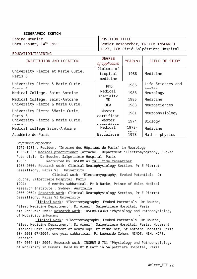
BIOGRAPHIC SKETCHSabine MeunierBorn January 14th 1955
POSITION TITLESenior Researcher, CR ICM INSERM U 1127, ICM Pitié-Salpêtrière Hospital
EDUCATION/TRAINING
INSTITUTION AND LOCATIONDEGREE
(if applicable)YEAR(s) FIELD OF STUDY
University Pierre et Marie Curie, Paris 6Diploma of
tropical medicine
1988 Medicine
University Pierre & Marie Curie, Paris 6 PhD 1986 Life Sciences and health
Medical College, Saint-Antoine Medical specialty
1986 Neurology
Medical College, Saint-Antoine MD 1985 Medicine
University Pierre & Marie Curie, Paris 6 DEA 1983 Neurosciences
University Pierre &Marie Curie, Paris 6Master
certificate1981 Neurophysiology
University Pierre & Marie Curie, Paris 6 Master Certificate
1974 Biology
Medical college Saint-Antoine Medical study 1973-1979
Medicine
Académie de Paris Baccalauréat 1973 Math - physics
Professional experience1979-1985 : Resident (Interne des Hôpitaux de Paris) in Neurology1986-1988: Medical practitioner (attaché), Department “Electromyography, Evoked Potentials” Dr Bouche, Salpetriere Hospital, Paris1988: Recruited by INSERM as full time researcher1988-2000: Research work: Clinical Neurophysiology Section, Pr E Pierrot-Deseilligny, Paris VI University Clinical work: “Electromyography, Evoked Potentials” Dr Bouche, Salpetriere Hospital, Paris1994: 6 months sabbatical, Pr D Burke, Prince of Wales Medical Research Institute”, Sydney, Australia2000-2002: Research work: Clinical Neurophysiology Section, Pr E Pierrot-Deseilligny, Paris VI University
Clinical work: “Electromyography, Evoked Potentials” Dr Bouche, ‘Sleep Medicine Department’, Dr Arnulf; Salpetriere Hospital, Paris01/ 2003-07/ 2003: Research work: INSERM/E0349 “Physiology and Pathophysiology of Motricity inHumans”
Clinical work: “Electromyography, Evoked Potentials” Dr Bouche, ‘Sleep Medicine Department’, Dr Arnulf; Salpetriere Hospital, Paris; Movement Disorder Unit, Department of Neurology, Pr Vidailhet, St Antoine Hospital Paris 08/ 2003-07/2004: one year sabbatical, Pr Leonardo Cohen, NINDS, NIH, HCPS, Bethesda07/ 2004-11/ 2004: Research work: INSERM U 731 “Physiology and Pathophysiology of Motricity in Humans” held by Dr R Katz in Salpetriere Hospital, Paris11/2004-06/2006: on sabbatical, adjunct research investigator in Dr Hallett’s lab, HMCS, NINDS, NIH, Bethesda, MD, USA. 07/2006-12/2009: INSERM U 731 “Physiology and Pathophysiology of Motricity in Humans” held by Dr R Katz in Salpetriere Hospital, Paris.01/2010 to now: CR-ICM, INSERM U1127, CNRS UMR7225, team “"Movement disorders and basal ganglia: pathophysiology and experimental therapeutics" Director Pr M Vidailhet., ICM (Brain and Spinal cord Institute)From 2006 to now: In the frame of the ICM technical platform « Gait, Equilibrium, Posture, Movement, TMS and Navigated Brain Stimulation (NBS) » I am leading the NBS platform. This facility is open to any research team doing clinical or neuroscience research.
MemberSociety for NeurosciencesInternational Laboratory of Neuroimagery and Modelization (France-Canada) (LINeM)
Welter_ETF 16
Visiting professor: Comprehensive Care Centre for Movement Disorder, Sree Chitra Tirunal Institute for Medical Sciences and Technology (SCTIMST), Trivandrum, Kérala, India
Welter_ETF 17
BIOGRAPHICAL SKETCHNAMEKarachi, Carine
POSITION TITLENeurosurgeon (MCU-PH)
eRA COMMONS USER NAME (credential, e.g., agency login)EDUCATION/TRAINING (Begin with baccalaureate or other initial professional education, such as nursing, include postdoctoral training and residency training if applicable.)
INSTITUTION AND LOCATIONDEGREE
(if applicable)
MM/YY FIELD OF STUDY
University of Paris VI, France MD 1997 MedicineUniversity of Paris VI, France Habilitation 2004 NeurosurgeryUniversity of Paris VI, France Ph.D. 2006 NeurobiologyColumbia University Postdoc 2009 Neurosciences
A. Positions1991-1997 Internship, Pitié-Salpetrière Hospital, Paris1997-2004 Neurosurgery Residency, Paris2002-2006 Doctoral (PhD) student: Inserm U679, Paris2009-2010 Post doctoral research fellow: Michael Goldberg, Columbia University, New York, USA2011-present Assistant professor, Neurosurgery, Pitié-Salpetrière Hospital, Paris
B. Honors & Awards1999-2000 French Foundation for Medical Research 2002-2003 French Association for Gilles de la Tourette syndrome2009 Fondation Bettencourt Schueller award for young researchers2009-2010 French Foundation for Medical Research
C. Selected Peer-reviewed PublicationsGrabli D, Karachi C, Folgoas E, Monfort M, Tande D, Clark S, Civelli O, Hirsch EC, Francois C (2013) Gait disorders in parkinsonian monkeys with pedunculopontine nucleus lesions: a tale of two systems. The Journal of neuroscience : the official journal of the Society for Neuroscience 33:11986-11993.Xu BY, Karachi C, Goldberg ME (2012) The postsaccadic unreliability of gain fields renders it unlikely that the motor system can use them to calculate target position in space. Neuron 76:1201-1209.Palminteri S, Justo D, Jauffret C, Pavlicek B, Dauta A, Delmaire C, Czernecki V, Karachi C, Capelle L, Durr A, Pessiglione M (2012) Critical roles for anterior insula and dorsal striatum in punishment-based avoidance learning. Neuron 76:998-1009.*Karachi C, *Andre A, Bertasi E, Bardinet E, Lehericy S, Bernard FA (2012) Functional parcellation of the lateral mesencephalus. The Journal of neuroscience : the official journal of the Society for Neuroscience 32:9396-9401.*Grabli D, *Karachi C, Welter ML, Lau B, Hirsch EC, Vidailhet M, Francois C (2012) Normal and pathological gait: what we learn from Parkinson's disease. Journal of neurology, neurosurgery, and psychiatry 83:979-985.Buot A, Welter ML, Karachi C, Pochon JB, Bardinet E, Yelnik J, Mallet L (2012) Processing of emotional information in the human subthalamic nucleus. Journal of neurology, neurosurgery, and psychiatry.Bardinet E, Belaid H, Grabli D, Welter ML, Vidal SF, Galanaud D, Derrey S, Dormont D, Cornu P, Yelnik J, Karachi C (2011) Thalamic stimulation for tremor: can target determination be improved? Movement disorders : official journal of the Movement Disorder Society 26:307-312.Karachi C, Grabli D, Bernard FA, Tande D, Wattiez N, Belaid H, Bardinet E, Prigent A, Nothacker HP, Hunot S, Hartmann A, Lehericy S, Hirsch EC, Francois C (2010) Cholinergic mesencephalic
Welter_ETF 18

neurons are involved in gait and postural disorders in Parkinson disease. The Journal of clinical investigation 120:2745-2754.
Welter_ETF 19

BIOGRAPHICAL SKETCHNAMELau, Brian
POSITION TITLETenured researcher (chargé de recherché 1st class CNRS)eRA COMMONS USER NAME (credential, e.g., agency
login)EDUCATION/TRAINING (Begin with baccalaureate or other initial professional education, such as nursing, include postdoctoral training and residency training if applicable.)
INSTITUTION AND LOCATIONDEGREE
(if applicable)
MM/YY FIELD OF STUDY
University of California, Berkeley B.A. 1998Molecular & Cell Biology, Neurobiology
New York University Ph.D. 2007 NeuroscienceColumbia University Postdoc 2011 Neuroscience
A. Positions1999-2000 Staff research associate, Department of Molecular & Cell Biology, UC Berkeley, Berkeley, CA2007-2011 Postdoctoral fellow, Department of Neuroscience, Columbia University, New York, NY2012 Postdoctoral fellow, Institute of the Brain and Spine, Paris, France2013-present Junior group leader, Institute of the Brain and Spine , Paris, France
B. Honors & Awards1997 Howard Hughes Medical Institute Biology Fellows award2000-2003 US Department of Defense NDSEG Fellowship2005-2006 Dean’s Dissertation Fellowship, New York University2008-2011 Helen Hay Whitney Postdoctoral Fellowship2012 Mairie de Paris Research Grant2012 Paris Ecole des Neuroscience Postdoctoral Fellowship
C. Selected Peer-reviewed PublicationsC.J. Peck*, B. Lau* and C.D. Salzman (2013). The primate amygdala combines information about space and value. Nature Neurosci, 16(3): 340-48, 2013. *equal contribution, co-first authorshipD. Grabli, C. Karachi, M.-L. Welter, B. Lau, E.C. Hirsch, M. Vidailhet and C. François (2012). Normal and pathological gait: What we learn from Parkinson’s disease. J Neurol, Neurosurg & Psych, 83(10): 979-85, 2012.S.E. Morrison, A. Saez, B. Lau and C.D. Salzman (2011). Different time courses for learning-related changes in amygdala and orbitofrontal cortex. Neuron, 71(6): 1127-1140, 2011.Rutledge, R. B., S. C. Lazzaro, B. Lau, et al. (2009). "Dopaminergic drugs modulate learning rates and perseveration in Parkinson's patients in a dynamic foraging task." J Neurosci 29(48): 15104-15114.Lau, B. and P. W. Glimcher (2008). "Value representations in the primate striatum during matching behavior." Neuron 58(3): 451-463.Lau, B. and P. W. Glimcher (2007). "Action and outcome encoding in the primate caudate nucleus." J Neurosci 27(52): 14502-14514.Bayer, H. M., B. Lau, et al. (2007). "Statistics of midbrain dopamine neuron spike trains in the awake primate." J Neurophysiol 98(3): 1428-1439.Lau, B., G. B. Stanley, et al. (2002). "Computational subunits of visual cortical neurons revealed by artificial neural networks." Proc Natl Acad Sci U S A 99(13): 8974-8979.
Welter_ETF 20

D. Research Support2014- ANRAnatomo-functional characterization of the cortical-subthalamic pathway and its relevance for the treatment of OCD by deep-brain stimulationRole: Co-investigator2013- Carnot InstituteDeveloping low-cost, high-density electrophysiological recording systemsRole: Co-investigator2013-present Fondation de FranceFeedback-based deep brain stimulation for the treatment of Parkinson’s diseaseRole: Co-investigator2013-present ATIP-AvenirThe subthalamic nucleus and motor control: understanding function and dysfunction in Parkinson’s DiseaseRole: PI
Welter_ETF 21
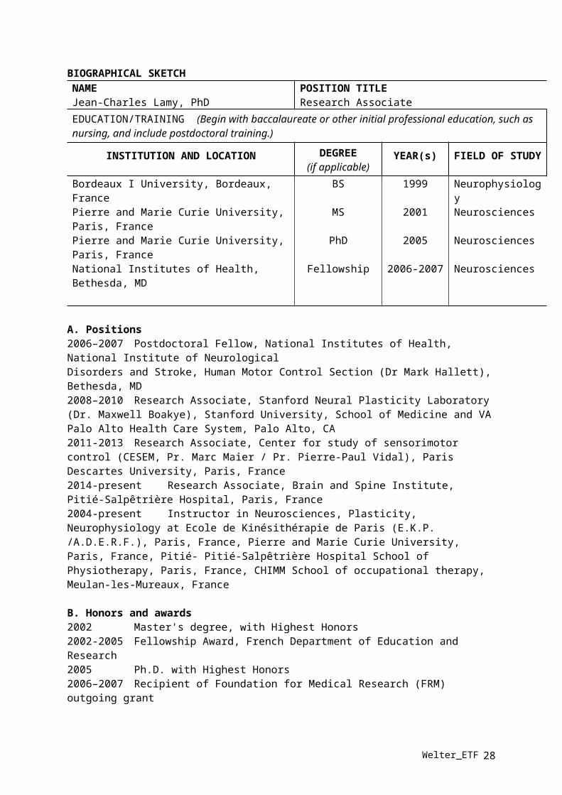
BIOGRAPHICAL SKETCHNAMEJean-Charles Lamy, PhD
POSITION TITLEResearch Associate
EDUCATION/TRAINING (Begin with baccalaureate or other initial professional education, such as nursing, and include postdoctoral training.)
INSTITUTION AND LOCATION DEGREE(if applicable)
YEAR(s) FIELD OF STUDY
Bordeaux I University, Bordeaux, France BS 1999 NeurophysiologyPierre and Marie Curie University, Paris, France MS 2001 NeurosciencesPierre and Marie Curie University, Paris, France PhD 2005 NeurosciencesNational Institutes of Health, Bethesda, MD Fellowship 2006-2007 Neurosciences
A. Positions2006–2007 Postdoctoral Fellow, National Institutes of Health, National Institute of NeurologicalDisorders and Stroke, Human Motor Control Section (Dr Mark Hallett), Bethesda, MD2008–2010 Research Associate, Stanford Neural Plasticity Laboratory (Dr. Maxwell Boakye), Stanford University, School of Medicine and VA Palo Alto Health Care System, Palo Alto, CA2011-2013 Research Associate, Center for study of sensorimotor control (CESEM, Pr. Marc Maier / Pr. Pierre-Paul Vidal), Paris Descartes University, Paris, France2014-present Research Associate, Brain and Spine Institute, Pitié-Salpêtrière Hospital, Paris, France2004-present Instructor in Neurosciences, Plasticity, Neurophysiology at Ecole de Kinésithérapie de Paris (E.K.P. /A.D.E.R.F.), Paris, France, Pierre and Marie Curie University, Paris, France, Pitié- Pitié-Salpêtrière Hospital School of Physiotherapy, Paris, France, CHIMM School of occupational therapy, Meulan-les-Mureaux, France
B. Honors and awards2002 Master's degree, with Highest Honors2002-2005 Fellowship Award, French Department of Education and Research2005 Ph.D. with Highest Honors2006–2007 Recipient of Foundation for Medical Research (FRM) outgoing grant2008 Recipient of France-Stanford Center for Interdisciplinary Studies grant2011 Recipient of Foundation for Medical Research (FRM) ingoing grant2013 Recipient of France-Stanford Center for Interdisciplinary Studies grant
C. 5 selected peer-reviewed publications related to the project (in chronological order) (among 19)1. Lamy, J.C., Boakye, M. «Seeking significance for transcutaneous spinal direct current stimulation» - Clin Neurophysiol (2013), 124(6):1049-50.2. Lamy, J.C., Boakye, M. «BDNF Val66Met polymorphism alters spinal plasticity in humans: a spinal direct current stimulation study» J Neurophysiol (2013), 110(1):109-16. 3. Lamy, J.C., Ho, C., Badel, A., Arrigo, R.T., Boakye, M. «Modulation of Soleus H-reflex by spinal DC stimulation in humans» J Neurophysiol (2012), 108(3):906-144. Raoul, S., Roualdes, V., Déligny, C., Leduc, D., Lamy, J.C., Lackmy, A., Nguyen, J., Damier, P., Katz, R. «Subthalamic nucleus stimulation reverses spinal motoneurone activity in Parkinsonian patients » Brain (2012), 135(1): 139-47.5. Meunier, S., Russmann, H., Shamim, E.A., Lamy, J.C., Hallett, M. «Plasticity of cortical inhibition in dystonia is impaired after motor learning and Paired-Associative Stimulation» Eur J Neurosci (2012), 35(6): 975-86.
C. Research Support.
Welter_ETF 22

Present1. Maier (PI) and Mas (PI)French Foundation for Medical ResearchRole of fluoxetin in the recovery of motor functions after strokeRole: Co-InvestigatorCompleted1. Hallett (PI) 9/1/06 - 8/31/07French Foundation for Medical Research and NIH Intramural ProgramImpaired Motor Learning and LTP/LTD-like Plasticity in Dystonia are Associated with Abnormal Modulation of Cortical Excitability by Somatosensory VolleysRole: Co-Investigator2. Hallett (PI) 9/1/07 – 12/18/07NIH/NINDS Intramural ProgramPlasticity in Cervical DystoniaRole: Co-Investigator3. Boakye (PI) 3/1/08 - 12/31/10Department of Veterans Affairs / Merit ReviewMultimodal Study of Sensorimotor Plasticity after Spinal Cord InjuryRole: Co-Investigator4. Boakye (PI) 3/1/08 – 12/31/10Department of Veterans Affairs / Merit ReviewImpact of BDNF Val66Met in activity-dependent plasticityRole: Co-Investigator5. Maier (PI) 1//1/11 - 12/31/13Institut de Recherche sur la Moelle Epinière (IRME)Corticospinal excitability in cerebral palsyRole: Co-Investigator
Welter_ETF 23

8. Copies of relevant abstracts and/or articles that have been published or submitted
Russmann H, Lamy JC, Shamim EA, Meunier S, Hallett M. Associative plasticity in intracortical inhibitory circuits in human motor cortex. Clin Neurophysiol. 2009 Jun;120(6):1204-12. doi: 10.1016/j.clinph.2009.04.005. Epub 2009 May 10.OBJECTIVE: Paired associative stimulation (PAS) is a transcranial magnetic stimulation technique inducing Hebbian-like synaptic plasticity in the human motor cortex (M1). PAS is produced by repetitive pairing of a peripheral nerve shock and a transcranial magnetic stimulus (TMS). Its effect is assessed by a change in size of a motor evoked response (MEP). MEP size results from excitatory and inhibitory influences exerted on cortical pyramidal cells, but no robust effects on inhibitory networks have been demonstrated so far. METHOD: In 38 healthy volunteers, we assessed whether a PAS intervention influences three intracortical inhibitory circuits: short (SICI) and long (LICI) intracortical inhibitions reflecting activity of GABA(A) and GABA(B) interneurons, respectively, and long afferent inhibition (LAI) reflecting activity of somatosensory inputs. RESULTS: After PAS, MEP sizes, LICI and LAI levels were significantly changed while changes of SICI were inconsistent. The changes in LICI and LAI lasted 45 min after PAS. Their direction depended on the delay between the arrival time of the afferent volley at the cortex and the TMS-induced cortical activation during the PAS. CONCLUSIONS: PAS influences inhibitory circuits in M1. SIGNIFICANCE: PAS paradigms can demonstrate Hebbian-like plasticity at selected inhibitory networks as well as excitatory networks.
Popa T, Russo M, Meunier S. Long-lasting inhibition of cerebellar output. Brain Stimul. 2010 Jul;3(3):161-9. doi: 10.1016/j.brs.2009.10.001. Epub 2009 Oct 31.OBJECTIVE: The cerebellar influence on the motor cortex output is exerted mostly though the cerebellothalamocortical pathway (CTC). One way to explore this pathway is by the means of transcranial magnetic stimulation (TMS). A single-pulse conditioning magnetic stimulation delivered over the lateral cerebellum was shown to diminish the excitability of the contralateral motor cortex 5 milliseconds later (cerebellocortical inhibition [CBI]), most likely through transynaptic activation of cerebellar Purkinje cells, which in turn inhibit the tonic activity of the CTC. Repetitive TMS (rTMS) delivered over the lateral cerebellum was shown to induce a long-lasting change of the cortical excitability, as well, but the mechanism and time course of this effect are still debated. METHODS: We tested the time course of the effects of rTMS on the CBI in five paradigms: (1) 1 Hz rTMS, (2) continuous theta burst stimulation (cTBS), and (3) intermittent TBS (iTBS) over the right cerebellum, (4) 1 Hz rTMS over the cervical nerve roots, and (5) 1 Hz rTMS over the left cerebellum. Surface electromyography was recorded from the right first dorsal interosseous (FDI) and adductor digiti minimi. A double-cone coil was used for single-pulse cerebellar stimulation, whereas a figure-of-eight coil was used for the rTMS. The stimulus intensity was set at 90% of the M1 resting motor threshold for 1 Hz rTMS, and at 80% of the M1 active motor threshold for TBS. Both types of cerebellar stimulation were performed under magnetic resonance image (MRI)-guided neuronavigation centered over the right VIII B lobule, and stimulation intensities were adjusted for cerebellar cortex depth. A figure-of-eight coil was used for left motor cortex stimulation. RESULTS: There was significant CBI suppression to the left motor cortex up to 30 minutes after the 900 stimuli of 1 Hz rTMS over either cerebellar hemisphere, and after 600 stimuli of cTBS over the right cerebellum, but not after 600 stimuli of iTBS over the right cerebellum, or after 900 of 1 Hz rTMS stimuli delivered over the cervical nerve roots. The 1 Hz rTMS over the left cerebellum significantly reduced the CBI in the right FDI 10 minutes after the end of the intervention. The amplitudes of the unconditioned cortical motor-evoked potentials were not significantly changed. CONCLUSIONS: Our findings suggest that repetitive cerebellar stimulation operate at a cerebellar level, rather then at a cortical level.
Welter_ETF 24

Bardinet E, Belaid H, Grabli D, Welter ML, Vidal SF, Galanaud D, Derrey S, Dormont D, Cornu P, Yelnik J, Karachi C. Thalamic stimulation for tremor: can target determination be improved? Mov Disord. 2011 Feb 1;26(2):307-12. doi: 10.1002/mds.23448. Epub 2010 Dec 13.High frequency stimulation of the ventral intermedius nucleus (Vim) of the thalamus is successfully used for the treatment of postural tremor. Target coordinates are most commonly calculated using a statistical method. Here, we compare a statistical and an individual targeting method, using an histology-based three-dimensional deformable brain atlas which allows localization of the Vim on individual patient's MR images by adaptation of the atlas onto the patient's brain. Twenty-nine consecutive patients had electrodes implanted in the Vim uni-or bilaterally for severe essential tremor. Thirty-five targets were determined by calculating the statistical target and then using the deformable atlas to compute the individual target. Pythagorean distance between these targets was calculated. Statistical and individual targets were compared by double blind evaluation of perioperative stimulation effects. For most cases (n = 24), the Pythagorean distance was higher than 1.5 mm. In 79% of these cases, the definitive electrode was implanted using the position of the individual target. For the remaining cases (n = 11, distance < 1.5 mm), the definitive electrode was implanted according to the statistical target location in 73% of the cases. As a whole, when individual target was used, it was located at least 2 mm more medial than the statistical one in 86% cases. These results suggest that Vim target determination based on a statistical method might be inaccurate. In particular, laterality might be overestimated, leading to nonoptimal clinical results. In clinical practice, this means that microelectrode exploration during Vim surgery should include at least one trajectory more medial than the statistical target.
Meunier S, Russmann H, Shamim E, Lamy JC, Hallett M. Plasticity of cortical inhibition in dystonia is impaired after motor learning and paired-associative stimulation. Eur J Neurosci. 2012 Mar;35(6):975-86. doi: 10.1111/j.1460-9568.2012.08034.x.Artificial induction of plasticity by paired associative stimulation (PAS) in healthy volunteers (HV) demonstrates Hebbian-like plasticity in selected inhibitory networks as well as excitatory networks. In a group of 17 patients with focal hand dystonia and a group of 19 HV, we evaluated how PAS and the learning of a simple motor task influence the circuits supporting long-interval intracortical inhibition (LICI, reflecting activity of GABA(B) interneurons) and long-latency afferent inhibition (LAI, reflecting activity of somatosensory inputs to the motor cortex). In HV, PAS and motor learning induced long-term potentiation (LTP)-like plasticity of excitatory networks and a lasting decrease of LAI and LICI in the motor representation of the targeted or trained muscle. The better the motor performance, the larger was the decrease of LAI. Although motor performance in the patient group was similar to that of the control group, LAI did not decrease during the motor learning as it did in the control group. In contrast, LICI was normally modulated. In patients the results after PAS did not match those obtained after motor learning: LAI was paradoxically increased and LICI did not exhibit any change. In the normal situation, decreased excitability in inhibitory circuits after induction of LTP-like plasticity may help to shape the cortical maps according to the new sensorimotor task. In patients, the abnormal or absent modulation of afferent and intracortical long-interval inhibition might indicate maladaptive plasticity that possibly contributes to the difficulty that they have to learn a new sensorimotor task.
Buot A, Welter ML, Karachi C, Pochon JB, Bardinet E, Yelnik J, Mallet L. Processing of emotional information in the human subthalamic nucleus. J Neurol Neurosurg Psychiatry. 2013 Dec;84(12):1331-8. doi: 10.1136/jnnp-2011-302158. Epub 2012 Oct 25.BACKGROUND: The subthalamic nucleus (STN) is an efficient target for treating patients with Parkinson's disease as well as patients with obsessive-compulsive disorder (OCD) using high frequency stimulation (HFS). In both Parkinson's disease and OCD patients, STN-HFS can trigger abnormal behaviours, such as hypomania and impulsivity. METHODS: To investigate if this structure processes emotional information, and whether it depends on motor demands, we
Welter_ETF 25

recorded subthalamic local field potentials in 16 patients with Parkinson's disease using deep brain stimulation electrodes. Recordings were made with and without dopaminergic treatment while patients performed an emotional categorisation paradigm in which the response varied according to stimulus valence (pleasant, unpleasant and neutral) and to the instruction given (motor, non-motor and passive). RESULTS: Pleasant, unpleasant and neutral stimuli evoked an event related potential (ERP). Without dopamine medication, ERP amplitudes were significantly larger for unpleasant compared with neutral pictures, whatever the response triggered by the stimuli; and the magnitude of this effect was maximal in the ventral part of the STN. No significant difference in ERP amplitude was observed for pleasant pictures. With dopamine medication, ERP amplitudes were significantly increased for pleasant compared with neutral pictures whatever the response triggered by the stimuli, while ERP amplitudes to unpleasant pictures were not modified. CONCLUSIONS: These results demonstrate that the ventral part of the STN processes the emotional valence of stimuli independently of the motor context and that dopamine enhances processing of pleasant information. These findings confirm the specific involvement of the STN in emotional processes in human, which may underlie the behavioural changes observed in patients with deep brain stimulation.
Popa T, Russo M, Vidailhet M, Roze E, Lehéricy S, Bonnet C, Apartis E, Legrand AP, Marais L, Meunier S, Gallea C. Cerebellar rTMS stimulation may induce prolonged clinical benefits in essential tremor, and subjacent changes in functional connectivity: an open label trial. Brain Stimul. 2013 Mar;6(2):175-9. doi: 10.1016/j.brs.2012.04.009. Epub 2012 May 12.BACKGROUND: Cerebello-thalamo-cortical (CTC) pathways dysfunction is involved in pathological oscillations causing tremor in essential tremor (ET). Low-frequency (1 Hz) repetitive transcranial magnetic stimulation (rTMS) of the cerebellum can effectively modulate the cerebellar output. OBJECTIVE: As one session of rTMS can induce a brief improvement, we hypothesized that repeated sessions might have a cumulative and potentially long-term therapeutic effect on ET. We assessed, in an open label trial, the efficacy of one-week rTMS treatment on tremor and on the motor-CTC dysfunction in ET patients. METHODS: Resting-state fMRI functional connectivity was used as an indicator of CTC network integrity in 11 ET patients and 11 healthy subjects. Resting-state fMRI connectivity was quantified at baseline in patients and control subjects between the cerebellum and the motor network, and between the cerebellum and the default brain network (DBN) taken as control. The fMRI study was repeated in patients after 5 days of bilateral 1 Hz rTMS applied to the posterior cerebellar cortex. Tremor was assessed clinically (Fahn-Tolosa-Marin scale) and quantified using electromyographic and accelerometric recordings at baseline (day 1, before the cerebellar stimulation) and after the end of the cerebellar stimulation period at day 5, day 12 and day 29. RESULTS: Repeated rTMS over the cerebellum significantly improved total and specific (tremor, drawing, functional disability) scores, and reduced tremor amplitude (P < 0.006). It also re-established the defective information processing in the CTC network (P(Delta|y) > 0.909), but not in the DBN. The effects persisted for 3 weeks after the last session. CONCLUSION: Cerebellar stimulation could be an effective treatment option for patients with severe essential tremor.
Welter ML, Schüpbach M, Czernecki V, Karachi C, Fernandez-Vidal S, Golmard JL, Serra G, Navarro S, Welaratne A, Hartmann A, Mesnage V, Pineau F, Cornu P, Pidoux B, Worbe Y, Zikos P, Grabli D, Galanaud D, Bonnet AM, Belaid H, Dormont D, Vidailhet M, Mallet L, Houeto JL, Bardinet E, Yelnik J, Agid Y. Optimal target localisation of subthalamic stimulation in Parkinson’s disease patients. Neurology, in press. Objectives: To further determine the causes of variable outcome from subthalamic deep brain stimulation (DBS-STN) in Parkinson’s disease (PD) patients.Methods: Data was obtained from our cohort of 309 PD patients operated for DBS-STN between 1996 and 2009. We examined the relationship between the one-year motor, cognitive and psychiatric outcomes and 1) preoperative PD clinical features, 2) MRI parameters, 3) surgical procedure, 4) therapeutic contacts locations.
Welter_ETF 26

Results: Pre- and post-operative results were obtained in 262 PD patients. The best motor outcome was obtained when stimulating contacts were located within the STN as compared to the zona incerta (64% versus 49% improvement). Eighteen percent of the patients presented a postoperative cognitive decline which was found to be principally related to the surgical procedure. Other factors predictive of poor cognitive outcome were perioperative confusion and psychosis. Nineteen patients showed a stimulation-induced hypomania which was related to both the form of the disease (younger age, shorter disease duration, higher levodopa responsiveness) and ventral contact location. Postoperative depression was more frequent in patients already showing preoperative depressive and/or residual axial motor symptoms. Conclusion: In this homogenous cohort of PD patients, we showed that 1) the STN is the best target to improve motor symptoms, 2) postoperative cognitive deficit is mainly related to the surgery itself, 3) stimulation-induced hypomania is related to a combination of both the disease characteristics and a more ventral STN location.
Welter_ETF 27
9. Completed conflict of interest questionnaire
CONFLICT OF INTEREST QUESTIONNAIRE
Grant Proposal Submissions
Pursuant to the purposes and interests of the policy adopted by the IETF Board of Directors requiring disclosure of certain interests, a copy of which has been furnished to me, I hereby state that I or relatives have the following affiliations or interests and have taken part in the following transactions which, when considered in conjunction with my relationship to the International Essential Tremor Foundation (IETF) might create or be a conflict of interest. (Write “none” where applicable).
1. Advisory Board or Panel AffiliationNone
2. Consulting, Speakers Bureau or Contractual ServicesConsulting fees from TEVA-Lundbeck and Medtronic
3. Research/Grant SupportResearch grants from the ICM Foundation, ANR and the patient’s association ‘France Parkinson’
4. Financial or Material Support not otherwise listed.None
I hereby acknowledge the information given is complete and accurate to the best of my knowledge and belief. I understand that failure to accurately disclose a potential interest may cause revocation of my grant award if approved. I also understand that if any of the above circumstances change, that I am to complete a new questionnaire.
WELTER Marie-Laure, MD, PhD
Signature Date 28/02/2014
PO Box 14005 | Lenexa, Kansas 66285-4005 | USA | 888.387.3667 (toll free) | 913.341.3880 (local) | essentialtremor.org ©2007 IETF, GrantConflict5222013
Welter_ETF 28