eprints.whiterose.ac.ukeprints.whiterose.ac.uk/...of_the...resubmitted.docxweb viewexploring the...
TRANSCRIPT

Exploring the role of the posterior middle temporal gyrus in semantic cognition: Integration
of anterior temporal lobe with executive processes
James Davey1, Hannah E. Thompson1, Glyn Hallam1, Theodoros Karapanagiotidis1, Charlotte
Murphy1, Irene De Caso1, Katya Krieger-Redwood1, Boris C. Bernhardt2, Jonathan Smallwood1,
Elizabeth Jefferies1*
1Deparment of Psychology and York Neuroimaging Centre, University of York, UK
2McConnell Brain Imaging Centre, Montreal Neurological Institute and Hospital,
McGill University, Montreal, QC, H3A 2B4, Canada
*Corresponding author
Email: [email protected]
Postal address: Professor Beth Jefferies,
Department of Psychology,
University of York,
York,
YO10 5DD
United Kingdom
+44 1904 324368
1

Abstract
Making sense of the world around us depends upon selectively retrieving information
relevant to our current goal or context. However, it is unclear whether selective semantic
retrieval relies exclusively on general control mechanisms recruited in demanding non-
semantic tasks, or instead on systems specialised for the control of meaning. One
hypothesis is that the left posterior middle temporal gyrus (pMTG) is important in the
controlled retrieval of semantic (not non-semantic) information; however this view remains
controversial since a parallel literature links this site to event and relational semantics. In a
functional neuroimaging study, we demonstrated that an area of pMTG implicated in
semantic control by a recent meta-analysis was activated in a conjunction of (i) semantic
association over size judgements and (ii) action over colour feature matching. Under these
circumstances the same region showed functional coupling with the inferior frontal gyrus –
another crucial site for semantic control. Structural and functional connectivity analysis
demonstrated that this site is at the nexus of networks recruited in automatic semantic
processing (the default mode network) and executively demanding tasks (the multiple-
demand network). Moreover, in both task and task-free contexts, pMTG exhibited
functional properties that were more similar to ventral parts of inferior frontal cortex,
implicated in controlled semantic retrieval, than more dorsal inferior frontal sulcus,
implicated in domain-general control. Finally, the pMTG region was functionally correlated
at rest with other regions implicated in control-demanding semantic tasks, including inferior
frontal gyrus and intraparietal sulcus. We suggest that pMTG may play a crucial role within a
large-scale network that allows the integration of automatic retrieval in the default mode
network with executively-demanding goal-oriented cognition, and that this could support
our ability to understand actions and non-dominant semantic associations, allowing
semantic retrieval to be ‘shaped’ to suit a task or context.
Keywords: Semantic control, memory retrieval, default mode network, multi demand
network, posterior middle temporal gyrus.
2

Introduction
Across our lifetime we acquire a large body of conceptual knowledge, only a subset of which
is relevant for any given task or context; thus automatic spreading activation within
semantic representations is often insufficient for efficient semantic cognition (Thompson-
Schill et al., 1997; Badre et al., 2005; Jefferies, 2013). Automatic spreading activation can
facilitate the retrieval of features and associations that are dominant for a particular
concept (e.g., carrot-peel). When semantic retrieval needs to be focussed on aspects of
knowledge that are not the strongest response for the inputs, additional control
mechanisms can be engaged to guide semantic retrieval. For example, control is needed to
recover weak associations (carrot-horse) and to match words on the basis of specific
sensory-motor features, such as actions or colour (e.g., carrot with traffic cone), since the
functional characteristics of these concepts are more central to their meaning (Thompson-
Schill et al., 1997; Badre et al., 2005; Whitney et al., 2011; Noonan et al., 2013; Davey et al.,
2015a).
Different brain regions have been implicated in the representation and controlled
retrieval of semantic information. The ventral anterior temporal lobes (ATL) have been
argued to form a key repository of conceptual information, following studies of patients
with semantic dementia (SD). These patients have relatively focal bilateral atrophy focussed
on ATL, associated with a gradual deterioration of knowledge and multimodal semantic
deficits, first affecting fine-grained distinctions between concepts, and then eroding more
basic distinctions (Mummery et al., 2000; Hodges and Patterson, 2007; Patterson et al.,
2007). Deficits in SD patients suggest a loss of central semantic information (Bozeat et al.,
2000; Jefferies and Lambon Ralph, 2006) and studies employing inhibitory transcranial
magnetic stimulation (TMS) in healthy participants have provided converging evidence for a
necessary role of this region in comprehension (Pobric et al., 2007; Pobric et al., 2010).
Functional magnetic resonance imaging (fMRI) studies reveal activation of ATL during
diverse semantic judgements (Binder et al., 2009; Visser et al., 2010; Rice et al., 2015).
Finally, analyses of inter-regional signal correlations during task free (i.e. resting-state)
functional scans have shown that ATL is part of a large scale assembly that includes medial
prefrontal and posterior cingulate cortices, commonly referred to as the default mode
3

network (DMN, Raichle et al., 2001; Buckner et al., 2008; Yeo et al., 2011; Jackson et al.,
2016).
Converging neuroscientific methods have also identified brain regions beyond ATL
which are important for multimodal semantics, specifically left inferior frontal gyrus (LIFG)
and posterior middle temporal gyrus (pMTG). These regions are thought to contribute to the
control of semantic retrieval. Patients with semantic aphasia (SA), who have lesions affecting
these regions following stroke, fail the same range of verbal and non-verbal semantic tasks
as SD patients; however, unlike SD cases, they often retrieve information that is irrelevant or
inappropriate for the task, show strong effects of cues and miscues, and perform poorly in
the face of strong distracters or ambiguous meanings (Thompson-Schill et al., 2002; Jefferies
and Lambon Ralph, 2006; Jefferies et al., 2008; Corbett et al., 2009; Jefferies et al., 2010) .
Converging evidence from fMRI (Poldrack et al., 1999; Badre et al., 2005; Snijders et al.,
2010; Noonan et al., 2013; Davey et al., 2015b) and TMS (Hoffman et al., 2010; Whitney et
al., 2011; Davey et al., 2015a) supports the view that both of these regions contribute to
semantic control. Indeed, in a recent neuroimaging meta-analysis, LIFG and pMTG were the
sites activated most strongly and consistently across many different contrasts designed to
tap semantic control (Noonan et al., 2013). In addition, when high-control semantic tasks
were contrasted with demanding phonological judgements, pMTG and the anterior part of
LIFG showed a specifically semantic response, suggesting that these two regions lie outside
of the multiple-demand network (MDN), which is recruited during executively-demanding
tasks across domains (Duncan, 2010).
These findings therefore provide some evidence that semantic cognition may be
underpinned by at least three component processes, supported by distinct brain networks.
(1) Domain-general executive control implemented by the MDN (Duncan, 2010) and the
fronto-parietal control system (Power and Petersen, 2013) may support the capacity to
engage and sustain a particular type of semantic retrieval in line with the task instructions,
as well as the application of top-down constraints to support goal-driven aspects of
cognition beyond semantics (Duncan and Owen, 2000; Duncan, 2010; Fedorenko et al.,
2013; Noonan et al., 2013). For example, in a feature-matching task (in which globally
unrelated words must be linked together on the basis that they both have a particular
feature specified in the task instructions), there is a need to apply a pre-specified goal
4

during semantic retrieval, and the implementation of this goal may involve the executive
system. (2) Activation is thought to spread automatically between highly-related concepts
within the representational system (underpinning semantic priming effects for strong
associates). This allows dominant features and associations to be retrieved in the absence of
executive control, and is supported by ATL and potentially other regions in the DMN (Wirth
et al., 2011; Lau et al., 2013; Power and Petersen, 2013; Jackson et al., 2016) . (3) A third
network might support situations in which there is no explicit goal to indicate which aspect
of knowledge should be brought to the fore, but the pattern of retrieval that is required for
success is not the dominant one given the stimuli – i.e., semantic retrieval must be
controlled to identify and sustain a linking context. The retrieval of relatively weak global
associations is a good example of such a task: here, the instructions do not establish which
types of associations or features should be the focus for retrieval – instead, it is necessary to
establish a linking context from the concepts themselves and retrieve features relevant to
this context.
Figure 1 illustrates the spatial distribution for these three putative networks (MDN,
DMN, and semantic control) from prior published investigations. This figure shows that
regions implicated in semantic control by the meta-analysis of Noonan et al. (2013, in green)
are only partially overlapping with the MDN (from Fedorenko et al., 2013, in red). Non-
overlapping areas in LIFG and pMTG appear to be important for demanding semantic tasks
(relative to easier semantic judgements) but not executive control across domains.
Moreover, these semantic control regions are spatially intermediate between the MDN
(implicated in executive control) and the DMN (implicated in automatic retrieval, from Yeo
et al., 2011, in blue); this location could allow semantic control regions to integrate two
distributed networks that are anti-correlated at rest and yet both crucial for semantic
cognition, e.g., when semantic knowledge, not a task goal, defines the attentional focus.
5

Figure 1. Spatial maps of the Default Mode Network (DMN, blue, from Yeo et al., 2011), Multiple-Demand
Network (MDN, red, from Fedorenko et al., 2013) and Semantic Control Network (green, from Noonan et al.,
2013), presented on a rendered MNI-152 brain and on axial, coronal, and sagittal slices. The key for
overlapping areas between different networks is presented on the right hand side of the figure. Images are
shown with fully saturated colours to maximise the visibility of the overlapping regions. Regions implicated in
semantic control and also found in the MDN include dlPFC (dorsolateral prefrontal cortex), dIFG (dorsal inferior
frontal gyrus), pre-SMA (pre-supplementary motor area), IPS (intraparietal sulcus) and LOC (lateral occipital
cortex). Regions implicated in semantic control and also found in the DMN include vIFG (ventral inferior frontal
gyrus); vMPFC (ventral medial prefrontal cortex) and pMTG (posterior middle temporal gyrus).
The proposal that the control of semantic retrieval is partially distinct from executive
control is broadly consistent with functional dissociations that have been identified within
left inferior frontal cortex. Within the language domain, studies have reported a functional
gradient in left inferior frontal gyrus (IFG), with ventral anterior aspects of IFG implicated in
semantic control specifically, and dorsal posterior IFG contributing more broadly to
language control, including phonological tasks (Poldrack et al., 1999; Wagner et al., 2001a;
Wagner et al., 2001b; Devlin et al., 2003; Gough et al., 2005; Snyder et al., 2007). Dorsal IFG,
bordering inferior frontal sulcus (IFS), is recruited when participants select specific aspects
of knowledge in line with an externally-specified goal (i.e., instructions to match words by
6

colour or shape in the absence of a global semantic relationship; Badre et al., 2005). This
selection process may be important for many language tasks, such as lexical and
phonological retrieval. In contrast, ventral/anterior IFG shows an increased response when
weak and strong semantic associations are contrasted (e.g., salt-grain > salt-pepper) – i.e.,
when participants shape retrieval to converge on a distant link between two concepts in the
absence of an explicit goal. This ability to recover a non-dominant conceptual link does not
generalise easily to other aspects of language processing. Recent work using single-subject
analyses identified regions within the multiple-demand network, in dorsal and posterior
IFG/IFS, that respond to difficult verbal working memory judgements involving non-words
(Fedorenko et al., 2013): these regions are adjacent to, but spatially distinct from, areas of
IFG that show a greater response to easier meaning-based trials involving words in
sentences (Fedorenko et al., 2012; Blank et al., 2014). Moreover, analyses of resting-state
connectivity have implicated anterior aspects of prefrontal cortex in a cingulo-opercular
control system, which includes regions that display sustained activity during task-set
maintenance, whilst dorsal prefrontal regions couple with a fronto-parietal system engaged
by ongoing selection and implementation (Power and Petersen, 2013): this pattern may
relate to the functional distinction between anterior and dorsal LIFG. Thus, a more semantic
response in anterior/ventral parts of IFG may be broadly in line with the proposal that
anterior areas in IFG establish and maintain priorities for what is to be retrieved, while the
short-term process of selection itself is implemented in posterior regions of IFG (Badre and
D'Esposito, 2007). Badre and colleagues referred to this functional specialisation within IFG
as “controlled retrieval” and “selection” respectively (Badre et al., 2005).
The functional contribution of IFG has been considered in detail while the
significance of the second region identified by Noonan and colleagues, pMTG, remains
controversial. Although this site is implicated in semantic control, a parallel literature links
pMTG, together with angular gyrus (AG), to the comprehension of actions and events
(Johnson-Frey et al., 2005; Liljeström et al., 2008), and to relational semantics (Humphreys
and Lambon Ralph, 2014; Price et al., 2015), and these adjacent areas of temporoparietal
cortex can show a similar response to contrasts tapping event knowledge (Wagner et al.,
2005; Sachs et al., 2008; Kim, 2011). One theoretical account suggests that AG and/or pMTG
provide a “thematic hub”, capturing aspects of knowledge relating to the associations
7

between concepts – such as knowledge about which concepts are found and used together
(Schwartz et al., 2011). However, while pMTG often shows increased activation in harder
semantic tasks (Noonan et al., 2013), mid-AG typically shows deactivation in semantic and
other tasks relative to rest, especially for harder judgements (Binder et al., 2008;
Humphreys and Lambon Ralph, 2014; Humphreys et al., 2015). Moreover, these sites
showed a double dissociation in a recent TMS study (Davey et al., 2015a): inhibitory
stimulation of pMTG disrupted weak more than strong associations, while TMS to AG
showed the opposite pattern. These data are not easily reconciled with a simple “thematic
hub” account and suggest instead that pMTG and AG support different components of
semantic cognition, although ones that can at times function in a cooperative manner.
The current study focussed on understanding the functional contribution of pMTG to
semantic control and event/relational semantics: we explored the hypothesis that this
region acts to integrate information from the MDN and also the DMN, which are anti-
correlated at rest. First, we identified whether there are regions of pMTG that show a
common response to the retrieval of action features (contrasted with colour feature
judgements which have not been historically linked to pMTG; Badre et al., 2005) and global
associations (relative to feature judgements). Both of these event/relational contrasts
depend on retrieving information in line with a specific stimulus-driven context, as opposed
to the application of a specific goal specified in the instructions. We compared the location
of this response to regions implicated in semantic control and domain-general control in
previous meta-analyses and also used psychophysiological interaction (PPI) analysis to
understand the functional coupling of this region under these conditions. Second, to explore
whether pMTG could integrate executive and automatic mechanisms contributing to
semantic retrieval, we used resting state fMRI and diffusion MRI tractography to examine if
the spatial networks that corresponded to peaks in our easy and hard semantic decisions
(i.e., the contrasts of relatively global associations over the harder feature selection and vice
versa), converged on the region of cortex identified as important in event/relational
semantics. Third, we considered whether the response in pMTG could be linked to the
proposed functional gradient in IFG (Badre et al., 2005) by examining whether
ventral/anterior as opposed to dorsal/posterior IFG had greater resting state connectivity
with this region.
8

Method
Participants
This research was approved by the ethics committee of the York Neuroimaging
Centre, University of York, UK. All participants were right-handed, native English speaking,
with normal or corrected-to-normal vision. For the experimental task, we recruited 22
neurologically healthy participants from the University of York (Cohort 1). Two participants
were removed due to movement artefacts during fMRI data acquisition (Mean age = 24.8,
SD = 3.8, range 21 – 35 years, 9 males). Resting-state scans were collected at York for two
cohorts (Mean Age = 21.3, SD = 2.7, Range 18 – 31 years, 38 males); 39 participants were
recruited into Cohort 2, with 48 recruited into Cohort 3. These two samples were collected
as part of different projects; however the resting-state scan was collected before task-based
scans in both cases and the data sets are combined in the analysis below. For Cohort 3, we
also obtained diffusion MRI. Finally, we also used two publicly-available data sets to provide
independent confirmation of the resting-state connectivity patterns observed in this study:
(i) 141 participants from the Nathan Kline Institute (NKI)/Rockland Enhanced Sample
(Nooner et al., 2012) were used to relate the connectivity patterns of ventral/anterior and
dorsal/posterior IFG to the response we observed in pMTG. Full details of this sample can be
found in Gorgolewski et al. (2014). (ii) Data from Yeo, Krienen and colleagues (2011),
implemented in Neurosynth (Yarkoni, 2011), was used in a final step to investigate the
functional connectivity of the pMTG site we obtained across different analyses. We
compared the functional behaviour of the pMTG across these different cohorts to minimise
the duration of specific testing sessions and to allow us to capitalize on the power of large
scale publicly available data sets.
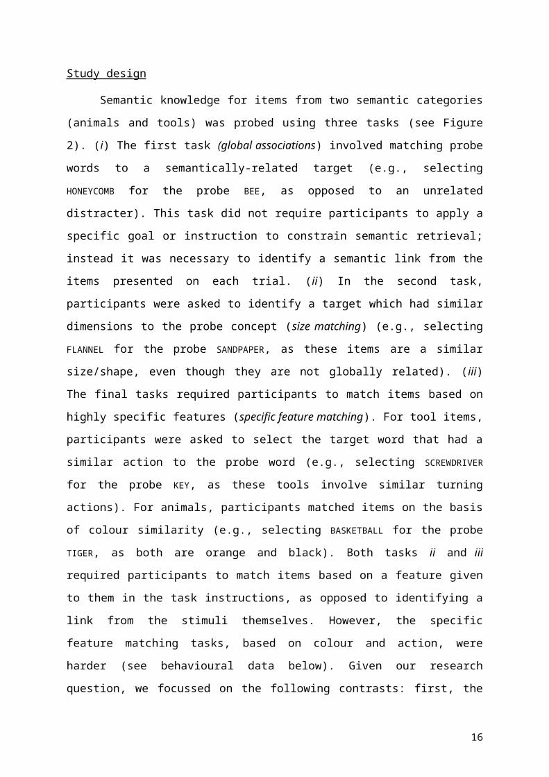
Study design
Semantic knowledge for items from two semantic categories (animals and tools) was
probed using three tasks (see Figure 2). (i) The first task (global associations) involved
matching probe words to a semantically-related target (e.g., selecting HONEYCOMB for the
probe BEE, as opposed to an unrelated distracter). This task did not require participants to
apply a specific goal or instruction to constrain semantic retrieval; instead it was necessary
9

to identify a semantic link from the items presented on each trial. (ii) In the second task,
participants were asked to identify a target which had similar dimensions to the probe
concept (size matching) (e.g., selecting FLANNEL for the probe SANDPAPER, as these items are a
similar size/shape, even though they are not globally related). (iii) The final tasks required
participants to match items based on highly specific features (specific feature matching). For
tool items, participants were asked to select the target word that had a similar action to the
probe word (e.g., selecting SCREWDRIVER for the probe KEY, as these tools involve similar
turning actions). For animals, participants matched items on the basis of colour similarity
(e.g., selecting BASKETBALL for the probe TIGER, as both are orange and black). Both tasks ii and
iii required participants to match items based on a feature given to them in the task
instructions, as opposed to identifying a link from the stimuli themselves. However, the
specific feature matching tasks, based on colour and action, were harder (see behavioural
data below). Given our research question, we focussed on the following contrasts: first, the
conjunction of action > colour features (localising voxels that respond to action
understanding) and global associations > size features (localising voxels that respond to
relational judgements), expected to converge in pMTG. By looking at the conjunction of
these two sets of contrasts, we can rule out task difficulty as a confound in our localisation
of the pMTG (since the global association task was easier than size matching). Secondly, we
contrasted the hardest feature selection tasks (action and colour matching) with easier
global associations, to identify brain regions responding differentially to control demands
(and the reverse contrast for more automatic spreading activation).
The experiment was organised into a total of 36 blocks divided equally among the 6
experimental conditions (i.e., the 3 tasks probed using 2 categories). There were 5 trials per
block resulting in 30 trials per experimental condition. Before each block commenced, an
instruction slide was presented stating the task to be performed (global, size, action, or
colour) for 1000ms. A reminder of the instructions was also present on each trial in
parentheses under the probe word. A two-alternative forced choice paradigm (Figure 2) was
used; participants were instructed to match the centrally presented probe word to one of
two potential targets. Probe words were presented for 1000ms, followed by the response
options which remained on screen until a response was recorded via button press, with
maximum trial duration set to 4500ms. The inter-trial interval was 4000-6000ms, with 10
10

seconds of rest between each experimental block. One null event was present in each
experimental block to increase the amount of rest which was used as a baseline in the
analysis of the fMRI data; the screen was blank for 4500ms plus jitter (4000-6000ms) with
the location of the null event randomised in each experimental block. Before the fMRI
experiment, participants were given a practice session consisting of two blocks for each
condition. The task was presented in the scanner using NBS Presentation version 16
(Neurobehavioral Systems inc., 2013). Participants viewed the words via a front silvered
mirror and responded using a Lumina Response Pad (Cedrus Corporation), placed in their
left hand.
Figure 2. Example trial structure for all conditions in the task-based fMRI study. The study employed a 2x3
design, with three types of judgements (about global semantic associations, size feature matching and specific
feature matching) for animal and tool concepts.
11

Stimuli
A copy of the stimuli used in this experiment is provided using the Open Science
Framework (OSF, https://osf.io/); https://osf.io/5pq8z/ . All words used in the experiment
were concrete nouns denoting manipulable objects or animals. There were 30 animal
probes and 30 man-made probes, repeated across the three tasks (global association, size
matching and specific-feature matching), each with a unique target word for each task. The
distracters in all conditions were target items from other trials that did not have overlapping
features or a global association with the probe. No restrictions were placed on the number
of times a word could be used (mean number of repetitions = 2, SD = 1.4, range = 6);
however the number of repetitions was equivalent between conditions. For the words in
each trial (probe, target, and distracter) we collected measures of familiarity, imageability,
manipulability, lexical frequency, word length, and number of words, averaging across all
the words in a single trial, and compared trials across relevant conditions. Ratings of
familiarity and imageability were taken from the MRC psycholinguistic database (Wilson,
1987). Ratings of manipulability, familiarity, and imageability were also collected on a 7
point scale (1 – low, 7 – high) from a separate cohort of 11 healthy adult participants who
did not take part in the scanning sessions (familiarity and imageability ratings were collected
for targets with missing values in existing databases). Lexical frequency was taken from the
SUBLEX-UK database (van Heuven et al., 2014). Table 1 contains the psycholinguistic
variables for the experimental conditions. Trials were matched across conditions for word
length (action vs. colour: t(58) = 1.1, p = .276; global vs. size:, t(58) = 1.89, p = .076 ; global
vs. feature: t(58) = 1.46, p = .179). They were also matched for number of letters (action vs.
colour: t(58) = .14, p = .886; global vs. size: t(58) = .23, p = .818; global vs. feature: t(58)
= .75, p = .449), and lexical frequency (action vs. colour: t(58)= 1.48, p = .114; global vs. size:
t(58)= 1.13, p = .261; global vs. feature: t(58)= 1.43, p = .156). Manipulability ratings were
higher for action than colour trials as expected (t(58) = 9.47, p <.001). Imageability was also
higher for colour than action trials (t(58) = 7.61, p <.001). No significant differences were
observed for manipulability and imageability for global vs. size (manipulability: t(58) = .027,
p = .979; imageability: t(58)= 1.55, p = .123), or for global vs. feature (manipulability:
t(58)= .50, p = .616; imageability: t(58)= .322, p = .748). Finally, a different set of 13
participants rated the extent to which they found it necessary to generate a spatiotemporal
12

context to complete the specific feature matching conditions (e.g., colour and action
judgements), on a seven point scale (1 – not very useful, 7 – retrieving the context was very
helpful). Retrieval of a spatiotemporal context was significantly more important for tool
action trials (M = 4.13, SD = .50) than animal colour trials (M = 2.49, SD = .43; t(28) = 13.87 p
<.001).
13

Table 1. Psycholinguistic variables for the stimuli used in the task-based fMRI study
Word length Number of words Manipulability Lexical frequency Familiarity Imageability
Condition Mean S.D. Mean S.D. Mean S.D. Mean S.D. Mean S.D. Mean S.D.
Global association 6.92 1.71 1.23 .26 4.77 .61 3.98 .39 5.74 .57 5.96 .51
Size feature 7.45 1.52 1.24 .27 4.77 .53 3.89 .43 5.88 .55 6.09 .42
Specific feature: action 7.13 1.64 1.27 .37 4.35 .44 3.94 .06 5.93 .46 6.30 .24
Specific feature: colour 7.63 1.87 1.28 .22 5.29 .33 3.81 .37 5.75 .49 5.67 .39
14

MRI acquisition
Structural and functional data were acquired using a 3T GE HDx Excite MRI scanner
utilising an eight-channel phased array head coil (GE) tuned to 127.4 MHz, at the York
Neuroimaging Centre, University of York. Structural MRI acquisition in all participants was
based on a T1-weighted 3D fast spoiled gradient echo sequence (TR = 7.8 ms, TE = minimum
full, flip angle 20°, matrix size = 256 x 256, 176 slices, voxel size = 1.13 x 1.13 x 1 mm 3). Task-
based and resting-state activity was recorded from the whole brain using single-shot 2D
gradient-echo echo planar imaging (EPI) with a flip angle = 90°, matrix size = 64 x 64, and
field of view (FOV) = 192 x 192 mm2. Other scan parameters slightly varied for task-based
fMRI in Cohort 1 (TR = 2000 ms, TE = 30 ms, 32 slices, voxel size = 3 x 3 x 4.5 mm 3, 12 min),
resting-state fMRI for Cohort 2 (TR = 2000 ms, TE = minimum full, 32 slices with 0.5 mm gap,
voxel size = 3 x 3 x 3 mm3, 7 min) and resting-state fMRI for Cohort 3 (TR = 3000 ms, TE =
minimum full, 60 slices, voxel size = 3 x 3 x 3 mm3, 9 min). An intermediary FLAIR scan with
the same orientation as the functional scans was collected to improve the co-registration
between subject-specific structural and functional scans. In Cohort 3, we also collected
diffusion weighted MRI data using a 2D single-shot pulsed gradient spin-echo EPI sequence
(TR = 15000ms, TE = 86ms, matrix = 96 x 96, 59 slices, voxel size = 2 x 2 x 2 mm 3; b = 1000
s/mm2, 45 diffusion directions, 7 B0 volumes, 13 minutes). Parameters of the independent
(NKI)/Rockland Enhanced Sample are described in detail by Gorgolewski et al. (2014) and
Smallwood et al. (2016).
Data pre-processing and analysis
a) Task-based fMRI. Analyses were conducted at the first and higher level using FSL-
FEAT version 4.1.9 (Smith et al., 2004; Woolrich et al., 2009; Jenkinson et al., 2012). Pre-
processing included slice timing correction, linear motion correction (Jenkinson et al., 2002),
high-pass temporal filtering (sigma = 100s), brain extraction (Smith, 2002), linear co-
registration to the corresponding T1-weighted image followed by linear co-registration to
MNI152 standard space (Jenkinson and Smith, 2001), spatial smoothing using a Gaussian
kernel with full-width-half-maximum (FWHM) of 5mm and grand-mean intensity
normalisation of the entire 4D dataset by a single multiplicative factor.
15
Pre-processed time series data were modelled using a general linear model
correcting for local autocorrelation (Woolrich et al., 2001) using a block design. The linear
model included the six experimental conditions modelling block start time and block
duration. fMRI scanning was split into two separate scanner runs collected sequentially;
both runs were analysed independently at the lower level then combined using a fixed-
effects higher-level analysis. Six contrasts were defined; individual conditions > rest
(animal/tool global, animal/tool size, tool action, animal colour). We focussed our
subsequent analysis on the comparison of easy global associations vs. harder specific
feature selection (building on the approach of Badre et al., 2005) and on a conjunction of
action > colour and global associations > size to identify regions engaged by event/relational
semantics (Nichols et al., 2005). All analyses were cluster corrected using a z-statistic
threshold of 2.3 to define contiguous clusters. Multiple comparisons were controlled using
Gaussian Random Field Theory at a threshold of p <.05.
b) Psychophysiological Interaction (PPI). A conjunction analysis of action > colour and global
> size revealed an area of pMTG that responded to event semantics. We used this pMTG
region as a mask and extracted the time-course (for each participant and each run) within
this area to examine psychophysiological interactions (PPI; O'Reilly et al., 2012) between the
pMTG and other brain regions involved in event semantics. The extracted time-course of
pMTG and the interaction were included in a GLM model as explanatory variables (at the
lower level, for each participant and each task individually, for each run). As with the
functional analysis, the two runs were combined, and the results were submitted to a group
level analysis, with the same cluster-forming threshold and significance level (Z = 2.3, p
< .05). The contrasts included in this analysis were action > colour and global > size as before
– and, as with the functional data, a formal conjunction of these contrasts was conducted.
c) Resting-state fMRI. Pre-processing steps were as for task fMRI, apart from the
addition of Gaussian low pass temporal filtering, with sigma = 2.8s, and spatial smoothing
using a Gaussian kernel with full-width-half-maximum (FWHM) of 6mm. We extracted the
time series from 3mm spheres placed at regions of interest (ROIs, see below) and used
these as explanatory variables in connectivity analyses at the single subject level. In each
analysis, we entered 11 nuisance regressors; the top five principal components extracted
from white matter (WM) and cerebrospinal fluid (CSF) masks based on the CompCor
16

method (Behzadi et al., 2007) and six head motion parameters. WM and CSF masks were
generated from each individual's high resolution structural image (Zhang et al., 2001). No
global signal regression was performed, following the method implemented in Murphy et al.
(2009). At the group-level, analyses were carried out using FMRIB's Local Analysis of Mixed
Effects (FLAME1), the same cluster correction method used for the functional fMRI was used
at the group level.
d) Diffusion MRI. Subject-wise diffusion MRI processing was carried out in native
diffusion space using FSL (version 4.1.9). Pre-processing of the DTI data involved eddy-
current distortion correction and motion correction using FDT v2.0 (part of FSL), as well as
brain extraction using BET. A probabilistic diffusion model was then fitted on the corrected
data using BEDPOSTX: the Bayesian estimation of diffusion parameters obtained using
sampling techniques toolbox (Behrens et al., 2003). BEDPOSTX uses Monte Carlo Markov
chain sampling to generate parameters for probabilistic tractography. Up to 2 fibres were
modelled per voxel using a burn-in of 1000 iterations before starting the sampling of
diffusion parameters. Next, probabilistic tractography was performed to reconstruct fibres
passing through our seed masks using PROBTRACKX. This technique repeatedly samples
from the diffusion parameters calculated in BEDPOSTX to build a distribution of the likely
tracts from each seed region. The seed masks were transformed from MNI standard space
to diffusion space using nonlinear registration. 5000 sample tracts were generated per seed
voxel. We used the standard parameters of a curvature threshold of 0.2 (corresponding to a
minimum angle of approximately ±80 degrees), a step length of 0.5mm and a maximum
number of steps of 2000. No waypoint or termination masks were included. The resulting
individual maps were transformed back to MNI standard space, thresholded at 0.02% of
total samples sent from the mask and concatenated into a single 4D file. Nonparametric
voxelwise statistical testing was performed using FSL Randomize with 25000 permutations
in order to get a group tractography map (Nichols and Holmes, 2002). The resulting maps
were thresholded at p < 0.01, Family-Wise Error (FWE) corrected, using the Threshold-Free
Cluster Enhancement (TFCE) technique (Smith and Nichols, 2009).
17

Selection of seeds and ROIs
For the psychophysiological interaction (PPI) analysis we used the cluster generated
by the conjunction of action > colour and global > size mask. For the resting state analysis
we used two functional peaks from our task-based fMRI analyses (data from Cohort 1) as
seeds in the analyses of resting state connectivity and diffusion MRI (data from Cohorts 2
and 3): one functional peak was linked to relatively automatic semantic retrieval (easy global
associations > hard feature selection, in inferior ATL, MNI co-ordinates -48 2 -38) and one
was linked to executive control (hard feature selection > easy global associations, in IFS, MNI
co-ordinates -42 28 16). In a further resting-state connectivity analysis (using the NKI data),
we placed seeds at peaks responding to different aspects of semantic control taken from
Badre et al. (2005), allowing us to link the response in pMTG to the previously-reported
functional distinction between “selection” in dorsal/posterior IFG (MNI -48 18 18) and
“controlled retrieval” in ventral/anterior IFG (MNI -51 27 3).
We also performed an ROI analysis of the task-based fMRI data using 8mm spheres,
focused on these ventral and dorsal LIFG peaks from Badre et al. (2005) and the pMTG peak
for semantic control taken from the Noonan et al. (2013) meta-analysis (-58 -49 -8). The
FEATquery tool in FSL was used to extract average percentage signal change across all the
voxels in each ROI for all six conditions across participants.
Following Simmons et al., (2011) we report the design choices that our study
depends on. The sample size of 22 for the functional data (Cohort 1) was based on the
assumption that approximately 20 participants with useable data would be necessary to
provide a stable measurement of the semantic processes in question. We used samples of
approximately 50 participants for the diffusion MRI and 90 participants for the resting state
analysis (Cohorts 2 and 3) reflecting the data that was available; moreover, prior studies
conducted in our laboratory that have successfully revealed positive results with samples
that range from 40 - 90 participants (e.g. Smallwood et al., 2013; Smallwood et al., 2016).
For the NKI data, we used the same participants as in a previous investigation (Gorgolewski
et al., 2014), since the data was already available. We did not perform a formal power
calculation for any of these decisions.
18

The participants in Cohorts 2 and 3 who provided resting state and diffusion MRI
subsequently performed a behavioural battery of tasks in the laboratory. These measures
were not directly related to the current experimental question and were not explored in the
current study. The relationship between these measures and individual variation in cognitive
performance is an ongoing focus in our laboratory (for examples in the public record see
Baker et al., 2015; Konishi et al., 2015; Smallwood et al., 2016).
Results
Behavioural results
Behavioural performance (reaction time, accuracy and response efficiency) is shown
in Table 2. A 2 (category; animals vs. tools) by 3 (task; global association, size feature, and
specific feature) repeated-measures analysis of variance (ANOVA) was conducted on
response efficiency, revealing no significant differences between item categories (F (1,29) =
1.33, p = .258), and a significant main effect of task (F (2,58) = 19.28, p < .001),
demonstrating poorest performance in the specific feature condition (M = 2313), followed
by the size feature (M = 2088), and global association (M = 1655) matching conditions. No
significant interaction was observed (F (2,58) = .42, p = .662). This pattern of results justifies
the comparison of specific feature matching vs. global associations as a way of identifying
regions responding to difficult judgements (cf. Badre et al., 2005).
Table 2 - Behavioural results (RT, accuracy, and response efficiency)
Condition Response efficiency RT Accuracy
Mean SE Mean (milliseconds) SE Mean
(% correct) SE
Animal global 1636.8 59.9 1565.1 52.5 .96 .01
Tool global 1984.2 94.2 1594.7 62.7 .97 .01
Animal size 2292.7 154.3 1830.5 52.2 .94 .01
Tool size 1674.9 84.5 1896.7 45.8 .90 .02
Animal colour 2192.8 112.2 1866.9 50.1 .86 .03
19

Tool action 2333.4 118.5 2031.7 61.1 .90 .02
Footnote: SE = standard error
Neuroimaging results
The unthresholded statistical maps can be found on Neurovault;
http://neurovault.org/collections/WLSYBFYI/ . This collection contains the uncorrected z-
statistic maps for the 6 experimental conditions (animal/tool global, animal/tool size, animal
colour, and tool action) contrasted against rest.
We identified the region of pMTG important for event/relational semantics through a
conjunction of two contrasts that commonly involved generating a spatiotemporal or
thematic context to identify a link between items: (i) Global semantic associations (i.e.,
whether CHICKEN goes with EGG) were compared with feature decisions about object size (i.e.,
whether a TORTOISE is the same size as a HELMET), since global associations to both animals
and tools require a linking context to be recovered, while size matching does not. (ii)
Decisions about action features (i.e., whether the motion used by a KEY is similar to a
SCREWDRIVER), were compared with decisions about colour features (i.e., whether a TIGER is
the same colour as a BASKETBALL), since tool action judgements involved generating a
spatiotemporal framework to support retrieval, while animal colour judgements did not.
While these contrasts are different in many ways, pMTG was expected to respond to both
since it has been implicated in understanding actions and thematic associations.
Importantly, a response to this conjunction cannot be explained in terms of global task
difficulty – since the global association task was easier than the size judgement task,
according to behavioural performance.
The result of these analyses is presented in Figure 3. When compared to size
judgements, global associations activated posterior aspects of the temporal lobes extending
from the lateral occipital cortex, along the middle temporal gyrus into the anterior temporal
lobes. Activation was also observed in the left frontal lobe focused on middle frontal gyrus,
superior frontal gyrus and frontal pole (contrast shown in blue in top panel of Figure 3).
Relative to colour judgements, action judgements activated a large left temporoparietal
cluster including inferior lateral occipital cortex, posterior middle and inferior temporal
20

gyrus, supramarginal gyrus, superior parietal lobule and angular gyrus. A second inferior
frontal cluster revealed activation in precentral gyrus, LIFG (opercularis and triangularis) and
frontal orbital cortex (contrast shown in red in top panel of Figure 3). The formal
conjunction analysis between these contrasts revealed the hypothesised pattern of shared
activation in pMTG, indicating that this region was common to both action and relational
semantic judgements (shown in green in Figure 3, both top and bottom panels).
Figure 3. Functional activation for the conjunction of global semantic decisions and action features (top row),
and the contrast between easy and hard semantic decisions (bottom row). The top row displays activation for
global semantic associations > size feature selection (blue), and for tool action feature selection > animal
colour feature selection (red). The conjunction revealing the shared activation between these contrasts
(event/relational semantics) is shown in green. The bottom row shows the contrast of hard > easy semantic
decisions (specific feature selection trials > easier global association judgements, in red) and easy > hard
semantic decisions (the reverse contrast, in blue). The event semantics conjunction from the top row is
overlaid (in green). All contrasts and conjunctions are cluster corrected for multiple comparisons (inclusion
threshold z = 2.3, cluster significance = p < .05).
21

To understand if this region of pMTG was independent of regions that were
exclusively activated by either difficult or easy tasks we compared the conjunction in Figure
3 (shown in green), with the contrasts of specific feature selection over global associations
(hard > easy semantic decisions, shown in red in Figure 3, bottom panel) and global
semantic associations over specific feature selection (easy > hard semantic decisions, shown
in blue in Figure 3, bottom panel). Consistent with prior studies (Duncan and Owen, 2000;
Fedorenko et al., 2013), hard > easy decisions engaged regions of the MDN, whereas easy >
hard decisions activated regions in the DMN. The conjunction of action and relational
judgements (in green in Figure 3) fell between these networks suggesting that the pMTG’s
role in processing actions and relations is consistent with a possible role for this site in the
integration of information from the multiple-demand network and DMN.
Next, to assess the possibility that the pMTG is part of a network of regions that is
important for the functional integration of information from the MDN and the DMN, we
conducted a psychophysiological interaction (PPI) analysis to characterise its functional
connectivity during active task processing. We used the region of the pMTG that responded
to the conjunction of Action > Colour and Global > Size as a seed region and at the group
level explored the pattern of functional connectivity that was common to both forms of
event/relational semantics. The results of this analysis are presented in Figure 4: there was
greater functional connectivity during event/relational semantics in a large region of inferior
prefrontal cortex. Comparisons of this spatial map with the DMN revealed a region of
overlap in the ventral inferior frontal gyrus, while a comparison with the MDN revealed
overlap in more ventral regions of lateral prefrontal cortex.
22

Figure 4. The rendered image shows the results of the psychophysiological interaction (PPI) analysis for the
region showing common activation for the global semantic decisions and action features (Green). The axial
slices illustrate regions of overlap between this spatial map and the default mode network (DMN) and
the multiple demand network (MDN). For ease of visual comparison the grey box presents the overlap
between the meta-analysis of Semantic control and the same pair of large-scale networks. The PPI was cluster
corrected for multiple comparisons (inclusion threshold z = 2.3, cluster significance = p < .05).
During tasks that rely on event/relational semantics, the pMTG shows increased
activation and heightened communication with regions in the MDN and DMN, indicating a
role for posterior regions of the temporal lobe in the integration between these two large-
scale networks. To understand whether this integrative role arises because pMTG is at the
nexus of the DMN and MDN we performed a seed based resting state fMRI connectivity
analysis in an independent set of participants (Cohorts 2 and 3). We placed seeds around
the peak response in IFS using the contrast of feature selection > global associations (to
identify a region in the MDN implicated in executive control ; -42 28 16). We used the
reverse contrast of global associations > feature selection in the ATL (to identify a site in the
DMN implicated in semantic representation and automatic retrieval; -48 2 -38). A formal
23

conjunction between these connectivity maps identified two regions common to both
networks, in LIFG and pMTG (overlapping with the cluster found in the functional study; see
Figure 5). This spatial pattern was similar to the functional recruitment observed in the PPI
analysis (see sub panel). In addition, we examined the fibre tracts from our ATL and IFS seed
regions using diffusion tractography. Overlapping white matter (WM) tracts were observed
beneath our conjunction pMTG cluster (see Figure 5, right-hand column). These results
suggest that the region of pMTG implicated in event/relational semantics, and in semantic
control by the wider literature (Kim, 2011; Noonan et al., 2013; Davey et al., 2015b), has a
pattern of functional and structural connectivity that would allow it to coordinate networks
implicated in automatic semantic retrieval and executive control
24

Figure 5. Resting state functional connectivity and structural connectivity (tractography) for functional peaks
identified for hard semantic judgements in a key executive region (-42 28 16, inferior frontal sulcus - IFS, in red)
and easy semantic judgements in a region linked to semantic representation (-48 2 -38, anterior temporal lobe
- ATL, in blue). The conjunction for the two connectivity patterns is displayed in green. All contrasts and
conjunctions are cluster corrected to control for multiple comparisons (inclusion threshold z = 2.3, cluster
significance = p <.05). The left-hand column shows the seed regions, columns 2 and 3 show resting state
connectivity and white matter (WM) fibre tracts identified using diffusion MRI for each seed and their overlap
are displayed on the right. We present the spatial maps from the Noonan et al. (2013) meta-analysis and from
the prior PPI analysis to facilitate visual comparison of these three networks. Note the colour bar does not
refer to the DTI Images which were corrected using randomise and are presented as fully saturated maps.
We next related the region of pMTG implicated in the analyses above to functional
gradients previously reported for control-demanding semantic tasks in LIFG. Our aim was to
investigate whether this region shows a pattern of connectivity compatible with a large-
scale distributed network underpinning a particular aspect of semantic control (i.e., Badre et
al.’s 2005 distinction between “controlled retrieval” in anterior/ventral IFG, and “selection”
in posterior/dorsal IFG). We reasoned that if pMTG is implicated in specific aspects of
controlled semantic retrieval (e.g., the generation and application of a spatiotemporal or
25

thematic context to identify a link between concepts), as opposed to difficult semantic
judgements in general, it may show stronger connectivity to anterior/ventral portions of
IFG. Our prior PPI analysis yielded a cluster of activity that extended from the dorsal region
corresponding to Badre et al.’s (2005) peaks for selection (-48 18 18) to the ventral region
corresponding to their peak for controlled retrieval (-51 27 3) indicating that functionally the
pMTG increased communicated with both of these regions during event semantics. To
understand if the resting profile of the pMTG was more similar to the region important in
controlled retrieval, we calculated the differences in the functional connectivity profiles of
the two seeds using the NKI dataset. Regions that showed greater connectivity with
anterior/ventral IFG (compared with posterior/dorsal IFG) included anterior temporal lobes,
medial prefrontal cortex, angular gyrus and a region of pMTG that overlapped with our
previous task-based analysis (see Figure 6 top panel). This illustrates that although during
event/relational semantics, pMTG communicates with both dorsal and ventral regions of
the inferior frontal cortex, at rest this region of posterior temporal cortex is more
functionally coupled to ventral regions of inferior frontal cortex.
26

Figure 6. The upper panel shows the difference in resting-state connectivity between the dorsal (red) and
ventral (green) inferior frontal gyrus (IFG) peaks from Badre et al. (2005), presented on MNI-152 axial and
coronal slices. All difference maps are cluster corrected to control for multiple comparisons (inclusion
threshold z = 2.3, cluster significance = p < .05). Colour bars represent the strength of the difference in the
connectivity profiles between the two seed regions in LIFG. The lower panel shows the percent signal change
for all experimental conditions extracted from 8mm regions of interest (ROIs) for dorsal and ventral IFG from
Badre et al. (2005) and for posterior middle temporal gyrus (pMTG) from Noonan et al (2013 ). For ease of
visual presentation we present the overlap between the seed regions in IFG and the resting state connectivity
presented in Figure 5 in the grey box. The black bars represent percentage signal change for the animal
conditions, and the grey bars represent the signal change for the tool conditions. Error bars correspond to the
standard error, with p values for between-condition t-tests presented below. G = global associations. S = size
feature matching. F = specific feature matching.
We also performed an regions of interest (ROI) analysis of the task-based data to
examine the correspondence between the pattern of response in anterior/ventral and
posterior/dorsal IFG with the functional recruitment in pMTG, localised using the peak for
semantic control from the meta-analysis of Noonan et al. (2013). This analysis revealed
similarities between pMTG and anterior/ventral IFG – with both sites responding more to (i)
27

global associations vs. size feature judgements, as well as (ii) action feature matching vs.
colour feature matching (see Figure 6 bottom panel). In contrast, posterior/dorsal IFG
responded most strongly to the two specific feature selection tasks and showed no
preference for the global association task over size matching: thus, percentage signal
change in this dorsal region largely mirrored the difficulty of the conditions.
Our final analysis directly examined the similarity between the functional architecture
of the pMTG region, identified by our analyses as important in event semantics, with the
network associated with semantic control by the meta-analysis of Noonan et al. (2013). We
calculated the conjunction of the previous three analyses (the task-based conjunction for
action/relational semantic decisions; the resting-state connectivity conjunction for ATL and
IFS; and the resting-state connectivity contrast of ventral IFG > dorsal IFG). This identified a
common region centred on -57 -55 3 (Figure 7 left panel). We entered this coordinate in
Neurosynth (Yarkoni, 2011; Yarkoni et al., 2011) to examine its resting-state functional
connectivity. The resulting map overlapped with the Noonan et al. (2013) meta-analysis in
inferior frontal gyrus and also showed connectivity with temporal parietal junction and pre-
SMA (Figure 7 right panel). The connectivity map for pMTG was also compared with the
DMN and the multiple-demand network (Figure 8) and it extended into both of these
networks, including overlap with the MDN in IFS, precentral gyrus (PCG), dorsal IFG, IPS and
LOC, plus overlap with the DMN in ventral IFG and MTG.
28

Figure 7. The left hand panel shows overlapping clusters in posterior middle temporal gyrus (pMTG) across
different contrasts and analyses; (i) the event semantics task-based conjunction (green, reproduced from
Figure 3), (ii) the inferior frontal sulcus (IFS) and anterior temporal lobe (ATL) connectivity conjunction (red,
reproduced from Figure 5), and (iii) the difference between ventral left inferior frontal gyrus (vLIFG) and dorsal
LIFG (dLIFG) connectivity (blue, reproduced from Figure 6). In the main figure, the top row displays the
Neurosynth functional connectivity pattern for a seed corresponding to the centre of gravity (COG) for the
cluster where all three contrasts overlap. The bottom row compares this pattern for pMTG (in green) with the
Noonan et al. (2013) semantic control meta-analysis from Figure 1 (in red). Regions that fall within both maps
are shown in yellow. Images are shown with fully saturated colours to maximise the visibility of the
overlapping regions.
29

Figure 8. Functional connectivity of posterior middle temporal gyrus (pMTG; in green, reproduced from Figure
7) contrasted with the multiple-demand network (MDN; in red) and default mode network (DMN; in blue). It
can be seen that the connectivity of the pMTG intersects with the MDN and DMN in similar regions as the
Noonan meta-analysis (see subpanel). Images are shown with fully saturated colours to maximise the visibility
of the overlapping regions. vIFG = ventral inferior frontal gyrus; LOC = lateral occipital cortex; IFS = inferior
frontal sulcus; dIFG/PCG = dorsal inferior frontal gyrus/precentral gyrus; IPS = intraparietal sulcus. For ease of
visual comparison in the grey panel we show the spatial maps generated through our prior three analyses, as
well the Noonan meta-analysis.
30

Discussion
This study set out to better characterise the functional role of the posterior middle
temporal gyrus (pMTG) in semantic processing. Left pMTG is thought to play a key role in
both controlled aspects of semantic retrieval and the comprehension of events, relations
and actions – but the specific relationship between structure and function remains an open
question. Although the field of semantic cognition is converging on a component process
account, involving conceptual representations plus control mechanisms which can shape the
pattern of retrieval to suit the task or context, the brain mechanisms that underpin this
capacity remain poorly understood. Our analysis suggests that pMTG is a functional nexus
drawing together two well-documented large-scale networks implicated in automatic
semantic processing and executive control, and thus allowing more controlled patterns of
retrieval. In task-based fMRI, we identified pMTG through the conjunction of judgements
about global semantic associations and action features and found that under these
conditions it showed greater functional coupling with inferior regions of the left frontal
prefrontal cortex that included aspects of both the DMN and the MDN. This region
overlapped with an area implicated in semantic control by a recent meta-analysis (Noonan
et al., 2013). Resting state functional connectivity in an independent data set revealed that
the same region was intrinsically coupled at rest to seed regions exhibiting peak activity in
hard > easy semantic judgements (in inferior frontal sulcus, IFS, within the multiple-demand
executive network) and easy > hard decisions (in anterior temporal lobe; ATL). A similar
analysis using diffusion MRI data demonstrated that long-range connections from IFS and
ATL overlapped in white matter adjacent to pMTG. Together these findings show that in
topological terms, pMTG is located at the intersection of the DMN and MDN, a position that
would allow it to integrate information from otherwise anti-correlated large-scale systems.
Our findings also link the functions of pMTG to previously-reported dissociations
between different aspects of semantic control within left inferior frontal gyrus (LIFG; Badre
et al., 2005). Anterior ventral LIFG is implicated in ‘controlled retrieval’, while posterior
dorsal LIFG is involved in the ‘selection’ of relevant information (Badre et al., 2005). Our
study suggests that this distinction extends to the posterior temporal cortex: pMTG was
differentially linked to ventral IFG, consistent with previous work which identified strong
connectivity between these regions for semantically demanding sentences (Snijders et al.,
31

2010). In contrast, posterior inferior temporal gyrus (ITG) and lateral occipital cortex (LOC;
below but adjacent to pMTG) were coupled with IFS and implicated in demanding feature
selection tasks. Our ROI analysis of the task-based data revealed that both ventral IFG (i.e.,
the site implicated in controlled retrieval by Badre et al., 2005) and pMTG (the site linked to
semantic control by Noonan et al., 2013) showed a pattern of functional recruitment
consistent with a role in event comprehension – i.e., a stronger response to action vs. colour
judgments and to global vs. size judgements. In contrast, dorsal IFG showed a stronger
response in the hardest feature matching trials, irrespective of the need to identify a
spatiotemporal or thematic context: both colour and action features elicited more activation
in this region than global semantic associations. pMTG also showed stronger connectivity
with ventral than dorsal IFG in resting-state analyses. This pattern is consistent with the
view that dorsal IFG bordering IFS is implicated in the application of a goal to semantic
selection, while ventral IFG and pMTG allows retrieval to be shaped to suit a context defined
by the stimuli themselves (i.e., link is not specified by the task instructions). Global
associations, especially those tapping relatively weak, non-dominant relationships (which
elicit more activation than strong associations in pMTG across multiple studies; Badre et al.,
2005; Gold et al., 2005; Noonan et al., 2013; Krieger-Redwood et al., 2015), require retrieval
mechanisms to determine a context which links these items together and applied to ensure
retrieval stays focussed on the association being probed. Action feature matching also
requires a spatiotemporal context to be determined and used to guide the retrieval of
actions. Finally, we used the conjunction of the location of the pMTG from each of these
analyses as a seed region in a publicly available data set, yielding a pattern of connectivity
that was consistent with a meta-analysis of studies of semantic control (Noonan et al.,
2013).
Together these lines of structural and functional evidence constrain the possible role
of pMTG in semantic cognition. The spatial correspondence between event semantics and a
meta-analysis of semantic control rules out simple interpretations of this region as
supporting only one or other of these aspects of processing. Likewise, our demonstration
that pMTG is a convergence zone for both the DMN and the MDN, while occupying a spatial
location that is independent of regions recruited by easy and hard decisions, indicates that
this region is not an exclusive member of either network. Our PPI analysis shows that during
32

event semantics the pMTG becomes functionally coupled to both regions of the DMN and
the MDN, providing evidence that the pMTG is in communication with multiple networks
when semantic retrieval must be shaped to fit different contexts. Contextual shaping of
retrieval is common to both event semantics and semantic control, and could be made
possible by the location of the pMTG as a nexus between the DMN and the MDN. Since
unguided spreading activation in ATL recovers strong associations and features, even when
these are irrelevant, this process may lead to problems in retrieval when the appropriate
target is not the most dominant aspect of our knowledge. By integrating information from
the DMN and MDN the pMTG may allow a set of features to be maintained that can help
shape ongoing spreading activation to suit a particular spatiotemporal or thematic context,
as occurs in both weak associations, and tasks that depend upon spatiotemporal context
such as event semantics. Our analysis highlights that pMTG is spatially well-placed to
provide this constraint on semantic retrieval since it is located between systems that are
important for automatic semantic retrieval and top-down goal-directed cognition.
Our study is also informative regarding the functional similarities and differences
between angular gyrus (AG) and pMTG. Consistent with the hypothesis that AG/pMTG form
a convergence zone for thematic knowledge, both of these sites have been implicated in
event semantics and thematic associations, as opposed to object knowledge (Schwartz et
al., 2011). Nevertheless, pMTG has previously been implicated alongside LIFG in semantic
control, while AG tends to show task-related deactivation along with aspects of ATL,
especially for non-semantic tasks and harder semantic tasks (Humphreys et al., 2015). While
AG showed a response to easy global associations over hard feature selection in this study,
and strong resting state connectivity to ATL, pMTG did not show the same profile – the
response of this region was not explicable in terms of overall difficulty (or reverse difficulty)
and it showed connectivity at rest to both ATL and IFS. Our data, therefore, add to a growing
body of evidence that while AG and pMTG sometimes co-activate during semantic tasks,
they do not always do so, and this may be in part a consequence of their differential
patterns of functional connectivity.
In summary, the current study helps constrain component process accounts of
semantic cognition. There is an emerging consensus that the brain regions reliably activated
by semantic tasks can show different response profiles – for example, meta-analytic
33

investigations of neuroimaging evidence, work with patient populations and TMS studies in
normal participants suggest brain regions implicated in automatic semantic retrieval and
representation can be dissociated from others implicated in more controlled semantic
processing (Jefferies and Lambon Ralph, 2006; Jefferies, 2013; Noonan et al., 2013;
Humphreys and Lambon Ralph, 2014). In addition, only a subset of the regions implicated in
semantic control contribute to executively-demanding non-semantic judgements (Noonan
et al., 2013). In line with this literature, our data provide converging evidence for at least
three components of semantic cognition. First, a network engaging ATL, AG, medial PFC and
PCC, broadly corresponding to the so-called default mode network, responds to more
strongly to relatively easy global associations, suggesting that it preferentially supports
automatic spreading activation within semantic representations. Second, a network
including IFS, dorsal posterior IFG, IPS, pre-SMA and ITG/LOC, corresponding to the multiple-
demand executive network, is recruited for demanding feature decisions suggesting that it
might be important for the top down allocation of effort to match current environmental
demands. Finally, pMTG and aIFG formed a third network showing common recruitment in
situations when semantic retrieval must be shaped in a flexible way to suit a spatiotemporal
or thematic context (even for global associations; i.e., in the absence of task instructions
that require goal-driven retrieval). Thus, we propose that that pMTG facilitates integration
of information from regions in the DMN (that underpin more automatic aspects of semantic
cognition) and those in the MDN (that contribute to executively-demanding judgements),
together with more ventral aspects of IFG. This third component of semantic cognition relies
on the integration of DMN and MDN that would otherwise remain anti-correlated, allowing
the retrieval of semantic representations to be shaped to fit the current demands.
Acknowledgements. EJ was supported by grants from BBSRC (BB/J006963/1) and the European Research Council (SEMBIND – 283530). This study was made possible through the support of a grant from the John Templeton Foundation to JS, “Prospective Psychology Stage 2: A Research Competition” to Martin Seligman. The opinions expressed in this publication are those of the author(s) and do not necessarily reflect the views of the John Templeton Foundation. HET was supported by a grant part-funded by the Wellcome Trust [ref: 105624] through the Centre for Chronic Diseases and Disorders (C2D2) at the University of York.
34

References
Badre D, D'esposito M (2007) Functional magnetic resonance imaging evidence for a hierarchical organization of the prefrontal cortex. Journal of Cognitive Neuroscience 19:2082-2099.
Badre D, Poldrack RA, Paré-Blagoev EJ, Insler RZ, Wagner AD (2005) Dissociable controlled retrieval and generalized selection mechanisms in ventrolateral prefrontal cortex. Neuron 47:907-918.
Baker DH, Karapanagiotidis T, Coggan DD, Wailes-Newson K, Smallwood J (2015) Brain networks underlying bistable perception. Neuroimage 119:229-234.
Behrens TEJ, Woolrich MW, Jenkinson M, Johansen-Berg H, Nunes RG, Clare S, Matthews PM, Brady JM, Smith SM (2003) Characterization and Propagation of Uncertainty in Diffusion-Weighted MR Imaging. Magnetic Resonance in Medicine 50:1077-1088.
Behzadi Y, Restom K, Liau J, Liu TT (2007) A component based noise correction method (CompCor) for BOLD and perfusion based fMRI. NeuroImage 37:90-101.
Binder JR, Swanson SJ, Hammeke TA, Sabsevitz DS (2008) A comparison of five fMRI protocols for mapping speech comprehension systems. Epilepsia 49:1980-1997.
Binder JR, Desai RH, Graves WW, Conant LL (2009) Where Is the Semantic System? A Critical Review and Meta-Analysis of 120 Functional Neuroimaging Studies. Cerebral Cortex 19:2767-2796.
Blank I, Kanwisher N, Fedorenko E (2014) A functional dissociation between language and multiple-demand systems revealed in patterns of BOLD signal fluctuations. Journal of neurophysiology 112:1105-1118.
Bozeat S, Lambon Ralph MA, Patterson K, Garrard P, Hodges JR (2000) Non-verbal semantic impairment in semantic dementia. Neuropsychologia 38:1207-1215.
Buckner RL, Andrews-Hanna JR, Schacter DL (2008) The Brain's Default Network: Anatomy, Function, and Relevance to Disease. Annals of the New York Academy of Sciences 1124:1-38.
Corbett F, Jefferies E, Lambon Ralph MA (2009) Exploring multimodal semantic control impairments in semantic aphasia: Evidence from naturalistic object use. Neuropsychologia 47:2721-2731.
Davey J, Cornelissen PL, Thompson HE, Sonkusare S, Hallam G, Smallwood J, Jefferies E (2015a) Automatic and Controlled Semantic Retrieval: TMS Reveals Distinct Contributions of Posterior Middle Temporal Gyrus and Angular Gyrus. Journal of Neuroscience 35:15230-15239.
Davey J, Rueschemeyer S-A, Costigan A, Murphy N, Krieger-Redwood K, Hallam G, Jefferies E (2015b) Shared neural processes support semantic control and action understanding. Brain and language 142:24-35.
Devlin JT, Matthews PM, Rushworth MFS (2003) Semantic processing in the left inferior prefrontal cortex: a combined functional magnetic resonance imaging and transcranial magnetic stimulation study. Journal of Cognitive Neuroscience 15:71-84.
Duncan J (2010) The multiple-demand (MD) system of the primate brain: mental programs for intelligent behaviour. Trends in Cognitive Sciences 14:172-179.
Duncan J, Owen AM (2000) Common regions of the human frontal lobe recruited by diverse cognitive demands. Trends in Neurosciences 23:475-483.
Fedorenko E, Duncan J, Kanwisher N (2012) Language-selective and domain-general regions lie side by side within Broca’s area. Current Biology 22:2059-2062.
Fedorenko E, Duncan J, Kanwisher N (2013) Broad domain generality in focal regions of frontal and parietal cortex. Proceedings of the National Academy of Sciences of the United States of America 110:16616-16621.
Gold BT, Balota DA, Kirchhoff BA, Buckner RL (2005) Common and dissociable activation patterns associated with controlled semantic and phonological processing: evidence from FMRI adaptation. Cerebral Cortex 15:1438-1450.
Gorgolewski KJ, Lurie D, Urchs S, Kipping JA, Craddock RC, Milham MP, Margulies DS, Smallwood J (2014) A Correspondence between Individual Differences in the Brain's Intrinsic Functional
35

Architecture and the Content and Form of Self-Generated Thoughts. PLoS ONE 9:e97176-e97176.
Gough PM, Nobre AC, Devlin JT (2005) Dissociating linguistic processes in the left inferior frontal cortex with transcranial magnetic stimulation. The Journal of Neuroscience 25:8010-8016.
Hodges JR, Patterson K (2007) Semantic dementia: a unique clinicopathological syndrome. Lancet Neurology 6:1004-1014.
Hoffman P, Jefferies E, Lambon Ralph MA (2010) Ventrolateral prefrontal cortex plays an executive regulation role in comprehension of abstract words: convergent neuropsychological and repetitive TMS evidence. The Journal of Neuroscience 30:15450-15456.
Humphreys GF, Lambon Ralph MA (2014) Fusion and Fission of Cognitive Functions in the Human Parietal Cortex. Cerebral Cortex.
Humphreys GF, Hoffman P, Visser M, Binney RJ, Lambon Ralph MA (2015) Establishing task- and modality-dependent dissociations between the semantic and default mode networks. Proceedings of the National Academy of Sciences 112:201422760-201422760.
Jackson RL, Hoffman P, Pobric G, Ralph MaL (2016) The Semantic Network at Work and Rest: Differential Connectivity of Anterior Temporal Lobe Subregions. The Journal of Neuroscience 36:1490-1501.
Jefferies E (2013) The neural basis of semantic cognition: Converging evidence from neuropsychology, neuroimaging and TMS. Cortex 49:611-625.
Jefferies E, Lambon Ralph MA (2006) Semantic impairment in stroke aphasia versus semantic dementia: a case-series comparison. Brain 129:2132-2147.
Jefferies E, Patterson K, Lambon Ralph MA (2008) Deficits of knowledge versus executive control in semantic cognition: insights from cued naming. Neuropsychologia 46:649-658.
Jefferies E, Rogers TT, Hopper S, Lambon Ralph M (2010) "Pre-semantic" cognition revisited: critical differences between semantic aphasia and semantic dementia. Neuropsychologia 48:248-261.
Jenkinson M, Smith S (2001) A global optimisation method for robust affine registration of brain images. Medical Image Analysis 5:143-156.
Jenkinson M, Bannister P, Brady M, Smith S (2002) Improved Optimization for the Robust and Accurate Linear Registration and Motion Correction of Brain Images. NeuroImage 17:825-841.
Jenkinson M, Beckmann CF, Behrens TEJ, Woolrich MW, Smith SM (2012) FSL. NeuroImage 62:782-790.
Johnson-Frey SH, Newman-Norlund R, Grafton ST (2005) A distributed left hemisphere network active during planning of everyday tool use skills. Cerebral cortex (New York, NY : 1991) 15:681-695.
Kim H (2011) Differential neural activity in the recognition of old versus new events: An Activation Likelihood Estimation Meta-Analysis. Human Brain Mapping 34.
Konishi M, Mclaren DG, Engen H, Smallwood J (2015) Shaped by the past: the default mode network supports cognition that is independent of immediate perceptual input. PloS one 10:e0132209.
Krieger-Redwood K, Teige C, Davey J, Hymers M, Jefferies E (2015) Conceptual control across modalities: graded specialisation for pictures and words in inferior frontal and posterior temporal cortex. Neuropsychologia.
Lau EF, Gramfort A, Hämäläinen MS, Kuperberg GR (2013) Automatic semantic facilitation in anterior temporal cortex revealed through multimodal neuroimaging. The Journal of Neuroscience 33:17174-17181.
Liljeström M, Tarkiainen A, Parviainen T, Kujala J, Numminen J, Hiltunen J, Laine M, Salmelin R (2008) Perceiving and naming actions and objects. NeuroImage 41:1132-1141.
36

Mummery CJ, Patterson K, Price CJ, Ashburner J, Frackowiak RS, Hodges JR (2000) A voxel-based morphometry study of semantic dementia: relationship between temporal lobe atrophy and semantic memory. Annals of Neurology 47:36-45.
Murphy K, Birn RM, Handwerker DA, Jones TB, Bandettini PA (2009) The impact of global signal regression on resting state correlations: Are anti-correlated networks introduced? NeuroImage 44:893-905.
Nichols T, Brett M, Andersson J, Wager T, Poline JB (2005) Valid conjunction inference with the minimum statistic. NeuroImage 25:653-660.
Nichols TE, Holmes AP (2002) Nonparametric permutation tests for functional neuroimaging: A primer with examples. Human Brain Mapping 15:1-25.
Noonan K, Jefferies E, Visser M, Lambon Ralph MA (2013) Going beyond inferior prefrontal involvement in semantic control: evidence for the additional contribution of dorsal angular gyrus and posterior middle temporal cortex. Journal of Cognitive Neuroscience 25:1824-1850.
Nooner KB et al. (2012) The NKI-Rockland sample: A model for accelerating the pace of discovery science in psychiatry. Frontiers in Neuroscience 6:1-11.
O'reilly JX, Woolrich MW, Behrens TE, Smith SM, Johansen-Berg H (2012) Tools of the trade: psychophysiological interactions and functional connectivity. Soc Cogn Affect Neurosci 7:604-609.
Patterson K, Nestor PJ, Rogers TT (2007) Where do you know what you know? The representation of semantic knowledge in the human brain. Nature Reviews Neuroscience 8:976-987.
Pobric G, Jefferies E, Lambon Ralph M (2007) Anterior temporal lobes mediate semantic representation: mimicking semantic dementia by using rTMS in normal participants. Proceedings of the National Academy of Sciences of the United States of America 104:20137-20141.
Pobric G, Jefferies E, Lambon Ralph MA (2010) Category-specific versus category-general semantic impairment induced by transcranial magnetic stimulation. Current Biology 20:964-968.
Poldrack RA, Wagner AD, Prull MW, Desmond JE, Glover GH, Gabrieli JD (1999) Functional specialization for semantic and phonological processing in the left inferior prefrontal cortex. NeuroImage 10:15-35.
Power JD, Petersen SE (2013) Control-related systems in the human brain. Current Opinion in Neurobiology 23:223-228.
Price AR, Bonner MF, Peelle JE, Grossman M (2015) Converging Evidence for the Neuroanatomic Basis of Combinatorial Semantics in the Angular Gyrus. 35:3276-3284.
Raichle ME, Macleod AM, Snyder AZ, Powers WJ, Gusnard DA, Shulman GL (2001) A default mode of brain function. Proceedings of the National Academy of Sciences of the United States of America 98:676-682.
Rice GE, Lambon Ralph MA, Hoffman P (2015) The Roles of Left Versus Right Anterior Temporal Lobes in Conceptual Knowledge : An ALE Meta-analysis of 97 Functional Neuroimaging Studies.1-18.
Sachs O, Weis S, Krings T, Huber W, Kircher T (2008) Categorical and thematic knowledge representation in the brain: neural correlates of taxonomic and thematic conceptual relations. Neuropsychologia 46:409-418.
Schwartz MF, Kimberg DY, Walker GM, Brecher A, Faseyitan OK, Dell GS, Mirman D, Coslett HB (2011) Neuroanatomical dissociation for taxonomic and thematic knowledge in the human brain. Proceedings of the National Academy of Sciences 108:8520-8524.
Simmons JP, Nelson LD, Simonsohn U (2011) False-positive psychology undisclosed flexibility in data collection and analysis allows presenting anything as significant. Psychological science:0956797611417632.
Smallwood J, Gorgolewski KJ, Golchert J, Ruby FJ, Engen HG, Baird B, Vinski MT, Schooler JW, Margulies DS (2013) The default modes of reading: modulation of posterior cingulate and
37

medial prefrontal cortex connectivity associated with comprehension and task focus while reading. Frontiers in human neuroscience 7.
Smallwood J, Karapanagiotidis T, Ruby F, Medea B, De Caso I, Konishi M, Wang H-T, Hallam G, Margulies DS, Jefferies E (2016) Representing Representation: Integration between the Temporal Lobe and the Posterior Cingulate Influences the Content and Form of Spontaneous Thought. PloS one 11:e0152272.
Smith SM (2002) Fast robust automated brain extraction. Human Brain Mapping 17:143-155.Smith SM, Nichols TE (2009) Threshold-free cluster enhancement: addressing problems of
smoothing, threshold dependence and localisation in cluster inference. NeuroImage 44:83-98.
Smith SM, Jenkinson M, Woolrich MW, Beckmann CF, Behrens TEJ, Johansen-Berg H, Bannister PR, De Luca M, Drobnjak I, Flitney DE, Niazy RK, Saunders J, Vickers J, Zhang Y, De Stefano N, Brady JM, Matthews PM (2004) Advances in functional and structural MR image analysis and implementation as FSL. NeuroImage 23 S208-219.
Snijders TM, Petersson KM, Hagoort P (2010) Effective connectivity of cortical and subcortical regions during unification of sentence structure. NeuroImage 52:1633-1644.
Snyder HR, Feigenson K, Thompson-Schill SL (2007) Prefrontal cortical response to conflict during semantic and phonological tasks. Journal of Cognitive Neuroscience 19:761-775.
Thompson-Schill SL, D'esposito M, Aguirre GK, Farah MJ (1997) Role of left inferior prefrontal cortex in retrieval of semantic knowledge: A reevaluation. Proceedings of the National Academy of Sciences of the United States of America 94:14792-14797.
Thompson-Schill SL, Jonides J, Marshuetz C, Smith EE, D'esposito M, Kan IP, Knight RT, Swick D (2002) Effects of frontal lobe damage on interference effects in working memory. Cognitive, affective & behavioral neuroscience 2:109-120.
Van Heuven WJB, Mandera P, Keuleers E, Brysbaert M (2014) SUBTLEX-UK: A new and improved word frequency database for British English. Quarterly Journal of Experimental Psychology (2006) 67:1176-1190.
Visser M, Embleton KV, Jefferies E, Parker GJ, Lambon Ralph MA (2010) The inferior, anterior temporal lobes and semantic memory clarified: novel evidence from distortion-corrected fMRI. Neuropsychologia 48:1689-1696.
Wagner AD, Maril A, Bjork RA, Schacter DL (2001a) Prefrontal contributions to executive control: fMRI evidence for functional distinctions within lateral Prefrontal cortex. NeuroImage 14:1337-1347.
Wagner AD, Paré-Blagoev EJ, Clark J, Poldrack RA (2001b) Recovering meaning: left prefrontal cortex guides controlled semantic retrieval. Neuron 31:329-338.
Wagner AD, Shannon BJ, Kahn I, Buckner RL (2005) Parietal lobe contributions to episodic memory retrieval. Trends in cognitive sciences 9:445-453.
Whitney C, Kirk M, O'sullivan J, Lambon Ralph MA, Jefferies E (2011) The neural organization of semantic control: TMS evidence for a distributed network in left inferior frontal and posterior middle temporal gyrus. Cerebral Cortex (New York, NY : 1991) 21:1066-1075.
Wilson M (1987) MRC Psycholinguistic Database: Machine-usable dictionary, version 2.00: Science and Engineering Research Council.
Wirth M, Jann K, Dierks T, Federspiel A, Wiest R, Horn H (2011) Semantic memory involvement in the default mode network: A functional neuroimaging study using independent component analysis. NeuroImage 54:3057-3066.
Woolrich MW, Ripley BD, Brady M, Smith SM (2001) Temporal autocorrelation in univariate linear modeling of FMRI data. NeuroImage 14:1370-1386.
Woolrich MW, Jbabdi S, Patenaude B, Chappell M, Makni S, Behrens T, Beckmann C, Jenkinson M, Smith SM (2009) Bayesian analysis of neuroimaging data in FSL. NeuroImage 45:S173-186.
Yarkoni T (2011) neurosynth.org, http://www.neurosynth.org/
38

Yarkoni T, Poldrack RA, Nichols TE, Van Essen DC, Wager TD (2011) Large-scale automated synthesis of human functional neuroimaging data. Nature Methods 8:665-670.
Yeo BTT, Krienen FM, Sepulcre J, Sabuncu MR, Lashkari D, Hollinshead M, Roffman JL, Smoller JW, Zöllei L, Polimeni JR, Fischl B, Liu H, Buckner RL (2011) The organization of the human cerebral cortex estimated by intrinsic functional connectivity. Journal of Neurophysiology 106:1125-1165.
Zhang Y, Brady M, Smith S (2001) Segmentation of Brain MR Images Through a Hidden Markov Random Field Model and the Expectation-Maximization Algorithm. IEEE Transactions on Medical Imaging 20:45-57.
39