spiral.imperial.ac.uk · web viewsahu, b., pihlajamaa, p., dubois, v., kerkhofs, s., claessens, f....
TRANSCRIPT
CELL-LINEAGE SPECIFICITY AND ROLE OF AP-1 IN THE PROSTATE FIBROBLAST
ANDROGEN RECEPTOR CISTROME
Damien A. Leach1,2, Vasilios Panagopoulos1, Claire Nash3, Charlotte Bevan2, Axel A. Thomson3,
Luke A. Selth4*, Grant Buchanan1*
1 The Basil Hetzel Institute for Translational Health Research, The University of Adelaide, SA,
Australia
2 Department of Surgery and Cancer, Imperial College London, United Kingdom
3 Division of Urology, Department of Surgery, McGill University Health Centre, Montreal, Canada.
4 Dame Roma Mitchell Cancer Research Laboratories and Freemasons Foundation Centre for Mens'
Health, School of Medicine, The University of Adelaide, Adelaide, SA Australia.
* Reprints and correspondence: Grant Buchanan, Department of Radiation Oncology, Canberra
Teaching Hospital, Yamba Drive, Garran ACT 2605, Australia. Telephone: +61-2-6244-2241.
Facsimile: +61-2-6244-2276. E-mail: [email protected]. Luke A. Selth, Dame Roma
Mitchell Cancer Research Laboratories, The University of Adelaide, SA 5001, Australia. Telephone:
+61-8-8222-3618. Facsimile: +61-8-8222-3217. E-mail: [email protected].
Keywords: prostate cancer, androgen receptor, stroma, fibroblasts, chromatin immunoprecipitation
Conflict of Interest: The authors disclose no potential conflicts of interest.
1
ABSTRACT
Androgen receptor (AR) signalling in fibroblasts is important in prostate development and
carcinogenesis, and is inversely related to prostate cancer mortality. However, the molecular
mechanisms of AR action in fibroblasts and other non-epithelial cell types are largely unknown. The
genome-wide DNA binding profile of AR in human prostate fibroblasts was identified by chromatin
immunoprecipitation sequencing (ChIP-Seq), and found to be common to other fibroblast lines but
disparate from AR cistromes of prostate cancer cells and tissue. Although AR binding sites specific to
fibroblasts were less well conserved evolutionarily than those shared with cancer epithelia, they were
likewise correlated with androgen regulation of fibroblast gene expression. Whereas FOXA1 is the
key pioneer factor of AR in cancer epithelia, our data indicated that AP-1 likely plays a more
important role in the AR cistrome in fibroblasts. The specificity of AP-1 and FOXA1 to binding in
these cells is demonstrated using immunoblot and immunohistochemistry. Importantly, we find the
fibroblast cistrome is represented in whole tissue/in vivo ChIP-seq studies at both genomic and
resulting protein levels, highlighting the importance of the stroma in whole tissue -omic studies. This
is the first nuclear receptor ChIP-seq study in prostatic fibroblasts, and provides novel insight into the
action of fibroblast AR in prostate cancer.
2
1. INTRODUCTION
Androgens are important drivers of prostatic homeostasis and carcinogenesis, and are a key
therapeutic target in progressive prostate cancer. The actions of androgens are mediated like other
hormones via its cognate nuclear receptor, in this case the androgen receptor (AR). The AR acts as a
potent transcription factor in prostate epithelial cells, and binds to specific sites on chromatin to
regulate genes involved in growth, differentiation and survival (Murashima et al., 2015). In general,
the targeting of AR to specific regions of chromatin regions relies on the presence of an androgen
response element (AREs) in DNA, and on the action of accessory pioneer transcription factors. The
classic AR pioneer factor in prostatic epithelial cells is FOXA1, which stimulates prostate cancer
growth and can be used as a prognostic marker of disease (Gerhardt et al., 2012, Wang et al., 2009a,
Robinson et al., 2014).
Numerous studies have determined the genome-wide chromatin interaction patterns of AR (termed
cistromes) in various epithelial cancer cell lines, normally by chromatin immunoprecipitation coupled
to next-generation sequencing (ChIP-seq) (Wang et al., 2009a, Decker et al., 2012, Sahu et al., 2011,
Jin et al., 2014, Chan et al., 2015). More recently, AR cistromes have been investigated in patient
tissues (Sharma et al., 2013, Yu et al., 2010, Xu et al., 2012). AR action in the stroma has been far
less studied. The cancer stroma is composed primarily of fibroblasts with an activated-
myofibroblastic phenotype, which surround and interact with tumour cells. This interaction provides a
number of physiological cues that regulate cancer proliferation and progression. Importantly,
fibroblast AR signalling is inversely associated with cancer progression (Wikstrom et al., 2009, Li et
al., 2008, Ricciardelli et al., 2005, Henshall et al., 2001, Olapade-Olaopa et al., 1999) and we have
recently reported that loss of AR expression in stroma is associated with poor patient outcome (Leach
et al., 2015). The mechanisms underlying the protective action of AR in stroma are yet to be fully
elucidated, but it is possible that AR signalling in fibroblasts is highly distinct from that in cancer
3

cells. More specifically, we have estimated that only 10% of the AR-regulated genome in fibroblasts
is also regulated in cancer cells (Leach et al., 2015). This may in part be reflective of the cell lineage
specific expression and action of AR coregulators, including pioneer factors (Chmelar et al., 2007,
Leach et al., 2014).
Given the apparent protective effects of AR signalling in fibroblasts, it is pertinent to identify the
different gene targets and DNA binding sites in this setting. This study used ChIP-seq to capture for
the first time the genome wide complement of AR binding sites in human prostatic fibroblasts. The
AR cistrome in fibroblasts correlated well with the transcriptome of embryonic prostate mesenchyme
tissue and primary human prostate fibroblasts, but was highly distinct from that of cancer cells.
Moreover, the fibroblast cistrome/transcriptome appears more strongly dependent on the AP-1
pioneer factor rather than FOXA1, which classically regulates AR binding in prostate cancer
epithelial cells. Collectively, our data provides the first global insight into the interactions of AR with
DNA in myofibroblasts, and provides a valuable resource for future studies into the important role of
transcriptional regulation in myofibroblasts to carcinogenesis.
4
2. MATERIALS and METHODS
2.1. Cell Lines
Telomerase immortalized human prostatic myofibroblast cells expressing AR (PShTert-AR)
(Li et al., 2008) were maintained in RPMI 1640 supplemented with 5% FBS. Human prostate cancer
epithelial C4-2B cells (Wu et al., 1994) and WPMY-1 human prostatic fibroblasts described (Li et al.,
2008, Need et al., 2009, Trotta et al., 2012) were maintained in RPMI 1640 + 10% FBS. For
treatments, stripped media was used, which contained PRF-RPMI (Gibco, LifeTechnologies, USA)
supplemented with 5% dextran coated charcoal (DCC) striped FBS (Equitech-Bio, Tx, USA).
WPMY-1 fibroblasts were transfected with AR expression construct pCMV-AR3.1 using
lipofectamine 2000 (Lifetechnologies, Vic, Australia). Transfection mix was replaced after six hours
with fresh plating media, and after 24 hours cells were collected. All cell lines were authenticated via
Short Tandem Repeat testing in 2014, completed at CellBank Australia (NSW, Australia, December
2014). For siRNA treatment, fibroblast were transfect with siRNA against JUN (Ambion, Thermo-
Fisher, UK) using RNAiMAX lipofectamine (Thermo-Fisher, UK) in striped media for three days
prior to experimental use.
2.2. Chromatin Immunoprecipitation (ChIP)
PShTert-AR fibroblasts, WPMY-1 fibroblasts, and C4-2B cells were seeded into 15cm tissue
dishes at 1x10⁶, 2x10⁶, and 5x10⁶ respectively per dish, in PRF-RPMI supplemented with charcoal
stripped FBS (striped media) and incubated for two days. Plating media was removed and cells were
treated with 10nM DHT or equivalent vehicle control supplemented striped media. After four hours,
treatment media was removed and cells were cross-linked in 1% formaldehyde in PBS for 20 minutes.
Cells were then washed and collected in PBS containing in protease inhibitors, then diluted in lysis
buffer (10% SDS, 0.5M EDTA, 0.5M TrisHCl pH8.1, protease inhibitor (Roche, Mannheim,
Germany)) before eight sonication bursts (high) of 30 seconds (interrupted by 90 seconds breaks) at
5

4ºC using Diagenode Bioruptor NGS. Cell lysates were then separated into input control (frozen for
later use) and test samples. Test samples were diluted in dilution buffer (10% SDS, Triton-X, 0.5M
EDTA, 0.5M TrisHCl pH8.1, 5M NaCl, protease inhibitor) pre-cleared for one hour with cleaned
sepharose beads which had been blocked with BSA. The Test samples were incubated with 4µg of
anti-AR antibody (N-20X; Santa Cruz Biotechnology, CA USA) or non-specific IgG control
overnight at 4ºC with gentle rotation. DNA and bound protein were pulled from the lysate using fresh,
pre-blocked sepharose-G beads for one hour at 4ºC with rotation. The now protein and DNA coated
beads were gently washed with low salt (10% SDS, Triton X-100, 0.5M EDTA, 0.5 TrisHCL pH8.1),
high salt (10% SDS, Triton X-100, 0.5M EDTA, 0.5 TrisHCl pH8.1), LiCl (1M LiCl, 10% Igepal,
10% Deoxycholate, 0.5M EDTA, 0.5M TrisHCl pH8.1), and TE (0.5M TrisHCl pH8.1, 0.5M EDTA)
buffers. DNA protein complexes were then eluted from the beads using vortexing and elution buffer
(10%SDS, 1M NaHCO3). The eluted test samples and thawed inputs were supplemented with 5M
NaCl and incubated overnight at 65ºC to reverse crosslinking. Samples were incubated with 0.5M
EDTA, 1M TrisHCl (pH 6.5), and protein K for one hour at 45ºC. DNA was then collected from
samples via phenol chloroform extraction, with the DNA extracted resolved via overnight incubation
at -20ºC with 2.5x volume of 100% ethanol and 1ul glycogen. DNA was then washed, dried, and
resuspended in pure H2O.
2.3. Sequencing and bioinformatics
For Sequencing, Input and AR pulldowns from 12 independent experiments using PShTert-
AR cells were poled to create two samples (six validated experiments in each pooled sample) and
concentrated by ethanol precipitation. Library creation and sequencing using an Illumina HiSeq2500
at 1× 50 bp was undertaken at the ACRF Cancer Genomics Facility, Adelaide, South Australia.
Peak calls were made as per previously detailed (Chan et al., 2015) by merging data from
multiple samples then calling using both Model-based analysis of ChIP-Seq (MACs) and HOMER
algorithms. Galaxy was used to determine overlap between data sets, whilst Cistrome was used to
6
quantitate conservation (Liu et al., 2011). Enriched motifs were identified using by scanning for
known motifs (JASPAR) or de novo motifs (Gibbs). Binding and expression target analysis (BETA)
(Wang et al., 2013) using previously published data for AR regulated genes in fibroblasts (Leach et
al., 2015) to correlate potential for binding sites to affect gene transcription.
Tag-sequencing expression datasets of matched primary normal and cancer associated
prostate fibroblasts (NPF and CAF respectively) as well as human foetal prostate tissue from a
previously published study from our group were re-analysed (Orr et al., 2012). Briefly, tag sequences
were aligned with Bowtie to build hg19/GRCh37 (Ensembl release 75, Feb 2014) using GeneProf
(Halbritter et al., 2012), and uniquely mapped, non-redundant reads with a transcripts per million
(tpm) frequency greater than three for EMB and six for NPF/CAF were retained. Fold difference
between NPF and CAF was calculated and a threshold of ≥ 2 fold was used to select NPF or CAF
enriched transcripts. These datasets were compared to the transcript validated PShTERT-AR cistrome
using BioVenn (Hulsen et al., 2008).
Heat mapping of pioneer factor expression in PShTert-AR and WPMY-1 fibroblasts
(GSE47203, GSE47354 (Leach et al., 2014, Leach et al., 2015)), NPF and CAFs (GSE681664 (Doldi
et al., 2015)), and C4-2Bs cancer cells was performed using GENE-E (Broad Institute, MA, USA)
2.4. RT-pPCR
In each reaction well, 2µl of ChIP DNA (test samples as neat, input samples dilute 1:125)
`was added to a mixture of 2.2µl of H2O, 0.4µl of 5µM of each forward and reverse primer
(Supplementary table 1) and 5µl of SYBR Green (Bio-Rad Laboratories, Vic, Australia). Cycling
conditions were 5 minutes at 95ºC, then 40 cycles of 95ºC for 15 seconds, 60ºC for 30 seconds, 72ºC
for 15 seconds; followed by a melt curve. Primers used are listed in supplementary table 4.
2.5. Immunoblot
7
Protein lysates in RIPA buffer were prepared as previously described (Need et al., 2009) and
immunostained with anti-AR (N20, Santa Cruz Biotechnology), anti-FOXA1 (Abcam, UK), anti-JUN
(MAB374, Enzo Lifesciences, NY, USA), anti-ATP11B (Sigma-Aldrich), and anti-CTDSPL (Sigma-
Aldrich) anti-alpha tubulin (05-829, Millipore, MA, USA), anti GAPDH (Millipore) or anti-β-actin
(A1978, Sigma-Aldrich, Australia). Primary antibodies were detected with goat anti-rabbit, and rabbit
anti-mouse HRP labeled secondary antibodies (Dako Laboratories, CA, USA) and ECL (GE
healthcare, UK).
2.6. Immunohistochemistry
Prostate cancer samples were as described previously (Leach et al., 2015).
Immunohistochemistry was performed using heat induced epitope retrieval and probing samples with
anti-FOXA1 (Abcam), anti-JUN (Enzo Lifesciences, NSW, Australia), anti-ATP11B (Sigma-
Aldrich), and anti-CTDSPL (Sigma-Aldrich) antisera and detected using the LSAB+ System-HRP kit
(Dako Laboratories). Due to the staining patterns observed two scoring methods were employed.
FOXA1 and JUN proteins were only observed in the nucleus or absent, with little observable variable
in intensity. For each antibody, we compared the number of FOXA2 or JUN positive nuclei and
presented as a percentage of the total number of nuclei content (Smith et al., 2016). ATP11B and
CTDSPL immunostaining was more varied in intensity and scored according (0, no staining; 1, low;
2, moderate; 3, strong). For each patient sample the intensity score from 4 fields of view was
summated and averaged between the dual cores used for each patient sample (Leach et al., 2015).
Supplementary immunohistochemistry images for ATP11B, CTDSPL, FOXA1, and JUN
from prostate cancer samples were also used from an online database (Human Protein Atlas available
from www.proteinatlas.org, (Uhlen et al., 2015))
8

3. RESULTS
3.1. AR CHROMATIN INTERACTIONS IN FIBROBLASTS
Primary fibroblasts rapidly lose AR expression ex-vivo (Cano et al., 2007, Shaw et al., 2006),
this makes them impractical for use in experiments exploring AR signalling where large cell numbers
and multiple passages are needed. In order to circumvent this and study the genome-wide interaction
of AR with chromatin in fibroblasts we used a human prostate myofibroblasts, PShTert-AR, which
have been engineered to stably express AR to a similar level as PCa cell lines and are a prime model
line for AR signalling in fibroblasts (Li et al., 2008, Leach et al., 2015, Leach et al., 2014)
(Supplementary Fig. 1A). ChIP-seq analysis of biological duplicates, each of which were pooled
randomly from multiple replicate ChIPs, was performed. Examples of the duplicate AR ChIP-seq
tracks are shown in Figure 1A for two genomic loci known to be AR-binding sites in cancer cells:
FKBP5 (FK506 binding protein 5) and TIPARP (TCDD-inducible poly [ADP-ribose] polymerase)
(Magee et al., 2006, Febbo et al., 2005, Siddique et al., 2011). Importantly, the biological replicates
show a high degree of concordance by correlation analyses of the raw sequence data (Supplementary
Fig. 2), and by AR binding affinity around a preliminary set of captured sites (Fig. 1B). To increase
the sensitivity of final peak calling, the independent biological replicates were combined and peaks
were called using MACS (Zhang et al., 2008) and HOMER (Heinz et al., 2010). Only peaks called by
both programs were considered in the final cistromes. This strategy of consolidating multiple
sequencing replicates has been used successfully in other AR ChIP-seq studies to increase the depth
of analysis (Chan et al., 2015, Sahu et al., 2014). The final AR cistrome in PShTert-AR fibroblasts
consisted of 5612 binding sites.
9

ChIP-seq results were validated by RT-qPCR for several sites on independently generated PShTert-
AR ChIP samples (Fig. 2A). Suggesting fibroblast generalizability, these were also concordant with
AR binding in human prostate fibroblast WPMY cells transiently transfected with AR (Fig. 2A;
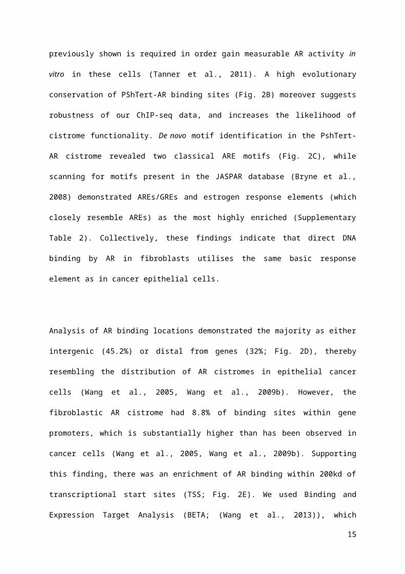
Supplementary Fig. 1B), which as previously shown is required in order gain measurable AR activity
in vitro in these cells (Tanner et al., 2011). A high evolutionary conservation of PShTert-AR binding
sites (Fig. 2B) moreover suggests robustness of our ChIP-seq data, and increases the likelihood of
cistrome functionality. De novo motif identification in the PshTert-AR cistrome revealed two classical
ARE motifs (Fig. 2C), while scanning for motifs present in the JASPAR database (Bryne et al., 2008)
demonstrated AREs/GREs and estrogen response elements (which closely resemble AREs) as the
most highly enriched (Supplementary Table 2). Collectively, these findings indicate that direct DNA
binding by AR in fibroblasts utilises the same basic response element as in cancer epithelial cells.
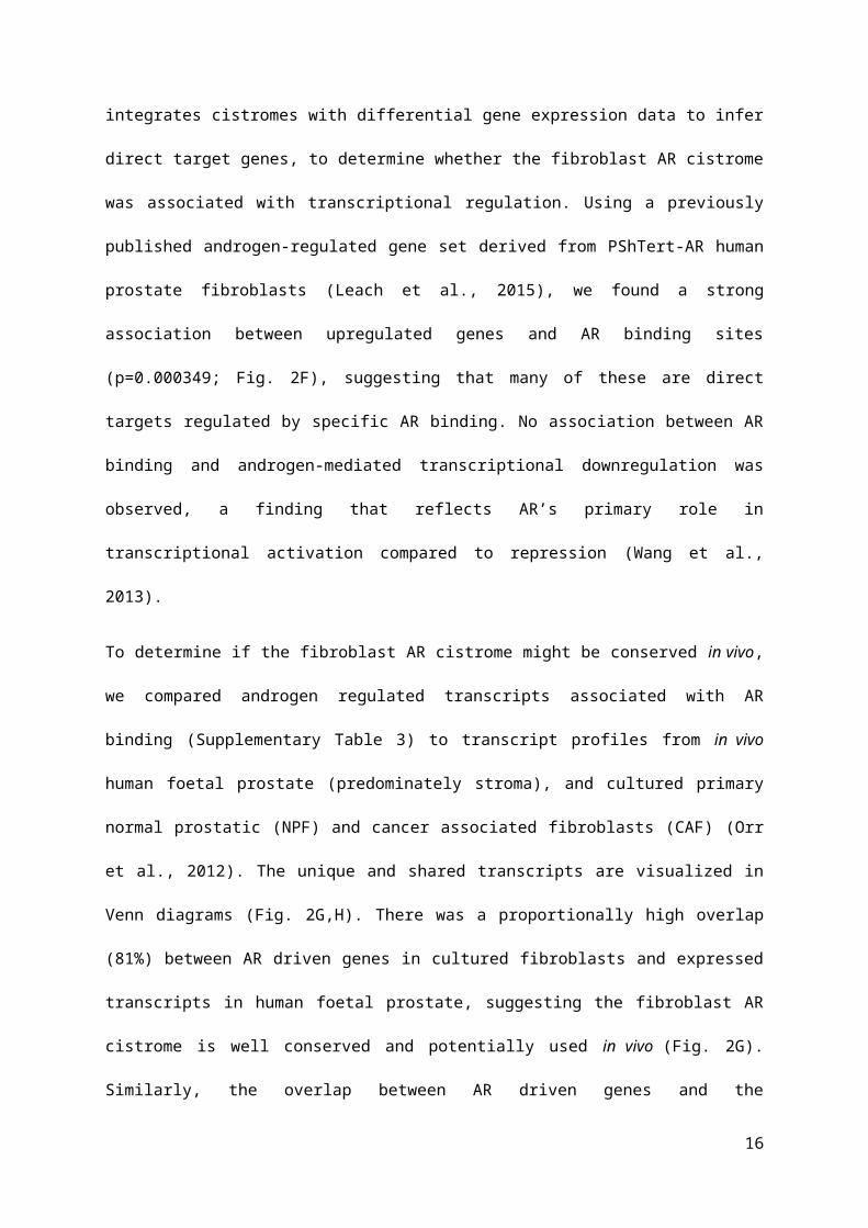
Analysis of AR binding locations demonstrated the majority as either intergenic (45.2%) or distal
from genes (32%; Fig. 2D), thereby resembling the distribution of AR cistromes in epithelial cancer
cells (Wang et al., 2005, Wang et al., 2009b). However, the fibroblastic AR cistrome had 8.8% of
binding sites within gene promoters, which is substantially higher than has been observed in cancer
cells (Wang et al., 2005, Wang et al., 2009b). Supporting this finding, there was an enrichment of AR
binding within 200kd of transcriptional start sites (TSS; Fig. 2E). We used Binding and Expression
Target Analysis (BETA; (Wang et al., 2013)), which integrates cistromes with differential gene
expression data to infer direct target genes, to determine whether the fibroblast AR cistrome was
associated with transcriptional regulation. Using a previously published androgen-regulated gene set
derived from PShTert-AR human prostate fibroblasts (Leach et al., 2015), we found a strong
association between upregulated genes and AR binding sites (p=0.000349; Fig. 2F), suggesting that
many of these are direct targets regulated by specific AR binding. No association between AR binding
and androgen-mediated transcriptional downregulation was observed, a finding that reflects AR’s
primary role in transcriptional activation compared to repression (Wang et al., 2013).
10

To determine if the fibroblast AR cistrome might be conserved in vivo, we compared androgen
regulated transcripts associated with AR binding (Supplementary Table 3) to transcript profiles from
in vivo human foetal prostate (predominately stroma), and cultured primary normal prostatic (NPF)
and cancer associated fibroblasts (CAF) (Orr et al., 2012). The unique and shared transcripts are
visualized in Venn diagrams (Fig. 2G,H). There was a proportionally high overlap (81%) between AR
driven genes in cultured fibroblasts and expressed transcripts in human foetal prostate, suggesting the
fibroblast AR cistrome is well conserved and potentially used in vivo (Fig. 2G). Similarly, the overlap
between AR driven genes and the transcriptomes of primary NPF and CAF was 71% and 73%
respectively (Fig. 2H).
3.2. COMPARISON OF THE FIBROBLAST CISTROME TO CANCER CELL CISTROMES
We next compared our fibroblast ChIP-seq data with several publically available prostate
cancer cell cistrome data sets. The degree of overlap varied from less than 1% to upward of 8.8%
(Table 1), which is consistent with microarray expression data showing that ~10% of genes are
regulated by androgens in both fibroblasts and cancer cells (Leach et al., 2015).
We next generated a set of “fibroblast–specific” AR binding sites by subtracting each of the published
prostate cancer cell cistromes from the fibroblast cistrome, yielding 4,251 binding events (i.e.
4251/5612 or 75.7% of the total cistrome; Fig. 3A). A number of these were validated as bona fide
fibroblast-specific or shared sites by ChIP-qPCR (Fig. 3B). The conservation of the fibroblast-specific
peak set was lower than that of the shared peak set (Fig. 3C). This is perhaps expected, as the shared
sites are found across multiple cell lineages and are thus more likely to be conserved. Quantitative
assessment of the two peak sets revealed that shared peaks were also slightly stronger (Fig. 3D). The
fibroblast-specific and shared peak sets showed a similar association with genomic elements (Fig 3E).
Importantly, there was a strong correlation between fibroblast specific binding sites and androgen-
upregulated genes in these cells, supporting their functionality (p=0.00463; Fig. 3F).
11

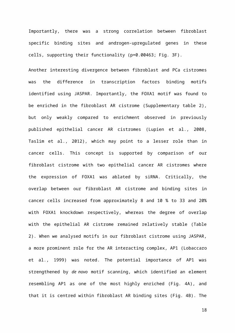
Another interesting divergence between fibroblast and PCa cistromes was the difference in
transcription factors binding motifs identified using JASPAR. Importantly, the FOXA1 motif was
found to be enriched in the fibroblast AR cistrome (Supplementary table 2), but only weakly
compared to enrichment observed in previously published epithelial cancer AR cistromes (Lupien et
al., 2008, Taslim et al., 2012), which may point to a lesser role than in cancer cells. This concept is
supported by comparison of our fibroblast cistrome with two epithelial cancer AR cistromes where
the expression of FOXA1 was ablated by siRNA. Critically, the overlap between our fibroblast AR
cistrome and binding sites in cancer cells increased from approximately 8 and 10 % to 33 and 20%
with FOXA1 knockdown respectively, whereas the degree of overlap with the epithelial AR cistrome
remained relatively stable (Table 2). When we analysed motifs in our fibroblast cistrome using
JASPAR, a more prominent role for the AR interacting complex, AP1 (Lobaccaro et al., 1999) was
noted. The potential importance of AP1 was strengthened by de novo motif scanning, which identified
an element resembling AP1 as one of the most highly enriched (Fig. 4A), and that it is centred within
fibroblast AR binding sites (Fig. 4B). The AP1 complex data is predominately composed of JUN and
FOS, but array data suggested JUN had by far the greater levels in fibroblast cell lines and primary
fibroblasts (Fig. 4C, GSE47203, GSE47354, GSE68166) (Leach et al., 2014, Doldi et al., 2015).
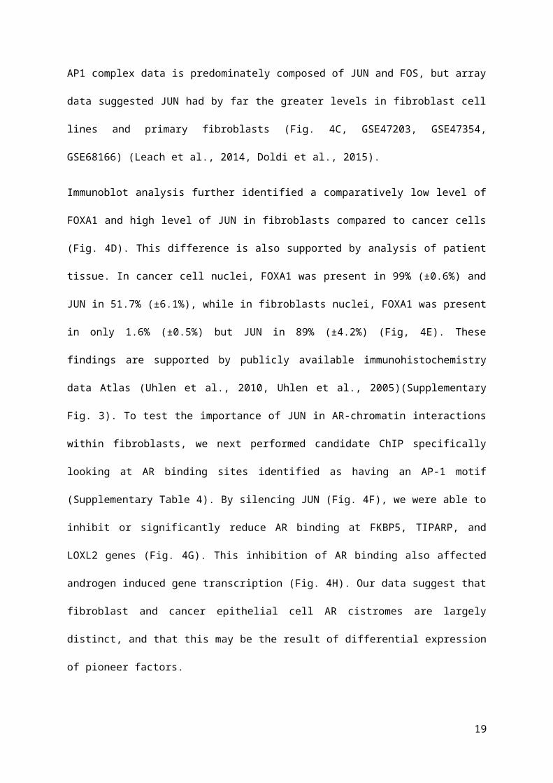
Immunoblot analysis further identified a comparatively low level of FOXA1 and high level of JUN in
fibroblasts compared to cancer cells (Fig. 4D). This difference is also supported by analysis of patient
tissue. In cancer cell nuclei, FOXA1 was present in 99% (±0.6%) and JUN in 51.7% (±6.1%), while
in fibroblasts nuclei, FOXA1 was present in only 1.6% (±0.5%) but JUN in 89% (±4.2%) (Fig, 4E).
These findings are supported by publicly available immunohistochemistry data Atlas (Uhlen et al.,
2010, Uhlen et al., 2005)(Supplementary Fig. 3). To test the importance of JUN in AR-chromatin
interactions within fibroblasts, we next performed candidate ChIP specifically looking at AR binding
sites identified as having an AP-1 motif (Supplementary Table 4). By silencing JUN (Fig. 4F), we
were able to inhibit or significantly reduce AR binding at FKBP5, TIPARP, and LOXL2 genes (Fig.
4G). This inhibition of AR binding also affected androgen induced gene transcription (Fig. 4H). Our
12

data suggest that fibroblast and cancer epithelial cell AR cistromes are largely distinct, and that this
may be the result of differential expression of pioneer factors.
3.3. THE FIBROBLAST CISTROME IN WHOLE TISSUE CHIP-SEQ
There is an increasing focus on in vivo/primary tissue ChIP-seq to generate individual
biologically relevant cistromic data (Collas and Dahl, 2008, Sharma et al., 2013). With this in mind,
we hypothesised that fibroblast AR binding sites could influence the results of whole tissue prostate
ChIP-seq data. To test this, we compared our whole fibroblast and fibroblast-specific cistromes with
three publicly available whole tissue ChIP-seq datasets (Sharma et al., 2013, Yu et al., 2010, Xu et al.,
2012) (Table 3). The degree of overlap between whole-tissue AR cistromes and our fibroblast AR
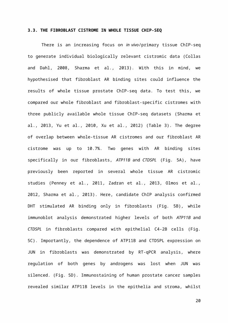
cistrome was up to 10.7%. Two genes with AR binding sites specifically in our fibroblasts, ATP11B
and CTDSPL (Fig. 5A), have previously been reported in several whole tissue AR cistromic studies
(Penney et al., 2011, Zadran et al., 2013, Olmos et al., 2012, Sharma et al., 2013). Here, candidate
ChIP analysis confirmed DHT stimulated AR binding only in fibroblasts (Fig. 5B), while immunoblot
analysis demonstrated higher levels of both ATP11B and CTDSPL in fibroblasts compared with
epithelial C4-2B cells (Fig. 5C). Importantly, the dependence of ATP11B and CTDSPL expression on
JUN in fibroblasts was demonstrated by RT-qPCR analysis, where regulation of both genes by
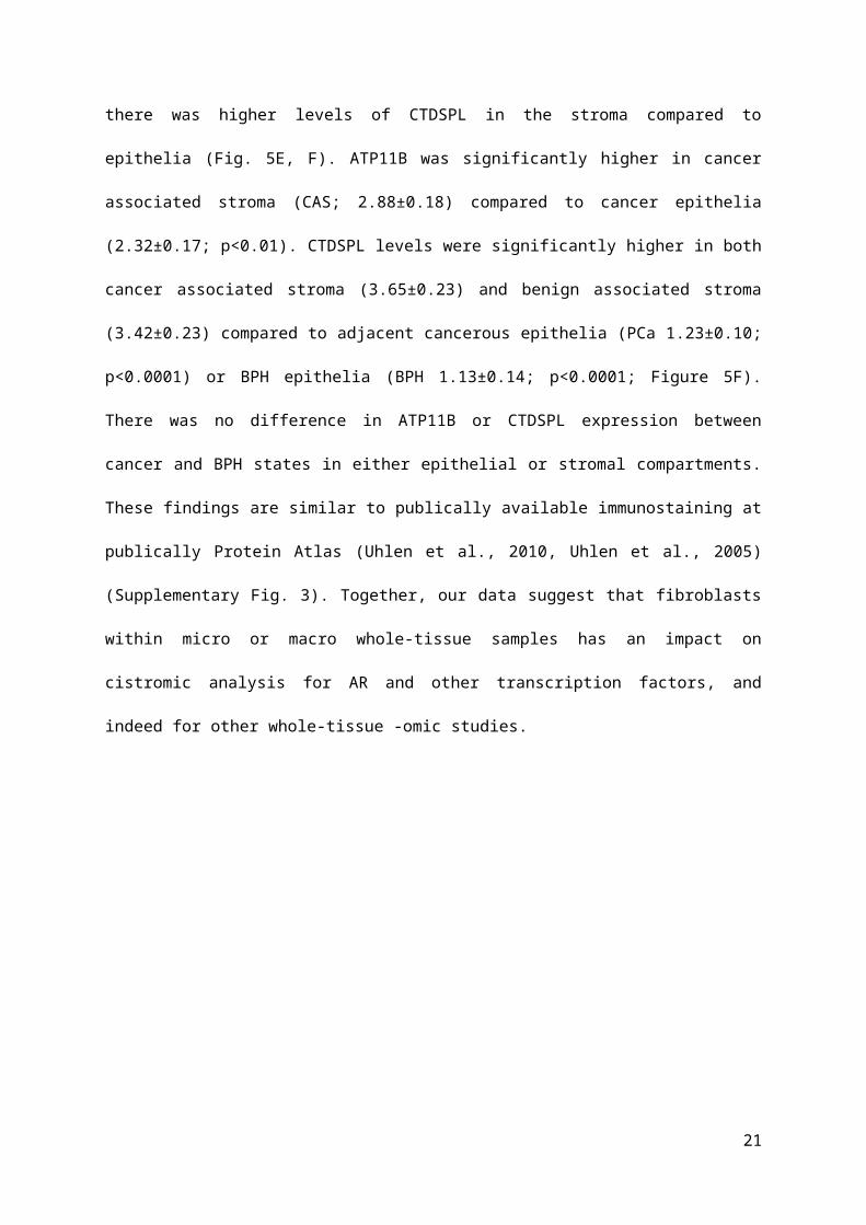
androgens was lost when JUN was silenced. (Fig. 5D). Immunostaining of human prostate cancer
samples revealed similar ATP11B levels in the epithelia and stroma, whilst there was higher levels of
CTDSPL in the stroma compared to epithelia (Fig. 5E, F). ATP11B was significantly higher in cancer
associated stroma (CAS; 2.88±0.18) compared to cancer epithelia (2.32±0.17; p<0.01). CTDSPL
levels were significantly higher in both cancer associated stroma (3.65±0.23) and benign associated
stroma (3.42±0.23) compared to adjacent cancerous epithelia (PCa 1.23±0.10; p<0.0001) or BPH
epithelia (BPH 1.13±0.14; p<0.0001; Figure 5F). There was no difference in ATP11B or CTDSPL
expression between cancer and BPH states in either epithelial or stromal compartments. These
findings are similar to publically available immunostaining at publically Protein Atlas (Uhlen et al.,
2010, Uhlen et al., 2005) (Supplementary Fig. 3). Together, our data suggest that fibroblasts within
13

micro or macro whole-tissue samples has an impact on cistromic analysis for AR and other
transcription factors, and indeed for other whole-tissue -omic studies.
14
4. DISCUSSION
Androgen receptor regulation of gene transcription is a highly co-ordinated process that encompasses
ligand-receptor binding, receptor-chromatin interactions, and synchronization of coregulatory proteins
and transcriptional machinery. These processes have been reported to be tissue specific (Goi et al.,
2013, Pihlajamaa et al., 2014), but research has focussed on AR signalling in malignant epithelial
cells and largely ignored the stromal compartment. We have previously shown that the AR-regulated
transcriptome in prostate fibroblasts is highly distinct from that in epithelial cancer cells. Here, we
report a detailed analysis of the AR cistrome in human prostatic fibroblasts, unveiling unique binding
events and providing evidence for a difference in primary pioneer factor influence from FOXA1 in
cancer epithelial cells versus AP-1 in fibroblasts.
A potential key factor in dictating cell lineage specificity of the AR cistrome is the expression and
influence of pioneer factors. One of the major pioneer factors associated with AR in PCa is FOXA1,
which directs up to 70% of AR binding events (Taslim et al., 2012). FOXA1 binds to closed
chromatin to permit access for other TFs and it directly interacts with the AR DNA-binding domain
(Cirillo et al., 2002, Cirillo et al., 1998, Belikov et al., 2009, Gao et al., 2003). We report here that
FOXA1 is minimally expressed in fibroblasts, whereas the AP-1 component JUN is highly expressed.
The AP-1 complex consists of different homo/hetero dimers combinations of JUN and FOS (Eckert et
al., 2013). When AP-1 proteins are forcedly expressed in PCa cells, they are reported to inhibit
androgen up-regulation of PSA (Sato et al., 1997) as well as reversing the proliferative effect of
androgens (Church et al., 2005). Thus, AP-1 appears to function differently to FOXA1 in AR
mediated transcription. Our data suggests that JUN/AP-1 is the predominant pioneer factor for AR in
fibroblasts, and that it contributes to a cistrome distinct from that seen in malignant epithelial cells.
This distinct fibroblast cistrome is strongly associated with androgen-upregulated genes that mediate
the anti-proliferative action of AR (Leach et al., 2015, Tanner et al., 2011). It has been reported that
pioneer factor expression varies between tissue and cell types, and may contribute to specificity of the
AR transcriptome (Pihlajamaa et al., 2014). This idea is supported by the reported conversion of cell
15
phenotypes by altering expression of key transcription factors (Sekiya and Suzuki, 2011). Pioneer
factors such as AP-1 are able to mediate differentiation cellular cues (Oh et al., 2014, Guo et al.,
2012). Our data linking AP-1 to AR in fibroblasts is further supported by the known role of AP-1
components in regulating mesenchymal traits, such as collagen production and mesenchymal type
movement (Qin et al., 2014a, Qin et al., 2014b, Selvaraj et al., 2015, Peng et al., 2015, Zhao et al.,
2014). Further investigation will be required to fully appreciate the nuances of cell specificity of AR-
pioneer factor-chromatin interactions, and the specific role of AP-1.
Targeting AR signalling is the mainstay treatment option in metastatic prostate cancer, but it is not
curative. New methods of targeting androgen signalling are being developed, one of which is
potentially inhibiting AR co-factor activity (Fitzgerald et al., 2014). Indeed, a number of studies have
suggested targeting AP-1 could be a useful therapeutic strategy (Ouyang et al., 2008, Chen et al.,
2006, Kavitha et al., 2014), even for localised disease (Cooperberg et al., 2007). Our previous studies
have demonstrated that loss of AR expression and activity in the stroma is associated with poor
patient outcome. As such, any targeting of AR signalling regulators or effectors in an intact primary
tumour setting needs to consider the potential for pro-tumourigenic outcomes if those factors are also
expressed in fibroblasts. The potential for unintended outcomes is further highlighted by the
observation that altering the expression of pioneer factors in the stroma affects interactions with the
adjacent epithelia (Szabowski et al., 2000, Pillebout et al., 2003, Chang et al., 2006, Katanov et al.,
2015, Yates and Rayner, 2002). The elucidation of AR action in the stroma, and thereby cell specific-
mechanisms of AR signalling, may inform the design and development of novel approaches to target
androgen action in prostate cancer.
This is the first report of nuclear receptor ChIP-seq in a mesenchymal cell type, and indeed the first
cistrome of any factor in prostatic fibroblasts. We have used hTERT immortalized prostate fibroblasts
to ensure stable AR expression. Although immortalization can be associated with gene changes,
16

previous studies have reported that immortalized fibroblast behave very similar to primary cells for up
to 150 passages (Milyavsky et al., 2003), which well exceeds the average passage number of 35 used
in PShTert-AR fibroblasts here. In addition, studies in epithelial cells report that WT-AR in
immortalized cells is still functional, able to interact with DNA, and retains similar activity to the
original non-immortalized cells (Gu et al., 2004, Kim et al., 2007). We have previously show that
genes regulated by PShTert-AR cells in response to androgens are also regulated in primary
fibroblast cultures and alternate prostate fibroblast lines transfected with AR (Tanner et al., 2011,
Leach et al., 2015). Furthermore, we report here that the vast majority (70-80%) of androgen
regulated genes associated with AR binding sites are also regulated by androgens in human non-
immortalized CAFs and NPFs and fetal human prostate (in vivo). Importantly, we show here that the
relatively modest overlap between fibroblast and epithelial cancer AR cistromes is correlated with
divergence in genome-wide androgen-regulated transcriptional (Leach et al., 2015). These data
strongly imply that cell specificity of AR transcriptional responses derive from differential AR
binding, with a role for FOXA1 in epithelia and AP1 in fibroblasts.
Understanding cell-specific AR binding and action is highly relevant to prostate cancer, particularly
since AR action in the stroma is required for normal prostatic development as well as being involved
in carcinogenesis. First, we and others have shown that AR regulates key fibroblast pathways
involved in cancer proliferation, progression, and invasiveness (Leach et al., 2015, Ricke et al., 2012,
Tanner et al., 2011). In the current study, the combination of ChIP-seq and expression data provides a
more detailed insight into AR regulation in fibroblasts, which will be important if we are to
manipulate AR-regulated fibroblast factors for therapeutic gain (Sluka and Davis, 2013). Second, we
believe our data is important for the interpretation of whole tissue cistrome data. Although RNA-
based analysis of tissue samples acknowledges that some degree of cell type isolation/sorting should
be performed to ensure outcomes are based on cancer cell expression and not stromal contamination
(Chmelar et al., 2007, Smith et al., 2009, Pena-Llopis and Brugarolas, 2013), in vivo ChIP-seq
analysis has not yet discriminated different cell types. Our data suggests that whole tissue capture of
AR DNA binding, which are discussed only in terms of epithelial cancer cells (Sharma et al., 2013,
17
Yu et al., 2010, Xu et al., 2012), are likely to contain a significant proportion of fibroblast AR binding
events. Third, genetic alterations and AR variants (Thompson et al., 1993, Fukino et al., 2007, Guo et
al., 2009, Buchanan et al., 2001) have been extensively studied in the context of cancer epithelial cells
but not in fibroblasts. Based on the opposing roles of stromal AR on cancer progression, we
hypothesize that inactivating mutations or inhibitory variants in prostate stroma will have an equally
important negative impact on patient outcome as activating mutations in epithelia. Fourth, the role of
chromatin-bound AR in fibroblasts may be relevant in understanding ongoing AR function following
epithelial-mesenchymal transition (EMT) (Jacob et al., 2014, Kong et al., 2015, Das et al., 2014),
which is an important intermediary in metastatic progression and cancer cell dissemination. The effect
of AR ablation on stromal AR signalling in vivo, and its impact on epithelial cell behaviour warrants
investigation. Overall, the current study provides a solid base for future analysis of AR-DNA
interactions in the stromal compartment, and considerable insight into how alteration of such
interactions might affect cancer development and progression.
In conclusion, we have shown that the AR cistrome in fibroblasts is fundamentally different from
prostate cancer epithelial cells. Our study also highlights the potential for pioneer factor expression to
have a role in controlling the cellular specificity of AR binding and divergence in transcriptional
profiles. Overall, we believe that this work provides new insight into the mechanisms and action of
AR in non-epithelial cells within the cancer microenvironment, which will help us to appreciate the
overall action of androgens in the prostate and other tissues.
18
References
BELIKOV, S., ASTRAND, C. & WRANGE, O. 2009. FoxA1 binding directs chromatin structure and the functional response of a glucocorticoid receptor-regulated promoter. Mol Cell Biol, 29, 5413-25.
BRYNE, J. C., VALEN, E., TANG, M. H., MARSTRAND, T., WINTHER, O., DA PIEDADE, I., KROGH, A., LENHARD, B. & SANDELIN, A. 2008. JASPAR, the open access database of transcription factor-binding profiles: new content and tools in the 2008 update. Nucleic Acids Res, 36, D102-6.
BUCHANAN, G., IRVINE, R. A., COETZEE, G. A. & TILLEY, W. D. 2001. Contribution of the androgen receptor to prostate cancer predisposition and progression. Cancer Metastasis Rev, 20, 207-23.
CANO, P., GODOY, A., ESCAMILLA, R., DHIR, R. & ONATE, S. A. 2007. Stromal-epithelial cell interactions and androgen receptor-coregulator recruitment is altered in the tissue microenvironment of prostate cancer. Cancer Res, 67, 511-9.
CHAN, S. C., SELTH, L. A., LI, Y., NYQUIST, M. D., MIAO, L., BRADNER, J. E., RAJ, G. V., TILLEY, W. D. & DEHM, S. M. 2015. Targeting chromatin binding regulation of constitutively active AR variants to overcome prostate cancer resistance to endocrine-based therapies. Nucleic Acids Res.
CHANG, H. J., PARK, J. S., KIM, M. H., HONG, M. H., KIM, K. M., KIM, S. M., SHIN, B. A., AHN, B. W. & JUNG, Y. D. 2006. Extracellular signal-regulated kinases and AP-1 mediate the up-regulation of vascular endothelial growth factor by PDGF in human vascular smooth muscle cells. Int J Oncol, 28, 135-41.
CHEN, S. Y., CAI, C., FISHER, C. J., ZHENG, Z., OMWANCHA, J., HSIEH, C. L. & SHEMSHEDINI, L. 2006. c-Jun enhancement of androgen receptor transactivation is associated with prostate cancer cell proliferation. Oncogene, 25, 7212-23.
CHMELAR, R., BUCHANAN, G., NEED, E. F., TILLEY, W. & GREENBERG, N. M. 2007. Androgen receptor coregulators and their involvement in the development and progression of prostate cancer. Int J Cancer, 120, 719-33.
CHURCH, D. R., LEE, E., THOMPSON, T. A., BASU, H. S., RIPPLE, M. O., ARIAZI, E. A. & WILDING, G. 2005. Induction of AP-1 activity by androgen activation of the androgen receptor in LNCaP human prostate carcinoma cells. Prostate, 63, 155-68.
CIRILLO, L. A., LIN, F. R., CUESTA, I., FRIEDMAN, D., JARNIK, M. & ZARET, K. S. 2002. Opening of compacted chromatin by early developmental transcription factors HNF3 (FoxA) and GATA-4. Mol Cell, 9, 279-89.
CIRILLO, L. A., MCPHERSON, C. E., BOSSARD, P., STEVENS, K., CHERIAN, S., SHIM, E. Y., CLARK, K. L., BURLEY, S. K. & ZARET, K. S. 1998. Binding of the winged-helix transcription factor HNF3 to a linker histone site on the nucleosome. EMBO J, 17, 244-54.
COLLAS, P. & DAHL, J. A. 2008. Chop it, ChIP it, check it: the current status of chromatin immunoprecipitation. Front Biosci, 13, 929-43.
COOPERBERG, M. R., BROERING, J. M., KANTOFF, P. W. & CARROLL, P. R. 2007. Contemporary trends in low risk prostate cancer: risk assessment and treatment. J Urol, 178, S14-9.
DAS, R., GREGORY, P. A., HOLLIER, B. G., TILLEY, W. D. & SELTH, L. A. 2014. Epithelial plasticity in prostate cancer: principles and clinical perspectives. Trends Mol Med, 20, 643-51.
DECKER, K. F., ZHENG, D., HE, Y., BOWMAN, T., EDWARDS, J. R. & JIA, L. 2012. Persistent androgen receptor-mediated transcription in castration-resistant prostate cancer under androgen-deprived conditions. Nucleic Acids Res, 40, 10765-79.
DOLDI, V., CALLARI, M., GIANNONI, E., D'AIUTO, F., MAFFEZZINI, M., VALDAGNI, R., CHIARUGI, P., GANDELLINI, P. & ZAFFARONI, N. 2015. Integrated gene and miRNA expression analysis of
19
prostate cancer associated fibroblasts supports a prominent role for interleukin-6 in fibroblast activation. Oncotarget, 6, 31441-60.
ECKERT, R. L., ADHIKARY, G., YOUNG, C. A., JANS, R., CRISH, J. F., XU, W. & RORKE, E. A. 2013. AP1 transcription factors in epidermal differentiation and skin cancer. J Skin Cancer, 2013, 537028.
FEBBO, P. G., LOWENBERG, M., THORNER, A. R., BROWN, M., LODA, M. & GOLUB, T. R. 2005. Androgen mediated regulation and functional implications of fkbp51 expression in prostate cancer. J Urol, 173, 1772-7.
FITZGERALD, K. A., EVANS, J. C., MCCARTHY, J., GUO, J., PRENCIPE, M., KEARNEY, M., WATSON, W. R. & O'DRISCOLL, C. M. 2014. The role of transcription factors in prostate cancer and potential for future RNA interference therapy. Expert Opin Ther Targets, 18, 633-49.
FUKINO, K., SHEN, L., PATOCS, A., MUTTER, G. L. & ENG, C. 2007. Genomic instability within tumor stroma and clinicopathological characteristics of sporadic primary invasive breast carcinoma. JAMA, 297, 2103-11.
GAO, N., ZHANG, J., RAO, M. A., CASE, T. C., MIROSEVICH, J., WANG, Y., JIN, R., GUPTA, A., RENNIE, P. S. & MATUSIK, R. J. 2003. The role of hepatocyte nuclear factor-3 alpha (Forkhead Box A1) and androgen receptor in transcriptional regulation of prostatic genes. Mol Endocrinol, 17, 1484-507.
GERHARDT, J., MONTANI, M., WILD, P., BEER, M., HUBER, F., HERMANNS, T., MUNTENER, M. & KRISTIANSEN, G. 2012. FOXA1 promotes tumor progression in prostate cancer and represents a novel hallmark of castration-resistant prostate cancer. Am J Pathol, 180, 848-61.
GOI, C., LITTLE, P. & XIE, C. 2013. Cell-type and transcription factor specific enrichment of transcriptional cofactor motifs in ENCODE ChIP-seq data. BMC Genomics, 14 Suppl 5, S2.
GU, Y., KIM, K. H., KO, D., NAKAMURA, K., YASUNAGA, Y., MOUL, J. W., SRIVASTAVA, S., ARNSTEIN, P. & RHIM, J. S. 2004. A telomerase-immortalized primary human prostate cancer clonal cell line with neoplastic phenotypes. Int J Oncol, 25, 1057-64.
GUO, L., SANS, M. D., HOU, Y., ERNST, S. A. & WILLIAMS, J. A. 2012. c-Jun/AP-1 is required for CCK-induced pancreatic acinar cell dedifferentiation and DNA synthesis in vitro. Am J Physiol Gastrointest Liver Physiol, 302, G1381-96.
GUO, Z., YANG, X., SUN, F., JIANG, R., LINN, D. E., CHEN, H., CHEN, H., KONG, X., MELAMED, J., TEPPER, C. G., KUNG, H. J., BRODIE, A. M., EDWARDS, J. & QIU, Y. 2009. A novel androgen receptor splice variant is up-regulated during prostate cancer progression and promotes androgen depletion-resistant growth. Cancer Res, 69, 2305-13.
HALBRITTER, F., VAIDYA, H. J. & TOMLINSON, S. R. 2012. GeneProf: analysis of high-throughput sequencing experiments. Nat Methods, 9, 7-8.
HEINZ, S., BENNER, C., SPANN, N., BERTOLINO, E., LIN, Y. C., LASLO, P., CHENG, J. X., MURRE, C., SINGH, H. & GLASS, C. K. 2010. Simple combinations of lineage-determining transcription factors prime cis-regulatory elements required for macrophage and B cell identities. Mol Cell, 38, 576-89.
HENSHALL, S. M., QUINN, D. I., LEE, C. S., HEAD, D. R., GOLOVSKY, D., BRENNER, P. C., DELPRADO, W., STRICKER, P. D., GRYGIEL, J. J. & SUTHERLAND, R. L. 2001. Altered expression of androgen receptor in the malignant epithelium and adjacent stroma is associated with early relapse in prostate cancer. Cancer Res, 61, 423-7.
HULSEN, T., DE VLIEG, J. & ALKEMA, W. 2008. BioVenn - a web application for the comparison and visualization of biological lists using area-proportional Venn diagrams. BMC Genomics, 9, 488.
JACOB, S., NAYAK, S., FERNANDES, G., BARAI, R. S., MENON, S., CHAUDHARI, U. K., KHOLKUTE, S. D. & SACHDEVA, G. 2014. Androgen receptor as a regulator of ZEB2 expression and its implications in epithelial-to-mesenchymal transition in prostate cancer. Endocr Relat Cancer, 21, 473-86.
20
JIN, H. J., ZHAO, J. C., WU, L., KIM, J. & YU, J. 2014. Cooperativity and equilibrium with FOXA1 define the androgen receptor transcriptional program. Nat Commun, 5, 3972.
KATANOV, C., LERRER, S., LIUBOMIRSKI, Y., LEIDER-TREJO, L., MESHEL, T., BAR, J., FENIGER-BARISH, R., KAMER, I., SORIA-ARTZI, G., KAHANI, H., BANERJEE, D. & BEN-BARUCH, A. 2015. Regulation of the inflammatory profile of stromal cells in human breast cancer: prominent roles for TNF-alpha and the NF-kappaB pathway. Stem Cell Res Ther, 6, 87.
KAVITHA, C. V., DEEP, G., GANGAR, S. C., JAIN, A. K., AGARWAL, C. & AGARWAL, R. 2014. Silibinin inhibits prostate cancer cells- and RANKL-induced osteoclastogenesis by targeting NFATc1, NF-kappaB, and AP-1 activation in RAW264.7 cells. Mol Carcinog, 53, 169-80.
KIM, K. H., DOBI, A., SHAHEDUZZAMAN, S., GAO, C. L., MASUDA, K., LI, H., DRUKIER, A., GU, Y., SRIKANTAN, V., RHIM, J. S. & SRIVASTAVA, S. 2007. Characterization of the androgen receptor in a benign prostate tissue-derived human prostate epithelial cell line: RC-165N/human telomerase reverse transcriptase. Prostate Cancer Prostatic Dis, 10, 30-8.
KONG, D., SETHI, S., LI, Y., CHEN, W., SAKR, W. A., HEATH, E. & SARKAR, F. H. 2015. Androgen receptor splice variants contribute to prostate cancer aggressiveness through induction of EMT and expression of stem cell marker genes. Prostate, 75, 161-74.
LEACH, D. A., NEED, E. F., TOIVANEN, R., TROTTA, A. P., PALENTHORPE, H. M., TAMBLYN, D. J., KOPSAFTIS, T., ENGLAND, G. M., SMITH, E., DREW, P. A., PINNOCK, C. B., LEE, P., HOLST, J., RISBRIDGER, G. P., CHOPRA, S., DEFRANCO, D. B., TAYLOR, R. A. & BUCHANAN, G. 2015. Stromal androgen receptor regulates the composition of the microenvironment to influence prostate cancer outcome. Oncotarget.
LEACH, D. A., NEED, E. F., TROTTA, A. P., GRUBISHA, M. J., DEFRANCO, D. B. & BUCHANAN, G. 2014. Hic-5 influences genomic and non-genomic actions of the androgen receptor in prostate myofibroblasts. Mol Cell Endocrinol, 384, 185-99.
LI, Y., LI, C. X., YE, H., CHEN, F., MELAMED, J., PENG, Y., LIU, J., WANG, Z., TSOU, H. C., WEI, J., WALDEN, P., GARABEDIAN, M. J. & LEE, P. 2008. Decrease in stromal androgen receptor associates with androgen-independent disease and promotes prostate cancer cell proliferation and invasion. J Cell Mol Med, 12, 2790-8.
LIU, T., ORTIZ, J. A., TAING, L., MEYER, C. A., LEE, B., ZHANG, Y., SHIN, H., WONG, S. S., MA, J., LEI, Y., PAPE, U. J., POIDINGER, M., CHEN, Y., YEUNG, K., BROWN, M., TURPAZ, Y. & LIU, X. S. 2011. Cistrome: an integrative platform for transcriptional regulation studies. Genome Biol, 12, R83.
LOBACCARO, J. M., POUJOL, N., TEROUANNE, B., GEORGET, V., FABRE, S., LUMBROSO, S. & SULTAN, C. 1999. Transcriptional interferences between normal or mutant androgen receptors and the activator protein 1--dissection of the androgen receptor functional domains. Endocrinology, 140, 350-7.
LUPIEN, M., EECKHOUTE, J., MEYER, C. A., WANG, Q., ZHANG, Y., LI, W., CARROLL, J. S., LIU, X. S. & BROWN, M. 2008. FoxA1 translates epigenetic signatures into enhancer-driven lineage-specific transcription. Cell, 132, 958-70.
MAGEE, J. A., CHANG, L. W., STORMO, G. D. & MILBRANDT, J. 2006. Direct, androgen receptor-mediated regulation of the FKBP5 gene via a distal enhancer element. Endocrinology, 147, 590-8.
MILYAVSKY, M., SHATS, I., EREZ, N., TANG, X., SENDEROVICH, S., MEERSON, A., TABACH, Y., GOLDFINGER, N., GINSBERG, D., HARRIS, C. C. & ROTTER, V. 2003. Prolonged culture of telomerase-immortalized human fibroblasts leads to a premalignant phenotype. Cancer Res, 63, 7147-57.
MURASHIMA, A., KISHIGAMI, S., THOMSON, A. & YAMADA, G. 2015. Androgens and mammalian male reproductive tract development. Biochim Biophys Acta, 1849, 163-170.
NEED, E. F., SCHER, H. I., PETERS, A. A., MOORE, N. L., CHEONG, A., RYAN, C. J., WITTERT, G. A., MARSHALL, V. R., TILLEY, W. D. & BUCHANAN, G. 2009. A novel androgen receptor amino
21
terminal region reveals two classes of amino/carboxyl interaction-deficient variants with divergent capacity to activate responsive sites in chromatin. Endocrinology, 150, 2674-82.
OH, I. Y., ALBEA, D. M., GOODWIN, Z. A., QUIGGLE, A. M., BAKER, B. P., GUGGISBERG, A. M., GEAHLEN, J. H., KRONER, G. M. & DE GUZMAN STRONG, C. 2014. Regulation of the dynamic chromatin architecture of the epidermal differentiation complex is mediated by a c-Jun/AP-1-modulated enhancer. J Invest Dermatol, 134, 2371-80.
OLAPADE-OLAOPA, E. O., MACKAY, E. H., TAUB, N. A., SANDHU, D. P., TERRY, T. R. & HABIB, F. K. 1999. Malignant transformation of human prostatic epithelium is associated with the loss of androgen receptor immunoreactivity in the surrounding stroma. Clin Cancer Res, 5, 569-76.
OLMOS, D., BREWER, D., CLARK, J., DANILA, D. C., PARKER, C., ATTARD, G., FLEISHER, M., REID, A. H., CASTRO, E., SANDHU, S. K., BARWELL, L., OOMMEN, N. B., CARREIRA, S., DRAKE, C. G., JONES, R., COOPER, C. S., SCHER, H. I. & DE BONO, J. S. 2012. Prognostic value of blood mRNA expression signatures in castration-resistant prostate cancer: a prospective, two-stage study. Lancet Oncol, 13, 1114-24.
ORR, B., RIDDICK, A. C., STEWART, G. D., ANDERSON, R. A., FRANCO, O. E., HAYWARD, S. W. & THOMSON, A. A. 2012. Identification of stromally expressed molecules in the prostate by tag-profiling of cancer-associated fibroblasts, normal fibroblasts and fetal prostate. Oncogene, 31, 1130-42.
OUYANG, X., JESSEN, W. J., AL-AHMADIE, H., SERIO, A. M., LIN, Y., SHIH, W. J., REUTER, V. E., SCARDINO, P. T., SHEN, M. M., ARONOW, B. J., VICKERS, A. J., GERALD, W. L. & ABATE-SHEN, C. 2008. Activator protein-1 transcription factors are associated with progression and recurrence of prostate cancer. Cancer Res, 68, 2132-44.
PENA-LLOPIS, S. & BRUGAROLAS, J. 2013. Simultaneous isolation of high-quality DNA, RNA, miRNA and proteins from tissues for genomic applications. Nat Protoc, 8, 2240-55.
PENG, B., ZHU, H., MA, L., WANG, Y. L., KLAUSEN, C. & LEUNG, P. C. 2015. AP-1 Transcription Factors c-FOS and c-JUN Mediate GnRH-Induced Cadherin-11 Expression and Trophoblast Cell Invasion. Endocrinology, 156, 2269-77.
PENNEY, K. L., SINNOTT, J. A., FALL, K., PAWITAN, Y., HOSHIDA, Y., KRAFT, P., STARK, J. R., FIORENTINO, M., PERNER, S., FINN, S., CALZA, S., FLAVIN, R., FREEDMAN, M. L., SETLUR, S., SESSO, H. D., ANDERSSON, S. O., MARTIN, N., KANTOFF, P. W., JOHANSSON, J. E., ADAMI, H. O., RUBIN, M. A., LODA, M., GOLUB, T. R., ANDREN, O., STAMPFER, M. J. & MUCCI, L. A. 2011. mRNA expression signature of Gleason grade predicts lethal prostate cancer. J Clin Oncol, 29, 2391-6.
PIHLAJAMAA, P., SAHU, B., LYLY, L., AITTOMAKI, V., HAUTANIEMI, S. & JANNE, O. A. 2014. Tissue-specific pioneer factors associate with androgen receptor cistromes and transcription programs. EMBO J, 33, 312-26.
PILLEBOUT, E., WEITZMAN, J. B., BURTIN, M., MARTINO, C., FEDERICI, P., YANIV, M., FRIEDLANDER, G. & TERZI, F. 2003. JunD protects against chronic kidney disease by regulating paracrine mitogens. J Clin Invest, 112, 843-52.
QIN, Z., ROBICHAUD, P., HE, T., FISHER, G. J., VOORHEES, J. J. & QUAN, T. 2014a. Oxidant exposure induces cysteine-rich protein 61 (CCN1) via c-Jun/AP-1 to reduce collagen expression in human dermal fibroblasts. PLoS One, 9, e115402.
QIN, Z., VOORHEES, J. J., FISHER, G. J. & QUAN, T. 2014b. Age-associated reduction of cellular spreading/mechanical force up-regulates matrix metalloproteinase-1 expression and collagen fibril fragmentation via c-Jun/AP-1 in human dermal fibroblasts. Aging Cell, 13, 1028-37.
RICCIARDELLI, C., CHOONG, C. S., BUCHANAN, G., VIVEKANANDAN, S., NEUFING, P., STAHL, J., MARSHALL, V. R., HORSFALL, D. J. & TILLEY, W. D. 2005. Androgen receptor levels in prostate cancer epithelial and peritumoral stromal cells identify non-organ confined disease. Prostate, 63, 19-28.
22
RICKE, E. A., WILLIAMS, K., LEE, Y. F., COUTO, S., WANG, Y., HAYWARD, S. W., CUNHA, G. R. & RICKE, W. A. 2012. Androgen hormone action in prostatic carcinogenesis: stromal androgen receptors mediate prostate cancer progression, malignant transformation and metastasis. Carcinogenesis.
ROBINSON, J. L., HICKEY, T. E., WARREN, A. Y., VOWLER, S. L., CARROLL, T., LAMB, A. D., PAPOUTSOGLOU, N., NEAL, D. E., TILLEY, W. D. & CARROLL, J. S. 2014. Elevated levels of FOXA1 facilitate androgen receptor chromatin binding resulting in a CRPC-like phenotype. Oncogene, 33, 5666-74.
SAHU, B., LAAKSO, M., OVASKA, K., MIRTTI, T., LUNDIN, J., RANNIKKO, A., SANKILA, A., TURUNEN, J. P., LUNDIN, M., KONSTI, J., VESTERINEN, T., NORDLING, S., KALLIONIEMI, O., HAUTANIEMI, S. & JANNE, O. A. 2011. Dual role of FoxA1 in androgen receptor binding to chromatin, androgen signalling and prostate cancer. EMBO J, 30, 3962-76.
SAHU, B., PIHLAJAMAA, P., DUBOIS, V., KERKHOFS, S., CLAESSENS, F. & JANNE, O. A. 2014. Androgen receptor uses relaxed response element stringency for selective chromatin binding and transcriptional regulation in vivo. Nucleic Acids Res, 42, 4230-40.
SATO, N., SADAR, M. D., BRUCHOVSKY, N., SAATCIOGLU, F., RENNIE, P. S., SATO, S., LANGE, P. H. & GLEAVE, M. E. 1997. Androgenic induction of prostate-specific antigen gene is repressed by protein-protein interaction between the androgen receptor and AP-1/c-Jun in the human prostate cancer cell line LNCaP. J Biol Chem, 272, 17485-94.
SEKIYA, S. & SUZUKI, A. 2011. Direct conversion of mouse fibroblasts to hepatocyte-like cells by defined factors. Nature, 475, 390-3.
SELVARAJ, N., BUDKA, J. A., FERRIS, M. W., PLOTNIK, J. P. & HOLLENHORST, P. C. 2015. Extracellular signal-regulated kinase signaling regulates the opposing roles of JUN family transcription factors at ETS/AP-1 sites and in cell migration. Mol Cell Biol, 35, 88-100.
SHARMA, N. L., MASSIE, C. E., RAMOS-MONTOYA, A., ZECCHINI, V., SCOTT, H. E., LAMB, A. D., MACARTHUR, S., STARK, R., WARREN, A. Y., MILLS, I. G. & NEAL, D. E. 2013. The androgen receptor induces a distinct transcriptional program in castration-resistant prostate cancer in man. Cancer Cell, 23, 35-47.
SHAW, A., PAPADOPOULOS, J., JOHNSON, C. & BUSHMAN, W. 2006. Isolation and characterization of an immortalized mouse urogenital sinus mesenchyme cell line. Prostate, 66, 1347-58.
SIDDIQUE, H. R., MISHRA, S. K., KARNES, R. J. & SALEEM, M. 2011. Lupeol, a novel androgen receptor inhibitor: implications in prostate cancer therapy. Clin Cancer Res, 17, 5379-91.
SLUKA, P. & DAVIS, I. D. 2013. Cell mates: paracrine and stromal targets for prostate cancer therapy. Nat Rev Urol, 10, 441-51.
SMITH, E., PALETHORPE, H. M., RUSZKIEWICZ, A. R., EDWARDS, S., LEACH, D. A., UNDERWOOD, T. J., NEED, E. F. & DREW, P. A. 2016. Androgen Receptor and Androgen-Responsive Gene FKBP5 Are Independent Prognostic Indicators for Esophageal Adenocarcinoma. Dig Dis Sci, 61, 433-43.
SMITH, M. J., CULHANE, A. C., DONOVAN, M., COFFEY, J. C., BARRY, B. D., KELLY, M. A., HIGGINS, D. G., WANG, J. H., KIRWAN, W. O., COTTER, T. G. & REDMOND, H. P. 2009. Analysis of differential gene expression in colorectal cancer and stroma using fluorescence-activated cell sorting purification. Br J Cancer, 100, 1452-64.
SZABOWSKI, A., MAAS-SZABOWSKI, N., ANDRECHT, S., KOLBUS, A., SCHORPP-KISTNER, M., FUSENIG, N. E. & ANGEL, P. 2000. c-Jun and JunB antagonistically control cytokine-regulated mesenchymal-epidermal interaction in skin. Cell, 103, 745-55.
TANNER, M. J., WELLIVER, R. C., JR., CHEN, M., SHTUTMAN, M., GODOY, A., SMITH, G., MIAN, B. M. & BUTTYAN, R. 2011. Effects of androgen receptor and androgen on gene expression in prostate stromal fibroblasts and paracrine signaling to prostate cancer cells. PLoS One, 6, e16027.
23
TASLIM, C., CHEN, Z., HUANG, K., HUANG, T. H., WANG, Q. & LIN, S. 2012. Integrated analysis identifies a class of androgen-responsive genes regulated by short combinatorial long-range mechanism facilitated by CTCF. Nucleic Acids Res, 40, 4754-64.
THOMPSON, T. C., TIMME, T. L., KADMON, D., PARK, S. H., EGAWA, S. & YOSHIDA, K. 1993. Genetic predisposition and mesenchymal-epithelial interactions in ras+myc-induced carcinogenesis in reconstituted mouse prostate. Mol Carcinog, 7, 165-79.
TROTTA, A. P., NEED, E. F., BUTLER, L. M., SELTH, L. A., O'LOUGHLIN, M. A., COETZEE, G. A., TILLEY, W. D. & BUCHANAN, G. 2012. Subdomain structure of the co-chaperone SGTA and activity of its androgen receptor client. J Mol Endocrinol, 49, 57-68.
UHLEN, M., BJORLING, E., AGATON, C., SZIGYARTO, C. A., AMINI, B., ANDERSEN, E., ANDERSSON, A. C., ANGELIDOU, P., ASPLUND, A., ASPLUND, C., BERGLUND, L., BERGSTROM, K., BRUMER, H., CERJAN, D., EKSTROM, M., ELOBEID, A., ERIKSSON, C., FAGERBERG, L., FALK, R., FALL, J., FORSBERG, M., BJORKLUND, M. G., GUMBEL, K., HALIMI, A., HALLIN, I., HAMSTEN, C., HANSSON, M., HEDHAMMAR, M., HERCULES, G., KAMPF, C., LARSSON, K., LINDSKOG, M., LODEWYCKX, W., LUND, J., LUNDEBERG, J., MAGNUSSON, K., MALM, E., NILSSON, P., ODLING, J., OKSVOLD, P., OLSSON, I., OSTER, E., OTTOSSON, J., PAAVILAINEN, L., PERSSON, A., RIMINI, R., ROCKBERG, J., RUNESON, M., SIVERTSSON, A., SKOLLERMO, A., STEEN, J., STENVALL, M., STERKY, F., STROMBERG, S., SUNDBERG, M., TEGEL, H., TOURLE, S., WAHLUND, E., WALDEN, A., WAN, J., WERNERUS, H., WESTBERG, J., WESTER, K., WRETHAGEN, U., XU, L. L., HOBER, S. & PONTEN, F. 2005. A human protein atlas for normal and cancer tissues based on antibody proteomics. Mol Cell Proteomics, 4, 1920-32.
UHLEN, M., FAGERBERG, L., HALLSTROM, B. M., LINDSKOG, C., OKSVOLD, P., MARDINOGLU, A., SIVERTSSON, A., KAMPF, C., SJOSTEDT, E., ASPLUND, A., OLSSON, I., EDLUND, K., LUNDBERG, E., NAVANI, S., SZIGYARTO, C. A., ODEBERG, J., DJUREINOVIC, D., TAKANEN, J. O., HOBER, S., ALM, T., EDQVIST, P. H., BERLING, H., TEGEL, H., MULDER, J., ROCKBERG, J., NILSSON, P., SCHWENK, J. M., HAMSTEN, M., VON FEILITZEN, K., FORSBERG, M., PERSSON, L., JOHANSSON, F., ZWAHLEN, M., VON HEIJNE, G., NIELSEN, J. & PONTEN, F. 2015. Proteomics. Tissue-based map of the human proteome. Science, 347, 1260419.
UHLEN, M., OKSVOLD, P., FAGERBERG, L., LUNDBERG, E., JONASSON, K., FORSBERG, M., ZWAHLEN, M., KAMPF, C., WESTER, K., HOBER, S., WERNERUS, H., BJORLING, L. & PONTEN, F. 2010. Towards a knowledge-based Human Protein Atlas. Nat Biotechnol, 28, 1248-50.
WANG, Q., CARROLL, J. S. & BROWN, M. 2005. Spatial and temporal recruitment of androgen receptor and its coactivators involves chromosomal looping and polymerase tracking. Mol Cell, 19, 631-42.
WANG, Q., LI, W., ZHANG, Y., YUAN, X., XU, K., YU, J., CHEN, Z., BEROUKHIM, R., WANG, H., LUPIEN, M., WU, T., REGAN, M. M., MEYER, C. A., CARROLL, J. S., MANRAI, A. K., JANNE, O. A., BALK, S. P., MEHRA, R., HAN, B., CHINNAIYAN, A. M., RUBIN, M. A., TRUE, L., FIORENTINO, M., FIORE, C., LODA, M., KANTOFF, P. W., LIU, X. S. & BROWN, M. 2009a. Androgen receptor regulates a distinct transcription program in androgen-independent prostate cancer. Cell, 138, 245-56.
WANG, S., SUN, H., MA, J., ZANG, C., WANG, C., WANG, J., TANG, Q., MEYER, C. A., ZHANG, Y. & LIU, X. S. 2013. Target analysis by integration of transcriptome and ChIP-seq data with BETA. Nat Protoc, 8, 2502-15.
WANG, X., KRUITHOF-DE JULIO, M., ECONOMIDES, K. D., WALKER, D., YU, H., HALILI, M. V., HU, Y. P., PRICE, S. M., ABATE-SHEN, C. & SHEN, M. M. 2009b. A luminal epithelial stem cell that is a cell of origin for prostate cancer. Nature, 461, 495-500.
WIKSTROM, P., MARUSIC, J., STATTIN, P. & BERGH, A. 2009. Low stroma androgen receptor level in normal and tumor prostate tissue is related to poor outcome in prostate cancer patients. Prostate, 69, 799-809.
24

WU, H. C., HSIEH, J. T., GLEAVE, M. E., BROWN, N. M., PATHAK, S. & CHUNG, L. W. 1994. Derivation of androgen-independent human LNCaP prostatic cancer cell sublines: role of bone stromal cells. Int J Cancer, 57, 406-12.
XU, K., WU, Z. J., GRONER, A. C., HE, H. H., CAI, C., LIS, R. T., WU, X., STACK, E. C., LODA, M., LIU, T., XU, H., CATO, L., THORNTON, J. E., GREGORY, R. I., MORRISSEY, C., VESSELLA, R. L., MONTIRONI, R., MAGI-GALLUZZI, C., KANTOFF, P. W., BALK, S. P., LIU, X. S. & BROWN, M. 2012. EZH2 oncogenic activity in castration-resistant prostate cancer cells is Polycomb-independent. Science, 338, 1465-9.
YATES, S. & RAYNER, T. E. 2002. Transcription factor activation in response to cutaneous injury: role of AP-1 in reepithelialization. Wound Repair Regen, 10, 5-15.
YU, J., YU, J., MANI, R. S., CAO, Q., BRENNER, C. J., CAO, X., WANG, X., WU, L., LI, J., HU, M., GONG, Y., CHENG, H., LAXMAN, B., VELLAICHAMY, A., SHANKAR, S., LI, Y., DHANASEKARAN, S. M., MOREY, R., BARRETTE, T., LONIGRO, R. J., TOMLINS, S. A., VARAMBALLY, S., QIN, Z. S. & CHINNAIYAN, A. M. 2010. An integrated network of androgen receptor, polycomb, and TMPRSS2-ERG gene fusions in prostate cancer progression. Cancer Cell, 17, 443-54.
ZADRAN, S., REMACLE, F. & LEVINE, R. D. 2013. miRNA and mRNA cancer signatures determined by analysis of expression levels in large cohorts of patients. Proc Natl Acad Sci U S A, 110, 19160-5.
ZHANG, Y., LIU, T., MEYER, C. A., EECKHOUTE, J., JOHNSON, D. S., BERNSTEIN, B. E., NUSBAUM, C., MYERS, R. M., BROWN, M., LI, W. & LIU, X. S. 2008. Model-based analysis of ChIP-Seq (MACS). Genome Biol, 9, R137.
ZHAO, C., QIAO, Y., JONSSON, P., WANG, J., XU, L., ROUHI, P., SINHA, I., CAO, Y., WILLIAMS, C. & DAHLMAN-WRIGHT, K. 2014. Genome-wide profiling of AP-1-regulated transcription provides insights into the invasiveness of triple-negative breast cancer. Cancer Res, 74, 3983-94.
Acknowledgments
This work was funded by the Prostate Cancer Foundation of Australia (GB, PG2210), seed funding
from the Freemasons Foundation Centre for Men’s Health (GB), and Cancer Australia (GB
25
APP1032970). The U.S. Department of Defense Prostate Cancer Research Program [Transformative
Impact Award W81XWH-13-2-0093 to LAS]; the Prostate Cancer Foundation of Australia [1012337,
1043482, and 2011/0452 to L.A.S]; the Ray and Shirl Norman Cancer Research Trust (LAS); the
Prostate Cancer Foundation [Young Investigator Award to LAS]. AAT and CN were supported by
Canadian Cancer Society grant (INNOV14-1 #702423).
Competing Interests
The authors declare no competing financial interests.
Corresponding authors
Correspondence to Luke Selth or Grant Buchanan. Grant Buchanan, The Basil Hetzel Institute for
Translational Health Research, The Queen Elizabeth Hospital, 28 Woodville Rd, Woodville
South SA 5011, Australia. Telephone: +61-8-8222-8447. Facsimile: +61-8-8222-6076. E-
mail: [email protected]. Luke A. Selth, Dame Roma Mitchell Cancer
Research Laboratories, The University of Adelaide, SA 5001, Australia. Telephone: +61-8-
8222-3618. Facsimile: +61-8-8222-3217. E-mail: [email protected].
26

FIGURE LEGENDS
Figure 1. PShTert-AR ChIP-seq samples. A) Gene track examples of ChIP-seq data at the loci on
chromosomes 3 and 16. AR binding sites are indicated in dual replicates of fibroblasts treated with
10nM DHT and probed with anti-AR antibody. B) Average ChIP-seq tag intensities expressed in
mapped reads per base pair per peak normalized per 10^ reads from duplicate PShTert-AR samples.
Figure 2. Validation and characterization of the AR cistrome in fibroblasts. A) RT-qPCR validation
of ChIP-seq AR binding sites in an independent set of PShTert-AR and WPMY fibroblasts, treated
with 10nM DHT or equivalent vehicle control. B) Conservation of fibroblast AR cistrome in
vertebrate species. Average PhastCon conservative scores relative to peaks called. The centre of each
binding site was set as zero. C) Sequence motif enrichment at AR binding sites identified de novo
using Gibbs Motif sequencing approach. D) Genomic location of AR binding sites in relation to target
genes as a percentage of total sites. E) Distribution of AR-binding sites within 200 kb upstream or
downstream of annotated transcription start sites. F) BETA analysis of AR binding and expression
data in PShTert-AR fibroblasts identified up-regulated (red), down-regulated (blue), and non-
differentially expressed genes as background (purple). G-H) Venn diagrams comparing overlap of
ChIP-seq derived PShTERT-AR cistrome with Tag-seq derived transcriptomes of (G) primary human
foetal prostate and (H) primary human prostate normal and cancer fibroblasts (NPF and CAF
respectively).
27

Figure 3. Specificity of AR cistrome between fibroblasts and cancer cells. A) Representative
diagrams of AR binding sites which are specific to fibroblasts or shared with prostate cancer cells. B)
RT-qPCR validation of AR binding sites specificity in independent ChIP samples from PShTert-AR,
WPMY, and C4-2B cells treated with 10nM DHT or equivalent vehicle control (VC). C) Average
PhastCon conservation scores relative to peak centre for fibroblast specific binding sites and common
binding sites shared with epithelial cancer. D) Read density around the centre of the highest point of
the peaks in the fibroblast specific and cancer shared binding sites. E) Genomic location of AR
binding sites in relation to target genes as a percentage of total sites for fibroblast specific AR
cistrome, and AR cistrome shared between fibroblast and prostate cancer cells. F) BETA analysis of
AR binding sites specific to fibroblasts and expression data in PShTert-AR fibroblasts identified up-
regulated (red), down-regulated (blue), and non-differentially expressed genes as background
(purple).
Figure 4. AP-1 as a major pioneer factor in the fibroblast AR cistrome. A) Gibbs de novo pioneer
factor motif analysis of the fibroblast specific binding sites identified higher enrichment of AP-1
motif than FOXA1 B) The number of AP-1 motifs up to 300 base pairs proximal to AR binding sites.
C) Heat map of FOXA1 and AP1 (JUN, FOS) genes in PShTert-AR and WPMY-1 fibroblast cell
lines, human fibroblasts isolated from areas adjacent to normal prostate (normal prostate fibroblasts;
NPF) or adjacent to cancer (cancer associated fibroblasts; CAFs), and C4-2B cancer cells. D)
Immunoblot analysis of pioneer factors in PShTert-AR and C4-2B cells treated with 10nM DHT or
equivalent vehicle control (V.C.). E) Representative images of immunohistochemical detection of
FOXA1 and JUN pioneer factor expression in prostate cancer cells (PCa) and stroma of human tissue
samples. The percentage of FOXA1 or JUN positive nuclei in the cancer cells and fibroblasts was
measured in five human samples. F) RT-qPCR analysis of JUN mRNA expression in fibroblasts
transfected with siRNA against JUN (siJUN), or a negative control (siNEG). Samples were
normalised to three housekeeping genes. Significance was detected using Student’s T-Test ** p<0.01
28

G) RT-qPCR of candidate ChIP analysis of PShTert-AR fibroblasts transfected with siRNA against
JUN (siJUN) or a negative control siRNA (siNEG), and treated with 10nM DHT or equivalent vehicle
control (V.C). Significance detected with Student’s T test, DHT vs V.C * p<0.05; siJUN vs siNEG #
p<0.05. H) RT-qPCR analysis of FKBP5 mRNA expression in PShTert-AR fibroblasts transfected in
triplicate with siRNA against JUN (siJUN) or a negative control siRNA (siNEG), and treated with
10nM DHT or equivalent vehicle control (V.C). Significance was detected using Student’s T-Test
DHT vs V.C * p<0.05, *** p<0.001; siJUN vs siNEG ## p<0.01, ### p<0.001.
Figure 5. The identification of whole tissue ARBS in fibroblast cistrome. A) Gene track examples
of ARBS located near the ATP11B and CTDSPL previously identified in whole tissue studies. B) RT-
qPCR validation of ARBS in ChIP samples from C4-2B and PShTert-AR cells treated with 10nM
DHT or equivalent vehicle control (VC). C) Immunoblot analysis of ATP11B and CTDSPL
expression in C4-2B and PShTert-AR cells treated with 10nM DHT or equivalent vehicle control
(V.C.). D) RT-qPCR analysis of ATP11B and CTDSPL mRNA expression in PShTert-AR fibroblasts
transfected in triplicate with siRNA against JUN (siJUN) or a negative control siRNA (siNEG), and
treated with 10nM DHT or equivalent vehicle control (V.C). Significance was detected using
Student’s T-Test, DHT vs V.C * p<0.05, *** p<0.001; siJUN vs siNEG ## p<0.01, ### p<0.001 E)
Representative images of immunohistochemical detection of ATP11B and CTDSPL in prostate cancer
(PCa) and BPH epithelia, cancer adjacent stroma (CAS) and BPH adjacent stroma (BAS) in human
tissue samples. F) Scoring results for ATP11B and CTDSPL proteins in the different compartments
and disease states. Significance detected with Mann-Whitney test, * p<0.05; *** p<0.001.
29
30
31
32
33

34
TABLES
Cell line Peaks Overlap #% of Fibroblast
cistrome% of PCa cistrome
Fibroblast cistrome PCa fibroblasts 5612 (Wang et al., 2009a) LNCaP 7712 485 8.64 6.29(Wang et al., 2009a) LNCaP-abl 6352 376 6.70 5.92(Decker et al., 2012) LNCaP/C4-2B (AxD) 7135 28 0.50 0.39(Decker et al., 2012) LNCaP/C4-2B (AI) 896 3 0.05 0.33(Sahu et al., 2011) LNCaP-1F5 (express rat GR) 6214 469 8.36 7.55(Jin et al., 2014) LNCaP 8811 600 10.69 6.81(Chan et al., 2015) CWR-R1 D1 (AR-FL) 12030 1060 18.89 8.81(Chan et al., 2015) CWR-R1 D567 (ARv567es) 3577 228 4.06 6.37
Table 1: Overlap of AR cistromes between fibroblast and cancer cells.
Cell line Peaks Overlap #% of Fibroblast
cistrome% of PCa cistrome
Fibroblast cistrome PCa fibroblasts 5612 (Sahu et al., 2011) ctrl LNCaP-1F5 (express rat GR) 6214 469 8.36 7.55(Sahu et al., 2011) FOXA1 kd LNCaP-1F5 (express rat GR) 17022 1852 33.00 10.88(Jin et al., 2014) ctrl LNCaP 8811 600 10.69 6.81(Jin et al., 2014) FOXA1 kd LNCaP 13222 1125 20.05 8.51
Table 2: Effect of FOXA1 expression on the AR cistrome overlap between fibroblast and cancer cells.
Cell line Peaks Overlap #% of Fibroblast
cistrome% of Tissue
cistromeFibroblast cistrome PCa fibroblasts 5612 (Sharma et al., 2013) Tissue (CRPC) 2484 157 2.8 6.32(Yu et al., 2010) Tissue 2236 239 4.3 10.7(Xu et al., 2012) Tissue 23110 44 0.8 0.19
Table 3: Overlap of fibroblast AR cistrome with whole tissue prostate cancer AR cistrome
35