viral vector-mediated selective and reversible blockade of...
TRANSCRIPT
ORIGINAL RESEARCH ARTICLEpublished: 11 October 2013
doi: 10.3389/fncir.2013.00162
Viral vector-mediated selective and reversible blockade ofthe pathway for visual orienting in miceThongchai Sooksawate1,2*, Kaoru Isa1, Ryosuke Matsui3, Shigeki Kato4, Masaharu Kinoshita1†,
Kenta Kobayashi5,6, Dai Watanabe3, Kazuto Kobayashi4 and Tadashi Isa1,5,6*
1 Department of Developmental Physiology, National Institute for Physiological Sciences, Okazaki, Japan2 Department of Pharmacology and Physiology, Faculty of Pharmaceutical Sciences, Chulalongkorn University, Bangkok, Thailand3 Department of Molecular and Systems Biology, Graduate School of Biostudies, Kyoto University, Kyoto, Japan4 Department of Molecular Genetics, Institute of Biomedical Sciences, Fukushima Medical University School of Medicine, Fukushima, Japan5 Section of Viral Vector Development, National Institute for Physiological Sciences, Okazaki, Japan6 Department of Life Sciences, The Graduate University for Advanced Studies (SOKENDAI), Hayama, Japan
Edited by:
Yasuo Kawaguchi, National Institutefor Physiological Sciences, Japan
Reviewed by:
Paul J. May, University ofMississippi Medical Center, USAMasahiko Takada, Kyoto University,Japan
*Correspondence:
Thongchai Sooksawate, Departmentof Pharmacology and Physiology,Faculty of Pharmaceutical Sciences,Chulalongkorn University, PhayathaiRoad, Patumwan, Bangkok 10330,Thailande-mail: [email protected];Tadashi Isa, Department ofDevelopmental Physiology, NationalInstitute for Physiological Sciences,Myodaiji, Okazaki 444-8585, Japane-mail: [email protected]†Present address:
Masaharu Kinoshita, Department ofPhysiology, Hirosaki UniversitySchool of Medicine, Hirosaki, Japan
Recently, by using a combination of two viral vectors, we developed a techniquefor pathway-selective and reversible synaptic transmission blockade, and successfullyinduced a behavioral deficit of dexterous hand movements in macaque monkeys byaffecting a population of spinal interneurons. To explore the capacity of this techniqueto work in other pathways and species, and to obtain fundamental methodologicalinformation, we tried to block the crossed tecto-reticular pathway, which is known tocontrol orienting responses to visual targets, in mice. A neuron-specific retrograde genetransfer vector with the gene encoding enhanced tetanus neurotoxin (eTeNT) tagged withenhanced green fluorescent protein (EGFP) under the control of a tetracycline responsiveelement was injected into the left medial pontine reticular formation. 7–17 days later,an adeno-associated viral vector with a highly efficient Tet-ON sequence, rtTAV16, wasinjected into the right superior colliculus. 5–9 weeks later, the daily administration ofdoxycycline (Dox) was initiated. Visual orienting responses toward the left side wereimpaired 1–4 days after Dox administration. Anti-GFP immunohistochemistry revealedthat a number of neurons in the intermediate and deep layers of the right superiorcolliculus were positively stained, indicating eTeNT expression. After the termination ofDox administration, the anti-GFP staining returned to the baseline level within 28 days.A second round of Dox administration, starting from 28 days after the termination of thefirst Dox administration, resulted in the reappearance of the behavioral impairment. Thesefindings showed that pathway-selective and reversible blockade of synaptic transmissionalso causes behavioral effects in rodents, and that the crossed tecto-reticular pathwayclearly controls visual orienting behaviors.
Keywords: superior colliculus, pontine reticular formation, orienting behavior, viral vector, Tet-ON, tetanus
neurotoxin, mouse
INTRODUCTIONTo study structure-function relationships within neural circuits,it is necessary to manipulate the activity of an “identified” pop-ulation of neuronal elements in these circuits. Recent advancesin molecular genetic techniques enabled such targeted manipula-tion by making transgenic animals with cell-specific promoters(Kobayashi et al., 1995; Watanabe et al., 1998). However, suchtechniques have generally been constrained to those animals inwhich the transgenic manipulation of gene expression is possi-ble (i.e., mouse, nematode, Drosophila, etc.) and to neuron typeswhose cell-specific promoter has been identified. An alternativepossibility is to use viral vectors to introduce particular genes. Toenable pathway specificity, lentivirus vectors pseudotyped withrabies virus glycoprotein (RVG) were developed as vectors spe-cific for retrograde transport (Kato et al., 2007). However, suchvectors were not sufficiently efficient to affect enough of a specificneuron population to induce behavioral effects, especially inlarger animals such as non-human primates. To overcome such
difficulties, Kobayashi and colleagues recently enhanced the ret-rograde transport efficiency of lentivirus vectors by pseudotypingwith a chimera of rabies virus and vesicular stomatitis virusglycoprotein (VSVG) [highly efficient retrograde gene transfer(HiRet) vector; Kato et al., 2011a]. We incorporated a tetracy-cline responsive element (TRE) and enhanced tetanus neurotoxin(eTeNT) tagged with enhanced green fluorescent protein (EGFP)into this retrograde vector (HiRet-TRE-EGFP.eTeNT). We alsoincorporated the newly developed efficient Tet-ON sequencertTAV16 into an adeno-associated type 2 virus vector (AAV-2)with a cytomegalovirus promoter (CMV) (AAV2-CMV-rtTAV16)(Kinoshita et al., 2012) as a switch to regulate the first con-struct. To study the function of a subpopulation of propriospinalneurons (PNs) whose cell bodies are located in the mid-cervicalsegments and project to hand/arm motor neurons in macaquemonkeys, we injected HiRet-TRE-EGFP.eTeNT into the ventralhorn of the C6–Th1 segments; one week later, we injected AAV2-CMV-rtTAV16 into the intermediate zone of the C2–C5 segments
Frontiers in Neural Circuits www.frontiersin.org October 2013 | Volume 7 | Article 162 | 1
NEURAL CIRCUITS
Sooksawate et al. Selective blockade of orienting pathway
where the PN cell bodies are located. 1–2 months later, we initi-ated the administration of doxycycline (Dox). We could observedeficits in reach and grasp movements. Acute electrophysiologicalexperiments clarified that approximately 90% of the transmis-sion through the PNs was blocked. Theoretically, this methodshould be available for universal use in the various pathways of thecentral nervous system without the need to identify cell-specificpromoters or develop genetically modified animals.
At the current stage, we still need to know the fundamen-tal properties of this technique and how to optimize it. For thispurpose, however, non-human primates are not the best ani-mal species because the number of available animals is limited,and behavioral and histological analyses are time consuming.Therefore, we decided to use mice to establish the fundamen-tal protocol of this technique. Moreover, we wished to selecta pathway whose function is easy to investigate using behav-ioral observations. We chose the crossed tectoreticular pathway(cTRNs), which originates from the intermediate and deep lay-ers of the midbrain superior colliculus (SC) and terminates inthe medial pontomedullary reticular formation contralateral tothe SC (Grantyn and Berthoz, 1987; Redgrave et al., 1990; Isaand Sasaki, 2002; Sooksawate et al., 2005, 2008). This pathwayis known to control the orienting response of the eyes, head,and body to visual targets (Wurtz and Albano, 1980; Sparks,1986; Dean et al., 1989; Isa and Sasaki, 2002; Isa and Hall, 2009),which is an innate behavior that does not require training. Inthis study, we focused on the time course of the effect of pathwayblockade through behavioral and histological examinations afterstarting Dox administration and after its offset. We also tested thereversibility of the effects by halting and repeating Dox admin-istration. This technique proved very feasible in mice, suggestingthat it could be used universally as a relatively convenient methodfor the pathway-specific manipulation of neural activity and geneexpression.
MATERIALS AND METHODSThe animal experimental procedures in this study were conductedin accordance with the Guidelines of the National Institutes ofHealth and the Ministry of Education, Culture, Sports, Science,and Technology of Japan, and were approved by the InstitutionalAnimal Care and Use Committee of the National Institutes ofNatural Sciences. All attempts were made to minimize the suf-fering and number of animals used in this study. The methodsand time schedule of the present experiments are summarized inFigure 1.
PREPARATION OF VIRAL VECTORSVector preparation was described by Kinoshita et al. (2012).However, a major difference was that we used a neuron-specifichighly efficient retrograde gene transfer lentivirus (NeuRet) vec-tor (Kato et al., 2011b) instead of the HiRet vector, because theNeuRet vector is specifically taken up by axons at the injectionsite.
NeuRet-TRE-EGFP.eTeNTThe NeuRet vector is a pseudotype of a human immunodeficiencyvirus type 1-based lentiviral vector with fusion glycoprotein
type C. In previous studies, it has been shown that pseudotyp-ing the lentiviral vector with the RVG changed the property ofthe vector specific for retrograde gene transfer (Kato et al., 2007).However, the efficiency of the gene transfer was not enoughto affect the majority of neurons projecting to the injectionsite. Later, it was found that replacing RVG with a chimericprotein of RVG and VSVG domains greatly enhanced the effi-ciency of retrograde gene transfer (highly efficient retrograde genetransfer vector or HiRet vector) (Kato et al., 2011a). Then, theuse of a different chimeric protein composing RVG and VSVG(fusion glycoprotein type C) switched the properties to neuron-specific infection (NeuRet) (Figure 1A; Kato et al., 2011b). In thepresent study, the envelope plasmid encoding fusion glycopro-tein type C, which was under the control of the cytomegalovirus(CMV) enhancer/chicken β-actin promoter, was used for vec-tor production. Chimeric EGFP.eTeNT.PEST was generated byfusing the human codon-optimized tetanus neurotoxin lightchain (eTeNT) with the EGFP derived from pEGFP-N1 vector(Clontech, Mountain View, CA, USA) and the PEST sequence ofornithine decarboxylase, as reported previously (Kinoshita et al.,2012). The transfer plasmid pLV-TRE-EGFP.eTeNT.PEST wasbased on pFUGW (a gift from D. Baltimore, California Instituteof Technology, Pasadena, CA, USA), and constructed by swap-ping the ubiquitin promoter-EGFP sequence with the TRE/CMVpromoter of the pTRE-Tight vector (Clontech, Mountain View,CA, USA) and EGFP.eTeNT.PEST. The NeuRet vector encod-ing EGFP.eTeNT was placed downstream of the TRE pro-moter (termed NeuRet-TRE-EGFP.eTeNT) and was prepared asdescribed previously (Inoue et al., 2012).
AAV2-CMV-rtTAV16The rtTA variant rtTAV16 was generated by introducing theV9I, G12S, F67S, F86Y, R171K, and A209T mutations intortTA2S-M2 of the pTet-ON advanced vector (Clontech, MountainView, CA, USA), as reported previously (Kinoshita et al., 2012).Plasmid pAAV2-CMV-rtTAV16 (Figure 1A) is based on pAAV-MCS (Agilent Technologies, Inc., Santa Clare, CA, USA), andwas constructed by inserting the CMV promoter sequence ofthe pTet-On advanced vector rtTAV16, the woodchuck hepatitisvirus posttranscriptional regulatory element WPRE sequence ofpFUGW, and the SV40 polyadenylation signal (SV40pA) of thepCMV-script vector (Agilent Technologies, Tokyo, Japan) into amultiple cloning site. The AAV vector for in vivo injection wasproduced as described previously (Kaneda et al., 2011).
INJECTIONS OF NeuRet-TRE-EGFP.eTeNT AND AAV2-CMV-rtTAV16We anesthetized 6–10-week-old male C57BL/6 mice with anintraperitoneal injection of a mixture of ketamine (60 mg/kgbody weight) and xylazine (10 mg/kg body weight). In addition,dexamethasone (5.5 mg/kg body weight) was injected intramus-cularly as premedication. The head of the mouse was fixed to thestereotaxic apparatus (Narishige, Tokyo, Japan) and injections ofthe vectors were made from the dorsal approach. NeuRet-TRE-EGFP.eTeNT (0.8–1.2 μL); titer, 3.3–12.1 × 10¹¹ copies/mL) wasinjected into the medial pontine reticular formation (MPRF)on the left side (Figures 1B,C) using a thin glass micropipette(tip diameter, 50–70 μm) inclined by 45◦caudally to the vertical
Frontiers in Neural Circuits www.frontiersin.org October 2013 | Volume 7 | Article 162 | 2
Sooksawate et al. Selective blockade of orienting pathway
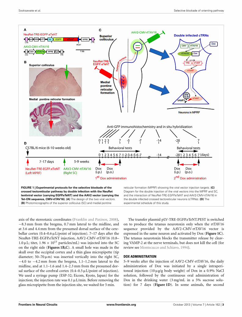
FIGURE 1 | Experimental protocols for the selective blockade of the
crossed tectoreticular pathway by double infection with the NeuRet
lentiviral vector (carrying EGFP.eTeNT) and the AAV2 vector (carrying the
Tet-ON sequence, CMV-rtTAV16). (A) The design of the two viral vectors.(B) Photomicrographs of the superior colliculus (SC) and medial pontine
reticular formation (MPRF) showing the viral vector injection targets. (C)
Diagram for the double injection of the viral vectors into the MPRF and SC,and the interaction of NeuRet-TRE-EGFP.eTeNT and AAV2-CMV-rtTAV16 inthe double infected crossed tectoreticular neurons (cTRNs). (D) Theexperimental schedule of this study.
axis of the stereotaxic coordinates (Franklin and Paxinos, 2008),−8.3 mm from the bregma, 0.7 mm lateral to the midline, andat 3.6 and 4.4 mm from the presumed dorsal surface of the cere-bellar cortex (0.4–0.6 μL/point of injection). 7–17 days after theNeuRet-TRE-EGFP.eTeNT injection, AAV2-CMV-rtTAV16 (0.8–1.0 μL; titer, 1.96 × 10¹³ particles/mL) was injected into the SCon the right side (Figures 1B,C). A small hole was made in theskull over the occipital cortex and a thin glass micropipette (tipdiameter; 50–70 μm) was inserted vertically into the right SC,−4.0 to −4.2 mm from the bregma, 1.1–1.2 mm lateral to themidline, and at 1.1–1.6 and 1.4–2.3 mm from the presumed dor-sal surface of the cerebral cortex (0.4–0.5 μL/point of injection).We used a syringe pump (ESP-32; Eicom, Kyoto, Japan) for theinjection; the injection rate was 0.1 μL/min. Before removing theglass micropipette from the injection site, we waited for 5 min.
The transfer plasmid pLV-TRE-EGFP.eTeNT.PEST is switchedon to produce the tetanus neurotoxin only when the rtTAV16sequence provided by the AAV2-CMV-rtTAV16 vector isexpressed in the same neuron and activated by Dox (Figure 1C).The tetanus neurotoxin blocks the transmitter release by cleav-ing VAMP-2 at the nerve terminals, but does not kill the cell (forreview see Montecucco and Schiavo, 1994).
DOX ADMINISTRATION5–9 weeks after the injection of AAV2-CMV-rtTAV16, the dailyadministration of Dox was initiated by a single intraperi-toneal injection (10 μg/g body weight) of Dox in a 0.9% NaClsolution, followed by the continuous oral administration ofDox in the drinking water (3 mg/mL in a 5% sucrose solu-tion) for 7 days (Figure 1D). In some animals, the second
Frontiers in Neural Circuits www.frontiersin.org October 2013 | Volume 7 | Article 162 | 3
Sooksawate et al. Selective blockade of orienting pathway
period of Dox administration (2nd Dox administration) wasconducted from 28 days after the offset of the first periodof Dox administration (1st Dox administration). One groupof mice received Dox continually for 21 days for histologicalanalysis.
BEHAVIORAL TESTS FOR VISUAL ORIENTING AND TURNING BEHAVIORWe utilized three tests to assess visual orienting and turningbehaviors: (1) the visual placing response, (2) the visual orientingresponse, and (3) a turning behavior test (Figure 1D). The behav-ioral tests began on the day before Dox administration, continuedduring Dox administration for 7 days, and after Dox administra-tion for 7 days. All tests were performed between 6.00 AM and3.00 PM.
Visual placing responseA modified visual placing response test was used to evaluate visualorienting behavior toward the affected and unaffected sides ofthe tested mouse. This test was modified from Metz and Schwab(2004) and Pinto and Enroth-Cugell (2000). In this test, themouse was suspended by holding its tail and then lowered toward
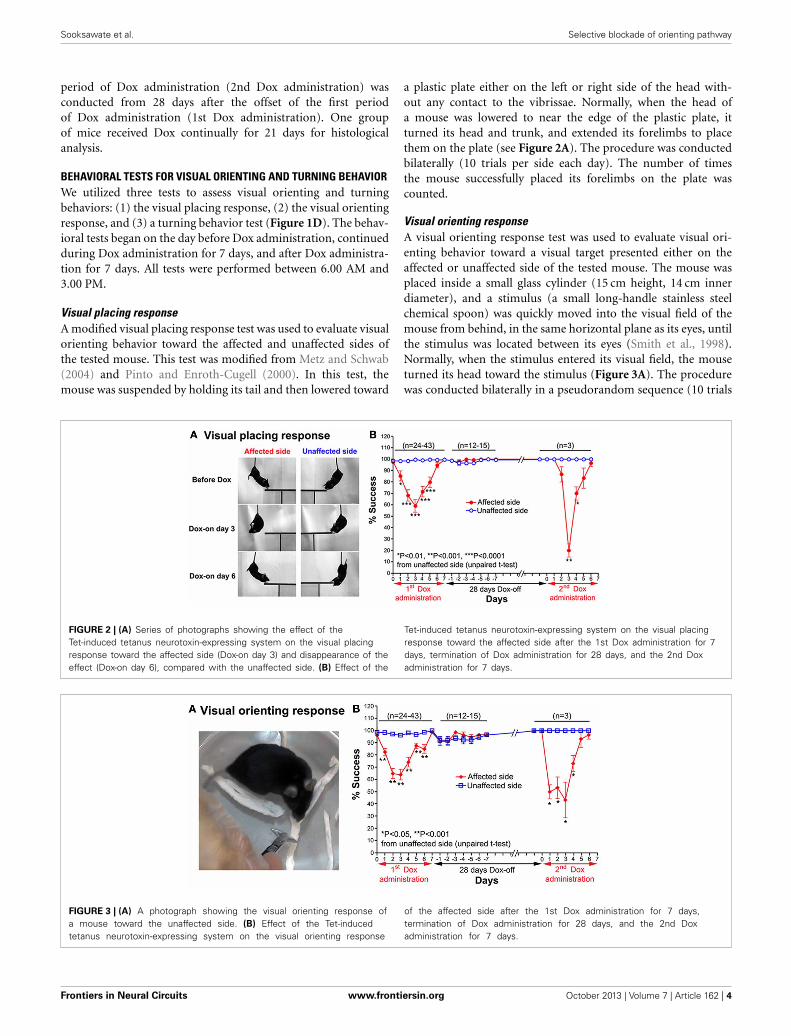
a plastic plate either on the left or right side of the head with-out any contact to the vibrissae. Normally, when the head ofa mouse was lowered to near the edge of the plastic plate, itturned its head and trunk, and extended its forelimbs to placethem on the plate (see Figure 2A). The procedure was conductedbilaterally (10 trials per side each day). The number of timesthe mouse successfully placed its forelimbs on the plate wascounted.
Visual orienting responseA visual orienting response test was used to evaluate visual ori-enting behavior toward a visual target presented either on theaffected or unaffected side of the tested mouse. The mouse wasplaced inside a small glass cylinder (15 cm height, 14 cm innerdiameter), and a stimulus (a small long-handle stainless steelchemical spoon) was quickly moved into the visual field of themouse from behind, in the same horizontal plane as its eyes, untilthe stimulus was located between its eyes (Smith et al., 1998).Normally, when the stimulus entered its visual field, the mouseturned its head toward the stimulus (Figure 3A). The procedurewas conducted bilaterally in a pseudorandom sequence (10 trials
FIGURE 2 | (A) Series of photographs showing the effect of theTet-induced tetanus neurotoxin-expressing system on the visual placingresponse toward the affected side (Dox-on day 3) and disappearance of theeffect (Dox-on day 6), compared with the unaffected side. (B) Effect of the
Tet-induced tetanus neurotoxin-expressing system on the visual placingresponse toward the affected side after the 1st Dox administration for 7days, termination of Dox administration for 28 days, and the 2nd Doxadministration for 7 days.
FIGURE 3 | (A) A photograph showing the visual orienting response ofa mouse toward the unaffected side. (B) Effect of the Tet-inducedtetanus neurotoxin-expressing system on the visual orienting response
of the affected side after the 1st Dox administration for 7 days,termination of Dox administration for 28 days, and the 2nd Doxadministration for 7 days.
Frontiers in Neural Circuits www.frontiersin.org October 2013 | Volume 7 | Article 162 | 4
Sooksawate et al. Selective blockade of orienting pathway
per side each day). The number of times the mouse successfullyoriented itself to the stimulus was counted.
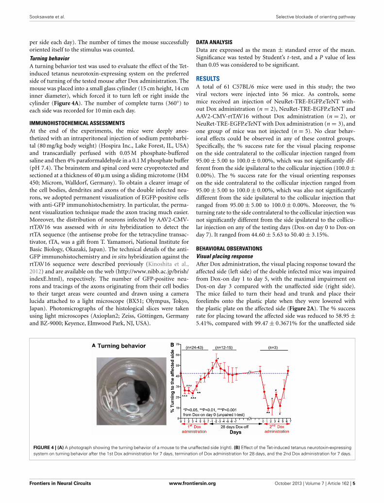
Turning behaviorA turning behavior test was used to evaluate the effect of the Tet-induced tetanus neurotoxin-expressing system on the preferredside of turning of the tested mouse after Dox administration. Themouse was placed into a small glass cylinder (15 cm height, 14 cminner diameter), which forced it to turn left or right inside thecylinder (Figure 4A). The number of complete turns (360◦) toeach side was recorded for 10 min each day.
IMMUNOHISTOCHEMICAL ASSESSMENTSAt the end of the experiments, the mice were deeply anes-thetized with an intraperitoneal injection of sodium pentobarbi-tal (80 mg/kg body weight) (Hospira Inc., Lake Forest, IL, USA)and transcardially perfused with 0.05 M phosphate-bufferedsaline and then 4% paraformaldehyde in a 0.1 M phosphate buffer(pH 7.4). The brainstem and spinal cord were cryoprotected andsectioned at a thickness of 40 μm using a sliding microtome (HM450; Microm, Walldorf, Germany). To obtain a clearer image ofthe cell bodies, dendrites and axons of the double infected neu-rons, we adopted permanent visualization of EGFP-positive cellswith anti-GFP immunohistochemistry. In particular, the perma-nent visualization technique made the axon tracing much easier.Moreover, the distribution of neurons infected by AAV2-CMV-rtTAV16 was assessed with in situ hybridization to detect thertTA sequence (the antisense probe for the tetracycline transac-tivator, tTA, was a gift from T. Yamamori, National Institute forBasic Biology, Okazaki, Japan). The technical details of the anti-GFP immunohistochemistry and in situ hybridization against thertTAV16 sequence were described previously (Kinoshita et al.,2012) and are available on the web (http://www.nibb.ac.jp/brish/indexE.html), respectively. The number of GFP-positive neu-rons and tracings of the axons originating from their cell bodiesto their target areas were counted and drawn using a cameralucida attached to a light microscope (BX51; Olympus, Tokyo,Japan). Photomicrographs of the histological slices were takenusing light microscopes (Axioplan2; Zeiss, Göttingen, Germanyand BZ-9000; Keyence, Elmwood Park, NJ, USA).
DATA ANALYSISData are expressed as the mean ± standard error of the mean.Significance was tested by Student’s t-test, and a P value of lessthan 0.05 was considered to be significant.
RESULTSA total of 61 C57BL/6 mice were used in this study; the twoviral vectors were injected into 56 mice. As controls, somemice received an injection of NeuRet-TRE-EGFP.eTeNT with-out Dox administration (n = 2), NeuRet-TRE-EGFP.eTeNT andAAV2-CMV-rtTAV16 without Dox administration (n = 2), orNeuRet-TRE-EGFP.eTeNT with Dox administration (n = 3), andone group of mice was not injected (n = 5). No clear behav-ioral effects could be observed in any of these control groups.Specifically, the % success rate for the visual placing responseon the side contralateral to the collicular injection ranged from95.00 ± 5.00 to 100.0 ± 0.00%, which was not significantly dif-ferent from the side ipsilateral to the collicular injection (100.0 ±0.00%). The % success rate for the visual orienting responseson the side contralateral to the collicular injection ranged from95.00 ± 5.00 to 100.0 ± 0.00%, which was also not significantlydifferent from the side ipsilateral to the collicular injection thatranged from 95.00 ± 5.00 to 100.0 ± 0.00%. Moreover, the %turning rate to the side contralateral to the collicular injection wasnot significantly different from the side ipsilateral to the collicu-lar injection on any of the testing days (Dox-on day 0 to Dox-onday 7). It ranged from 44.60 ± 5.63 to 50.40 ± 3.15%.
BEHAVIORAL OBSERVATIONSVisual placing responseAfter Dox administration, the visual placing response toward theaffected side (left side) of the double infected mice was impairedfrom Dox-on day 1 to day 5, with the maximal impairment onDox-on day 3 compared with the unaffected side (right side).The mice failed to turn their head and trunk and place theirforelimbs onto the plastic plate when they were lowered withthe plastic plate on the affected side (Figure 2A). The % successrate for placing toward the affected side was reduced to 58.95 ±5.41%, compared with 99.47 ± 0.3671% for the unaffected side
FIGURE 4 | (A) A photograph showing the turning behavior of a mouse to the unaffected side (right). (B) Effect of the Tet-induced tetanus neurotoxin-expressingsystem on turning behavior after the 1st Dox administration for 7 days, termination of Dox administration for 28 days, and the 2nd Dox administration for 7 days.
Frontiers in Neural Circuits www.frontiersin.org October 2013 | Volume 7 | Article 162 | 5
Sooksawate et al. Selective blockade of orienting pathway
(P < 0.0001, n = 38, unpaired t-test; Figure 2B). However, thisbehavioral effect was gradually reduced from Dox-on day 4 andcompletely disappeared on Dox-on days 6–7. After the termi-nation of Dox administration, no behavioral effect could beseen on the affected and unaffected sides for 7 days (Dox-offdays −1 to −7).
Visual orienting responseImpairment of the orienting response could also be found inthe visual orienting response test. After Dox administration, thevisual orienting response toward the affected side (left side) wasimpaired from days 1–6, while it was not impaired toward theunaffected side (right side) (Figure 3A). The % success rate ofthe affected side was reduced to 64.21 ± 4.473%, compared tothe 96.32 ± 0.793% of the unaffected side (P < 0.001, n = 38,unpaired t-test; Figure 3B). The behavioral effect was gradu-ally reduced from Dox-on day 4, and completely disappeared byDox-on day 7. After the termination of Dox administration, nobehavioral effect could be seen on the affected and unaffectedsides for 7 days (Dox-off days −1 to −7).
Turning behaviorBefore Dox administration and in the control groups, the micealmost equally preferred to turn toward the left and right.However, after Dox administration, the mice preferred to turnto the unaffected side (right) (Figure 4A). The % turning to theaffected side (left side) was reduced from Dox-on days 1 to 4,with the maximal reduction on day 3 (Figure 4B). The % turn-ing to the affected side was reduced to 23.92 ± 2.721% from the42.77 ± 1.824% of the controls on day 0 (P < 0.001, n = 38,unpaired t-test). Then, the % turning to the affected side grad-ually returned to the same level as before Dox administrationby Dox-on day 7. It is noteworthy that after the termination ofDox administration, % turning to the affected side (left side)increased to its highest level on Dox-off day −2 (P < 0.05, n =15, unpaired t-test), before returning to the same level as beforeDox administration on Dox-off days −5 to −7.
REVERSIBILITY OF THE DOX-ON EFFECTS28 days after the termination of the 1st Dox administration (for7 days), the 2nd Dox administration was started for another 7days. All of the behavioral tests for visual orienting were foundto be impaired after the 2nd Dox administration. The patternand time course of the impairments were almost the same asfor the 1st Dox administration. The % success rates for thevisual placing response (Figure 2B) and visual orienting response(Figure 3B) and % turning (Figure 4B) toward the affected sidewere reduced on Dox-on days 1–2 to days 4–5 and, then, grad-ually increased to the same levels as before Dox administrationon days 6–7. Although, the maximal effects of the 2nd Doxadministration on all behavioral tests appeared to be greater,they were not significantly different from the 1st Dox admin-istration (visual placing response, P = 0.0531; visual orientingresponse, P = 0.2123; turning behavior, P = 0.1544, unpairedt-test). These results indicate that the selective blockade of thecrossed tectoreticular pathway by double infection with NeuRet-TRE-EGFP.eTeNT and AAV2-CMV-rtTAV16 can be performedrepeatedly with an interval of 28 days.
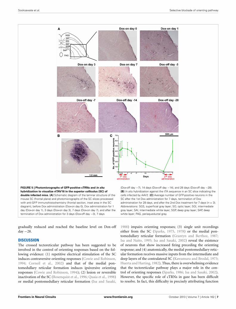
HISTOLOGICAL ANALYSISThe number of GFP-positive crossed tectoreticular neurons(cTRNs) in the intermediate and deep layers of the SC of thedouble infected mice increased sharply after the start of Doxadministration for 1 day and reached its maximum on Dox-onday 3 (Figures 5A,C). The maximum number of GFP-positivecTRNs was 458.8 ± 141.4 neurons (n = 6). After Dox adminis-tration was terminated, the number of GFP-positive cTRNs wasgradually reduced on Dox-off days −3 to −14, and had returnedto the baseline level by Dox-off day −28. To confirm the injec-tion site of AAV2 in the right SC, in situ hybridization of rtTAV16was performed. The injection sites were found to be located inthe intermediate to deep layers of the right SC (Figure 5B). Todemonstrate the reversibility of the Dox-on effects in these dou-bled infected mice, the 2nd Dox administration was started aftera 28-day Dox-off period. The number of GFP-positive cTRNs inthe intermediate and deep layers of the right SC increased againto 400.0 ± 218.3 (n = 3; Figure 5C). The histological results par-alleled the impairment of visual orienting behavior after the 2ndDox treatment in this group (Figures 2B, 3B, 4B).
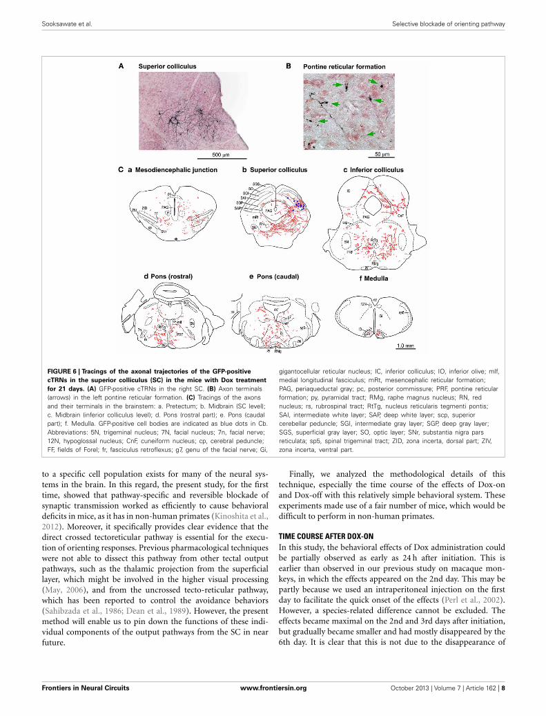
In another group of mice (n = 3), the period of Dox adminis-tration was extended to 21 days, which resulted in strong stainingof the cTRNs with anti-GFP immunohistochemistry that filledtheir distal dendrites. The axons from their cell bodies in theintermediate and deep layers of the right SC (Figure 6A) werelabeled all the way to their target areas. Such tracing was diffi-cult to perform in the mice with Dox administration for 7 days.The axons and terminals (Figures 6B,C) of these GFP-positivecTRNs could be found in the right mesodiencephalic junction[e.g., fields of Forel (FF), zona incerta (ZID, ZIV)] (Figure 6Ca),mesencephalic reticular formation (mRt) (Figure 6Cb), nucleusreticularis tegmenti pontis (RtTg) (Figures 6Cc,d), pontine retic-ular formation (PRF; including the injection site of NeuRet-TRE-EGFP.eTeNT) (Figures 6Cc,d,e), gigantocellular reticular nucleus(Gi) (Figure 6Ce), inferior olivary nuclei (IO) (Figure 6Cf), etc.Thus, in addition to the injection site of the NeuRet vector, posi-tive axons and terminals were found in many other target areas ofcollaterals that originated from the cTRNs (see Discussion).
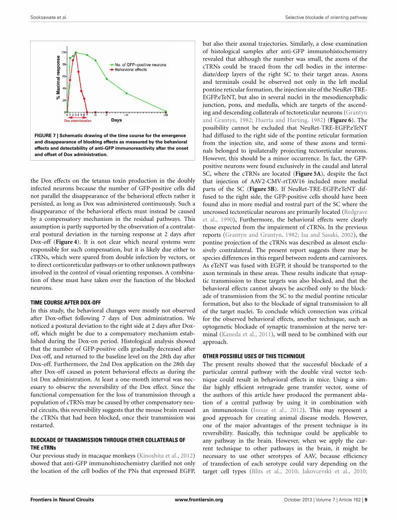
TIME COURSE OF THE DOX-ON AND DOX-OFF PERIODSTo compare the time course of the behavioral effects and theappearance of anti-GFP immunoreactivity in cTRNs for theDox-on and Dox-off periods, the results of the three behav-ioral tests were normalized to the maximal impairment andaveraged. Then, the normalized values were compared to thenormalized number of GFP-positive cTRNs during the Dox-onand Dox-off periods (Figure 7). The number of GFP-positivecTRNs increased sharply after the administration of Dox for1 day, reached its maximum on Dox-on day 3, and wasmaintained until the end of Dox administration on day 7.Although behavioral impairment was also found from Dox-on day 1, it increased slower than the appearance of GFP-positive cTRNs. In contrast to the number of GFP-positivecTRNs, the behavioral impairment, after reaching its maxi-mal effect on Dox-on day 3, was reduced to near the baselinelevel on Dox-on days 6–7. After Dox administration was ter-minated on day 7, the number of GFP-positive cTRNs was
Frontiers in Neural Circuits www.frontiersin.org October 2013 | Volume 7 | Article 162 | 6
Sooksawate et al. Selective blockade of orienting pathway
FIGURE 5 | Photomicrographs of GFP-positive cTRNs and in situ
hybridization to visualize rtTAV16 in the superior colliculus (SC) of
double infected mice. (A) Schematic diagram of the laminar structure of themouse SC (frontal plane) and photomicrographs of the SC slices processedwith anti-GFP immunohistochemistry (frontal section, inset area in the SCdiagram), before Dox administration (Dox-on day 0), Dox administration for 1day (Dox-on day 1), 3 days (Dox-on day 3), 7 days (Dox-on day 7), and after thetermination of Dox administration for 3 days (Dox-off day −3), 7 days
(Dox-off day −7), 14 days (Dox-off day −14), and 28 days (Dox-off day −28).(B) In situ hybridization against the tTA sequence in an SC slice indicating thecells infected by AAV2. (C) Average number of GFP-positive neurons in theSC after the 1st Dox administration for 7 days, termination of Doxadministration for 28 days, and after the 2nd Dox treatment for 7 days (n = 3).Abbreviations: SGS, superficial gray layer; SO, optic layer; SGI, intermediategray layer; SAI, intermediate white layer; SGP, deep gray layer; SAP, deepwhite layer; PAG, periaqueductal gray.
gradually reduced and reached the baseline level on Dox-offday −28.
DISCUSSIONThe crossed tectoreticular pathway has been suggested to beinvolved in the control of orienting responses based on the fol-lowing evidence: (1) repetitive electrical stimulation of the SCinduces contraversive orienting responses (Cowie and Robinson,1994; Corneil et al., 2002) and that of the medial pon-tomedullary reticular formation induces ipsiversive orientingresponses (Cowie and Robinson, 1994); (2) lesion or reversibleinactivation of the SC (Rosenquist et al., 1996; Quaia et al., 1998)or medial pontomedullary reticular formation (Isa and Sasaki,
1988) impairs orienting responses; (3) single unit recordingseither from the SC (Sparks, 1975, 1978) or the medial pon-tomedullary reticular formation (Grantyn and Berthoz, 1987;Isa and Naito, 1995; Isa and Sasaki, 2002) reveal the existenceof neurons that show increased firing preceding the orientingresponse; and (4) anatomically, the medial pontomedullary retic-ular formation receives massive inputs from the intermediate anddeep layers of the contralateral SC (Kawamura and Brodal, 1973;Huerta and Harting, 1982). Thus, there is overwhelming evidencethat the tectoreticular pathway plays a major role in the con-trol of orienting responses (Sparks, 1986; Isa and Sasaki, 2002).However, the specific role of cTRNs in gaze has been difficultto resolve. In fact, this difficulty in precisely attributing function
Frontiers in Neural Circuits www.frontiersin.org October 2013 | Volume 7 | Article 162 | 7
Sooksawate et al. Selective blockade of orienting pathway
FIGURE 6 | Tracings of the axonal trajectories of the GFP-positive
cTRNs in the superior colliculus (SC) in the mice with Dox treatment
for 21 days. (A) GFP-positive cTRNs in the right SC. (B) Axon terminals(arrows) in the left pontine reticular formation. (C) Tracings of the axonsand their terminals in the brainstem: a. Pretectum; b. Midbrain (SC level);c. Midbrain (inferior colliculus level); d. Pons (rostral part); e. Pons (caudalpart); f. Medulla. GFP-positive cell bodies are indicated as blue dots in Cb.Abbreviations: 5N, trigeminal nucleus; 7N, facial nucleus; 7n, facial nerve;12N, hypoglossal nucleus; CnF, cuneiform nucleus; cp, cerebral peduncle;FF, fields of Forel; fr, fasciculus retroflexus; g7, genu of the facial nerve; Gi,
gigantocellular reticular nucleus; IC, inferior colliculus; IO, inferior olive; mlf,medial longitudinal fasciculus; mRt, mesencephalic reticular formation;PAG, periaqueductal gray; pc, posterior commissure; PRF, pontine reticularformation; py, pyramidal tract; RMg, raphe magnus nucleus; RN, rednucleus; rs, rubrospinal tract; RtTg, nucleus reticularis tegmenti pontis;SAI, intermediate white layer; SAP, deep white layer; scp, superiorcerebellar peduncle; SGI, intermediate gray layer; SGP, deep gray layer;SGS, superficial gray layer; SO, optic layer; SNr, substantia nigra parsreticulata; sp5, spinal trigeminal tract; ZID, zona incerta, dorsal part; ZIV,zona incerta, ventral part.
to a specific cell population exists for many of the neural sys-tems in the brain. In this regard, the present study, for the firsttime, showed that pathway-specific and reversible blockade ofsynaptic transmission worked as efficiently to cause behavioraldeficits in mice, as it has in non-human primates (Kinoshita et al.,2012). Moreover, it specifically provides clear evidence that thedirect crossed tectoreticular pathway is essential for the execu-tion of orienting responses. Previous pharmacological techniqueswere not able to dissect this pathway from other tectal outputpathways, such as the thalamic projection from the superficiallayer, which might be involved in the higher visual processing(May, 2006), and from the uncrossed tecto-reticular pathway,which has been reported to control the avoidance behaviors(Sahibzada et al., 1986; Dean et al., 1989). However, the presentmethod will enable us to pin down the functions of these indi-vidual components of the output pathways from the SC in nearfuture.
Finally, we analyzed the methodological details of thistechnique, especially the time course of the effects of Dox-onand Dox-off with this relatively simple behavioral system. Theseexperiments made use of a fair number of mice, which would bedifficult to perform in non-human primates.
TIME COURSE AFTER DOX-ONIn this study, the behavioral effects of Dox administration couldbe partially observed as early as 24 h after initiation. This isearlier than observed in our previous study on macaque mon-keys, in which the effects appeared on the 2nd day. This may bepartly because we used an intraperitoneal injection on the firstday to facilitate the quick onset of the effects (Perl et al., 2002).However, a species-related difference cannot be excluded. Theeffects became maximal on the 2nd and 3rd days after initiation,but gradually became smaller and had mostly disappeared by the6th day. It is clear that this is not due to the disappearance of
Frontiers in Neural Circuits www.frontiersin.org October 2013 | Volume 7 | Article 162 | 8
Sooksawate et al. Selective blockade of orienting pathway
FIGURE 7 | Schematic drawing of the time course for the emergence
and disappearance of blocking effects as measured by the behavioral
effects and detectability of anti-GFP immunoreactivity after the onset
and offset of Dox administration.
the Dox effects on the tetanus toxin production in the doublyinfected neurons because the number of GFP-positive cells didnot parallel the disappearance of the behavioral effects rather itpersisted, as long as Dox was administered continuously. Such adisappearance of the behavioral effects must instead be causedby a compensatory mechanism in the residual pathways. Thisassumption is partly supported by the observation of a contralat-eral postural deviation in the turning response at 2 days afterDox-off (Figure 4). It is not clear which neural systems wereresponsible for such compensation, but it is likely due either tocTRNs, which were spared from double infection by vectors, orto direct corticoreticular pathways or to other unknown pathwaysinvolved in the control of visual orienting responses. A combina-tion of these must have taken over the function of the blockedneurons.
TIME COURSE AFTER DOX-OFFIn this study, the behavioral changes were mostly not observedafter Dox-offset following 7 days of Dox administration. Wenoticed a postural deviation to the right side at 2 days after Dox-off, which might be due to a compensatory mechanism estab-lished during the Dox-on period. Histological analysis showedthat the number of GFP-positive cells gradually decreased afterDox-off, and returned to the baseline level on the 28th day afterDox-off. Furthermore, the 2nd Dox application on the 28th dayafter Dox-off caused as potent behavioral effects as during the1st Dox administration. At least a one-month interval was nec-essary to observe the reversibility of the Dox effect. Since thefunctional compensation for the loss of transmission through apopulation of cTRNs may be caused by other compensatory neu-ral circuits, this reversibility suggests that the mouse brain reusedthe cTRNs that had been blocked, once their transmission wasrestarted.
BLOCKADE OF TRANSMISSION THROUGH OTHER COLLATERALS OFTHE cTRNsOur previous study in macaque monkeys (Kinoshita et al., 2012)showed that anti-GFP immunohistochemistry clarified not onlythe location of the cell bodies of the PNs that expressed EGFP,
but also their axonal trajectories. Similarly, a close examinationof histological samples after anti-GFP immunohistochemistryrevealed that although the number was small, the axons of thecTRNs could be traced from the cell bodies in the interme-diate/deep layers of the right SC to their target areas. Axonsand terminals could be observed not only in the left medialpontine reticular formation, the injection site of the NeuRet-TRE-EGFP.eTeNT, but also in several nuclei in the mesodiencephalicjunction, pons, and medulla, which are targets of the ascend-ing and descending collaterals of tectoreticular neurons (Grantynand Grantyn, 1982; Huerta and Harting, 1982) (Figure 6). Thepossibility cannot be excluded that NeuRet-TRE-EGFP.eTeNThad diffused to the right side of the pontine reticular formationfrom the injection site, and some of these axons and termi-nals belonged to ipsilaterally projecting tectoreticular neurons.However, this should be a minor occurrence. In fact, the GFP-positive neurons were found exclusively in the caudal and lateralSC, where the cTRNs are located (Figure 5A), despite the factthat injection of AAV2-CMV-rtTAV16 included more medialparts of the SC (Figure 5B). If NeuRet-TRE-EGFP.eTeNT dif-fused to the right side, the GFP-positive cells should have beenfound also in more medial and rostral part of the SC where theuncrossed tectoreticular neurons are primarily located (Redgraveet al., 1990), Furthermore, the behavioral effects were clearlythose expected from the impairment of cTRNs. In the previousreports (Grantyn and Grantyn, 1982; Isa and Sasaki, 2002), thepontine projection of the cTRNs was described as almost exclu-sively contralateral. The present report suggests there may bespecies differences in this regard between rodents and carnivores.As eTeNT was fused with EGFP, it should be transported to theaxon terminals in these areas. These results indicate that synap-tic transmission to these targets was also blocked, and that thebehavioral effects cannot always be ascribed only to the block-ade of transmission from the SC to the medial pontine reticularformation, but also to the blockade of signal transmission to allof the target nuclei. To conclude which connection was criticalfor the observed behavioral effects, another technique, such asoptogenetic blockade of synaptic transmission at the nerve ter-minal (Kaneda et al., 2011), will need to be combined with ourapproach.
OTHER POSSIBLE USES OF THIS TECHNIQUEThe present results showed that the successful blockade of aparticular central pathway with the double viral vector tech-nique could result in behavioral effects in mice. Using a sim-ilar highly efficient retrograde gene transfer vector, some ofthe authors of this article have produced the permanent abla-tion of a central pathway by using it in combination withan immunotoxin (Inoue et al., 2012). This may represent agood approach for creating animal disease models. However,one of the major advantages of the present technique is itsreversibility. Basically, this technique could be applicable toany pathway in the brain. However, when we apply the cur-rent technique to other pathways in the brain, it might benecessary to use other serotypes of AAV, because efficiencyof transfection of each serotype could vary depending on thetarget cell types (Blits et al., 2010; Jakovcevski et al., 2010;
Frontiers in Neural Circuits www.frontiersin.org October 2013 | Volume 7 | Article 162 | 9
Sooksawate et al. Selective blockade of orienting pathway
Markakis et al., 2010). The success of the current method was pri-marily based on the following factors: (1) development of highlyefficient retrograde gene transfer using NeuRet; (2) very effi-cient amplification of gene expression by the recently developedTet-ON sequence rtTAV16; and (3) very effective expression ofthe humanized tetanus neurotoxin eTeNT. Replacing eTeNT withother functional proteins, such as light-sensitive opsins for opto-genetic control of neural activity (for reviews see Yizhar et al.,2011; Tye and Disseroth, 2012), may open up a novel directionfor neural circuit analysis.
ACKNOWLEDGMENTSThis study is the result of “Highly Creative Animal ModelDevelopment for Brain Sciences” carried out under the StrategicResearch Program for Brain Sciences (to Dai Watanabe, KazutoKobayashi, and Tadashi Isa) and a Grant-in-Aid for ScientificResearch on Innovative Areas “Mesoscopic Neurocircuitry” (No.25115731 to Tadashi Isa) by the Ministry of Education, Culture,Sports, Science, and Technology of Japan. We thank KotomiShimizu for assistance with immunohistochemistry and in situhybridization.
REFERENCESBlits, B., Derks, S., Twisk, J., Ehlert,
E., Prins, J., and Verhaagen, J.(2010). Adeno-associated viral vec-tor (AAV)-mediated gene trans-fer in the red nucleus of theadult rat brain: comparative anal-ysis of the transduction propertiesof seven AAV serotypes and lentivi-ral vectors. J. Neurosci. Methods 185,257–263. doi: 10.1016/j.jneumeth.2009.10.009
Corneil, B. D., Olivier, E., and Munoz,D. P. (2002). Neck muscle responsesto stimulation of monkey supe-rior colliculus. I. Topographyand manipulation of stimulationparameters. J. Neurophysiol. 88,1980–1999.
Cowie, R. J., and Robinson, D. L.(1994). Subcortical contributionsto head movements in macaques.I. Contrasting effects of electri-cal stimulation of a medial pon-tomedullary region and the supe-rior colliculus. J. Neurophysiol. 72,2648–2664.
Dean, P., Redgrave, P., and Westby, G.W. (1989). Event or emergency? Tworesponse systems in the mammaliansuperior colliculus. Trends Neurosci.12, 137–147. doi: 10.1016/0166-2236(89)90052-0
Franklin, K. B. J., and Paxinos, G.(2008). The Mouse Brain: inStereotaxic Coordinates. 3rd Edn.New York, NY: Academic Press.
Grantyn, A., and Berthoz, A. (1987).Reticulo-spinal neurons participat-ing in the control of synergic eye andhead movements during orientingin the cat. I. Behavioral properties.Exp. Brain Res. 66, 339–354. doi:10.1007/BF00243309
Grantyn, A., and Grantyn, R. (1982).Axonal patterns and sites oftermination of cat superior col-liculus neurons projecting in thetecto-bulbo-spinal tract. Exp.Brain Res. 46, 243–256. doi:10.1007/BF00237182
Huerta, M. F., and Harting, J. K.(1982). Tectal control of spinal cordactivity: neuroanatomical demon-stration of pathways connecting the
superior colliculus with the cervi-cal spinal cord grey. Prog. Brain Res.57, 293–328. doi: 10.1016/S0079-6123(08)64135-7
Inoue, K., Koketsu, D., Kato, S.,Kobayashi, K., Nambu, A., andTakada, M. (2012). Immunotoxin-mediated tract targeting inthe primate brain: selectiveelimination of the cortico-subthalamic“hyperdirect” pathway.PLoS ONE 7:e39149. doi: 10.1371/journal.pone.0039149
Isa, T., and Hall, W. C. (2009).Exploring the superior collicu-lus in vitro. J. Neurophysiol. 102,2581–2593. doi: 10.1152/jn.00498.2009
Isa, T., and Naito, K. (1995). Activityof neurons in the medial pon-tomedullary reticular formationduring orienting movements inalert head-free cats. J. Neurophysiol.74, 73–95.
Isa, T., and Sasaki, S. (1988). Effectsof lesion of paramedian pon-tomedullary reticular formationby kainic acid injection on thevisually triggered horizontal ori-enting movements in the cat.Neurosci. Lett. 87, 233–239. doi:10.1016/0304-3940(88)90454-5
Isa, T., and Sasaki, S. (2002). Brainstemcontrol of head movements dur-ing orienting; organization of thepremotor circuits. Prog. Neurobiol.66, 205–241. doi: 10.1016/S0301-0082(02)00006-0
Jakovcevski, M., Guo, Y., Su, Q.,Gao, G., and Akbarian, S. (2010).rAAV9–a human-derived adeno-associated virus vector for efficienttransgene expression in mousecingulate cortex. Cold SpringHarb. Protoc. 4, pdb.prot5417. doi:10.1101/pdb.prot5417
Kaneda, K., Kasahara, H., Matsui,R., Katoh, T., Mizukami, H.,Ozawa, K., et al. (2011). Selectiveoptical control of synaptic trans-mission in the subcortical visualpathway by activation of viralvector-expressed halorhodopsin.PLoS ONE 6:e18452. doi:10.1371/journal.pone.0018452
Kato, S., Inoue, K., Kobayashi, K.,Yasoshima, Y., Miyachi, S., Inoue,S., et al. (2007). Efficient genetransfer via retrograde transport inrodent and primate brains usinga human immunodeficiency virustype 1-based vector pseudotypedwith rabies virus glycoprotein.Hum. Gene. Ther. 18, 1141–1151.doi: 10.1089/hum.2007.082
Kato, S., Kobayashi, K., Inoue, K.,Kuramochi, M., Okada, T.,Yaginuma, H., et al. (2011a).A lentiviral strategy for highlyefficient retrograde gene trans-fer by pseudotyping with fusionenvelope glycoprotein. Hum.Gene Ther. 22, 197–206. doi:10.1089/hum.2009.179
Kato, S., Kuramochi, M., Takasumi, K.,Kobayashi, K., Inoue, K., Takahara,D., et al. (2011b). Neuron-specificgene transfer through retrogradetransport of lentiviral vector pseu-dotyped with a novel type offusion envelope glycoprotein. Hum.Gene Ther. 22, 1511–1523. doi:10.1089/hum.2011.111
Kawamura, K., and Brodal, A. (1973).The tectopontine projection in thecat: an experimental anatomicalstudy with comments on path-ways for teleceptive impulses tothe cerebellum. J. Comp. Neurol.149, 371–390. doi: 10.1002/cne.901490306
Kinoshita, M., Matsui, R., Kato, S.,Hasegawa, T., Kasahara, H., Isa, K.,et al. (2012). Genetic dissection ofthe circuit for hand dexterity inprimate. Nature 487, 235–238. doi:10.1038/nature11206
Kobayashi, K., Morita, S., Sawada, H.,Mizuguchi, T., Yamada, K., Nagatsu,I., et al. (1995). Immunotoxin-mediated conditional disruptionof specific neurons in trans-genic mice. Proc. Natl. Acad.Sci. U.S.A. 92, 1132–1136. doi:10.1073/pnas.92.4.1132
Markakis, E. A., Vives, K. P., Bober, J.,Leichtle, S., Leranth, C., Beecham,J., et al. (2010). Comparative trans-duction efficiency of AAV vec-tor serotypes 1-6 in the substantia
nigra and striatum of the primatebrain. Mol. Ther. 18, 588–593. doi:10.1038/mt.2009.286
May, P. J. (2006). The mammaliansuperior colliculus: laminarstructure and connections. Prog.Brain Res. 151, 321–378. doi:10.1016/S0079-6123(05)51011-2
Metz, G. A., and Schwab, M. E. (2004).Behavioral characterization in acomprehensive mouse test bat-tery reveals motor and sensoryimpairments in growth-associatedprotein-43 null mutant mice.Neuroscience. 129, 563–574. doi:10.1016/j.neuroscience.2004.07.053
Montecucco, C., and Schiavo, G.(1994). Mechanism of action oftetanus and botulinum neuro-toxins. Mol. Microbiol. 13, 1–8.doi: 10.1111/j.1365-2958.1994.tb00396.x
Perl, A.-K. T., Tichelaar, J. W., andWhitsett, J. A. (2002). Conditiongene expression in the respiratoryepithelium of mouse. TransgenicRes. 11, 21–29. doi: 10.1023/A:1013986627504
Pinto, L. H., and Enroth-Cugell,C. (2000). Tests of the mousevisual system. Mamm. Genome.11, 531–536. doi: 10.1007/s003350010102
Quaia, C., Aizawa, H., Optican, L.M., and Wurtz, R. H. (1998).Reversible inactivation of monkeysuperior colliculus. II. Maps of sac-cadic deficits. J. Neurophysiol. 79,2097–2110.
Redgrave, P., Dean, P., and Westby,G. W. (1990). Organization of thecrossed tecto-reticulo-spinal projec-tion in rat - I. Anatomical evi-dence for separate output chan-nels to the periabducens area andcaudal medulla. Neuroscience 37,571–584. doi: 10.1016/0306-4522(90)90092-I
Rosenquist, A. C., Ciaramitaro, V. M.,Durmer, J. S., Wallace, S. F., andTodd, W. E. (1996). Ibotenic acidlesions of the superior colliculusproduce longer lasting deficits invisual orienting behavior thanaspiration lesions in the cat. Prog.
Frontiers in Neural Circuits www.frontiersin.org October 2013 | Volume 7 | Article 162 | 10
Sooksawate et al. Selective blockade of orienting pathway
Brain Res. 112, 117–130. doi:10.1016/S0079-6123(08)63324-5
Sahibzada, N., Dean, P., and Redgrave,P. (1986). Movements resemblingorientation or avoidance elicited byelectrical stimulation of the supe-rior colliculus in rats. J. Neurosci. 6,723–733.
Smith, D. R., Striplin, C. D., Geller,A. M., Mailman, R. B., Drago,J., Lawler, C. P., et al. (1998).Behavioural assessment ofmice lacking D1A dopaminereceptors. Neuroscience 86,135–146. doi: 10.1016/S0306-4522(97)00608-8
Sooksawate, T., Isa, K., and Isa, T.(2008). Cholinergic responsesin crossed tecto-reticular neu-rons of rat superior colliculus.J. Neurophysiol. 100, 2702–2711.doi: 10.1152/jn.90723.2008
Sooksawate, T., Saito, Y., and Isa,T. (2005). Electrophysiologicaland morphological properties ofidentified cross tecto-reticular neu-rons in the rat superior colliculus.
Neurosci. Res. 52, 174–184. doi:10.1152/jn.90723.2008
Sparks, D. L. (1975). Responseproperties of eye movement-related neurons in the monkeysuperior colliculus. Brain Res. 90,147–152. doi: 10.1016/0006-8993(75)90690-3
Sparks, D. L. (1978). Functional prop-erties of neurons in the mon-key superior colliculus: couplingof neuronal activity and saccadeonset. Brain Res. 156, 1–16. doi:10.1016/0006-8993(78)90075-6
Sparks, D. L. (1986). Translationof sensory signals into com-mands for control of saccadiceye movements: role of primatesuperior colliculus. Physiol. Rev. 66,118–171.
Tye, K. M., and Disseroth, K. (2012).Optogenetic investigation of neuralcircuits underlying brain disease inanimal models. Nat. Rev. Neurosci.13, 251–266. doi: 10.1038/nrn3171
Watanabe, D., Inokawa, H., Hashimoto,K., Suzuki, N., Kano, M.,
Shigemoto, R., et al. (1998).Ablation of cerebellar Golgi cellsdisrupts synaptic integrationinvolving GABA inhibition andNMDA receptor activation inmotor coordination. Cell 95,17–27. doi: 10.1016/S0092-8674(00)81779-1
Wurtz, R. H., and Albano, J. E. (1980).Visual-motor function of the pri-mate superior colliculus. Annu. Rev.Neurosci. 3, 189–226. doi: 10.1146/annurev.ne.03.030180.001201
Yizhar, O., Fenno, L. E., Davidson, T.J., Mogri, M., and Deisseroth,K. (2011). Optogenetics inneural systems. Neuron 71,9–34. doi: 10.1016/j.neuron.2011.06.004
Conflict of Interest Statement: Theauthors declare that the researchwas conducted in the absence of anycommercial or financial relationshipsthat could be construed as a potentialconflict of interest.
Received: 01 July 2013; accepted: 21September 2013; published online: 11October 2013.Citation: Sooksawate T, Isa K, MatsuiR, Kato S, Kinoshita M, Kobayashi K,Watanabe D, Kobayashi K and Isa T(2013) Viral vector-mediated selectiveand reversible blockade of the path-way for visual orienting in mice. Front.Neural Circuits 7:162. doi: 10.3389/fncir.2013.00162This article was submitted to the journalFrontiers in Neural Circuits.Copyright © 2013 Sooksawate, Isa,Matsui, Kato, Kinoshita, Kobayashi,Watanabe, Kobayashi and Isa. This isan open-access article distributed underthe terms of the Creative CommonsAttribution License (CC BY). The use,distribution or reproduction in otherforums is permitted, provided the origi-nal author(s) or licensor are credited andthat the original publication in this jour-nal is cited, in accordance with acceptedacademic practice. No use, distributionor reproduction is permitted which doesnot comply with these terms.
Frontiers in Neural Circuits www.frontiersin.org October 2013 | Volume 7 | Article 162 | 11