virtual 3d model of the lumbar spine - ubdiposit.ub.edu/dspace/bitstream/2445/44844/16/... · ·...
TRANSCRIPT

LSNA
Virtual 3D Model of the Lumbar Spine
Alberto Prats-Galino1, Miguel Angel Reina2, Marija Mavar Haramija1, Anna Puigdellivol-Sánchez1, Joan San Molina3 and José Antonio De Andrés4.
1 Laboratory of Surgical NeuroAnatomy (LSNA), Human Anatomy and Embryology Unit, Faculty of Medicine, Universitat de Barcelona, Barcelona, Spain. 2 Department of Clinical Medical Sciences and Applied Molecular Medicine Institute, CEU San Pablo University School of Medicine, Department of Anaesthesiology, Madrid-Montepríncipe University Hospital, Madrid, Spain. 3 Department of Medical Sciences, Faculty of Medicine. Universitat de Girona, Spain. 4 Department of Critical Care and Multidisciplinary Pain Management, General University Hospital, Valencia, Spain.

Virtual 3D Model of the Lumbar Spine
2
Table of Contents
1 Introduction ................................................................................................................................................................. 3
1.1 About the document ............................................................................................................................................ 3
Authors ......................................................................................................................................................................... 3
1.2 Objectives ............................................................................................................................................................. 4
2 Basic Instructions ......................................................................................................................................................... 5
2.1 Screen Working Area ........................................................................................................................................... 6
2.2 Default Views ....................................................................................................................................................... 8
2.3 Structure Selection ............................................................................................................................................. 10
2.4 Structure Validation Function ............................................................................................................................ 12
2.5 Transparency Graduation .................................................................................................................................. 12
2.6 “Show all” Option ............................................................................................................................................... 13
2.7 “Hide all” Option ................................................................................................................................................ 13
2.8 MRI Slices ........................................................................................................................................................... 14
2.9 MRI Slice Navigation .......................................................................................................................................... 14
2.10 Clipping Function ............................................................................................................................................... 16
2.11 Clipping Plane Orientation ................................................................................................................................. 16
2.12 Clipping Plane Offset .......................................................................................................................................... 17
2.13 Predetermined Custom Views and Short Explanations ..................................................................................... 19
2.14 “Disable selection” Option ................................................................................................................................. 19
2.15 Access the Instructions ...................................................................................................................................... 19
3 Table of Figures .......................................................................................................................................................... 20
4 Acknowledgements .................................................................................................................................................... 21
5 Licensing ..................................................................................................................................................................... 21

Virtual 3D Model of the Lumbar Spine
3
1 Introduction
1.1 About the document
Figure 1 Interactive 3D PDF document
Authors
Alberto Prats-Galino1, Miguel Angel Reina2, Marija Mavar Haramija1, Anna Puigdellivol-Sánchez1, Joan San Molina3 and José Antonio De Andrés4. 1 Laboratory of Surgical NeuroAnatomy (LSNA), Human Anatomy and Embryology Unit, Faculty of Medicine, Universitat de Barcelona, Barcelona, Spain. 2 Department of Clinical Medical Sciences and Applied Molecular Medicine Institute, CEU San Pablo University School of Medicine, Department of Anaesthesiology, Madrid-Montepríncipe University Hospital, Madrid, Spain. 3 Department of Medical Sciences, Faculty of Medicine. Universitat de Girona, Spain. 4 Department of Critical Care and Multidisciplinary Pain Management, General University Hospital, Valencia, Spain. Mail address for correspondence: Alberto Prats-Galino: [email protected] Miguel Angel Reina: [email protected]

Virtual 3D Model of the Lumbar Spine
4
This interactive model has been developed from 3D reconstructions of human magnetic resonance (MR) images. The authors have no conflict of interest to declare.
1.2 Objectives
The PDF format enclosing this three dimensional (3D) interactive anatomical model greatly simplifies its use, portability, compatibility and storage as the file size can be compressed and transferred across multiple platforms. The main fields of interest in regard to this specific 3D anatomical model include: Educational programs
Real 3D imaging support for teaching neuroaxial anatomy and regional anesthesia Visual aid in the development of new approaches in regional anesthetic techniques
Research programs
Review of patient data and analysis of anesthetic techniques Image inspection techniques of complications in regional anesthesia
Patient information
Complementary visual aid to instrumental techniques and surgical procedures relevant to patients This project allows users to examine 3D reconstructions from human MR images. We present a 3D
interactive anatomical model of interest in regional anesthesia and pain medicine (e.g. Figure 2 shows spinal medial approach illustrated by our model).
Figure 2 Example: Spinal medial approach
The technology used to support this interactive model has recently being applied in medicine, and enables us to produce interactive 3D models by means of a complex and strenuous technique of 3D image reconstruction from 2D images thanks to specific software (Amira 5.4.0 ©). The outcome is a simple and useful teaching, working and research tool. The use of this type of model is straightforward and does not

Virtual 3D Model of the Lumbar Spine
5
require previous experience, as it is an intuitive product, supported in PDF file format and can be opened in Windows and Mac computers. But above all, it may be used free of charge by every physician. The present project includes reconstructions of vertebrae, vertebral disks, vertebral arches, ligamentum flavum, supraspinous ligaments, interspinous ligaments, epidural fat, foraminal fat, dural sac, nerve root cuffs, sensory and motor nerve roots. It has a dynamic 360º view including partial or total views of each and all structures, including zoom function.
2 Basic Instructions
The PDF document runs under Adobe Acrobat 11 or superior. Navigation may be started from different points. One or more structures may be selected initially, as well as selecting preset options of 3D reconstruction images. Interaction with the screen is performed by mouse, allowing the model to move, offering views of the structures from different perspectives. It includes axial, sagittal and coronal MRI slices as reference. This interactive anatomical model allows cutting in axial, sagittal and coronal planes. Clipping may include all the structures reconstructed in the model. The other options are intuitive and easy to grasp.
Figure 3 Function description: click on a numbered button to jump to a corresponding chapter

Virtual 3D Model of the Lumbar Spine
6
The functionality of this pdf document is described in 15 short instructions, each one explaining a concrete
function/button. You can click on the blue numbered buttons on the Figure 3 to jump to a chapter describing
the corresponding function.
2.1 Screen Working Area
Full screen mode is invoked by Control + L key combination and exited by the ESC key.
Figure 4 Working area

Virtual 3D Model of the Lumbar Spine
7
Dynamic 360º view is obtained by mouse motion while holding down left mouse button.
Figure 5 360º rotation of the model
Virtual 3D Model of the Lumbar Spine
8
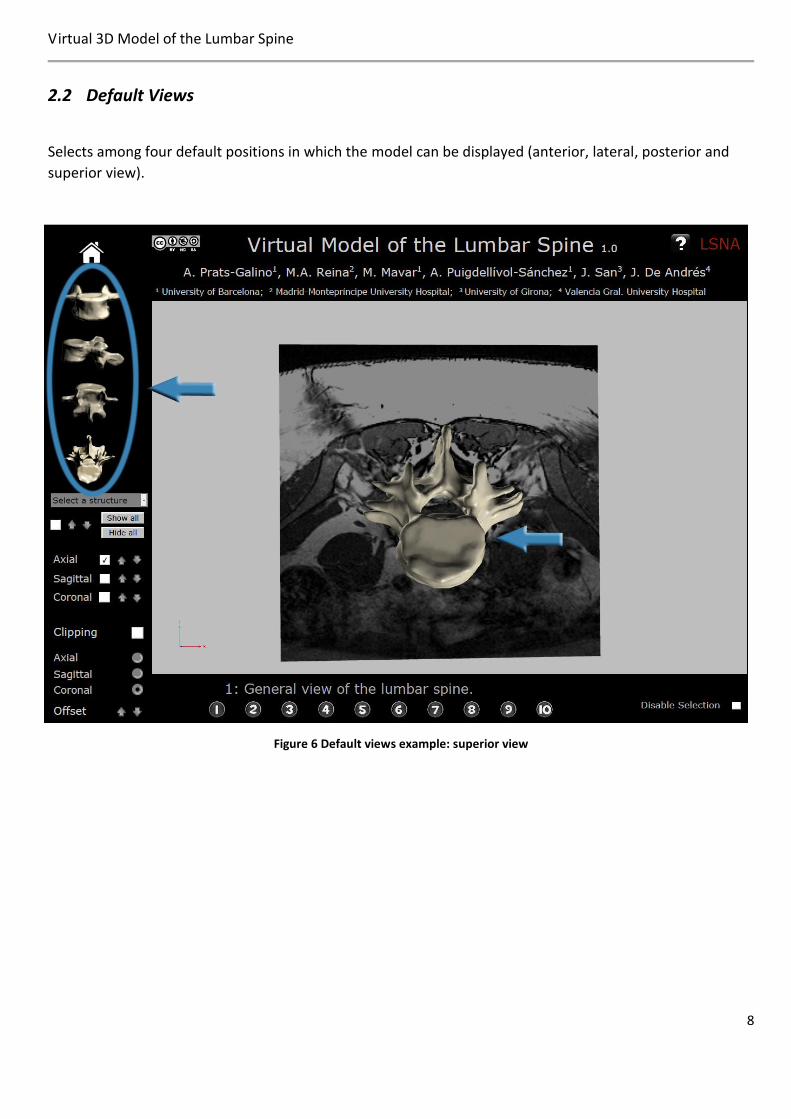
2.2 Default Views
Selects among four default positions in which the model can be displayed (anterior, lateral, posterior and
superior view).
Figure 6 Default views example: superior view

Virtual 3D Model of the Lumbar Spine
9
Figure 7 Default views example: lateral view

Virtual 3D Model of the Lumbar Spine
10
2.3 Structure Selection
Click on the structure of the drop-down menu to build the model up on the screen. Selection is validated by
clicking on function Nº 4 (show/hide selected structure). Structures may be successively incorporated to the
model on the screen.
Figure 8 Structure selection function
Figure 9 Structure selection example: epidural fat selected (in red)

Virtual 3D Model of the Lumbar Spine
11
Figure 10 Structure selection example: Epidural fat selected
Figure 11 Examples of different structures shown/hidden

Virtual 3D Model of the Lumbar Spine
12
2.4 Structure Validation Function
Structure validation button, when checked, the selected structure is added to the model in the working area. When
unchecked, the selected structure is hidden from the model.
Figure 12 Structure validation: show/hide selected structure
2.5 Transparency Graduation
Arrows modify the degree of transparency, allowing the structure selected on the screen to disappear
progressively as it changes its color.
Figure 13 Gradual change of the selected structure's transparency
Figure 14 Transparency graduation example: Semi-transparent L4 vertebra

Virtual 3D Model of the Lumbar Spine
13
2.6 “Show all” Option
This button displays all of the structures of the 3D model on the screen. Each superficial structure may be
selected and made transparent, allowing internal ones to appear on the screen. To remove structures from
the screen, select each structure by clicking on it and pressing on validation-function (2.4).
Alternatively, structures can be removed from the screen by selecting their name in the drop-down menu
(2.3) and clicking the structure validation function (2.4).
Figure 15 Show all: All structures of the model are made visible
2.7 “Hide all” Option
“Hide all” button removes all structures displayed in the working area of the screen.

Virtual 3D Model of the Lumbar Spine
14
2.8 MRI Slices
This functionality shows or hides MRI slices from the working area.
Axial, sagittal, and coronal options may be selected either independently or in combination.
Figure 16 Showing MRI slices in the working area
2.9 MRI Slice Navigation
Arrows allow navigating from one MRI slice to the next. There are coupled arrows for each of the
orientations: there are nine axial, seven sagittal and six coronal slices.
Figure 17 Axial MRI slices

Virtual 3D Model of the Lumbar Spine
15
Figure 18 Sagittal MRI slices
Figure 19 Coronal MRI slices

Virtual 3D Model of the Lumbar Spine
16
2.10 Clipping Function
This function displays cuts of the original 3D model, including the MRI slices selected by function 11.
Figure 20 Clipping: cutting the model by a clipping plane
2.11 Clipping Plane Orientation
This function selects different clipping plane orientations: axial, sagittal or coronal.
Figure 21 Clipping plane orientations

Virtual 3D Model of the Lumbar Spine
17
2.12 Clipping Plane Offset
This functionality allows navigating along successive cuts of the model (by moving the clipping plane).
Figure 22 Clipping plane offset: moving along the model
Figure 23 Clipping example: Axial clipping

Virtual 3D Model of the Lumbar Spine
18
Figure 24 Clipping example: Sagittal and coronal clipping

Virtual 3D Model of the Lumbar Spine
19
2.13 Predetermined Custom Views and Short Explanations
Different predetermined views are available, together with a short explanation of the current view. All
above mentioned functions may be applied to each view.
Figure 25 Custom views and comments
2.14 “Disable selection” Option
This option disables user from selecting structures by a mouse click.
It can be useful in different occasions: in presence of problems regarding performance of screen area due to
problems with graphical properties of a computer or similar, or simply if we want to rotate the model
without accidently highlighting the structures causing them to appear in color red.
Figure 26 Disable selection of the model
2.15 Access the Instructions
Instructions on how to use interactive PDF file can be accessed by clicking on a question mark button on the
top right.
Figure 27 Accessing instructions

Virtual 3D Model of the Lumbar Spine
20
3 Table of Figures
Figure 1 Interactive 3D PDF document ................................................................................................................................ 3
Figure 2 Example: Spinal medial approach .......................................................................................................................... 4
Figure 3 Function description: click on a numbered button to jump to a corresponding chapter ..................................... 5
Figure 4 Working area .......................................................................................................................................................... 6
Figure 5 360º rotation of the model .................................................................................................................................... 7
Figure 6 Default views example: superior view ................................................................................................................... 8
Figure 7 Default views example: lateral view ...................................................................................................................... 9
Figure 8 Structure selection function................................................................................................................................. 10
Figure 9 Structure selection example: epidural fat selected (in red) ................................................................................ 10
Figure 10 Structure selection example: Epidural fat selected ........................................................................................... 11
Figure 11 Examples of different structures shown/hidden ............................................................................................... 11
Figure 12 Structure validation: show/hide selected structure .......................................................................................... 12
Figure 13 Gradual change of the selected structure's transparency ................................................................................. 12
Figure 14 Transparency graduation example: Semi-transparent L4 vertebra ................................................................... 12
Figure 15 Show all: All structures of the model are made visible...................................................................................... 13
Figure 16 Showing MRI slices in the working area ............................................................................................................. 14
Figure 17 Axial MRI slices ................................................................................................................................................... 14
Figure 18 Sagittal MRI slices............................................................................................................................................... 15
Figure 19 Coronal MRI slices .............................................................................................................................................. 15
Figure 20 Clipping: cutting the model by a clipping plane ................................................................................................. 16
Figure 21 Clipping plane orientations ................................................................................................................................ 16
Figure 22 Clipping plane offset: moving along the model ................................................................................................. 17
Figure 23 Clipping example: Axial clipping ......................................................................................................................... 17
Figure 24 Clipping example: Sagittal and coronal clipping ................................................................................................ 18
Figure 25 Custom views and comments ............................................................................................................................ 19
Figure 26 Disable selection of the model........................................................................................................................... 19
Figure 27 Accessing instructions ........................................................................................................................................ 19

Virtual 3D Model of the Lumbar Spine
21
4 Acknowledgements
This work has been partly supported by the grants "Marató TV3 Project" [411/U/2011 - TITLE: Quantitative
analysis and computer aided simulation of minimally invasive approaches for intracranial vascular lesions.]
and "2012PID-UB/002 Project" [Grupo de Anatomía Virtual y de Simulación, Universitat de Barcelona].
We also thank Olga Fuentes1 for her technical assistance.
5 Licensing
The 3D Interactive Spine Virtual Model PDF file is distributed under CC BY-NC-SA 2.02 license, which requires
attribution to the authors, but allows derivate works without commercial use, provided that it is shared
under the same license as the original document.
JavaScript source code and original surface model geometry are not public and cannot be accessed or
modified.
In case of using any part of the mentioned document (e.g. image capture of the anatomical model)
according to CC BY-NC-SA 2.0 license please reference this work by the following citation:
- Prats-Galino A, Mavar M, Reina MA, Puigdellívol-Sánchez A, San-Molina J, De Andrés JA. Three-
dimensional interactive model of lumbar spinal structures. Anaesthesia 2014; 69:521.
The original 3D PDF document, together with the instructions and the license file, is freely available at
http://diposit.ub.edu/dspace/handle/2445/44844?locale=en.
1 Laboratory of Surgical NeuroAnatomy (LSNA), Human Anatomy and Embryology Unit, Faculty of Medicine, Universitat de
Barcelona, Barcelona.
2 http://creativecommons.org/licenses/by-nc-sa/2.0/