visual apparatus -...
TRANSCRIPT
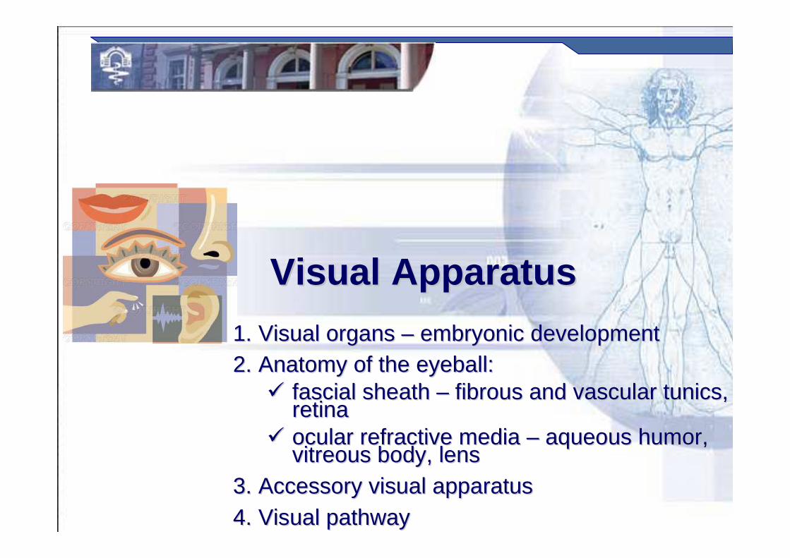
Visual ApparatusVisual Apparatus
1.1. Visual organs Visual organs –– embryonic development embryonic development 2.2. Anatomy of the eyeball:Anatomy of the eyeball:
�� fascial sheath fascial sheath –– fibrous and vascular tunics, fibrous and vascular tunics, retinaretina
�� ocular refractive media ocular refractive media –– aqueous humor, aqueous humor, vitreous body, lensvitreous body, lens
3.3. Accessory visual apparatusAccessory visual apparatus4.4. Visual pathwayVisual pathway
Prof. Dr. Nikolai LazarovProf. Dr. Nikolai Lazarov 2
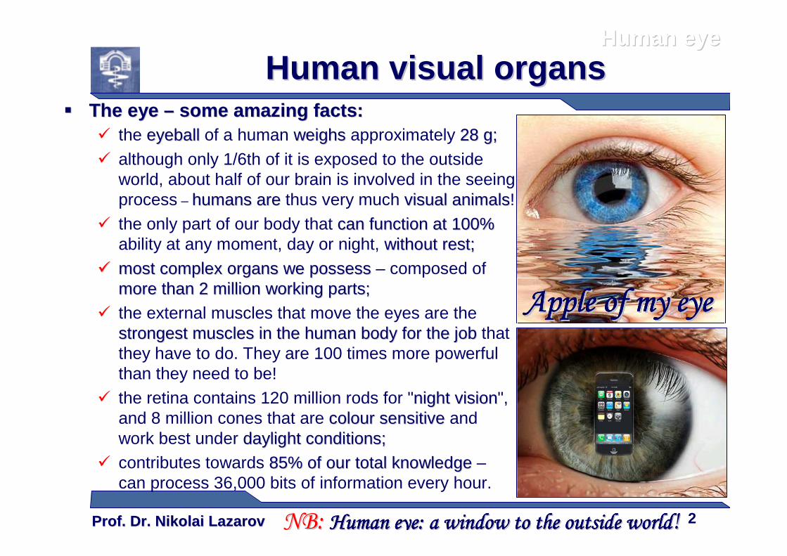
�� The eye The eye –– ssome amazing facts:ome amazing facts:� the eyeballeyeball of a human weighsweighs approximately 28 g28 g;;
� although only 1/6th of it is exposed to the outside world, about half of our brain is involved in the seeing process – hhumans areumans are thus very much visual animalsvisual animals!
� the only part of our body that can function at 100%can function at 100%ability at any moment, day or night, without restwithout rest;;
�� most complex organs most complex organs wewe possesspossess – composed of more than 2 million working partsmore than 2 million working parts;;
� the external muscles that move the eyes are the strongest muscles in the human body for the jobstrongest muscles in the human body for the job that they have to do. They are 100 times more powerful than they need to be!
� the retina contains 120 million rods for "night visionnight vision", and 8 million cones that are colour sensitivecolour sensitive and work best under daylight conditionsdaylight conditions;;
� contributes towards 85% of 85% of ourour total knowledgetotal knowledge –can process 36,000 bits of information every hour.
Human visual organsHuman visual organs
NB: NB: Human eye:Human eye:Human eye:Human eye:Human eye:Human eye:Human eye:Human eye: a window to the outside worlda window to the outside worlda window to the outside worlda window to the outside worlda window to the outside worlda window to the outside worlda window to the outside worlda window to the outside world!!
Human eyeHuman eye
Apple of my eyeApple of my eyeApple of my eyeApple of my eyeApple of my eyeApple of my eyeApple of my eyeApple of my eye
Prof. Dr. Nikolai LazarovProf. Dr. Nikolai Lazarov 3
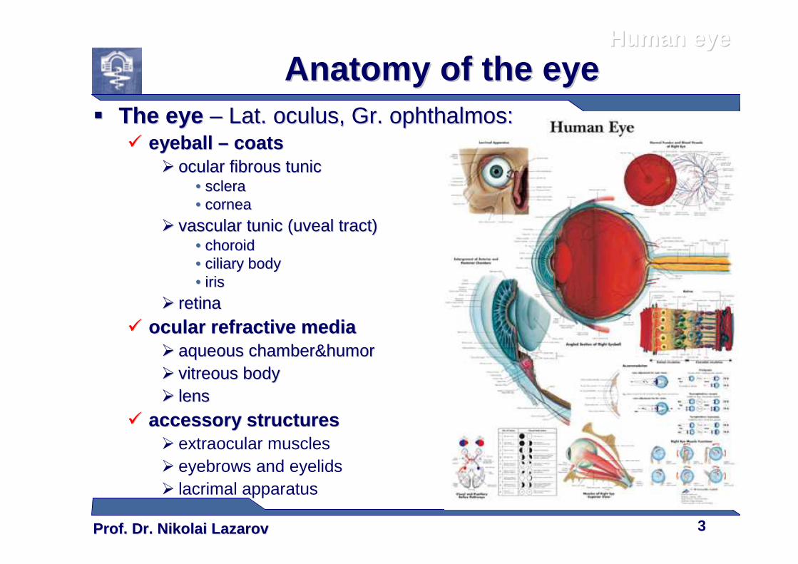
�� The eye The eye –– Lat. Lat. oculusoculus, Gr. , Gr. ophthalmosophthalmos::�� eyeball eyeball –– coatscoats
�� ocular fibrous tunicocular fibrous tunic•• sclerasclera•• corneacornea
�� vascular tunic (uveal tract)vascular tunic (uveal tract)•• choroid choroid •• ciliary bodyciliary body•• irisiris
�� retinaretina
�� ocular refractive mediaocular refractive media�� aqueous chamber&humoraqueous chamber&humor�� vitreous bodyvitreous body�� lenslens
�� accessory structuresaccessory structures� extraocular muscles� eyebrows and eyelids� lacrimal apparatus
Anatomy of the eyeAnatomy of the eyeHuman eyeHuman eye
Prof. Dr. Nikolai LazarovProf. Dr. Nikolai Lazarov 4
Human eyeHuman eye
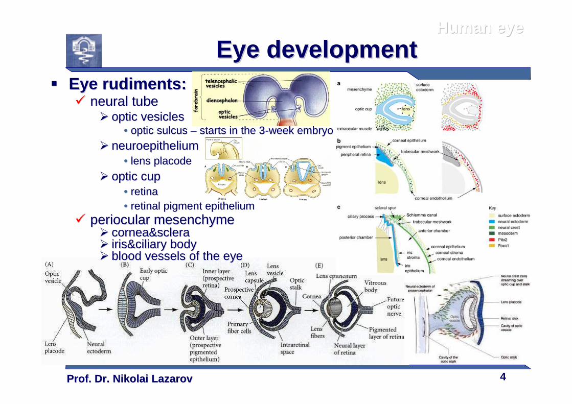
Eye developmentEye development�� Eye rudimentsEye rudiments ::
�� neural tubeneural tube�� optic vesiclesoptic vesicles
•• optic sulcus optic sulcus –– starts in the 3starts in the 3--week embryoweek embryo
�� neuroepitheliumneuroepithelium•• lens placode lens placode
�� optic cupoptic cup•• retinaretina•• retinal pigment epitheliumretinal pigment epithelium
�� periocular mesenchymeperiocular mesenchyme�� cornea&scleracornea&sclera�� iris&ciliary bodyiris&ciliary body�� blood vessels of the eyeblood vessels of the eye
Prof. Dr. Nikolai LazarovProf. Dr. Nikolai Lazarov 5
Human eyeHuman eye
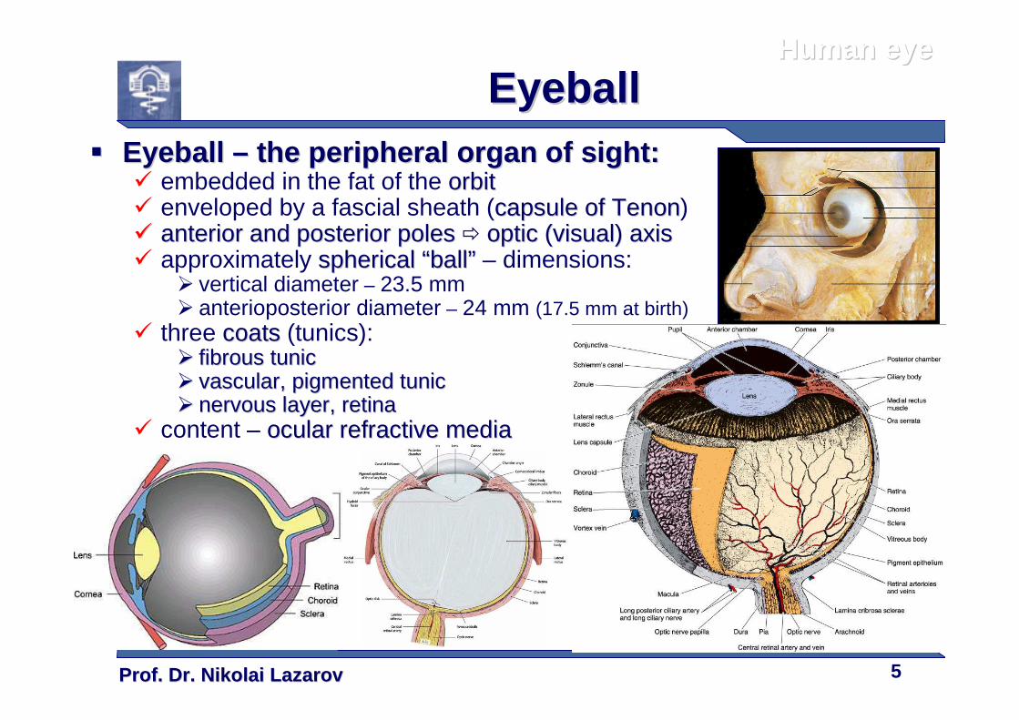
EyeballEyeball�� Eyeball Eyeball –– the peripheral organ of sightthe peripheral organ of sight ::
� embedded in the fat of the orbitorbit� enveloped by a fascial sheath (capsule of capsule of TenonTenon)�� anterior and posterior polesanterior and posterior poles � optic (visual) axisoptic (visual) axis� approximately spherical spherical ““ballball”” – dimensions:
� vertical diameter – 23.5 mm� anterioposterior diameter – 24 mm (17.5 mm at birth)
� three coatscoats (tunics):�� fibrous tunicfibrous tunic�� vascular, pigmented tunicvascular, pigmented tunic�� nervous layer, retinanervous layer, retina
� content – ocular refractive mediaocular refractive media
Prof. Dr. Nikolai LazarovProf. Dr. Nikolai Lazarov 6
Human eyeHuman eye
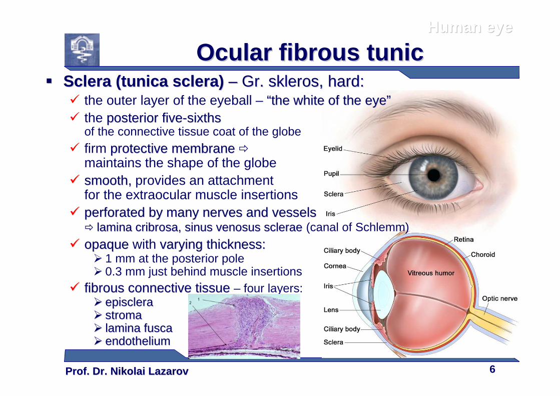
Ocular fibrous tunicOcular fibrous tunic�� Sclera (tunica sclera) Sclera (tunica sclera) –– Gr. Gr. sklerosskleros, hard:, hard:
� the outer layer of the eyeball – ““the white of the eyethe white of the eye””� the posterior fiveposterior five--sixthssixths
of the connective tissue coat of the globe
� firm protective membraneprotective membrane �
maintains the shape of the globe �� smooth, smooth, provides an attachment
for the extraocular muscle insertions �� perforated by many nerves and vesselsperforated by many nerves and vessels
�� lamina cribrosa, sinus venosus scleraelamina cribrosa, sinus venosus sclerae (canal of Schlemm)
�� opaqueopaque with varying varying thicknessthickness::� 1 mm at the posterior pole� 0.3 mm just behind muscle insertions
�� fibrous connective tissue fibrous connective tissue – four layers:�� episcleraepisclera�� stromastroma�� lamina fuscalamina fusca�� endotheliumendothelium
Prof. Dr. Nikolai LazarovProf. Dr. Nikolai Lazarov 7
Human eyeHuman eye
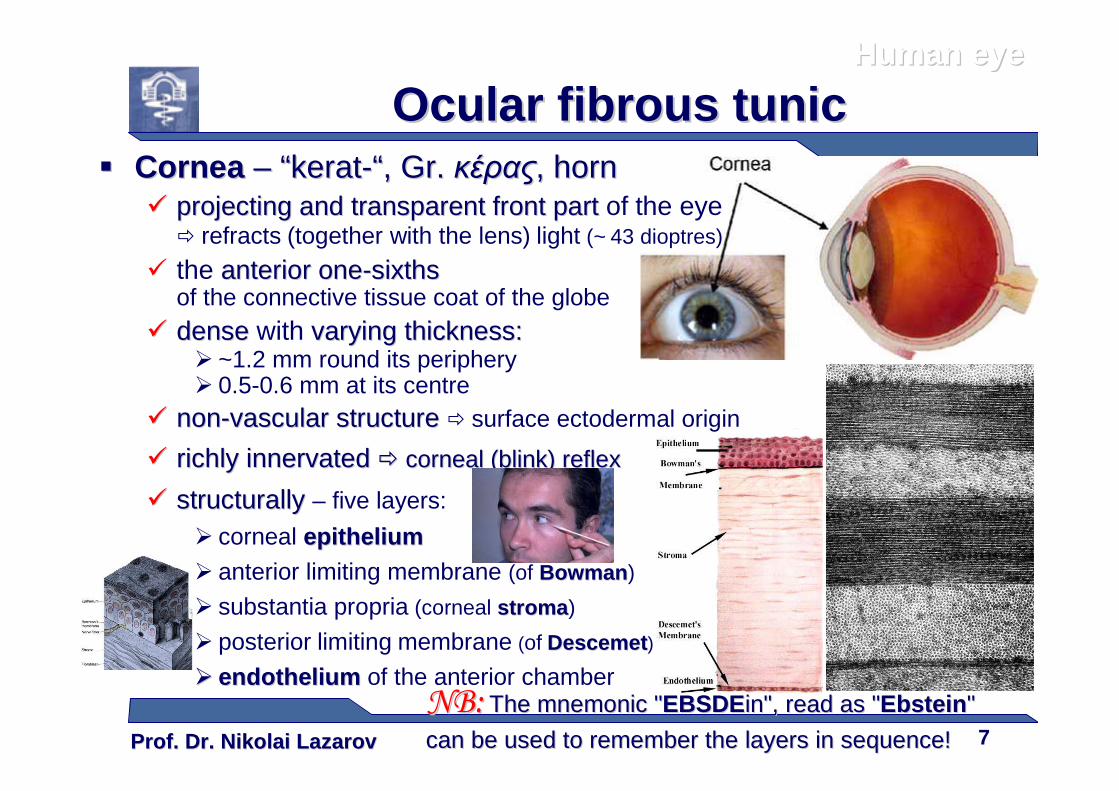
Ocular fibrous tunicOcular fibrous tunic�� Cornea Cornea –– ““keratkerat--““, Gr., Gr. κέραςκέρας, horn, horn
�� projecting and transparent front partprojecting and transparent front part of the eye � refracts (together with the lens) light (~ 43 dioptres)
� the anterioranterior oneone--sixthssixthsof the connective tissue coat of the globe
�� densedense with varying varying thicknessthickness::� ~1.2 mm round its periphery� 0.5-0.6 mm at its centre
�� nonnon--vascular structure vascular structure � surface ectodermal origin
�� richly innervated richly innervated �� corneal (blink) reflexcorneal (blink) reflex
�� structurally structurally – five layers:
� corneal epitheliumepithelium� anterior limiting membrane (of BowmanBowman)
� substantia propria (corneal stromastroma )
� posterior limiting membrane (of DescemetDescemet)
�� endotheliumendothelium of the anterior chamberNB:NB: The mnemonic "The mnemonic "EBSDEEBSDEin", read as "in", read as "EbsteinEbstein" "
can be used to remember the layers in sequencecan be used to remember the layers in sequence!!
Prof. Dr. Nikolai LazarovProf. Dr. Nikolai Lazarov 8
Human eyeHuman eye
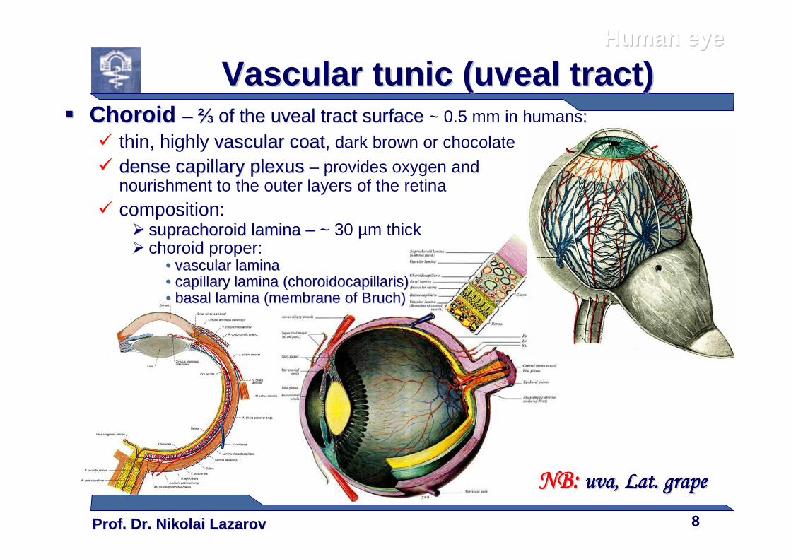
Vascular tunic (uveal tract)Vascular tunic (uveal tract)�� Choroid Choroid –– ⅔⅔ of the uveal tract surface of the uveal tract surface ~ 0.5 mm in humans::
� thin, highly vascular coatvascular coat, dark brown or chocolate
�� dense capillary plexusdense capillary plexus – provides oxygen and nourishment to the outer layers of the retina
� composition:�� suprachoroid laminasuprachoroid lamina – ~ 30 µm thick� choroid proper:
•• vascular laminavascular lamina•• capillary lamina (choroidocapillaris)capillary lamina (choroidocapillaris)•• basal lamina (membrane of basal lamina (membrane of BruchBruch))
NB:NB:NB:NB:NB:NB:NB:NB: uva, Lat. grapeuva, Lat. grapeuva, Lat. grapeuva, Lat. grapeuva, Lat. grapeuva, Lat. grapeuva, Lat. grapeuva, Lat. grape
Prof. Dr. Nikolai LazarovProf. Dr. Nikolai Lazarov 9
Human eyeHuman eye
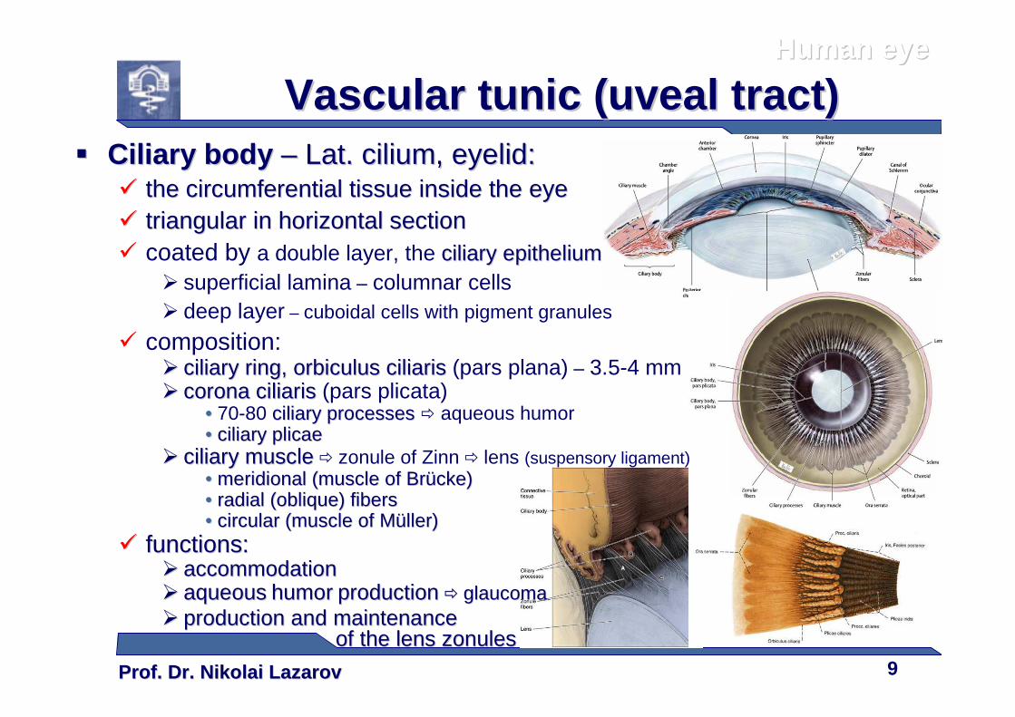
Vascular tunic (uveal tract)Vascular tunic (uveal tract)�� Ciliary bodyCiliary body –– Lat. Lat. ciliumcilium, eyelid:, eyelid:
�� the circumferential tissue inside the the circumferential tissue inside the eyeeye�� triangular in horizontal sectiontriangular in horizontal section� coated by a double layer, the ciliary epitheliumciliary epithelium
� superficial lamina – columnar cells� deep layer – cuboidal cells with pigment granules
� composition:�� ciliary ring, ciliary ring, orbiculus ciliarisorbiculus ciliaris (pars plana) – 3.5-4 mm�� corona ciliariscorona ciliaris (pars plicata)
•• 70-80 ciliary processes ciliary processes � aqueous humor•• ciliary plicaeciliary plicae
�� ciliary muscleciliary muscle � zonule of Zinn � lens (suspensory ligament)•• meridional (muscle of meridional (muscle of BrBrüückecke))•• radial (oblique) fibersradial (oblique) fibers•• circular (muscle of circular (muscle of MMüüllerller))
�� functions:functions:�� aaccommodationccommodation�� aqueousaqueous humorhumor productionproduction �� glaucomaglaucoma�� production and maintenance production and maintenance
of the lens zonulesof the lens zonules
Prof. Dr. Nikolai LazarovProf. Dr. Nikolai Lazarov 10
Human eyeHuman eye
Vascular tunic (uveal tract)Vascular tunic (uveal tract)
Iris eyeIris eyeIris eyeIris eyeIris eyeIris eyeIris eyeIris eye recognitionrecognition
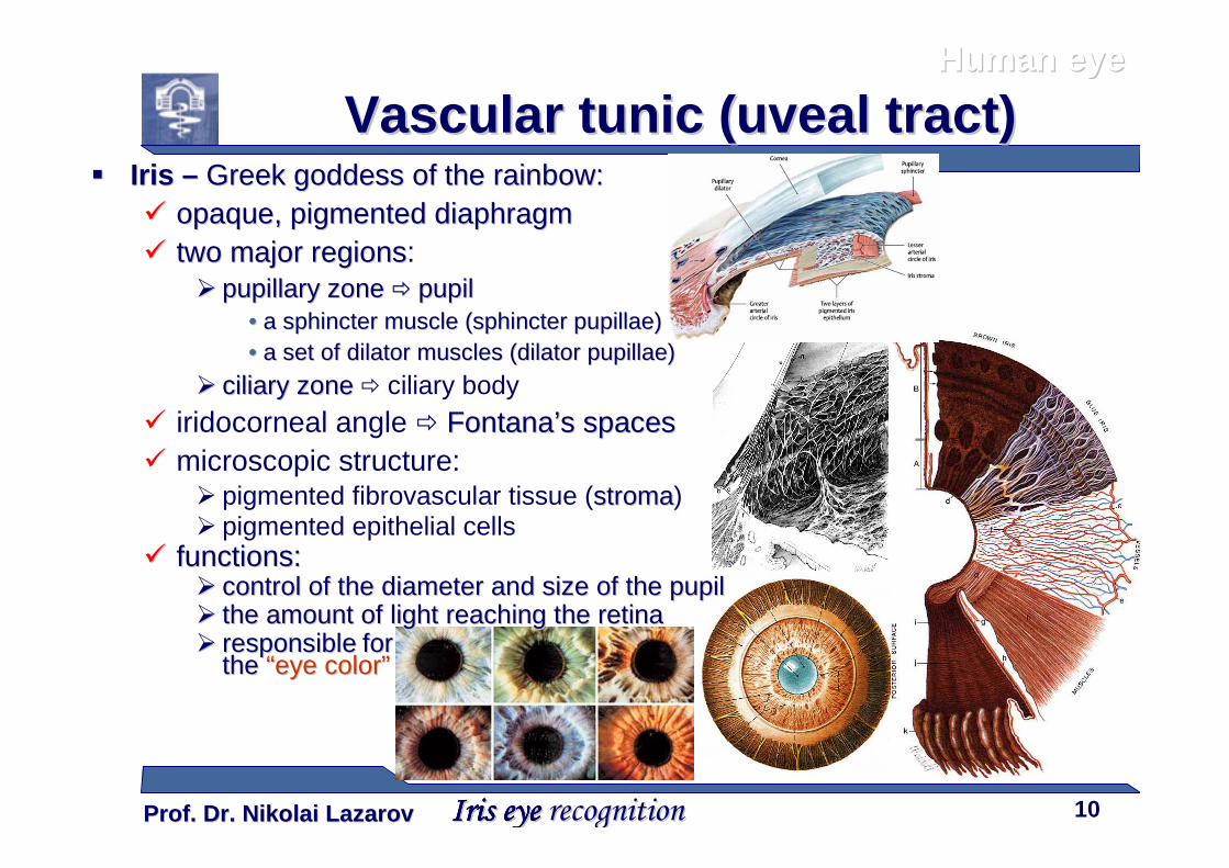
�� Iris Iris –– GreekGreek goddess of the rainbowgoddess of the rainbow::�� opaque, pigmented diaphragmopaque, pigmented diaphragm�� two major regionstwo major regions:
�� pupillary zonepupillary zone �� pupilpupil•• a a sphincter sphincter muscle (muscle (sphincter pupillaesphincter pupillae))•• a set of dilator muscles (a set of dilator muscles (dilator pupillaedilator pupillae))
�� ciliary zoneciliary zone � ciliary body
� iridocorneal angle � FontanaFontana’’s spacess spaces� microscopic structure:
� pigmented fibrovascular tissue (stromastroma)� pigmented epithelial cells
�� functions:functions:�� ccontrolontrol ofof the diameter and size of the the diameter and size of the pupilpupil�� the amount of light reaching the the amount of light reaching the retinaretina�� responsibleresponsible for for
the the ““eeye colorye color””
Prof. Dr. Nikolai LazarovProf. Dr. Nikolai Lazarov 11
Human eyeHuman eye
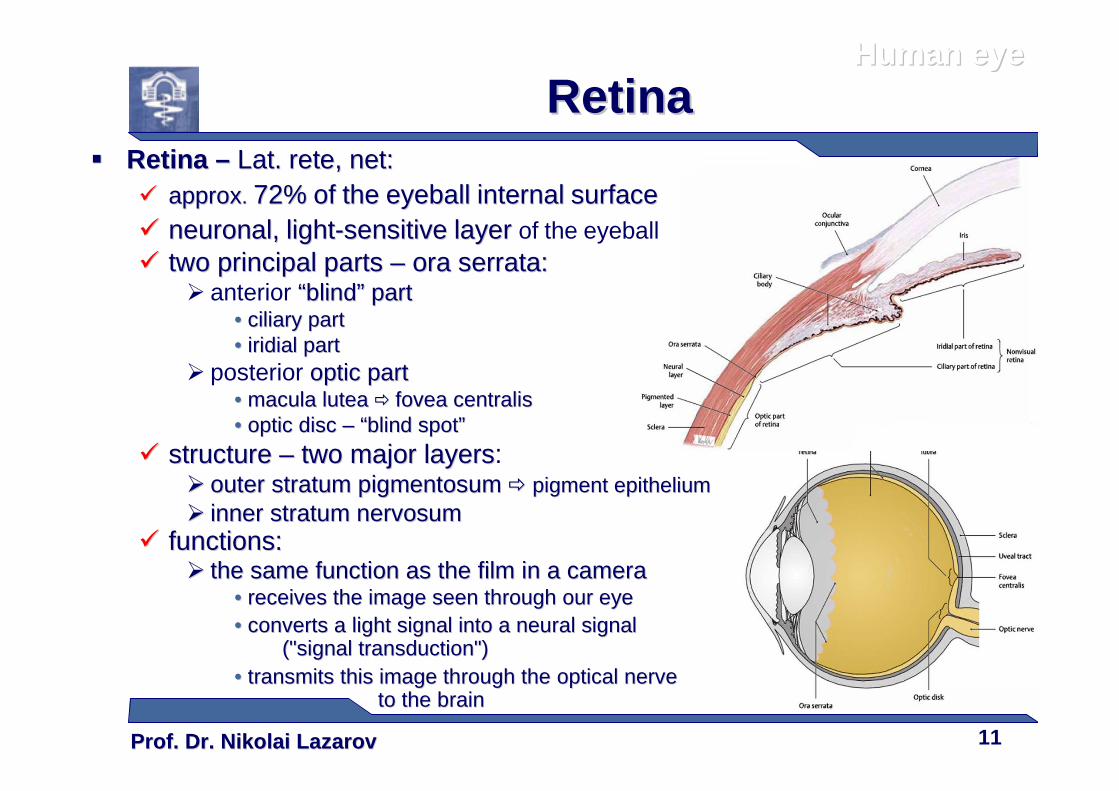
RetinaRetina�� Retina Retina –– Lat.Lat. rete, rete, net:net:
�� aapprpproxox.. 72%72% ofof thethe eyeballeyeball internalinternal surfacesurface�� neuronal,neuronal, lightlight--sensitivesensitive layerlayer of the eyeball�� two principal parts two principal parts –– ora serrata:ora serrata:
� anterior ““blindblind”” partpart•• ciliary partciliary part•• iridial partiridial part
� posterior optic partoptic part•• macula luteamacula lutea �� fovea centralisfovea centralis•• optic disc optic disc –– ““blind spotblind spot””
�� structure structure –– two major two major layerslayers:�� outer outer stratum pigmentosumstratum pigmentosum �� pigment epitheliumpigment epithelium�� inner inner stratum nervosumstratum nervosum
�� functions:functions:�� the same the same functionfunction as the film in a cameraas the film in a camera
•• recereceiives the image seen through ves the image seen through ourour eyeeye•• convertconvertss a light signal into a neural signal a light signal into a neural signal
("signal transduction")("signal transduction")•• transmits this image through the optical nervetransmits this image through the optical nerve
to the brainto the brain
Prof. Dr. Nikolai LazarovProf. Dr. Nikolai Lazarov 12
Human eyeHuman eye
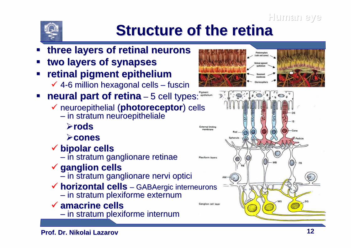
Structure of the retinaStructure of the retina�� three layers of three layers of retinal neuronsretinal neurons�� two layers of two layers of synapsessynapses�� retinal pigment epitheliumretinal pigment epithelium
�� 44--6 million hexagonal6 million hexagonal cells cells –– fuscinfuscin
�� neural part of retinaneural part of retina –– 5 cell types5 cell types::�� neuroepithelialneuroepithelial ((photoreceptorphotoreceptor ) ) cellscells
–– in in stratum neuroepithelialestratum neuroepitheliale��rodsrods��conescones
�� bipolar cells bipolar cells –– inin stratum ganglionare retinaestratum ganglionare retinae
�� ganglion cells ganglion cells –– inin stratum ganglionare nervi opticistratum ganglionare nervi optici
�� horizontal cells horizontal cells –– GABAergic interneurons GABAergic interneurons –– inin stratum plexiforme externum stratum plexiforme externum
�� amacrine cells amacrine cells –– inin stratum plexiforme internumstratum plexiforme internum
Prof. Dr. Nikolai LazarovProf. Dr. Nikolai Lazarov 13
Human eyeHuman eye
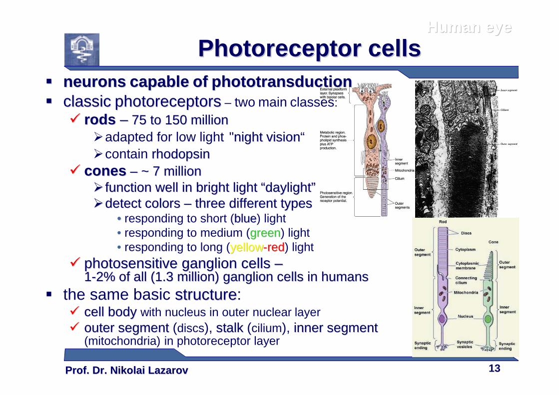
Photoreceptor cellsPhotoreceptor cells�� neuronsneurons capablecapable ofof phototransductionphototransduction�� classicclassic photoreceptorsphotoreceptors – two main classes:
�� rods rods –– 75 to 150 million75 to 150 million�adapted for low light "night visionnight vision“�contain rhodopsinrhodopsin
�� conescones –– ~ 7~ 7 millionmillion�� function well in bright lightfunction well in bright light ““daylightdaylight””��detect colorsdetect colors –– three different typesthree different types
•• responding to short (blueblue) light• responding to medium (greengreen) light• responding to long (yellowyellow--redred) light
�� photosensitive ganglion cells photosensitive ganglion cells ––11--2% of all (1.3 million) ganglion cells in humans2% of all (1.3 million) ganglion cells in humans
� the same basic structurestructure:�� cell bodycell body with nucleus in outer nuclear layer�� outer segmentouter segment (discs), stalkstalk (cilium), inner segmentinner segment
(mitochondria) in photoreceptor layer
Prof. Dr. Nikolai LazarovProf. Dr. Nikolai Lazarov 14
Human eyeHuman eye
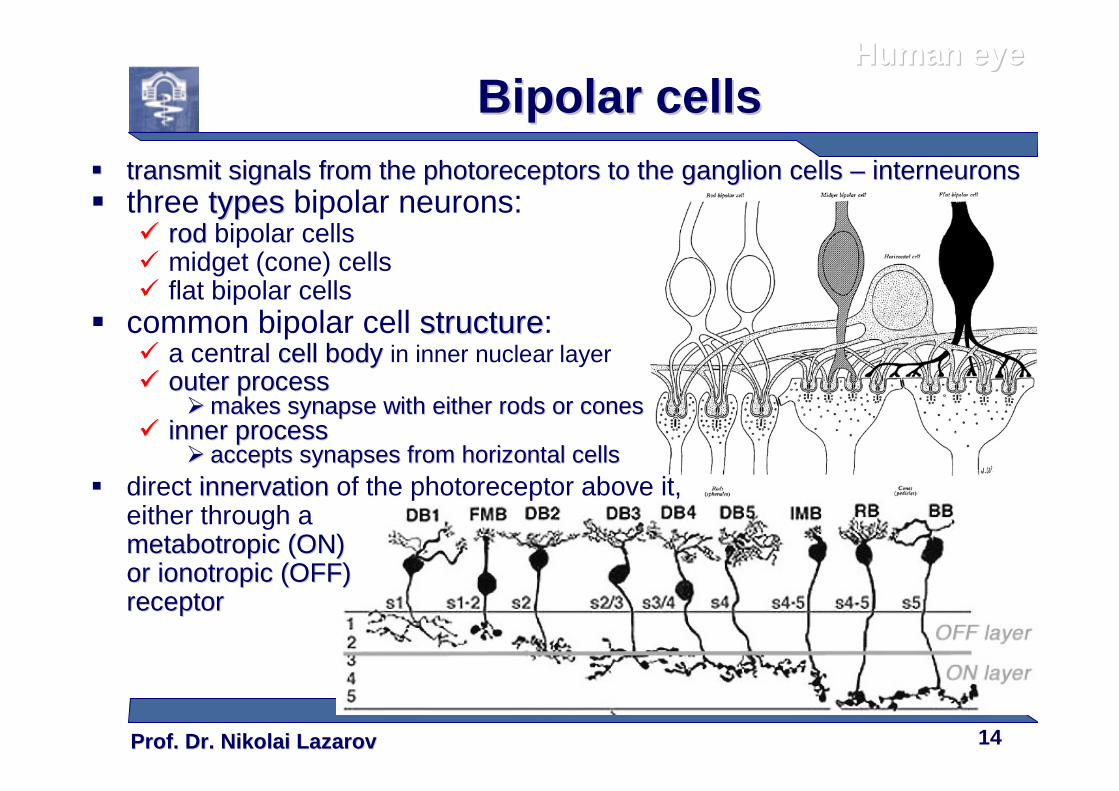
Bipolar cellsBipolar cells�� transmit signals from the photoreceptors to the ganglion cellstransmit signals from the photoreceptors to the ganglion cells –– interneuronsinterneurons� three typestypes bipolar neurons:
�� rodrod bipolar cells� midget (cone) cells� flat bipolar cells
� common bipolar cell structurestructure:� a central cell bodycell body in inner nuclear layer�� outer processouter process
�� makes synapse with either rods or conesmakes synapse with either rods or cones�� inner processinner process
�� accepts synapses from horizontal cellsaccepts synapses from horizontal cells� direct innervationinnervation of the photoreceptor above it,
either through ametabotropicmetabotropic (ON) (ON) or or ionotropicionotropic (OFF) (OFF) receptorreceptor
Prof. Dr. Nikolai LazarovProf. Dr. Nikolai Lazarov 15
Human eyeHuman eye
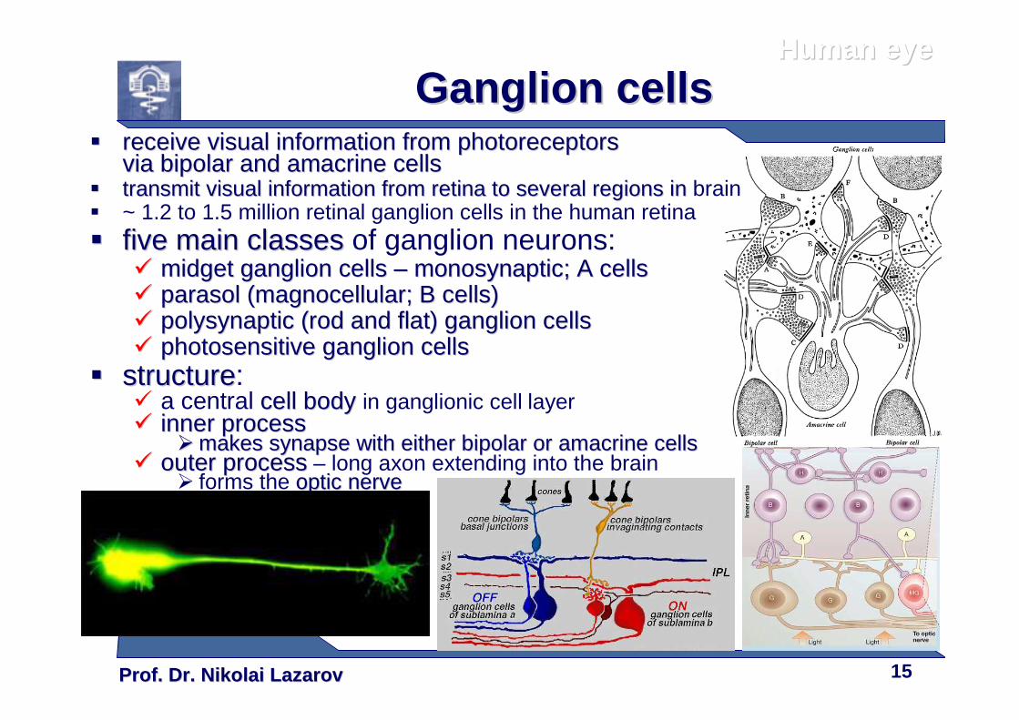
Ganglion cellsGanglion cells�� receive visual information from receive visual information from photoreceptorsphotoreceptors
via via bipolar bipolar and and amacrine cellsamacrine cells�� transmit visual information from retina to several regions in transmit visual information from retina to several regions in brain� ~ 1.2 to 1.5 million retinal ganglion cells in the human retina�� five main classesfive main classes of ganglion neurons:
�� midget ganglion cells midget ganglion cells –– monosynaptic; A cellsmonosynaptic; A cells�� pparasol (arasol (mmagnocellular; B cells)agnocellular; B cells)�� polysynaptic (rod and flat)polysynaptic (rod and flat) ganglion cellsganglion cells�� photosensitive ganglion cellsphotosensitive ganglion cells
�� structurestructure:� a central cell bodycell body in ganglionic cell layer�� inner processinner process
�� makes synapse with either bipolar or amacrine cellsmakes synapse with either bipolar or amacrine cells�� outer processouter process – long axon extending into the brain
� forms the optic nerveoptic nerve
Prof. Dr. Nikolai LazarovProf. Dr. Nikolai Lazarov 16
Human eyeHuman eye
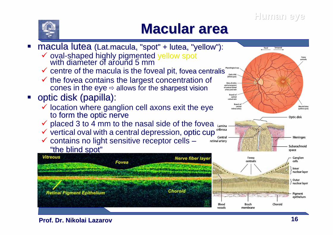
Macular areaMacular area�� macula lutea macula lutea ((Lat.Lat.maculamacula, "spot" + , "spot" + lutealutea, "yellow"), "yellow"):
� oval-shaped highly pigmented yellow spotyellow spotwith diameter of around 5 mm
� centre of the macula is the foveal pit, foveafovea centraliscentralis� the fovea contains the largest concentration of
cones in the eye � allows for the sharpest visionsharpest vision
�� optic disk (papilla)optic disk (papilla):� location where ganglion cell axons exit the eye
to form the form the optic nerveoptic nerve� placed 3 to 4 mm to the nasal side of the fovea� vertical oval with a central depression, opticoptic cupcup� contains no light sensitive receptor cells –
““the blind spotthe blind spot””
Prof. Dr. Nikolai LazarovProf. Dr. Nikolai Lazarov 17
Human eyeHuman eye
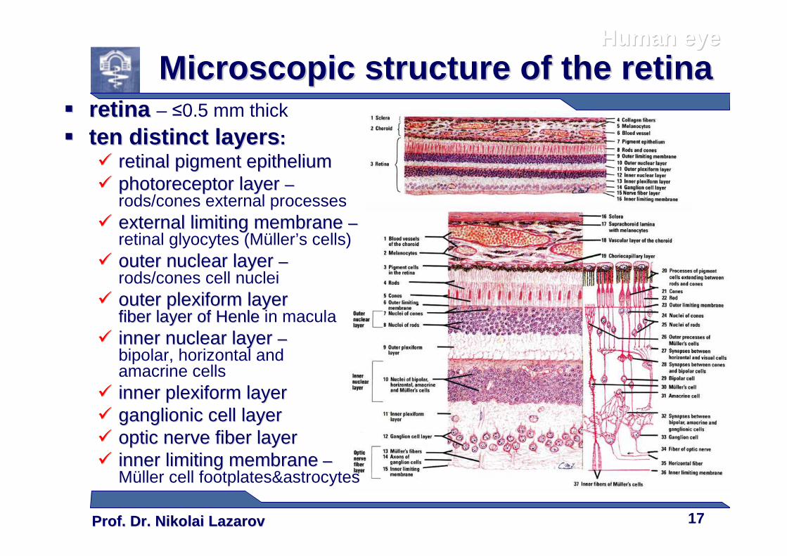
Microscopic structure of the retinaMicroscopic structure of the retina�� retina retina – ≤0.5 mm thick
�� ten distinct layersten distinct layers ::�� retinal pigment epitheliumretinal pigment epithelium�� photoreceptor layer photoreceptor layer ––
rods/cones external processes�� external limiting membrane external limiting membrane ––
retinal glyocytes (Müller’s cells)�� outer nuclear layer outer nuclear layer ––
rods/cones cell nuclei�� outer plexiform layer outer plexiform layer
fiber layer of fiber layer of HenleHenle in macula�� inner nuclear layer inner nuclear layer ––
bipolar, horizontal and amacrine cells
�� inner plexiform layerinner plexiform layer�� ganglionic cell layerganglionic cell layer�� optic nerve fiber layeroptic nerve fiber layer�� inner limiting membraneinner limiting membrane ––
Müller cell footplates&astrocytes
Prof. Dr. Nikolai LazarovProf. Dr. Nikolai Lazarov 18
Human eyeHuman eye
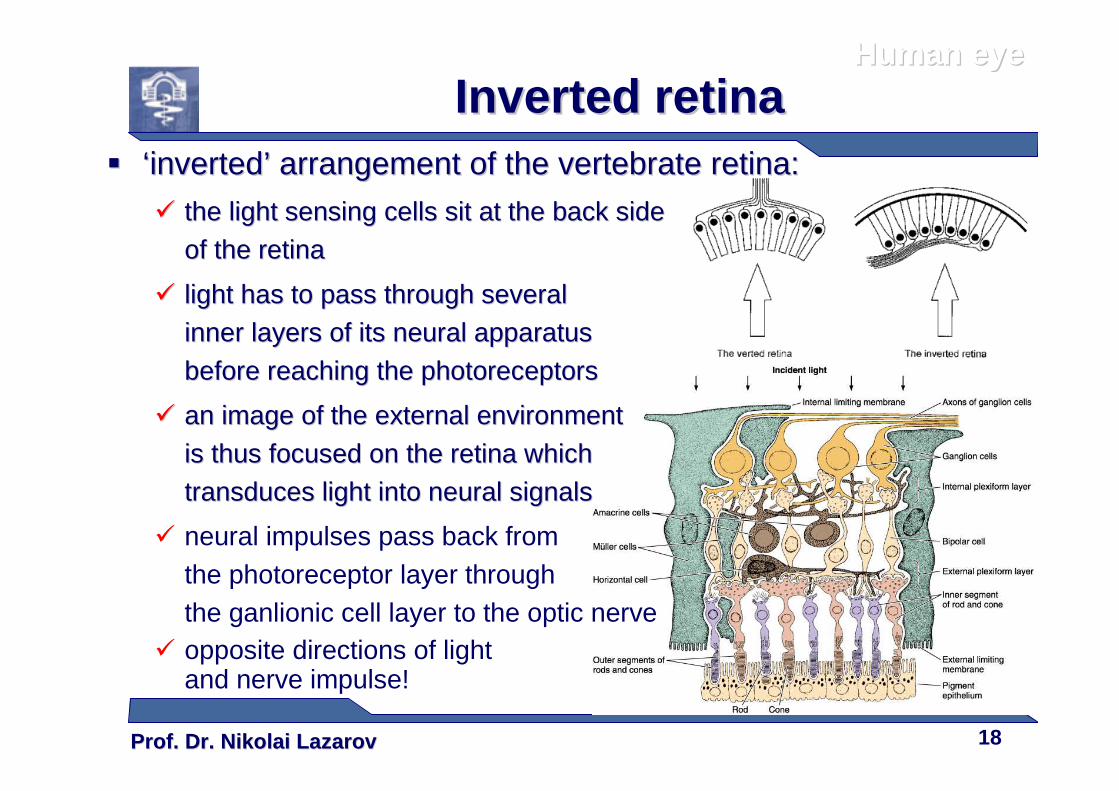
Inverted retinaInverted retina�� ‘‘invertedinverted’’ arrangement of the vertebrate retinaarrangement of the vertebrate retina::
�� the light sensing cells sit at the back side the light sensing cells sit at the back side
of the retinaof the retina
�� light has to pass through several light has to pass through several
inner layers of its neural apparatus inner layers of its neural apparatus
before reaching the photoreceptorsbefore reaching the photoreceptors
�� aan image of the external environment n image of the external environment
is thus focused on the retina which is thus focused on the retina which
transduces light into neural signalstransduces light into neural signals
� neural impulses pass back from the photoreceptor layer through the ganlionic cell layer to the optic nerve
� opposite directions of light and nerve impulse!
Prof. Dr. Nikolai LazarovProf. Dr. Nikolai Lazarov 19
Human eyeHuman eye
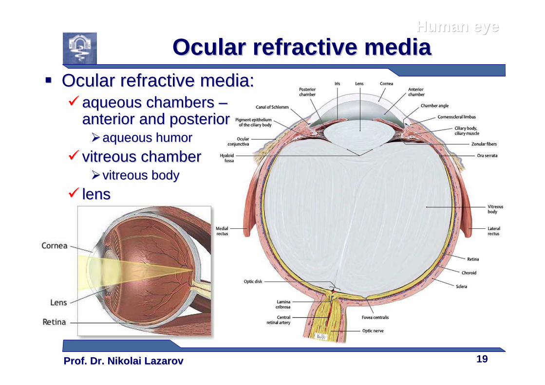
Ocular refractive mediaOcular refractive media�� Ocular refractive media:Ocular refractive media:
�� aqueous chambers aqueous chambers ––anterior and posterioranterior and posterior��aqueous humoraqueous humor
�� vitreous chambervitreous chamber��vitreous bodyvitreous body
�� lenslens
Prof. Dr. Nikolai LazarovProf. Dr. Nikolai Lazarov 20
Human eyeHuman eye
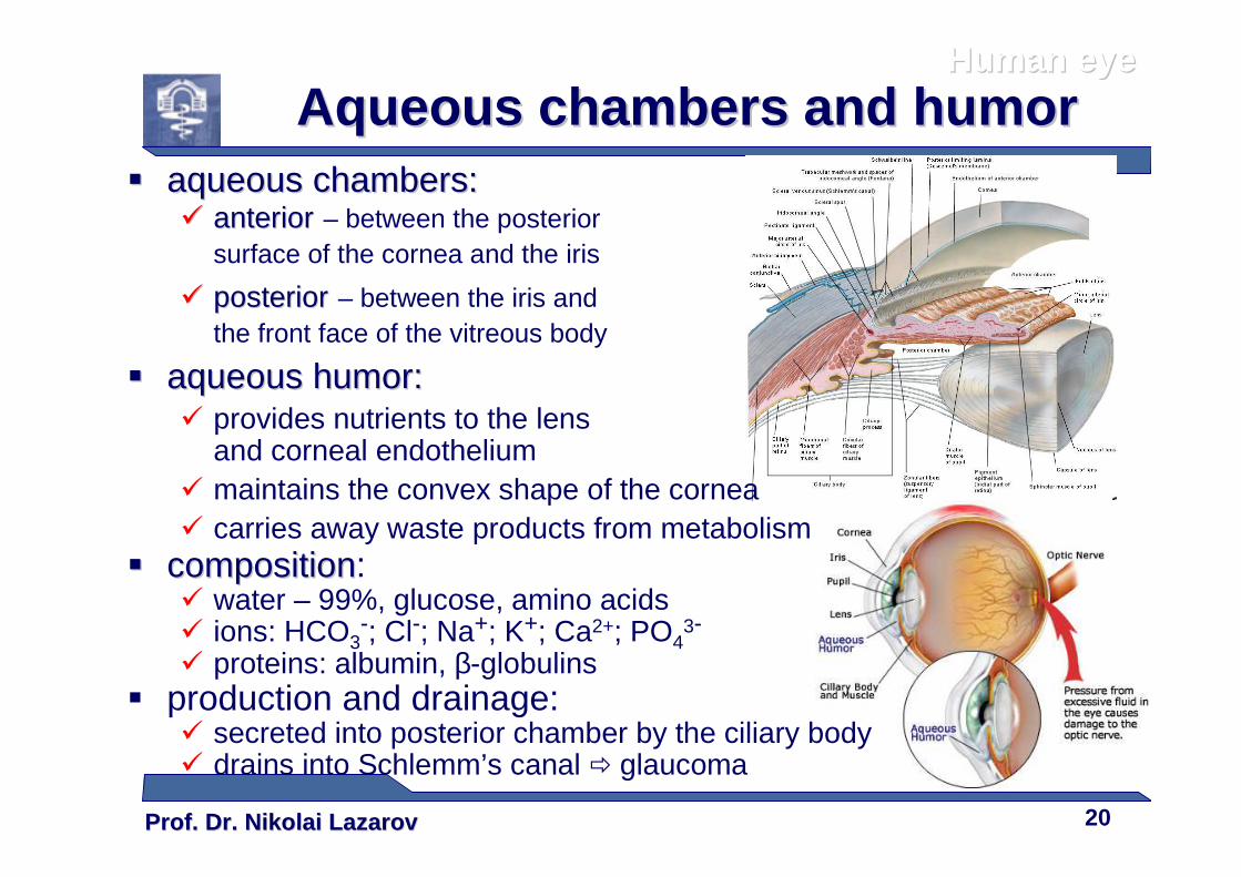
Aqueous chambers and humorAqueous chambers and humor�� aqueous chambers:aqueous chambers:
�� anterioranterior – between the posterior surface of the cornea and the iris
�� posteriorposterior – between the iris and the front face of the vitreous body
�� aqueous humor:aqueous humor:� provides nutrients to the lens
and corneal endothelium� maintains the convex shape of the cornea� carries away waste products from metabolism
�� compositioncomposition:� water – 99%, glucose, amino acids� ions: HCO3
-; Cl-; Na+; K+; Ca2+; PO43-
� proteins: albumin, β-globulins� production and drainage:
� secreted into posterior chamber by the ciliary body� drains into Schlemm’s canal � glaucoma
Prof. Dr. Nikolai LazarovProf. Dr. Nikolai Lazarov 21
Human eyeHuman eye
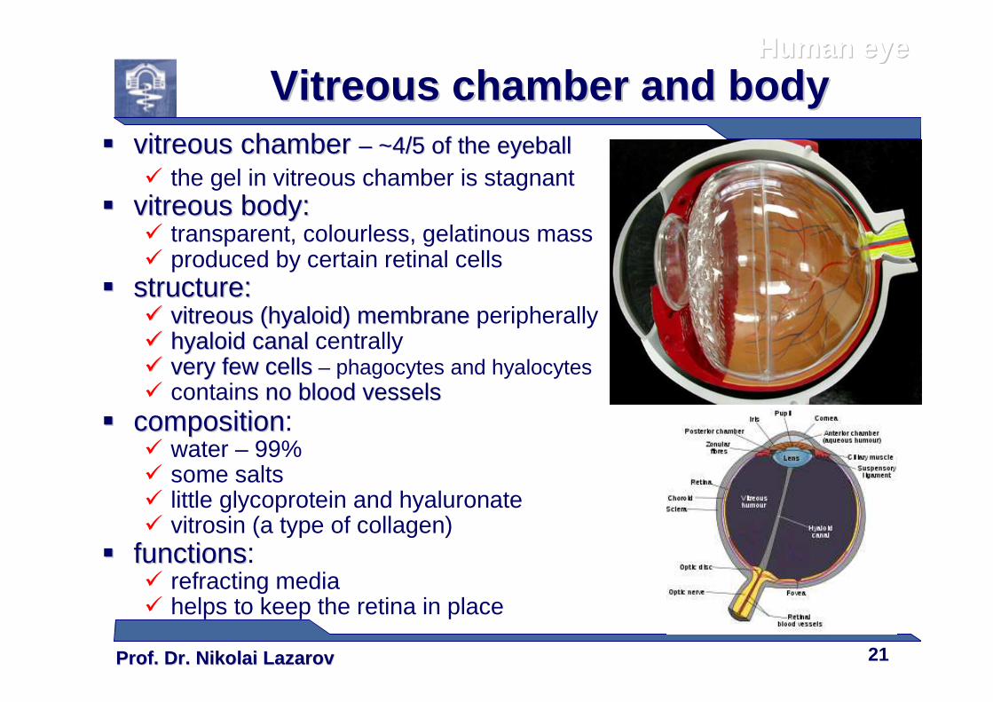
Vitreous chamber and bodyVitreous chamber and body�� vitreous chamber vitreous chamber –– ~4/5 of the eyeball~4/5 of the eyeball
� the gel in vitreous chamber is stagnant �� vitreous body:vitreous body:
� transparent, colourless, gelatinous mass� produced by certain retinal cells
�� structure:structure:�� vitreous (hyaloid) membranevitreous (hyaloid) membrane peripherally�� hyaloid canalhyaloid canal centrally�� very few cellsvery few cells – phagocytes and hyalocytes� contains no blood vesselsno blood vessels
�� compositioncomposition:� water – 99%� some salts� little glycoprotein and hyaluronate� vitrosin (a type of collagen)
�� functionsfunctions:� refracting media� helps to keep the retina in place
Prof. Dr. Nikolai LazarovProf. Dr. Nikolai Lazarov 22
Human eyeHuman eye
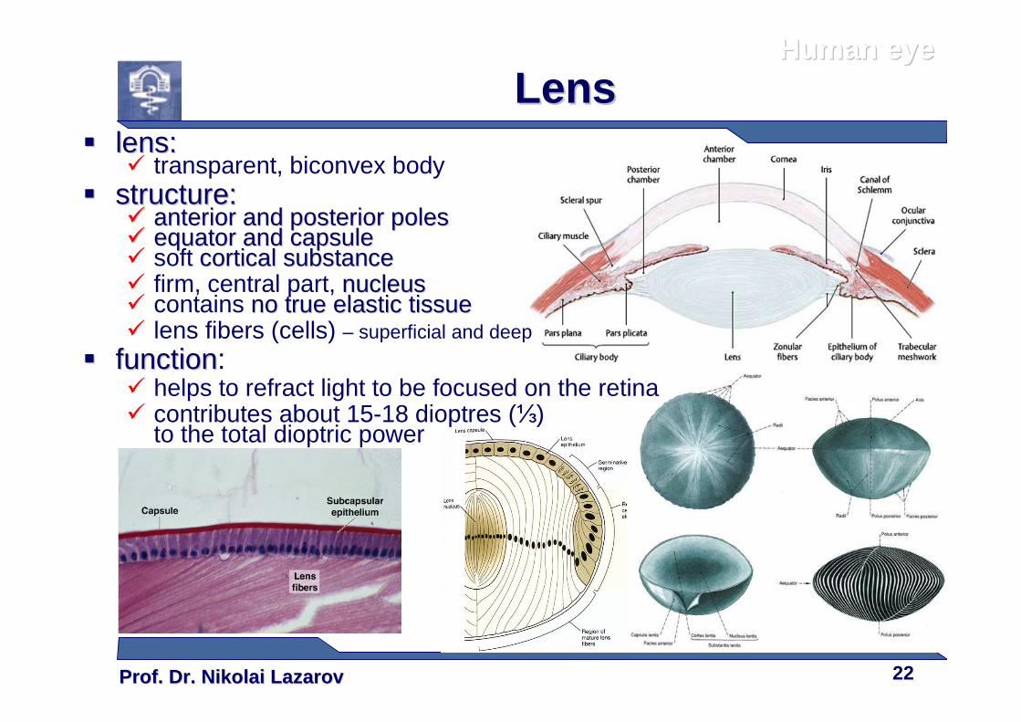
LensLens�� lens:lens:
� transparent, biconvex body�� structure:structure:
�� anterior and posterior polesanterior and posterior poles�� equator and capsuleequator and capsule� soft cortical substancecortical substance� firm, central part, nucleusnucleus� contains no true elastic tissueno true elastic tissue� lens fibers (cells) – superficial and deep
�� functionfunction:� helps to refract light to be focused on the retina� contributes about 15-18 dioptres (⅓)
to the total dioptric power
Prof. Dr. Nikolai LazarovProf. Dr. Nikolai Lazarov 23
Human eyeHuman eye
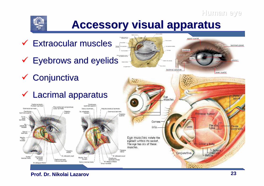
Accessory visual apparatusAccessory visual apparatus
�� Extraocular musclesExtraocular muscles
�� Eyebrows and eyelidsEyebrows and eyelids
�� ConjunctivaConjunctiva
�� Lacrimal apparatusLacrimal apparatus
Prof. Dr. Nikolai LazarovProf. Dr. Nikolai Lazarov 24
Human eyeHuman eye
Extraocular musclesExtraocular muscles
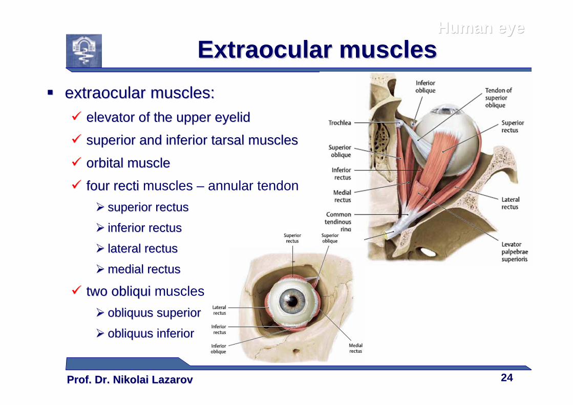
�� extraocular muscles:extraocular muscles:
�� elevator of the upper eyelidelevator of the upper eyelid
�� superior and inferior tarsal musclessuperior and inferior tarsal muscles
�� orbital muscleorbital muscle
�� fourfour rectirecti muscles – annular tendon
�� superior rectussuperior rectus
�� inferior rectusinferior rectus
�� lateral rectuslateral rectus
�� medial rectusmedial rectus
�� two obliquitwo obliqui muscles
�� obliquus superiorobliquus superior
�� obliquus inferiorobliquus inferior
Prof. Dr. Nikolai LazarovProf. Dr. Nikolai Lazarov 25
Human eyeHuman eye
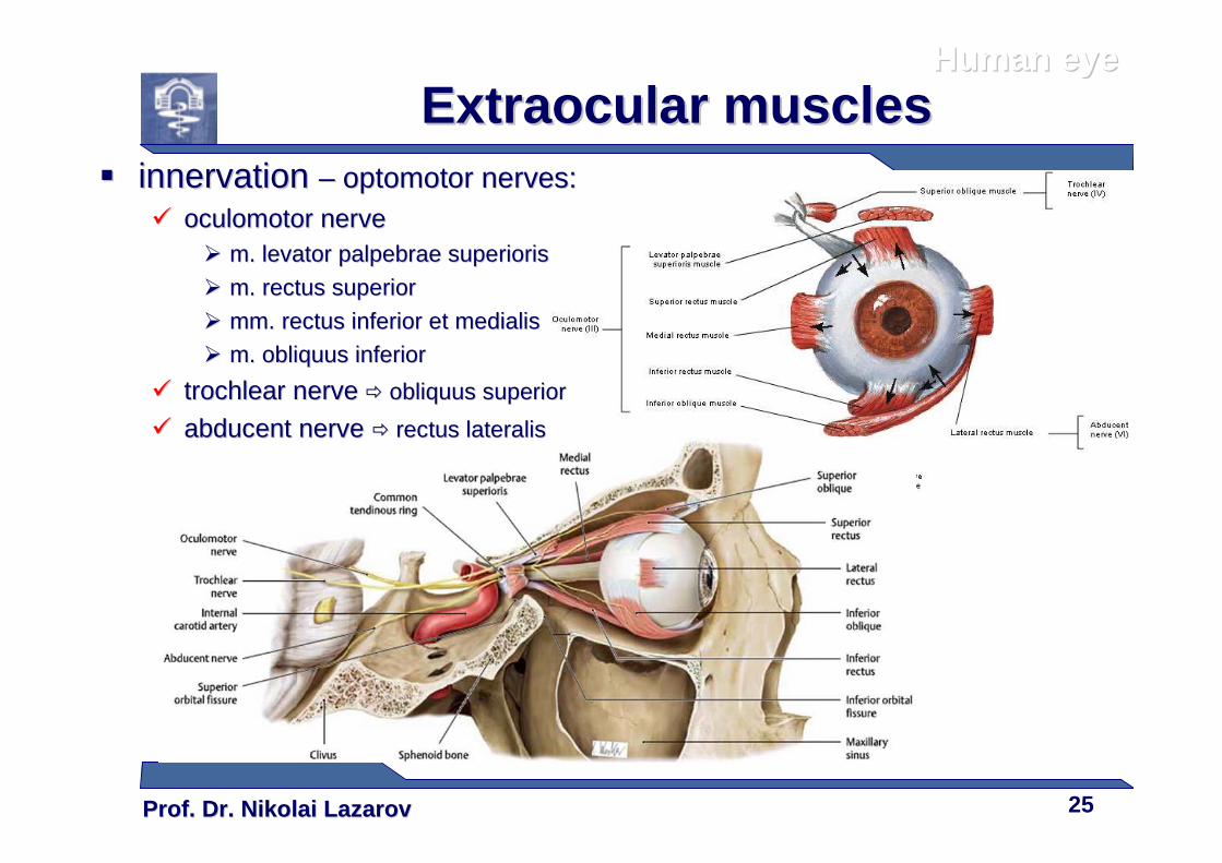
Extraocular musclesExtraocular muscles�� innervation innervation –– optomotor nerves:optomotor nerves:
�� oculomotor nerveoculomotor nerve�� m. levator palpebrae superiorism. levator palpebrae superioris
�� m. rectus superiorm. rectus superior
�� mm. rectus inferior et medialismm. rectus inferior et medialis
�� m. obliquus inferiorm. obliquus inferior
�� trochlear nervetrochlear nerve �� obliquus superiorobliquus superior
�� abducent nerve abducent nerve �� rectus lateralisrectus lateralis
Prof. Dr. Nikolai LazarovProf. Dr. Nikolai Lazarov 26
Human eyeHuman eye
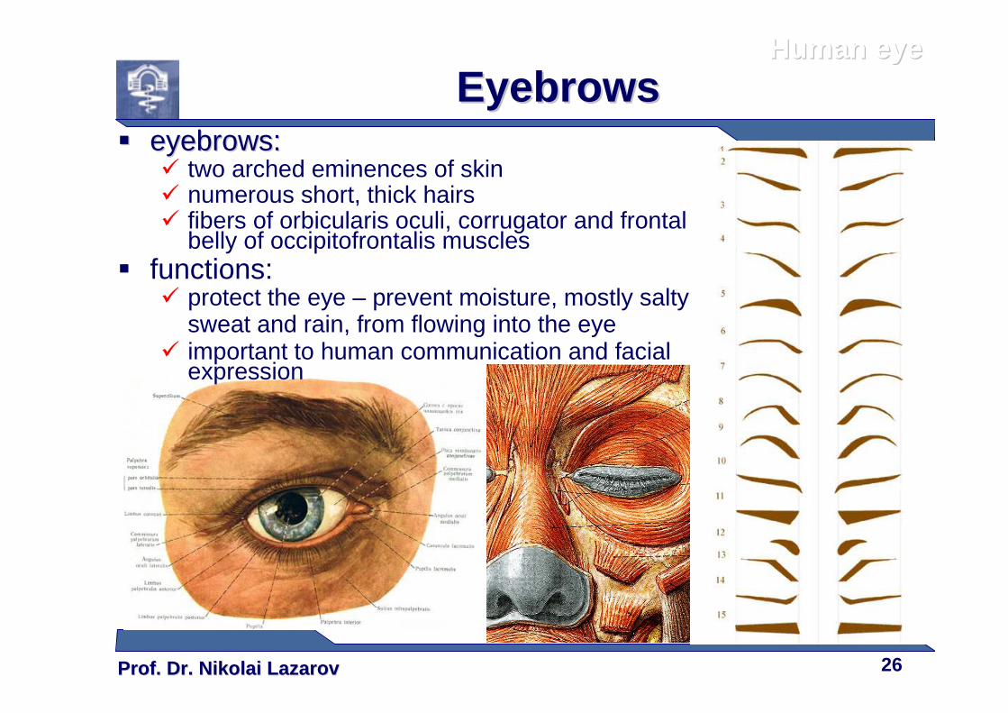
EyebrowsEyebrows�� eyebrows:eyebrows:
� two arched eminences of skin� numerous short, thick hairs� fibers of orbicularis oculi, corrugator and frontal
belly of occipitofrontalis muscles� functions:
� protect the eye – prevent moisture, mostly salty sweat and rain, from flowing into the eye
� important to human communication and facial expression
Prof. Dr. Nikolai LazarovProf. Dr. Nikolai Lazarov 27
Human eyeHuman eye
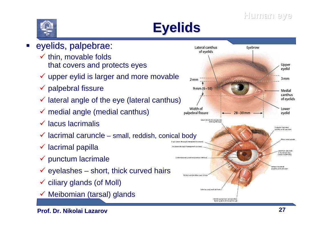
EyelidsEyelids�� eyelids, eyelids, palpebrae:palpebrae:
� thin, movable folds that covers and protects eyes
� upper eylid is larger and more movable
� palpebral fissure
� lateral angle of the eye (lateral canthus)
� medial angle (medial canthus)
� lacus lacrimalis
� lacrimal caruncle – small, reddish, conical body
� lacrimal papilla
� punctum lacrimale
� eyelashes – short, thick curved hairs
� ciliary glands (of Moll)
� Meibomian (tarsal) glands
Prof. Dr. Nikolai LazarovProf. Dr. Nikolai Lazarov 28
Human eyeHuman eye
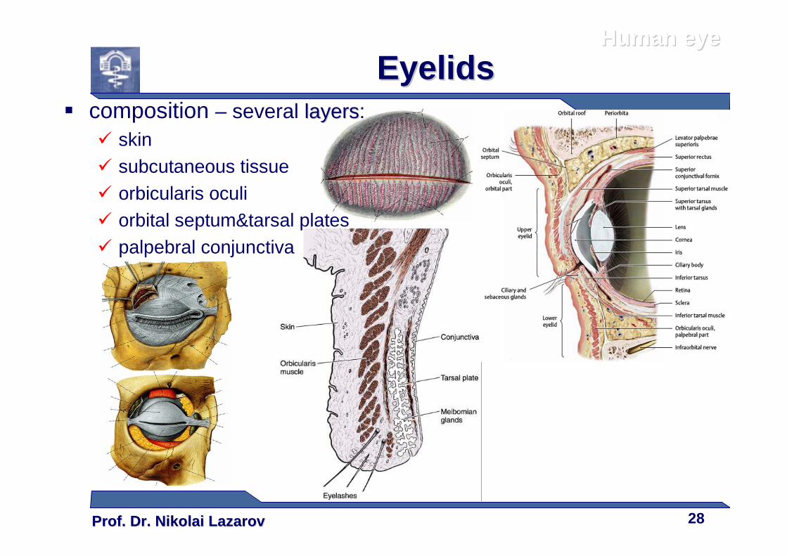
EyelidsEyelids� composition – several layerslayers:
� skin� subcutaneous tissue� orbicularis oculi� orbital septum&tarsal plates� palpebral conjunctiva
Prof. Dr. Nikolai LazarovProf. Dr. Nikolai Lazarov 29
Human eyeHuman eye
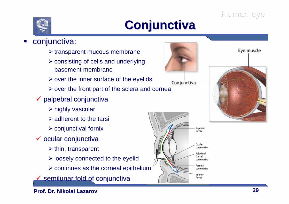
ConjunctivaConjunctiva�� conjunctiva:conjunctiva:
� transparent mucous membrane
� consisting of cells and underlying basement membrane
� over the inner surface of the eyelids
� over the front part of the sclera and cornea
�� palpebral conjunctivapalpebral conjunctiva� highly vascular
� adherent to the tarsi
� conjunctival fornix
�� ocular conjunctivaocular conjunctiva� thin, transparent
� loosely connected to the eyelid
� continues as the corneal epithelium
�� semilunar fold of conjunctivasemilunar fold of conjunctiva
Prof. Dr. Nikolai LazarovProf. Dr. Nikolai Lazarov 30
Human eyeHuman eye
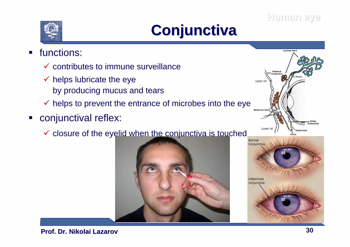
ConjunctivaConjunctiva� functions:
� contributes to immune surveillance
� helps lubricate the eyeby producing mucus and tears
� helps to prevent the entrance of microbes into the eye
� conjunctival reflex:
� closure of the eyelid when the conjunctiva is touched
Prof. Dr. Nikolai LazarovProf. Dr. Nikolai Lazarov 31
Human eyeHuman eye
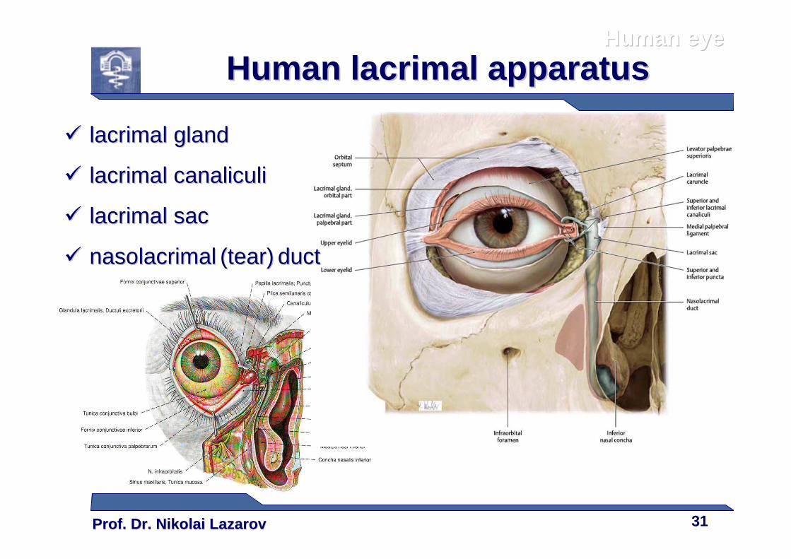
Human lacrimal apparatusHuman lacrimal apparatus
�� lacrimal glandlacrimal gland
�� lacrimal canaliculilacrimal canaliculi
�� lacrimal saclacrimal sac
�� nasolacrimalnasolacrimal (tear)(tear) ductduct
Prof. Dr. Nikolai LazarovProf. Dr. Nikolai Lazarov 32
Human eyeHuman eye
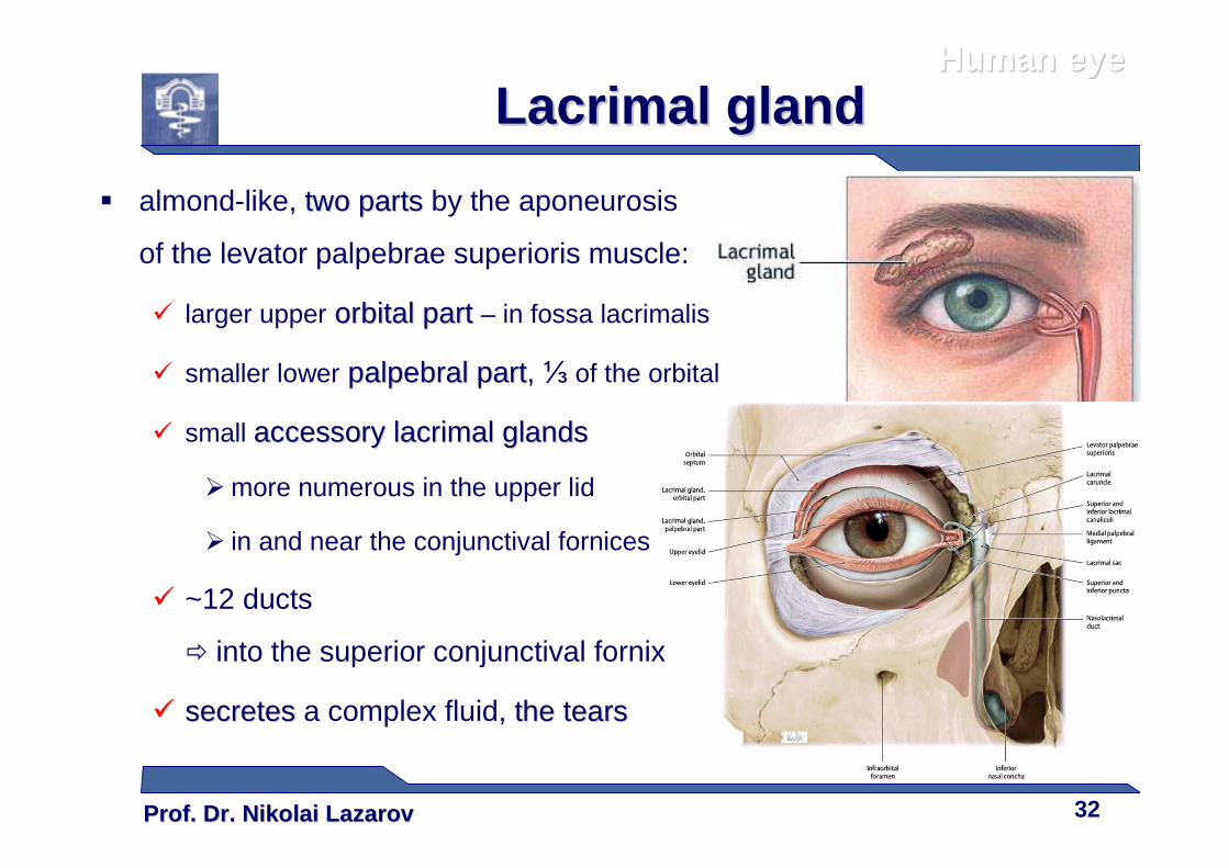
Lacrimal glandLacrimal gland
� almond-like, two parts two parts by the aponeurosis
of the levator palpebrae superioris muscle:
� larger upper orbital part orbital part – in fossa lacrimalis
� smaller lower palpebral partpalpebral part, ⅓ of the orbital
� small accessory lacrimal glandsaccessory lacrimal glands
� more numerous in the upper lid
� in and near the conjunctival fornices
� ~12 ducts
� into the superior conjunctival fornix
�� secretessecretes a complex fluid, the tearsthe tears
Prof. Dr. Nikolai LazarovProf. Dr. Nikolai Lazarov 33
Human eyeHuman eye
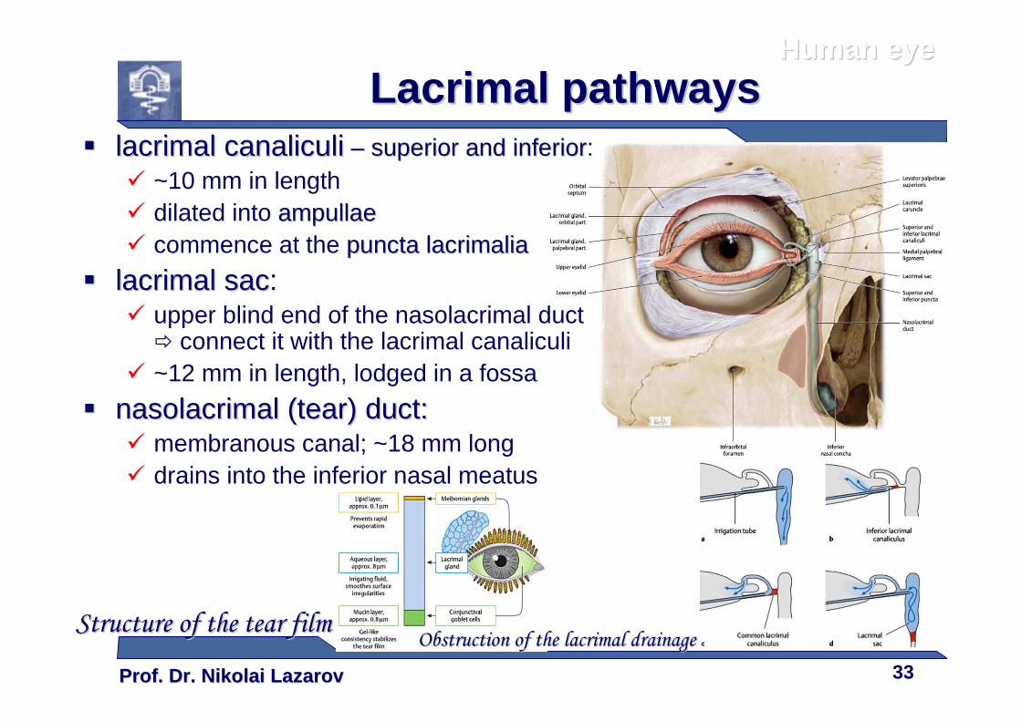
Lacrimal pathwaysLacrimal pathways�� lacrimal canaliculilacrimal canaliculi –– superior and inferiorsuperior and inferior:
� ~10 mm in length� dilated into ampullaeampullae� commence at the puncta lacrimaliapuncta lacrimalia
�� lacrimal saclacrimal sac:� upper blind end of the nasolacrimal duct
� connect it with the lacrimal canaliculi� ~12 mm in length, lodged in a fossa
�� nasolacrimal (tear) duct:nasolacrimal (tear) duct:� membranous canal; ~18 mm long� drains into the inferior nasal meatus
Structure of the tear filmStructure of the tear filmObstruction of the lacrimal drainageObstruction of the lacrimal drainage
Prof. Dr. Nikolai LazarovProf. Dr. Nikolai Lazarov 34
Human eyeHuman eye
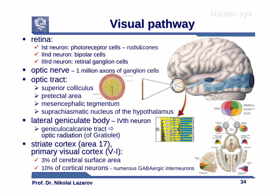
Visual pathwayVisual pathway�� retinaretina:
�� Ist neuron: photoreceptor cellsIst neuron: photoreceptor cells – rods&cones�� IInd neuron: bipolar cellsIInd neuron: bipolar cells�� IIIrd neuron: retinal ganglion cellsIIIrd neuron: retinal ganglion cells
�� optic nerveoptic nerve –– 1 million axons 1 million axons of ganglion cells
�� optic tract:optic tract:� superior colliculus� pretectal area� mesencephalic tegmentum� suprachiasmatic nucleus of the hypothalamus
�� lateral geniculate bodylateral geniculate body –– IVth neuronIVth neuron� geniculocalcarine tract �
optic radiationoptic radiation (of Gratiolet)�� striate cortex (area 17), striate cortex (area 17),
primary visual cortex (Vprimary visual cortex (V--I):I):� 3% of cerebral surface area� 10% of cortical neurons – numerous GABAergic interneurons
Prof. Dr. Nikolai LazarovProf. Dr. Nikolai Lazarov 35
Thank youThank you……