visualization of the cocaine-sensitive dopamine transporter with ligand-conjugated quantum dots
TRANSCRIPT

Published: April 26, 2011
r 2011 American Chemical Society 370 dx.doi.org/10.1021/cn200032r |ACS Chem. Neurosci. 2011, 2, 370–378
RESEARCH ARTICLE
pubs.acs.org/acschemicalneuroscience
Visualization of the Cocaine-Sensitive Dopamine Transporter withLigand-Conjugated Quantum DotsOleg Kovtun,† Ian D. Tomlinson,† Dhananjay S. Sakrikar,^ Jerry C. Chang,† Randy D. Blakely,||,^,#,3 andSandra J. Rosenthal*,†,‡,§,||,^,O
†Departments of Chemistry, ‡Physics and Astronomy, and §Chemical and Biomolecular Engineering and )Vanderbilt Institute ofNanoscale Science and Engineering, Vanderbilt University, Nashville, Tennessee 37235, United States^Departments of Pharmacology and #Psychiatry and 3Center for Molecular Neuroscience, Vanderbilt University School of Medicine,Nashville, Tennessee 37232, United StatesOJoint Faculty, Oak Ridge National Laboratory, Oak Ridge, Tennessee 37831, United States
bS Supporting Information
The neurotransmitter dopamine (DA) modulates a variety ofphysiological functions and behavioral responses including
attention, arousal, cognition, reward, and motor activity in thecentral nervous system (Figure 1).1�3 Impaired DA signaling hasbeen linked to a number of neurodegenerative and psychiatricdisorders such as attention-deficit hyperactivity disorder (ADHD),bipolar disorder, major depression, Tourette’s syndrome, Par-kinson’s disease, and schizophrenia.4�8 The synaptic DA con-centration influences postsynaptic DA signal transduction capa-city and is modulated by the activity of a presynaptic D2 DAreceptor, that modulates DA release, and the DA transporter(DAT),1,9 that clears DA to achieve DA inactivation andrecycling.10 DAT (SLC6A3) is a member of a family of Naþ-coupled solute transporters whose substrates include neuro-transmitters, nutrients, osmolytes, and amino acids. Several re-ports have demonstrated that experimental DAT deficiencyresults in pronounced changes in dopaminergic tone and loco-motor hyperactivity.10�12 In addition, DAT is the primary targetfor widely used psychostimulants, such as amphetamine andcocaine that acutely elevate synaptic DA concentrations. Cocaineis a competitive DAT inhibitor and attenuates DA clearance byoccupying the DA binding site on DAT, whereas amphetaminepromotes DAT-mediated DA efflux that also results in theincreased DA synaptic concentration.13 DAT activity has also
been demonstrated to be a subject of acute, dynamic regulationby several post-translational mechanisms, such as constitutiveendocytosis, protein-kinase-C (PKC)-dependent internalization,protein�protein interactions, and substrate-induced changes insurface expression level.13,14 The spatial organization and tem-poral control of these mechanisms remain largely unknown and,when disrupted, may influence risk for disorders linked tocompromised DA signaling.
The investigation of DAT regulation has thus far trailed similarefforts directed at membrane receptors and channels due anumber of important challenges. First, the lack of an efficientantibody against an extracellular epitope does not allow directlocalization and visualization of DAT molecules in living cellswithout prior chemical processing (fixation and permeabili-zation).15,16 Second, the use of popular fusion tags, such as greenfluorescent protein (GFP) and hemagglutinin (HA), requiresgenetic perturbation of DAT and thus does not allow directvisualization of endogenous DAT. Third, traditional autoradio-graphic, biochemical, and optical techniques to monitor DATexpression, function, and cellular distribution suffer from suboptimal
Received: March 30, 2011Accepted: April 26, 2011
ABSTRACT: The presynaptic dopamine (DA) transporter is responsible for DA inactiva-tion following release and is a major target for the psychostimulants cocaine andamphetamine. Dysfunction and/or polymorphisms in human DAT (SLC6A3) have beenassociated with schizophrenia, bipolar disorder, Parkinson’s disease, and attention-deficithyperactivity disorder (ADHD). Despite the clinical importance of DAT, many uncertaintiesremain regarding the transporter’s regulation, in part due to the poor spatiotemporalresolution of conventional methodologies and the relative lack of efficient DAT-specificfluorescent probes. We developed a quantum dot-based labeling approach that uses a DAT-specific, biotinylated ligand, 2-β-carbomethoxy-3-β-(4-fluorophenyl)tropane (IDT444),that can be bound by streptavidin-conjugated quantum dots. Flow cytometry and confocalmicroscopy were used to detect DAT in stably and transiently transfected mammalian cells.IDT444 is useful for quantum-dot-based fluorescent assays to monitor DAT expression,function, and plasma membrane trafficking in living cells as evidenced by the visualization ofacute, protein-kinase-C (PKC)-dependent DAT internalization.
KEYWORDS: Quantum dot, live cell imaging, single-cell analysis, dopamine transporter, cocaine analogue, trafficking

371 dx.doi.org/10.1021/cn200032r |ACS Chem. Neurosci. 2011, 2, 370–378
ACS Chemical Neuroscience RESEARCH ARTICLE
spatial and temporal resolution and are limited to providing en-semble-averaged information.17,18 Recently, a series of dye-conjugated fluorescent cocaine analogues has been developedand successfully used to directly visualize DAT in living cells forthe first time. Cha, Eriksen and colleagues used an organic dye-conjugated 2β-carbomethoxy-3β-(3,4-dichlorophenyl)tropane(RTI 111) ligand to visualize changes in DAT cellular movementin response to different stimuli via laser confocal microscopy.16
However, this ligand does not have the photostability propertiesto permit single-molecule resolution.
We have focused on developing new DAT-specific ligands forconjugation with nanometer-sized semiconductor nanocrystals,known as quantum dots (Qdots). Qdots offer several distinctadvantages over conventional fluorophores and permit visualiza-tion of membrane-associated proteins with high accuracy andtemporal resolution, with reported values as low as 10 nmwith 10ms integration time.19�23 Specifically, their excellent brightnessand superior resistance to photodegradation enable noninvasiveimaging of complex biological processes with high signal-to-noiseratio (SNR) over time scales from milliseconds to hours. Also,the broad absorption spectra and size-dependent, narrow, sym-metric emission spectra of Qdots considerably simplify multi-plexed, molecular imaging experiments. We have previouslyreported the synthesis of GBR12909- and GBR12935-basedDAT-specific ligands for conjugation with Qdots.24,25 In thiseffort, we sought to improve the design of the DAT ligand byincorporating a phenyltropane-based dopamine reuptake inhi-bitor parent compound (β-CFT, WIN 35,428) into the struc-ture. β-CFT is a structural analogue of cocaine, is 3�10� morepotent than cocaine, and is characterized by excellent structuralstability.26,27 Our choice of the parent compound is also validatedby multiple instances of the use of radiolabeled β-CFT to mapDAT distribution in the animal and human brain.28�31
Here, we present a relatively simple and rapid approach forQdot-based direct visualization of DAT in living cells that uses aDAT-specific, biotinylated 2-β-carbomethoxy-3-β-(4-fluorophenyl)-tropane (IDT444) in conjunction with streptavidin-conjugatedQdots (SavQdots). Using this approach, we demonstrate thespecificity of DAT Qdot labeling and the ability to detectDAT-expressing mammalian cells at a combination of low nano-molar concentrations of IDT444 and picomolar concentrationsof Qdots. To determine whether we could use our Qdot-basedapproach to capture DAT trafficking, we visualized acute, PKC-dependent internalization of DAT-Qdot complexes in responseto phorbol ester treatment. Finally, we show the advantages ofQdot photophysical properties in time-lapse image series acqui-sition over extended periods of time.
’RESULTS AND DISCUSSION
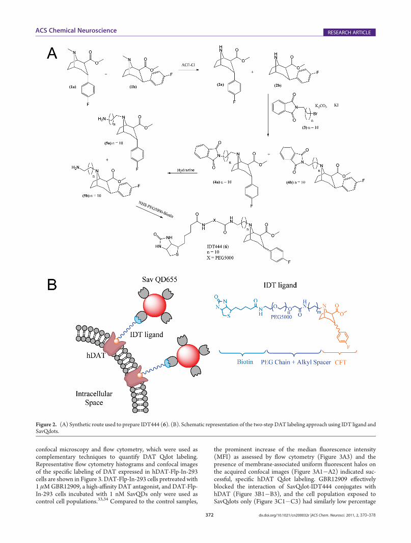
Cocaine Analogue Synthesis. To develop a DAT-specificprobe, a biotinylated cocaine analogue IDT444 (6) was synthe-sized (Figure 2A). The IDT444 ligand is composed of fourdistinct parts: (i) a high-affinity cocaine analogue, 2-β-carbo-methoxy-3-β-(4-fluorophenyl)tropane (β-CFT or WIN 35,428),first reported by Clarke et al., (ii) a short alkyl spacer, (iii) apoly(ethylene glycol) (PEG) chain (averageMW5000), and (iv)a biotin terminus (Figure 2B).26 β-CFT was chosen as the parentdrug due to its high affinity for DAT, excellent structural stability,and pharmacological properties closely resembling those ofcocaine.26,27 The hydrophobic alkyl linker was incorporated intothe IDT444 ligand by attaching it to the nitrogen atom on thetropane ring. The tropane nitrogen was chosen as the attachmentpoint based on previous studies, which showed that bulky groupsattached to the tropane N-position had no significant effect onthe phenyl tropane pharmacological properties and structuralstability.27�31 The short alkyl spacer was attached to the tropanenitrogen to increase flexibility and allow enhanced access to thebinding site. The PEG chain was used to ensure the IDT444ligand is soluble in aqueous buffers and possibly reduce anypotential nonspecific interactions with the cellular membrane.32
The biotin handle at the end of the PEG chain served as a bindingsite for SavQdots. Detailed synthetic steps are described in theSupporting Information.DAT Visualization in Flp-In-293 Cells in Suspension. The
HEK Flp-In-293 cell line was used as a model stable expressionsystem to investigate the interactions of our Qdot-based fluor-escent probes with DAT. To achieve DAT expression, a plasmidvector containing the FRT site linked to the hygromycinresistance gene and the DAT cDNA was integrated into thegenome via Flp recombinase-mediated DNA recombination atthe FRT site. DAT-expressing Flp-In-293 cells were selected inthe presence of 100 μg/mL hygromycin B.DAT-Flp-In-293 cells were subjected to a two-step solution-
based Qdot labeling protocol. Flp-In-293 cells were incubatedwith the IDT444 ligand and subsequently labeled with SavQdotsin solution to prevent the loss of cells due to detachment from theplate surface, reduce nonspecific Qdot interactions with theculture vessel, and enhance specific recognition of the biotiny-lated ligand. These cells were incubated with 100 nM IDT444phosphate-buffered saline (PBS) solution for 5 min at 37 �C,lifted off the culture plate by gentle pipetting, centrifuged, andresuspended in 1 nM SavQdot PBS solution for 5 min at 4 �C.After several wash steps to rinse away the unbound ligands andconjugates, specific DAT Qdot labeling was demonstrated by
Figure 1. Structures of DAT and its relevant substrates. A two-dimen-sional topology of DAT based on the leucine transporter (LeuT) isshown with 12 transmembrane segments, intracellularly oriented N- andC-termini, and substrate binding site. Structures of dopamine, cocaine,and cocaine analogue β-CFT are shown as well. The binding site fordopamine, cocaine, and cocaine analogues has been suggested to overlapand is buried deep between transmembrane segments 1, 3, 6, and 8.38
372 dx.doi.org/10.1021/cn200032r |ACS Chem. Neurosci. 2011, 2, 370–378
ACS Chemical Neuroscience RESEARCH ARTICLE
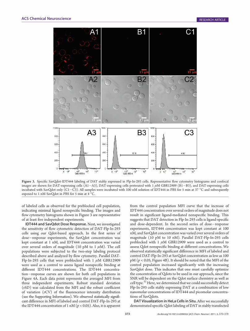
confocal microscopy and flow cytometry, which were used ascomplementary techniques to quantify DAT Qdot labeling.Representative flow cytometry histograms and confocal imagesof the specific labeling of DAT expressed in hDAT-Flp-In-293cells are shown in Figure 3. DAT-Flp-In-293 cells pretreated with1 μMGBR12909, a high-affinity DAT antagonist, and DAT-Flp-In-293 cells incubated with 1 nM SavQDs only were used ascontrol cell populations.33,34 Compared to the control samples,
the prominent increase of the median fluorescence intensity(MFI) as assessed by flow cytometry (Figure 3A3) and thepresence of membrane-associated uniform fluorescent halos onthe acquired confocal images (Figure 3A1�A2) indicated suc-cessful, specific hDAT Qdot labeling. GBR12909 effectivelyblocked the interaction of SavQdot-IDT444 conjugates withhDAT (Figure 3B1�B3), and the cell population exposed toSavQdots only (Figure 3C1�C3) had similarly low percentage
Figure 2. (A) Synthetic route used to prepare IDT444 (6). (B). Schematic representation of the two-step DAT labeling approach using IDT ligand andSavQdots.
373 dx.doi.org/10.1021/cn200032r |ACS Chem. Neurosci. 2011, 2, 370–378
ACS Chemical Neuroscience RESEARCH ARTICLE
of labeled cells as observed for the preblocked cell population,indicating minimal ligand nonspecific binding. The images andflow cytometry histograms shown in Figure 3 are representativeof at least five independent experiments.IDT444 and SavQdot Dose Response.Next, we investigated
the sensitivity of flow cytometric detection of DAT-Flp-In-293cells using our Qdot-based approach. In the first series ofdose�response experiments, the SavQdot concentration waskept constant at 1 nM, and IDT444 concentration was variedover several orders of magnitude (10 pM to 1 μM). The cellpopulations were subjected to the two-step labeling protocoldescribed above and analyzed by flow cytometry. Parallel DAT-Flp-In-293 cells that were preblocked with 1 μM GBR12909were used as a control to assess ligand nonspecific binding atdifferent IDT444 concentrations. The IDT444 concentra-tion�response curves are shown for both cell populations inFigure 4A. Each data point represents the averaged MFI fromthree independent experiments. Robust standard deviation(rSD) was calculated from the MFI and the robust coefficientof variation (rCV) of the fluorescence intensity distribution(see the Supporting Information). We observed statistically signifi-cant difference in MFI of labeled and control DAT-Flp-In-293 atthe IDT444 concentration of 1 nM (p < 0.05). Also, it is apparent
from the control population MFI curve that the increase ofIDT444 concentration over several orders of magnitude does notresult in significant ligand-mediated nonspecific binding. Thissuggests that DAT detection in Flp-In-293 cells is ligand-specificand dose-dependent. In the second series of dose�responseexperiments, IDT444 concentration was kept constant at 100nM, and SavQdot concentration was varied over several orders ofmagnitude (10 pM to 10 nM). Parallel DAT-Flp-In-293 cellspreblocked with 1 μM GBR12909 were used as a control toassess Qdot nonspecific binding at different concentrations. Weobserved statistically significant difference in MFI of labeled andcontrol DAT-Flp-In-293 at SavQdot concentration as low as 100pM (p < 0.05, Figure 4B). It should be noted that the MFI of thecontrol population increased significantly with the increasingSavQdot dose. This indicates that one must carefully optimizethe concentration of Qdots to be used in our approach, since theSNR will be dependent on the Qdot surface chemistry as well ascell type.32 Here, we determined that we could successfully detectFlp-In-293 cells stably expressing DAT at a combination of lownanomolar concentrations of IDT444 and picomolar concentra-tions of SavQdots.DATVisualization in HeLa Cells in Situ.After we successfully
demonstrated specific Qdot labeling of DAT in stably transfected
Figure 3. Specific SavQdot-IDT444 labeling of DAT stably expressed in Flp-In-293 cells. Representative flow cytometry histograms and confocalimages are shown for DAT-expressing cells (A1�A3), DAT-expressing cells pretreated with 1 μM GBR12909 (B1�B3), and DAT-expressing cellsincubated with SavQdot only (C1�C3). All samples were incubated with 100 nM solution of IDT444 in PBS for 5 min at 37 �C and subsequentlyexposed to 1 nM SavQdot in PBS for 5 min at 4 �C.

374 dx.doi.org/10.1021/cn200032r |ACS Chem. Neurosci. 2011, 2, 370–378
ACS Chemical Neuroscience RESEARCH ARTICLE
Flp-In-293 cells in suspension, we attempted to visualize Flp-In-293 cells in 8-well chamber slides in situ. This proved to be a very
challenging task, since Flp-In-293 cells are weakly adherent anddetach easily during gentle washes between labeling steps even incollagen- and fibronectin-coated chamber slides. Therefore wechose a relatively strongly adherent HeLa cell line to determinewhether we could use our approach to label living DAT-expres-sing cells in situ. HeLa cells were transiently transfected with apcDNA3 vector containing the human DAT cDNA in 8-wellchamber slides. Parallel HeLa cells were transfected with anempty (sham) pcDNA3 vector and served as a negative controlto evaluate nonspecific cell labeling. Just prior to confocalimaging, DAT-transfected HeLa cells were incubated with 100nM IDT444, washed 3� with warm imaging buffer, incubatedwith 1 nM SavQD, and washed 3� with warm imaging buf-fer, and Qdot-labeled HeLa cells were subsequently visualized(Figure 5A1,A2). Confocal images clearly demonsrate uniformplasma membrane labeling of DAT-transfected cells. No fluor-escence staining was observed for DAT-transfected HeLa cellspreincubated with 1 μMGBR12909 (Figure 5B1,B2) and sham-transfected HeLa cells (Figure 5C1,C2), indicating a low level ofnonspecific labeling. These data demonstrate that our Qdot-based approach can be used to specifically label DAT in livingcells in situ with high SNR.Visualization of PKC-Dependent DAT Internalization.
Many groups have demonstrated that PKC activation by phorbolesters, such as phorbol 12-myristate 13-acetate (PMA), results inacute down-regulation of DAT surface expression levels inseveral heterologous cell lines.35�37 To investigate whether wecould use our IDT444-SavQdot conjugates to visualize acuteDAT internalization, we incubated transiently transfected HeLacells with 100 nM IDT444 for 5 min at 37 �C, washed the cells3� with warm imaging medium, incubated the cells with 1 nMSavQdot for 2�3 min 37 �C, and washed the cells 3� with warmimaging medium. Qdot-labeled HeLa cells were then incubated
Figure 4. IDT444 (A) and SavQdot (B) dose response curves.Individual data points represent averaged MFI from three indepen-dent experiments. rSD was calculated from the MFI and rCV of thefluorescence intensity distribution (see the Supporting Information).
Figure 5. Specific SavQdot-IDT444 labeling of DAT stably expressed in living HeLa cells in situ. Representative confocal images are shown for DAT-pcDNA3 transiently transfected HeLa cells (A1,A2), DAT-pcDNA3 transiently transfected cells pretreated with 1 μMGBR12909 (B1,B2), and sham-pcDNA3 transiently transfected cells (C1,C2). Fluorescent (top) and overlay (bottom) images are shown. All samples were incubated with 100 nMsolution of IDT444 in the imaging buffer for 5 min at 37 �C and subsequently exposed to 1 nM SavQdot in the imaging buffer for 2�3 min at 37 �C.Images are representative of at least five independent experiments.

375 dx.doi.org/10.1021/cn200032r |ACS Chem. Neurosci. 2011, 2, 370–378
ACS Chemical Neuroscience RESEARCH ARTICLE
for 30 min at 37 �C in the presence or absence of 1 μM PMA.While we did not observe any apparent change in the distributionof membrane DAT-Qdot complexes in HeLa cells incubated inthe presence of vehicle (DMSO) only (Figure 6A1,A2), PMAtreatment led to the appearance of punctate intracellular fluor-escence, a clear sign of acute, PKC-dependent DAT-Qdotcomplex internalization consistent with the previous reports(Figure 6B1,B2). This experiment demonstrates the applicabilityand utility of our Qdot-based labeling approach in the investiga-tion of DAT regulation in living cells.Qdot Photostability. To demonstrate that our Qdot-based
labeling approach can be used as a tool to visualize DAT overextended periods of time, we compared SavQdots to a standardorganic dye FITC. Individual 8-bit images (512 � 512 pixels, 1Airy unit) were acquired every 30 s for 10 min at a scan speed of51.20 μs/pixel, with laser intensity set to 20% (Figure 7A).Fluorescence intensity of Qdot or FITC DAT membrane label-ing in Flp-In-293 cells was quantified, normalized, and plotted asa function of acquisition time (Figure 7B). Each data pointrepresents average integrated membrane fluorescence intensityfor 6 cells from two independent experiments. In the case ofFITC, bleaching occurs within 300 s (Inorm < 0.2), whereas theintensity of Qdot labeling undergoes minimal decrease within600 s. The photobleaching issue becomes important when one ismonitoring biological processes for longer periods of time at hightemporal resolution. Excellent photostability of Qdots makes ourQdot-based labeling approach an excellent candidate for mon-itoring changes in DAT surface expression level and cellularlocalization for prolonged periods of time at temporal resolutionhigher than that allowed by the use of traditional fluorophores.
’CONCLUSION
To summarize, our goal for the current effort was to develop aDAT-specific ligand and demonstrate specific labeling of cellsurface DAT using Qdot-based detection. The biotinylatedderivative of the β-CFT cocaine analogue, IDT444, was preparedby attaching the biotin-PEG-alkyl group to the tropane nitrogen.The ability to visualize mammalian cells stably expressing DATwith SavQdot-IDT444 conjugates was clearly demonstrated byflow cytometry and confocal microscopy for Flp-In-293 cells insuspension and HeLa cells in situ. We determined that DAT-expressing cells can be detected using a combination of lownanomolar concentrations of IDT444 and picomolar concentra-tions of SavQdots, as assessed by flow cytometry. The relativelyhigh sensitivity of our approach in combination with short sample
Figure 6. Visualization of DAT internalization in HeLa cells transientlyexpressing DAT using IDT444-SavQdot conjugates. PMA promotesinternalization of the DAT-Qdot complex in live HeLa cells transientlytransfected with DAT-pcDNA3. The cells were incubated with 100 nMsolution of IDT444 in the imaging buffer for 5 min at 37 �C andsubsequently exposed to 1 nM SavQdot in the imaging buffer for 2�3min at 37 �C before incubation in the absence (A1,A2) or presence of1 μMPMA (B1,B2) for 30 min at 37 �C. Images are representative of atleast three independent experiments.
Figure 7. Photostability comparison between Qdot655 and FITC. (A)Top row: DAT-Flp-In-293 cells labeled with IDT444 and SavFITC.Bottom row: DAT-Flp-In-293 cells labeled with IDT444 and SavQ-dot655. The time-lapse image series were acquired on the Zeiss LSM510 inverted confocal microscope with the 488 nm excitation laser, a650 nm long pass filter (Qdot), or a 505�550 nm band-pass filter(FITC), and Zeiss Plan-Apo oil immersion objective (63�, NA 1.40).Images (512 � 512 pixels, 1 Airy unit) were acquired every 30 s for 10min at a scan speed of 51.20 μs/pixel, with laser intensity set to 20%.Images at 0, 120, and 240 s are shown. (B) Quantitative analysis ofchanges in intensities of SavQdot655 and SavFITC. Membrane-asso-ciated intensity was quantified for each frame by integrating fluorescenceintensity over a manually drawn region encompassing the membranearea, with DIC images serving as a reference point (Metamorph).Intensity values were normalized with respect to the highest intensityvalue obtained (I/I0, where I0 = Imax).

376 dx.doi.org/10.1021/cn200032r |ACS Chem. Neurosci. 2011, 2, 370–378
ACS Chemical Neuroscience RESEARCH ARTICLE
preparation time renders IDT444 useful in a quantum-dot-basedfluorescent assay to monitor DAT expression, function, cellulardistribution, and regulation. Specifically, we demonstrated theapplicability and utility of our approach in the visualization ofacute, PKC-dependent DAT internalization. Due to its sensitivityand flexibility, the quantum dot-based fluorescent assay platformwe describe can potentially replace conventional biochemical andradiolabeled isotope-based approaches to study the distributionand regulation of DAT proteins, and may also be useful in high-throughput screening of novel DAT modulators. If issues relatedto blood-brain barrier penetration of Qdots can be overcome, it isnot unreasonable to consider whether our approach might notalso be of diagnostic utility for perturbations of in vivo DATlevels in addiction, neuropsychiatric, or neurodegenerative dis-orders. Finally, our Qdot-based approch should allow appropria-tion of the unique photophysical properties of Qdots to achievetime-lapse imaging of single DAT molecules in living cells forprolonged periods of time combined with improved temporalresolution, providing new opportunities to elucidate the molec-ular mechanisms supporting DAT regulation.
’METHODS
Materials. All reagents were purchased fromVWR or Sigma-Aldrichand used without further purification unless otherwise noted. AnalyticalTLC was performed on commercial plates obtained from SorbentTechnologies (Atlanta, GA) coated with silica gel with a UV indicator(cat. no. 1634126), and spots were located by UV light (254 and365 nm). Silica gel for flash chromatography was obtained from SorbentTechnologies (60 Å, 230�400mesh). Biotinylated PEG5000NHS esterwas obtained from Laysan Bio (Arab, AL). Streptavidin-conjugatedquantum dots with a maximum fluorescence emission at 655 nm(SavQdots655) were obtained as a 1 μM solution dissolved in boratebuffer at pH 8.5 from Invitrogen (Carlsbad, CA). GBR12909 wasobtained from Tocris Bioscience (Ellisville, MI). Streptavidin-conju-gated fluorescein isothiocyanate (SavFITC) was purchased from Invi-trogen (Carlsbad, CA).Ligand Synthesis. A mixture of the diastereoisomers (1a) and
(1b) was synthesized from tropanone using the method described byMeltzer et al., and these were demethylated with R-chloroethyl chlor-oformate to give a mixture of the boat (2a) and the chair (2b) in 87%yield (Figure 2A).28 According to Meltzer et al., cocaine analogues (1a)and (1b) both exhibited low nanomolar affinity for DAT; therefore, thebiotin-terminated linker arm was attached to both diastereoisomers inthe following manner. The alkyl spacer was then attached to the mixtureof diastereoisomers (2a) and (2b) by refluxing overnight in acetonitrilewith the phthalimide protected alkyl spacer (3) in the presence ofpotassium carbonate and potassium iodide. This gave the mixture ofdiasterioisomers (4a) and (4b). Compound (3) was synthesized aspreviously described by Tomlinson et al.39 The pthalimide protectinggroup was removed from (4a) and (4b) by stirring a solution of thedesired compound in the presence hydrazine mono hydrate to yield themixtures (5a) and (5b). The final intermediate was conjugated tobiotinylated PEG-NHS ester by stirring a solution of the mixture ofdiastereoisomers of phenyl tropane attached to the amino terminatedalkyl spacer with biotinylated PEG5000-NHS in methylene chloride for24 h. The solution was then evaporated, and the product was washedwith diethyl ether to give IDT444 (6). The product was further purifiedfrom excess unreacted intermediate (5a,b) by size-exclusion chroma-tography using PD-10 column (GE Biosciences). The final product wascharacterized by MALDI-TOF mass spectrometry (Supporting Infor-mation Figure S1). Detailed synthetic steps are described in the Sup-porting Information.
Cell LineMaintenance. The Flp-In 293 host cell line (Invitrogen)was grown in complete medium (D-MEM with 2 mM L-glutamine, 10%FBS, 1% pen/strep) supplemented with 100 μg/mL Hygromycin B in a37 �C incubator with 5% CO2. The wild-type human DAT (hDAT)cDNA cloned in the pcDNA5/FRT expression vector was transfectedinto Flp-In-293 cells using the Fugene 6 transfection reagent (Roche, NJ).After 48 h recovery, the cells were grown in medium with 100 μg/mLhygromycin B (100 μg/mL) added for several weeks to select forresistant cells where the cDNA construct had been recombined into theFlp-In site in the Flp-In-293 cells. Prior to fluorescent imaging and flowcytometry experiments, cells were seeded in 24-well polylysine-coatedculture plates (BD Biosciences, Bedford, MA). HeLa cell line (ATCC#CCL-2) was grown in complete medium (D-MEM with 2 mML-glutamine, 10% FBS, 1% penn/strep) in a 37 �C incubator with 5%CO2. For transient transfection experiments, HeLa cells were grown incomplete medium in the absence of pen/strep. Twenty-four hours afterseeding HeLa cells, nonexpressing (sham) and DAT-expressing pcDNA3vectors containing the SV40 origin and early promoter region forepisomal replication were introduced into HeLa cells. Fugene HD(Roche, NJ) was used to facilitate transient transfection according tothe manufacturer’s recommendations. The cells were allowed 24 h post-transfection for successful expression of DAT.Flp-In 293 Cell Labeling. A two-step labeling assay was utilized to
specifically target DAT with the IDT444 ligand and SavQdots655. Thecells were incubated in 24-well culture plates with a solution of thebiotinylated ligand (IDT444) in PBS (Gibco) for 10 min at 37 �C. Thenthe contents of each well were pipetted, transferred into 1.5 mLmicrofuge tubes (Millipore, Billerica, MA), pelleted by centrifugationat 2000 rpm for 5 min, and resuspended in a solution of SavQdots655 inPBS for 5 min at 4 �C. Qdot-labeled cells were washed two times bycentrifugation and resuspension in PBS at 4 �C, transferred to 5 mLpolystyrene round-bottom tubes (BD Biosciences, Bedford, MA), andassayed for SavQdot655 labeling with flow cytometry and confocalmicroscopy.Flow Cytometry. Qdot-labeled cells were analyzed on a BD LSRII
flow cytometer (BD Biosciences). Qdot655 fluorescence was detectedwith the 488 nm excitation laser on the FL3 channel (640 nm long passfilter). Forward (FSC) and side scatter (SSC) data were collected inlinear mode, while the FL3 channel data was collected in log mode.Twenty thousand events were collected per sample. FSC and SSCmeasurements were used to gate the viable cell region to assay for Qdotfluorescence (Supporting Information Figure S2). Median fluorescenceintensity (MFI) and robust standard deviation (rSD) were thendetermined for the gated cell population using Flow Jo (Tree Star,Ashland, OR). See the Supporting Information for detailed explanationof the statistical analysis in Flow Jo.Confocal Microscopy (Suspended Cells). Flow cytometry cell
samples were also used for confocal image acquisition. A 50 μL aliquot ofthe suspended cells was transferred onto the 0.13�0.17 mm thinmicroscope cover glass. DIC and fluorescent images were acquired onthe Zeiss LSM 510 inverted confocal microscope with the 488 nmexcitation laser, a 650 nm long pass filter (SavQdot655) or a505�550 nm band-pass filter (SavFITC), and Zeiss Plan-Apo oilimmersion objective (63�, NA 1.40).Confocal Microscopy (Adherent Cells). HeLa cells were
seeded in 8-well chamber slides (Nalge Nunc, Rochester, NY) andtransiently transfected as described above. Prior to confocal confocalmicroscopy, the old mediumwas aspirated from the chamber slide wells,and HeLa cells were washed 3�with warm imaging buffer (Phenol Red-free DMEM/5%FBS). HeLa cells were then incubated with 100 nMIDT444, washed 3� with warm imaging buffer, incubated with 1 nMSavQdot655, washed 3� with warm imaging buffer, and visualized in200 μL of warm imaging buffer at 37 �C. DIC and fluorescent imageswere acquired on the Zeiss LSM 510 inverted confocal microscope with
377 dx.doi.org/10.1021/cn200032r |ACS Chem. Neurosci. 2011, 2, 370–378
ACS Chemical Neuroscience RESEARCH ARTICLE
the 488 nm excitation laser, a 650 nm long pass filter, and Zeiss Plan-Apooil immersion objective (63�, NA 1.40).Qdot versus FITC Photostability. DAT-Flp-In-293 cells were
incubated with 100 nM IDT444 and resuspended in either 1 nMSavQdot or 10 μg/mL SavFITC. The time-lapse image series wereacquired on the Zeiss LSM 510 inverted confocal microscope with the488 nm excitation laser, a 650 nm long pass filter (Qdot) or a505�550 nm band-pass filter (FITC), and Zeiss Plan-Apo oil immer-sion objective (63�, NA 1.40). Images (512 � 512 pixels, 1 Airy unit)were acquired every 30 s for 10 min at a scan speed of 51.20 μs/pixel,with laser intensity set to 20%. Membrane-associated intensity wasquantified for each frame by integrating fluorescence intensity over amanually drawn region encompassing the membrane area, with DICimages serving as a reference point (Metamorph). Intensity values werenormalized with respect to the highest intensity value obtained (I/I0,where I0 = Imax).
’ASSOCIATED CONTENT
bS Supporting Information. Detailed synthetic steps andligand characterization, flow cytometry analysis, and Qdot label-ing of HeLa cells in suspension. This material is available free ofcharge via the Internet at http://pubs.acs.org.
’AUTHOR INFORMATION
Corresponding Author*E-mail: [email protected].
Funding SourcesConfocal imaging using Zeiss LSM 510 Meta was performed inpart through the use of the VUMCCell Imaging Shared Resourcesupported by NIH Grants CA68485, DK20593, DK58404,HD15052, DK59637, and EY08126. Flow cytometry experi-ments were performed in the VMC Flow Cytometry SharedResource supported by the Vanderbilt Ingram Cancer Center(P30 CA68485) and the Vanderbilt Digestive Disease ResearchCenter (DK058404). This work was supported by NIH GrantsEB003728 to S.J.R., and DA07390 and DA027739 to R.D.B.
’ACKNOWLEDGMENT
The authors would like to thank Dr. Michael R. Warnementand Dr. Michael A. Schreuder for helpful discussions, JaneWright and Qiao Han for help with cell line maintenance, andJosh Swartz for help with size-exclusion chromatography.
’REFERENCES
(1) Giros, B., and Caron,M. G. (1993)Molecular characterization ofthe dopamine transporter. Trends Pharmacol. Sci. 14 (2), 43–49.(2) Darvas, M., and Palmiter, R. D. (2010) Restricting dopaminergic
signaling to either dorsolateral or medial striatum facilitates cognition.J. Neurosci. 30 (3), 1158–1165.(3) Bannon, M. J. (2004) Dopamine. Nature Encyclopedia of Life
Sciences, Nature Publishing Group, retrieved May 6, 2010 from www.els.net.(4) Swanson, J. M., Flodman, P., Kennedy, J., Spence, M. A., Moyzis,
R., Schuck, S., Murias, M., Moriarity, J., Barr, C., Smith, M., and Posner,M. (2000) Dopamine genes and ADHD.Neurosci. Biobehav. Rev. 24 (1),21–25.(5) Greenwood, T. A., Alexander, M., Keck, P. E., McElroy, S.,
Sadovnick, A. D., Remick, R. A., and Kelsoe, J. R. (2001) Evidence forlinkage disequilibrium between the dopamine transporter and bipolardisorder. Am. J. Med. Genet. 105 (2), 145–151.
(6) Tremblay, L. K., Naranjo, C. A., Graham, S. J., Herrmann, N.,Mayberg, H. S., Hevenor, S., and Busto, U. E. (2005) Functionalneuroanatomical substrates of altered reward processing in majordepressive disorder revealed by a dopaminergic probe. Arch. Gen.Psychiatry 62 (11), 1228–1236.
(7) Sulzer, D. (2007) Multiple hit hypotheses for dopamine neuronloss in Parkinson’s disease. Trends Neurosci. 30 (5), 244–250.
(8) Swerdlow, N. R., and Koob, G. F. (1987) Dopamine, schizo-phrenia, mania, and depression: Toward a unified hypothesis of cortico-striatopallido-thalamic function. Behav. Brain Sci. 10 (02), 197–208.
(9) Usiello, A., Baik, J.-H., Rouge-Pont, F., Picetti, R., Dierich, A.,LeMeur, M., Piazza, P. V., and Borrelli, E. (2000) Distinct functions ofthe two isoforms of dopamine D2 receptors. Nature 408 (6809),199–203.
(10) Giros, B., Jaber, M., Jones, S. R., Wightman, R. M., and Caron,M. G. (1996) Hyperlocomotion and indifference to cocaine andamphetamine in mice lacking the dopamine transporter. Nature 379(6566), 606–12.
(11) Raul, R. G., Sara, R. J., and Marc, G. C. (1999) Functionalhyperdopaminergia in dopamine transporter knock-out mice. Biol.Psychiatry 46 (3), 303–311.
(12) Ralph, R. J., Paulus, M. P., Fumagalli, F., Caron, M. G., andGeyer, M. A. (2001) Prepulse Inhibition Deficits and PerseverativeMotor Patterns in Dopamine Transporter Knock-OutMice: DifferentialEffects of D1 andD2 Receptor Antagonists. J. Neurosci. 21 (1), 305–313.
(13) Schmitt, K. C., and Reith, M. E. A. (2010) Ann. N.Y. Acad. Sci.1187, 316–340.
(14) Eriksen, J., Jorgensen, T. N., and Gether, U. (2010) Regulationof dopamine transporter function by protein-protein interactions: newdiscoveries and methodological challenges. J. Neurochem. 113 (1),27–41.
(15) Furman, C. A., Chen, R., Guptaroy, B., Zhang, M., Holz, R. W.,and Gnegy, M. (2009) Dopamine and amphetamine rapidly increasedopamine transporter trafficking to the surface: live-cell imaging usingtotal internal reflection fluorescence microscopy. J. Neurosci. 29 (10),3328–3336.
(16) Eriksen, J., Rasmussen, S. G., Rasmussen, T. N., Vaegter, C. B.,Cha, J. H., Zou, M. F., Newman, A. H., and Gether, U. (2009)Visualization of dopamine transporter trafficking in live neurons byuse of fluorescent cocaine analogs. J. Neurosci. 29 (21), 6794–808.
(17) Haraguchi, T. (2002) Live cell imaging: approaches for study-ing protein dynamics in living cells. Cell Struct. Funct. 27 (5), 333–334.
(18) Adkins, E. M., Samuvel, D. J., Fog, J. U., Eriksen, J., Jayanthi,L. D., Vaegter, C. B., Ramamoorthy, S., and Gether, U. (2007)Membrane Mobility and Microdomain Association of the DopamineTransporter Studied with Fluorescence Correlation Spectroscopy andFluorescence Recovery after Photobleaching. Biochemistry 46 (37),10484–10497.
(19) Bruchez, M., Jr., Moronne, M., Gin, P., Weiss, S., and Alivisatos,A. P. (1998) Semiconductor Nanocrystals as Fluorescent BiologicalLabels. Science 281 (5385), 2013–2016.
(20) Chan, W. C. W, and Nie, S. (1998) Quantum Dot Bioconju-gates for Ultrasensitive Nonisotopic Detection. Science 281 (5385),2016–2018.
(21) Pinaud, F., Clarke, S., Sittner, A., and Dahan, M. (2010)Probing cellular events, one quantum dot at a time. Nat. Methods 7(4), 275–285.
(22) Rosenthal, S. J., Chang, J. C., Kovtun, O., McBride, J. R., andTomlinson, I. D. (2011) Biocompatible Quantum Dots for BiologicalApplications. Chem. Biol. 18 (1), 10–24.
(23) Rosenthal, S. J., Tomlinson, I., Adkins, E. M., Schroeter, S.,Adams, S., Swafford, L., McBride, J., Wang, Y., DeFelice, L. J., andBlakely, R. D. (2002) Targeting Cell Surface Receptors with Ligand-Conjugated Nanocrystals. J. Am. Chem. Soc. 124 (17), 4586–4594.
(24) Tomlinson, I. D., Mason, J., Burton, J. N., Blakely, R., andRosenthal, S. J. (2003) The design and synthesis of novel derivatives ofthe dopamine uptake inhibitors GBR 12909 and GBR 12935. High-affinity dopaminergic ligands for conjugation with highly fluorescent

378 dx.doi.org/10.1021/cn200032r |ACS Chem. Neurosci. 2011, 2, 370–378
ACS Chemical Neuroscience RESEARCH ARTICLE
cadmium selenide/zinc sulfide core/shell nanocrystals. Tetrahedron 59(40), 8035–8047.(25) Tomlinson, I. D., and Mason, J. N. et al. (2006) High affinity
inhibitors of the dopamine transporter (DAT): Novel biotinylatedligands for conjugation to quantum dots. Bioorg. Med. Chem. Lett. 16(17), 4664–4667.(26) Clarke, R. L., Daum, S. J., Gambino, A. J., Aceto, M. D., Pearl, J.,
Levitt, M., Cumiskey, W. R., and Bogado, E. F. (1973) Compoundsaffecting the central nervous system. 4. 3 Beta-phenyltropane-2-car-boxylic esters and analogs. J. Med. Chem. 16 (11), 1260–1267.(27) Carroll, F. I., Lewin, A. H., Boja, J. W., and Kuhar, M. J. (1992)
Cocaine receptor: biochemical characterization and structure�activityrelationships of cocaine analogues at the dopamine transporter. J. Med.Chem. 35 (6), 969–981.(28) Meltzer, P. C., Blundell, P., Zona, T., Yang, L., Huang, H.,
Bonab, A. A., Livni, E., Fischman, A., and Madras, B. K. (2003) ASecond-Generation 99mTechnetium Single Photon Emission Com-puted Tomography Agent That Provides in Vivo Images of theDopamine Transporter in Primate Brain. J. Med. Chem. 46 (16),3483–3496.(29) Davis, M. R., Votaw, J. R., Bremner, J. D., Byas-Smith, M. G.,
Faber, T. L., Voll, R. J., Hoffman, J. M., Grafton, S. T., Kilts, C. D., andGoodman, M. M. (2003) Initial Human PET Imaging Studies withthe Dopamine Transporter Ligand 18F-FECNT. J. Nucl. Med. 44 (6),855–861.(30) Rinne, J., Ruottinen, H., Bergman, J., Haaparanta, M., Sonninen,
P., and Solin, O. (1999) Usefulness of a dopamine transporter PETligand [18F]β-CFT in assessing disability in Parkinson’s disease.J. Neurol., Neurosurg. Psychiatry 67 (6), 737–741.(31) Harada, N., Ohba, H., Fukumoto, D., Kakiuchi, T., and
Tsukada, H. (2004) Potential of [(18)F]beta-CFT-FE (2beta-carbo-methoxy-3beta-(4-fluorophenyl)-8-(2-[(18)F]fluoroethyl)nortropane)as a dopamine transporter ligand: A PET study in the conscious monkeybrain. Synapse 54 (1), 37–45.(32) Bentzen, E. L., Tomlinson, I. D., Mason, J., Gresch, P.,
Warnement, M. R., Wright, D., Sanders-Bush, E., Blakely, R., andRosenthal, S. J. (2005) Surface modification to reduce nonspecificbinding of quantum dots in live cell assays. Bioconjugate Chem. 16 (6),1488–1494.(33) H€osli, E., and H€osli, L. (1997) Autoradiographic studies on the
uptake of 3H-dopamine by neurons and astrocytes in explant andprimary cultures of rat CNS: effects of uptake inhibitors. Int. J. Dev.Neurosci. 15 (1), 45–53.(34) Inazu, M., Kubota, N., Takeda, H., Zhang, J., Kiuchi, Y., Oguchi,
K., and Matsumiya, T. (1999) Pharmacological characterization ofdopamine transport in cultured rat astrocytes. Life Sci. 64 (24),2239–2245.(35) Blakely, R. D., and Bauman, A. L. (2000) Biogenic amine
transporters: regulation in flux. Curr. Opin. Neurobiol. 10, 328–336.(36) Daniels, G.M., and Amara, S. G. (1999) Regulated trafficking of
the human dopamine transporter. Clathrin-mediated internalization andlysosomal degradation in response to phorbol esters. J. Biol. Chem.274, 35794–35801.(37) Melikian, H. E., and Buckley, K. M. (1999) Membrane traffick-
ing regulates the activity of the human dopamine transporter. J. Neurosci.19, 7699–7710.(38) Kurian, M. A., Zhen, J., Cheng, S.-Y., Li, Y., Mordekar, S. R.,
Jardine, P., Morgan, N. V., Meyer, E., Tee, L., Pasha, S., Wassmer, E.,Heales, S. J. R., Gissen, P., Reith, M. E. A., and Maher, E. R. (2009)Homozygous loss-of-function mutations in the gene encoding thedopamine transporter are associated with infantile parkinsonism-dystonia. J. Clin. Invest. 119 (6), 1595–1603.(39) Tomlinson, I. D., Gies, A. P., Gresch, P. J., Dillard, J., Orndorff,
R. L., Sanders-Bush, E., Hercules, D. M., and Rosenthal, S. J. (2006)Universal polyethylene glycol linkers for attaching receptor ligands toquantum dots. Bioorg. Med. Chem. Lett. 16 (24), 6262–6266.