visualizing infection teaching the principles of clinical ... institutes of health • office of the...
TRANSCRIPT
NATIONAL INSTITUTES OF HEALTH • OFFICE OF THE DIRECTOR | VOLUME 24 ISSUE 2 • MARCH-APRIL 2016
Visualizing InfectionNew Ways to Image the Immune SystemBY BRANDON LEVY, NIMH
CONTENTS
FEATURES • |1| Visualizing Infection |1| Teaching the Principles of Clinical Research
|3| Sorcerer of the Sequencer: Bob Blakesley |5| NHGRI Seminar Series on Human Genome
Project |6| New Director for NCATS Stem Cell Translation Research Center: Ilyas Singec
DEPARTMENTS • |2| DDIR: Contributions to Public Health |4| Training Page: Visiting Fellows
|4| Abbreviations |12,13| SIG Beat: New; Periodontitis Seminar |8| Research Briefs
|16| Colleagues: Recently Tenured |19| Announcements |20| Photographic Moment: Safety Girl
CONTINUED ON PAGE 10
CONTINUED ON PAGE 14
Celebrating its 20th anniversary, the course “Introduction to the Principles and Practice of Clinical Research” (IPPCR) has an impressive title and focuses on a clear goal: providing instruction on the basics of high-quality, safe, ethical, and efficiently conducted clinical research.
The course, one of the longest-running educational programs at the National Institutes of Health (NIH), started with a simple conversation in the early 1990s between then–NIH
“We live in a dangerous world, constantly bombarded with bacteria, viruses, fungi, or parasites,” said Ronald Germain at the annual G. Burroughs Mider Lecture, held in December. “How does the immune system protect against adverse unpredictable disease entities at unanticipated sites in the body?”
Fighting off infection is an incredibly complex process. There’s not one type of immune cell, but many. They swarm. They dance. They work in sync. Germain, who’s chief of the Laboratory of Systems Biology and of the Lymphocyte Biology Section at the National Institute of Allergy and Infectious Disease (NIAID), uses cutting-edge imaging techniques to investigate the intricate movements and positioning of immune cells.
“In contrast to almost all other tissues in an adult, the immune system is unique [because] there are cells moving all around in many tissues and organs, touching each other, transmitting signals, then going apart and changing their function,” said Germain. “It’s impossible to access these key aspects of immune behavior without doing imaging.”
His lab was one of the first to use a technique called two-photon intravital imaging, which uses laser illumination to see deep into living tissue without causing the damage done by traditional confocal fluorescence microscopy. Germain collaborates with other scientists, such as
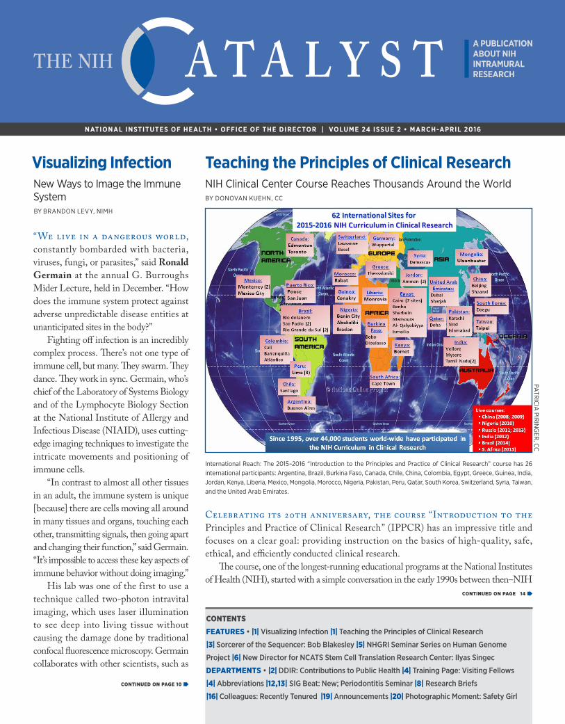
Teaching the Principles of Clinical ResearchNIH Clinical Center Course Reaches Thousands Around the WorldBY DONOVAN KUEHN, CC
PATR
ICIA
PIRIN
GER
, CC
International Reach: The 2015–2016 “Introduction to the Principles and Practice of Clinical Research” course has 26 international participants: Argentina, Brazil, Burkina Faso, Canada, Chile, China, Colombia, Egypt, Greece, Guinea, India, Jordan, Kenya, Liberia, Mexico, Mongolia, Morocco, Nigeria, Pakistan, Peru, Qatar, South Korea, Switzerland, Syria, Taiwan, and the United Arab Emirates.
2 THE NIH CATALYST MARCH-APRIL 2016
FROM THE DEPUTY DIRECTOR FOR INTRAMURAL RESEARCH
I have often used this space to remind the NIH intramural community of the important role that the intramural program plays in addressing urgent and compelling issues related to the public health. Three recent examples are worthy illustrations of the importance and impact of intramural involvement in mounting a rapid and effective research response to complex and difficult issues that have aroused public concern and threaten the public health: (1) the role the National Institute of Allergy and Infectious Diseases (NIAID) is playing in developing diagnostics and a vaccine that could help contain the current Zika epidemic; (2) a new protocol at the National Institute of Neurological Disorders and Stroke (NINDS) to help define, characterize, and potentially treat postinfectious myalgic encephalomyelopathy (previously known as chronic fatigue syndrome); and (3) the National Institute of Environmental Health Sciences’ (NIEHS) research related to the lead-contaminated water supply in Flint, Michigan.
NIAID Director Anthony Fauci has been a visible authority in interpreting events related to the Zika epidemic in South and Central America and the Caribbean to a concerned American and international audience. The key elements of this filovirus epidemic are the important role that mosquito vectors play (especially members of the genus Aedes); the relative mildness of the primary illness compared with its potentially devastating effects—especially microcephaly—on the developing
fetus when women are infected early in pregnancy; and its cross-reactivity with dengue, another filovirus, makes antibody-based epidemiological studies difficult.
As related by Hugh Auchincloss, NIAID deputy director and acting scientific director, the NIH intramural program is contributing to studies of the pathogenesis of Zika and its potential prevention in two
important ways. First, to allow Zika-specific antibody studies, NIAID is synthesizing a Zika antigen that is devoid of dengue cross-reacting epitopes. Second, a current intramural quadrivalent dengue vaccine is being modified to insert Zika open-reading frames. The production of this construct is straightforward, but the creation of a vaccine suitable for use will take at least a year. This delay, given the rapid spread of Zika in the Americas, will mean that such a vaccine will arrive after the main epidemic has passed. Most susceptible individuals will have been infected and developed natural immunity. Targeting the mosquito vector now appears to be the highest priority for prevention.
Av i Nat h , N I N D S c l i n i c a l director, is developing a c l inica l protocol to study postinfectious myalgic encephalomyelopathy–chronic fatigue
syndrome (ME-CFS). An important issue with this disorder is the need to carefully define the patient population that will be studied to increase the likelihood that a clear etiology can be determined. One typical pattern is the development of profound fatigue and postexertional weakness after an acute febrile illness.
Postviral asthenia, or weakness, is a well-established phenomenon, but in most people, the fatigue dissipates after a few weeks. In people with ME-CFS, however, the fatigue persists for many months or years. A reasonable hypothesis is that this weakness is the result of the activation of an immune-mediated brain dysfunction.
Nath’s protocol will explore this hypothesis through detailed
phenot y ping inc lud ing genet ic , metabolic, microbiological, neurological, immunological, and neuroendocrine studies of a pilot group of 40 ME-CFS patients and various control subjects. It is hoped that the application of the most advanced studies of this type will provide an indication as to what goes wrong in this disorder.
Finally, the NIEHS has taken a long-term leadership role for the Department of Health and Human Services in research related to the health effects of the lead-contaminated drinking water in Flint, Michigan. Linda Birnbaum, director of NIEHS, and John Bucher, scientific director of NIEHS’s National Toxicology Program, explained that Flint’s water source was changed about two years ago from the treated Detroit Water and Sewerage Department water (from the Detroit River
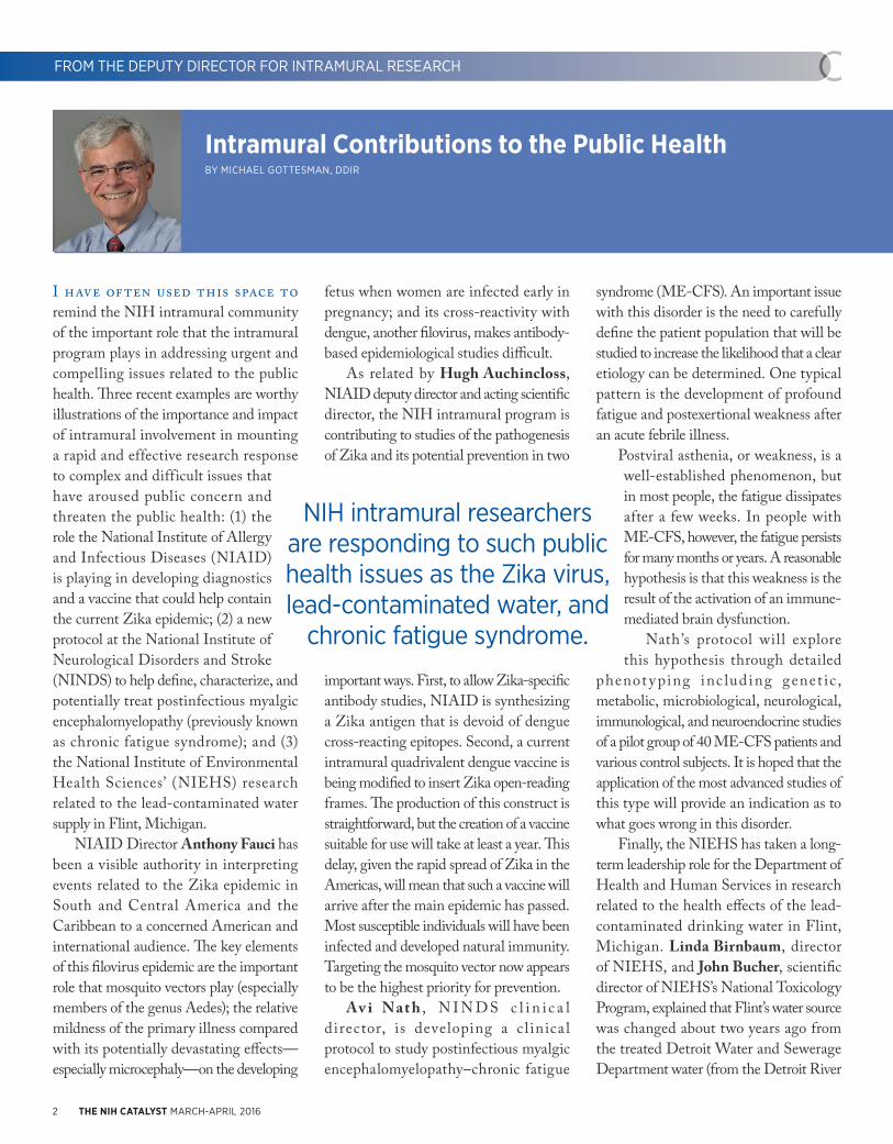
Intramural Contributions to the Public HealthBY MICHAEL GOTTESMAN, DDIR
FROM THE DEPUTY DIRECTOR FOR INTRAMURAL RESEARCH
NIH intramural researchers are responding to such public health issues as the Zika virus, lead-contaminated water, and
chronic fatigue syndrome.

http://irp.nih.gov/catalyst 3
FROM THE DEPUTY DIRECTOR FOR INTRAMURAL RESEARCH
and Lake Huron) to the highly corrosive Flint River. Because the Flint River water was not treated with orthophosphate to prevent leaching of lead from lead piping and lead soldering in public pipes and plumbing in Flint households, lead concentrations in some of the drinking water coming from some faucets reached 13,000 parts per billion (ppb); 15 ppb is the highest concentration allowed by the Environmental Protection Agency.
No level of exposure to lead is considered safe, however. NIEHS research has confirmed that blood-lead concentrations above 5 micrograms per deciliter (μg/dL) delay puberty, increase the incidence of attention deficit hyperactivity disorder (ADHD) and other problem behaviors, and decrease academic achievement and cognitive measures in children.
A full evaluation of blood-lead concentrations in children in Flint is underway, but a limited sample so far indicates a doubling or tripling of the numbers of children with concentrations greater than 5 μg/dL. Both Birnbaum and Bucher emphasized that environmental lead contamination in lower-income communities is not limited to Flint and represents an important contributor to health disparities as well as illustrating the need for environmental justice in oversight of environmental contaminants.
I think we should all be proud of these important leadership roles and contributions of our intramural colleagues to improving both global health and health within our own communities. In addition to the direct effects of developing new diagnostics and therapeutics, the trusted research that we do at NIH provides a firm foundation for important public-health policies.
G e n o m i c s r e s e a r c h i s a q u i n t e s s e n t i a l t e a m s c i e n c e . Contributing to each project are those who identify the scientif ic questions, collect biological samples, purify and sequence the DNA, and analyze the resulting data. The National Institutes of Health lost a key member of its broader genomics team with the December 31, 2015, retirement of Robert Blakesley, who was the director of the sequencing g r o u p a t t h e N I H I n t r a m u r a l Sequencing Center (NISC) in Rockville, Maryland.
Blakesley came to NIH in 2000 after having spent more than 20 years in the biotechnology industry, where he oversaw the creation of an automated DNA-sequencing machine for medical diagnostics and directed new product development.
“When I recruited him to NISC in 2000, I knew that we would benefit from someone with his seasoned private-sector experience, deep technical expertise, good judgment, and calm, mature personality,”
said Eric Green, director of the National Human Genome Research Institute (NHGRI), who had established NISC in 1997. In recognition of Blakeley’s contributions, Green awarded him the NHGRI Director’s Distinguished Service Award at the institute’s 2015 scientific symposium.
The scope of what Blakesley has accomplished on behalf of numerous intramural researchers is nothing short of epic. Between 1998 and 2012, NISC generated more than 57 million DNA-sequence reads for 76 researchers using the Sanger dideoxy sequencing method. That figure has spiked in the past six years with the introduction of next-generation DNA sequencing: During that time, NISC generated 2,151 billion sequence reads for 20,593 unique DNA samples contributed by 126 researchers at 15 NIH institutes and centers.
Blakesley’s achievement goes beyond the sheer quantity of sequence data: His group has attained a 90 percent success rate in generating sequence data from DNA samples accepted for study.
“He demand[ed] the highest possible quality all the time,” said NISC Deputy Director Alice Young, who has worked with Blakesley since 1990, first at Bethesda Research Labs (BRL; renamed Life Technologies, Inc., while he was still there) and now at NISC. “He takes a lot of personal pride in his work, so everybody who works with him has the same pride and ownership.”
Fifth-Generation CalifornianBlakesley is a fifth-generation Californian. His maternal great-great-grandfather was
FROM THE DEPUTY DIRECTOR FOR INTRAMURAL RESEARCH
http://irp.nih.gov/catalyst 3
Sorcerer of the SequencerBob Blakesley, Director of the NISC Sequencing Group, RetiresBY JEANNINE MJOSETH, NHGRI
FEATURE
CONTINUED ON PAGE 11
Robert Blakesley, a key member of NIH’s genomics team, retired recently.
ERN
ESTO
DEL
AG
UIL
A, N
HG
RI

THE TRAINING PAGE
4 THE NIH CATALYST MARCH-APRIL 2016
FROM THE FELLOWS COMMITTEEHelping Visiting Fellows Thrive at the NIHBY CRAIG MYRUM, NIA
The quest to solve the world’s most critical biomedical questions is a global venture that has sparked interactions among researchers around the world. Few other institutions make this more evident than the NIH, where nearly half of all postdoctoral fellows are international researchers. It is not always easy for these researchers to uproot themselves from the familiar surroundings of their home countries and start anew. Luckily, the members of the Visiting Fellows Committee (VFC) use their experiences to help other international fel lows transition to life at the NIH.
VFC, one of eight Fellows Committee (FelCom) subcommittees, offers valuable resources to international postdocs and even designates country representatives to help newly arrived compatriots. Members also run a brown-bag lunch series at which postdocs can discuss practical issues such as immigration, funding opportunities, and taxes. VFC also organizes a “Science Voices from Home” seminar, which aims to connect all NIH fellows with the international science community and identifies research opportunities abroad. The VFC even has its own quarterly newsletter that highlights activities; funding and research opportunities; NIH alumni; professional-development information; and stories on the postdoc life.
“VFC really has a strong sense of community, as we are all in the same situation, trying to navigate the social, cultural, and administrative USA. We feel united and help each other [as] others help[ed] us when we arrived,” says VFC co-chair Stephanie Olivier-Van Stichelen (National Institute of Diabetes and Digestive and Kidney Diseases). The VFC saw a surge in activity last year, thanks
to her and co-chair Fatima Ali-Rahmani (National Cancer Institute) and several new VFC members who were determined to improve and promote the organization. (Olivier-Van Stichelen is from France and Ali-Rahmani is from Pakistan.) Members meet frequently for monthly lunches, social networking, and other events like baseball or hockey games, museums, and ice-skating.
“Foreign scientists are often lacking networking opportunities—either because we don’t know anybody in the U.S. or because of language issues,” said Olivier-Van Stichelen. “These events allow us to discuss and socialize with people in the same situation and at the same time gives us the opportunity to experience the U.S. culture.”
For more information: Subscribe to the VFC-L
LISTSERV at https://list.nih.gov/cgi-bin/
wa.exe?A0=vfc-l; check out the VFC websites:
http://visitingfellows.tumblr.com or https://
www.training.nih.gov/felcom/visitingfellows2;
or contact the VFC co-chairs (Stephanie.
[email protected] or fatima.ali-
NIH ABBREVIATIONS
CBER: Center for Biologics Evaluation and Research, FDACC: NIH Clinical CenterCCR: Center for Cancer Research, NCICDC: Centers for Disease Control and PreventionCIT: Center for Information TechnologyDCEG: Division of Cancer Epidemiology and Genetics, NCIFAES: Foundation for Advanced Education in the SciencesFARE: Fellows Award for Research Excellence FelCom: Fellows CommitteeFDA: Food and Drug AdministrationFNL: Frederick National LaboratoryIRP: Intramural Research ProgramHHS: U.S. Department of Health and Human ServicesNCATS: National Center for Advancing Translational SciencesNCBI: National Center for Biotechnology InformationNCCIH: National Center for Complementary and Integrative HealthNCI: National Cancer InstituteNEI: National Eye InstituteNHGRI: National Human Genome Research InstituteNHLBI: National Heart, Lung, and Blood InstituteNIA: National Institute on AgingNIAAA: National Institute on Alcohol Abuse and AlcoholismNIAID: National Institute of Allergy and Infectious DiseasesNIAMS: National Institute of Arthritis and Musculoskeletal and Skin DiseasesNIBIB: National Institute of Biomedical Imaging and BioengineeringNICHD: Eunice Kennedy Shriver National Institute of Child Health and Human DevelopmentNIDA: National Institute on Drug AbuseNIDCD: National Institute on Deafness and Other Communication DisordersNIDCR: National Institute of Dental and Craniofacial ResearchNIDDK: National Institute of Diabetes and Digestive and Kidney DiseasesNIEHS: National Institute of Environmental Health SciencesNIGMS: National Institute of General Medical SciencesNIMH: National Institute of Mental HealthNIMHD: National Institute on Minority Health and Health DisparitiesNINDS: National Institute of Neurological Disorders and StrokeNINR: National Institute of Nursing ResearchNLM: National Library of MedicineOD: Office of the DirectorOITE: Office of Intramural Training and EducationOIR: Office of Intramural ResearchORS: Office of Research ServicesORWH: Office of Research on Women’s HealthOTT: Office of Technology Transfer
UPCOMING OITE EVENTS
POSTBACCALAUREATE POSTER DAY
Wed., April 20, 10:00 a.m-.3:30 p.m.
Natcher Conference Center (Bldg 45)
More information: https://www.training.
nih.gov/postbac_poster_day.
BUILD YOUR CAREER; SHAPE YOUR
FUTURE: NIH CAREER SYMPOSIUM
Friday, May 6, 8:30 a.m.-5:00 p.m.
Natcher Conference Center (Bldg 45)
More information: www.training.nih.gov
— More details on page 19 —

http://irp.nih.gov/catalyst 5http://irp.nih.gov/catalyst 5http://irp.nih.gov/catalyst 5http://irp.nih.gov/catalyst 5
“The Human Genome Project was a remarkable scientif ic endeavor. It reshaped biomedical research and paved the way for clinical advances that are already impacting patients’ lives,” said National Human Genome Research Institute (NHGRI) Director Eric Green at the launch of a new seminar series that commemorates the 25th anniversary of the launch of the Human Genome Project.
The series, entitled “A Quarter Century after the Human Genome Project’s Launch: Lessons Beyond the Base Pairs,” showcases the venture, its influence on biomedical research, and the future implications of genomics. It began on December 3, 2015, with a panel discussion moderated by Green and featuring former NHGRI Deputy Directors Elke Jordan (retired in 2002) and Mark Guyer (retired in 2014).
The Human Genome Project arose in part from the United States Department of Energy’s interest in the effects of low-frequency radiation on humans, particularly on their DNA. However, the effects were impossible to study without first building a map of the human genome, an endeavor that was controversial from the start.
“There were some who thought that this [undertaking] was too much and was not interesting research,” said Guyer. But “others thought that this had to be done.”
The key to bridging this gap was the idea that creating genomic maps of nonhuman model organisms would yield great scientific insight on its own, even if a blueprint of the human genome proved an unattainable goal. In 1988, the NIH created the Office of Human Genome
Research, which laid the groundwork for NIH’s contribution to the Human Genome Project. A year later, this office became the National Center for Human Genome Research, and Jordan was asked to become its deputy director by then–NIH Director James Wyngaarden.
“This [creation of a new center] was a big step because there was no telling whether this project would succeed [or] whether the project would even live because the money was coming from Congress, and who knew what Congress was going to decide,” Jordan said. “It was a risky thing, but it seemed like the most exciting thing I could do. So I said yes.”
The Human Genome Project was launched on October 1, 1990.
Much of the December panel discussion focused on the leadership vacuum created after the departure of James Watson, the first director of the National Center for Human Genome Research. Watson, who had been appointed to the position in 1990, left two years later because he opposed the efforts of then-NIH Director Bernadine Healy to patent segments of the human genome. Deputy Director for Intramural Research Michael Gottesman was acting director of the center from 1992 to 1993.
Fortunately, Watson’s replacement was quickly found in current NIH Director Francis Collins, who had been one of the center’s major grantees.
“The community was very concerned about whether the project would continue,” Jordan said. “It was very reassuring that [Collins] was someone who knew the program [and] was respected by the community.”
Collins served as director of the center and of NHGRI (the center gained
institute status in 1997) until 2008. Alan Guttmacher became acting director until 2009, when Green was appointed director. Collins became NIH director in 2009.
The panel also contemplated the enduring legacy of the Human Genome Project, which has led to the creation of many avenues of research, including targeted pharmaceuticals and precision medicine. The project also spurred the use of large-scale databases for scientific inquiry.
“When we first started, there was a lot of criticism of the project because it was going to change biology forever,” said Jordan. “We said, ‘This is just a project; we’ll finish it and then we’ll go on.’ We didn’t completely envision how much it would, indeed, change biology.”
At the series’ second event, held on January 28, 2016, genomicist Maynard Olson of the University of Washington (Seattle) gave a talk titled “Genomics Grows Up: What Have We Learned during the Past 25 Years?” Olson discussed his early research examining yeast genomes and the important new technologies that arose from studies in such model organisms.
“The challenges we face in genomics today are going to require dramatically new technology,” Olson said. “If we try to guess exactly what the technology is, we will surely get it wrong.”
The seminar series includes talks to be
presented on the following Thursdays: March
24, April 28, and May 26, 2016. The lectures
are held in Lipsett Amphitheater (Building
10), from 2:00 to 3:00 p.m. For links to videos
of each session, go to http://www.genome.
gov/27562713. For more information, contact
Kris Wetterstrand ([email protected]).
25 Years and 3 Billion Base Pairs LaterNHGRI Seminar Series Reflects on Human Genome ProjectBY BRANDON LEVY, NIMH
FEATURE

6 THE NIH CATALYST MARCH-APRIL 2016
NIH’s Regener ative Medicine Program (RMP) provides resources and new knowledge to stem-cell researchers to accelerate the development of novel medical applications and cell-based therapies for human disease. To move stem-cell technologies forward via a more centralized effort, NIH has launched a new Common Fund–supported Stem Cell Translation Laboratory (SCTL) within the National Center for Advancing Translat iona l Sciences (NCATS). SCTL will enable researchers across various disciplines and organizations to collaborate and advance the translation of regenerative-medicine. Ilyas Singec joined NCATS in 2015 as director of the SCTL.
Induced pluripotent stem cells (iPSCs) are adult somatic cells that have been epi-genetically reprogrammed to be in an embryonic stem-cell-like state and are able to differentiate into any cell type. New iPSC-based therapies hold great promise for mil-lions of people suffering from such ailments as Alzheimer disease, diabetes, muscular dys-trophy, Parkinson disease, and spinal-cord injury. But there’s a lack of reproducible and well-defined procedures to safely generate, characterize, and differentiate patient-specific iPSCs for preclinical and clinical use.
That’s where Singec and his SCTL staff come in. They are developing new resources and strategies that will help scientists accelerate the translation of iPSC research into cell therapies and drug discovery.
The SCTL is using a multidisciplinary collaborative team approach to (1) establish quality-control (QC) standards to define human pluripotency and differentiated cell types; (2) develop methods to assess heterogeneity in cultured cells derived from iPSCs; (3) develop standardized methods to produce mature cells meeting QC standards; and (4) discover, validate, and disseminate
small-molecule reagents to replace expensive recombinant proteins, xenogenic material, and undefined media components in cell-differentiation protocols. Current RMP resources (methods and cell lines) are listed on the Common Fund websites at http://commonfund.nih.gov/stemcells/methods and http://commonfund.nih.gov/stemcells/lines.
Singec received his M.D. and Ph.D. degrees respectively from the University of Bonn (Bonn, Germany) and the Albert Ludwig University of Freiburg (Freiburg im Breisgau, Germany) before joining the NIH in 2004 to work as a postdoctoral fellow with Ron McKay in the National Institute of Neurological Disorders and Stroke’s Labo-ratory of Molecular Biology (2004–2005).
After Singec left NIH, he held posi-tions of increasing responsibility in academic research and the pharmaceutical industry. From 2005 to 2008, he was a postdoctoral fellow at the Sanford-Burnham Medical Research Institute in La Jolla, California (recently renamed the Sanford Burnham Prebys Medical Discovery Institute), where he developed the first human iPSC cell lines (2008). He later became the director of Cell Reprogramming. Subsequently, he was a senior principal scientist, laboratory head, and head of cell technologies at Pfizer (Cam-bridge, Massachusetts) before returning to NIH in September 2015 as SCTL’s director.
Following is an edited interview with Singec. For more about him, visit https://ncats.nih.gov/staff/singeci.
What drew you into stem-cell research?As a physician-scientist, my professional goal is to help patients. At medical school, I first wanted to become a neurosurgeon. But I changed my mind when I started doing laboratory work for my doctoral
FEATURE
thesis in neuropathology. I was fascinated by basic questions in neuroscience such as neurotransmission and synaptic plasticity. My first project involved characterizing the molecular and cellular changes that occur in the hippocampus of patients with epilepsy. Around that time, I attended a neuroscience conference in Göttingen, Germany, and was impressed by lectures delivered by German-American biochemist Thomas Südhof, who shared the 2013 Nobel Prize in Physiology or Medicine for research on vesicle trafficking; American neuropsychiatrist Eric Kandel, who shared the 2000 Nobel Prize in Physiology or Medicine for his work on the physiological basis of memory storage in neurons; and American neuroendocrinologist Bruce McEwen.
Eventually, a Nature paper published in 1997 got me interested in adult neurogenesis and neural stem cells (Nature 386:493–495, 1997). Seeing the great potential of stem cells, particularly of pluripotent stem cells, I decided to join NINDS in 2004 for post-doctoral training.
New Director for the NCATS Stem Cell Translation LaboratoryInterview with Ilyas SingecBY JOSEPH TIANO, OD
Ilyas Singec, who did his postdoctoral training in NINDS more than a decade ago, is the new director of NCATS’s Stem Cell Translation Laboratory.
CO
UR
TESY: NC
ATS

http://irp.nih.gov/catalyst 7http://irp.nih.gov/catalyst 7
FEATURE
How have your previous positions prepared you to be the SCTL director?My positions in academia and industry helped me to get a clear understanding of the challenges and opportunities associated with the application of human stem cells for regenerative medicine and drug discovery. Going back to my earliest studies in Ger-many, I was always very independent and unbiased in my approach to science. At the same time, I always stayed close to labora-tory work and raw data and understood the importance of looking at discoveries with my own eyes, ideally through the micro-scope. I also carefully studied various human and rodent model systems including adult, embryonic, and reprogrammed stem cells. I think having hands-on expertise and trans-parency is critical to meeting the challenges ahead. In January 2008, I independently generated the first iPSC lines at the Sanford-Burnham. Soon I was able to produce more than 70 iPSC lines from patients with vari-ous neurological and psychiatric disorders. In parallel in 2008, I combined three small molecules—dorsomorphin, A83-01, [and] PNU-74654 [collectively, DAP]—to develop a chemically defined and highly efficient six-day neural-induction protocol. These small molecules transiently block BMP [bone morphogenic protein], TGF-beta [transforming growth factor–beta], and WNT signaling. Our “DAP protocol” is now being used by other groups. This idea was inspired by published research (J Cell Sci 117:1269–1280, 2004). Based on this experience, I understand the importance of leading and promoting collaborative team efforts and the commitment to innovation, high standards, and data sharing.
How will the SCTL benefit stem-cell researchers inside and outside the NIH?My SCTL team and I will take advantage of the unique resources and environment
provided by NIH Common Fund and NCATS in order to help advance the application of human pluripotent stem-cell biology. We envision collaborating with the intramural and extramural research commu-nities on specific projects and key questions so that the iPSC technology can be firmly established for personalized cell therapies, drug discovery, and toxicology testing. Once developed and externally validated, we hope that our assays and protocols will be widely implemented and used. Accordingly, all information and new resources will be shared with the public.
How can NIH researchers set up collaborations with the SCTL?NCATS has an open-minded scientific culture and track record for setting up successful collaborations. NCATS will operate the SCTL in a similar fashion and leverage collaborations by addressing important questions that align with SCTL goals. We will announce a process for intramural and extramural researchers to apply to become collaborators of the SCTL, including a review process to identify the most promising opportunities. For now, those interested should contact me personally at [email protected]. When setting up collaborations, we will discuss timelines, define deliverables, and assign shared responsibilities. This process is important given the laborious and costly trajectory typical for human stem-cell work. NCATS is already collaborating with intramural scientists doing stem-cell work. We will also start new collaborative projects with our NIH colleagues.
What equipment and techniques are available now or will be within the next 12 to 18 months?Over the last couple of years, visionary colleagues at NCATS have established
important technological platforms, new assays and small-molecule libraries for drug screening and translational research. The new stem-cell group is fortunate to be able to access these resources, which will play an important role in setting up collaborations. Moreover, NCATS has dedicated more than 4,400 square feet for a new state-of-the-art laboratory space for iPSC research [in Rock-ville, Maryland]. The newly renovated and outfitted stem-cell laboratory likely will be ready this fall. Apart from high-throughput and high-content screening, major invest-ments will be made in automated cell cul-ture, quantitative biology, and single-cell analysis.
How do you like to spend your free time?I always had an interest in fine arts, classi-cal music, and history. In my limited free time I am trying to explore the rich cultural program that the Washington, D.C., area has to offer. For instance, I recently enjoyed the Gustave Caillebotte exhibition at the National Gallery of Art. I often have Bach’s “Goldberg Variations” performed by Glenn Gould or the Chopin “Nocturnes” playing in the background. A personal goal for next year is to run another half-marathon.
The Regenerative Medicine Program is sup-
ported by the NIH Common Fund (https://
commonfund.nih.gov/stemcells/index), which
is designed to pursue major opportunities and
gaps in biomedical research that no single NIH
Institute could tackle alone. NCATS focuses
on what is common across diseases and the
translational process; it emphasizes innova-
tion and deliverables, relying on the power of
data and new technologies to develop, dem-
onstrate, and disseminate advancements in
translational science that bring about tangible
improvements in health. For more information,
visit https://ncats.nih.gov/stemcell or sign up
for updates at https://ncats.nih.gov/connect.

8 THE NIH CATALYST MARCH-APRIL 2016
TH
NIEHS: NATURAL PROTEIN POINTS TO NEW
INFLAMMATION TREATMENT
NIEHS researchers report that increasing
the concentration of tristetraprolin (TTP),
a naturally produced protein, in mice
significantly reduced inflammation or
protected the mice from it altogether. The
researchers genetically altered ZFP36, the
gene that codes for TTP, in mice to produce
higher than normal amounts of the protein.
The mice were then tested by inducing a
disease that has features similar to human
rheumatoid arthritis (RA), psoriasis, or
multiple sclerosis (MS). Mice with more
TTP in their bodies were resistant to the
inflammation that accompanied the induction
of disease.
The team also found evidence that TTP
exerts its beneficial effect by targeting
several messenger RNAs (mRNA) that
encode cytokines. TTP binds to mRNAs
and destabilizes them, resulting in lower
concentrations of cytokines and thus
decreased inflammation. The results suggest
that pharmaceutical compounds, or other
therapeutic methods that produce elevated
TTP in humans, may offer an effective
treatment for some inflammatory diseases,
such as RA, psoriasis, and MS. (NIH authors:
S. Patial, W.S. Lai, D.J. Stumpo, G.D. Hill, G.P.
Flake, and P.J. Blackshear, Proc Natl Acad
Sci U S A 113:1865–1870, 2016)
NHGRI, NINDS, NCI, NIA: T-CELL
TRANSCRIPTION FACTOR MAY OFFER NEW
PATHWAY FOR VACCINE RESEARCH
A team led by NHGRI scientists discovered
that the transcription factor T-cell factor–1
(TCF1) may show promise for the development
of vaccines. The researchers determined
that the presence of TCF1 is necessary for
the generation of white blood cells called T
follicular helper (TFH) cells in response to a
viral infection. These TFH cells then interact
with the B cells that actually produce the
antibodies. If the TCF1 transcription factor
is absent or weakened, the TFH cells—and
the antibodies—are either damaged or
nonexistent. Although further research is
needed, the findings “may help shed light on
pathways important for the development of
vaccines and immune therapies targeting viral
infections,” the authors wrote. (NIH authors:
T. Wu, E.A. Moseman, Y. Ji, B. Huang, C. Harly,
J.M. Sen, L. Gattinoni, D.B. McGavern, and P.L.
Schwartzberg, Cell Rep 12:2099–2110, 2015)
NIMH: CIRCUIT TWEAK BOOSTS SOCIAL
MEMORY IN MICE
NIMH researchers have boosted the staying
power of a social memory at least 80-fold by
stimulating a circuit they discovered in a mouse
brain. A male mouse would normally forget a
female mouse it had just met within an hour.
However, when the circuit was stimulated in a
male mouse, it instead remembered her at least
a week later. Researchers genetically primed
the circuit to respond to pulses of light in a
technique called optogenetics.
The study is the first to enhance social
memory by stimulating a specific circuit.
The enhancement worked only if the male’s
circuit was stimulated while the memory was
being formed, not recalled—and only during
its first encounter with the female mouse.
The memory remained strong even after the
male was distracted by the introduction of a
second female mouse, which would normally
degrade memory of the first one. Based on their
previous studies, the team knew that genetically
silencing the activity of a receptor for the social
behavior–related hormone vasopressin blocks
social memory. They also knew that brain
expression of the vasopressin 1b receptor is
confined mostly to a little-studied part of the
hippocampus called CA2 and that blocking
CA2 reduces social memory. So the researchers
set out to discover the upstream circuitry that
triggers release of vasopressin in CA2.
A prime candidate was a set of neurons that
project to CA2 from the paraventricular nucleus
(PVN) in the hypothalamus. The PVN integrates
information from external and internal
environments to orchestrate stress responses.
In the new study, the researchers confirmed
that vasopressin activity in CA2, triggered by
the circuit from PVN, is a key player in social
memory, although other vasopressin pathways
are also likely involved. The researchers believe
that if the same circuitry is at work in the human
brain, treatments based on similar targeted
brain-pathway stimulation might someday
help to improve the relationships of people
experiencing social-memory impairment due to
dementias and mental illnesses. (NIH authors:
A.S. Smith, S.K. Williams Avram, A. Cymerblit-
Sabba, J. Song, and W.S. Young, Mol Psychiatry
DOI:10.1038/mp.2015.189)
Stained sections of foot joints show that when researchers created two mouse models of rheumatoid arthritis, the wild-type mouse, left, experienced significant inflammation. Arrows point to the presence of inflammatory immune cells in tissues lining the joints. In contrast, the mouse with higher amounts of TTP, right, did not exhibit inflammation.
CATALYTIC RESEARCH
Intramural Research Briefs
8
CONTRIBUTORS: R. ARNETTE (NIEHS), BRANDON LEVY (NIMH)
CO
UR
TESY
: NIE
HS
8 THE NIH CATALYST MARCH-APRIL 2016
http://irp.nih.gov/catalyst 9
TH
http://irp.nih.gov/catalyst 9
NICHD: POVERTY MAY SLIGHTLY INCREASE
CHILDHOOD RISK OF NEUROLOGICAL
IMPAIRMENT
Children from low-income environments
seem to have a higher risk of neurological
i m p a i r m e n t t h a n t h o s e f r o m m o r e
economica l ly secure c i rcumstances ,
according to a multi-institutional study led
by an NICHD researcher. This neurological
impairment appears distinct from the risk
of cognitive and emotional delays known
to accompany early-life poverty. Increased
neurological impairment could increase
the risk for childhood learning difficulties,
attention deficit disorders, and psychological
conditions such as anxiety disorders and
schizophrenia. The researchers analyzed
data from 36,443 participants in the United
States Collaborative Perinatal Project, a study
of a socioeconomically diverse pregnancy
cohort conducted between 1959 and 1974.
Children in the study received comprehensive
neurological examinations at birth, 4 months,
1 year, and 7 years of age.
Beginning at age 4 months, the chance
of having a neurological abnormality was
higher in the most disadvantaged children
(12.8 percent) compared with the least
disadvantaged (9.3 percent). By age 7, the
likelihood of a neurological abnormality
increased to 20.2 percent among the most
disadvantaged compared with 13.5 percent
among the least disadvantaged.
Studies indicate that people living in
poverty are at higher risk for substance
abuse, anxiety, depression, and child abuse,
and the authors theorize that these factors
could explain the higher rates of neurological
impairment their study found for children
raised in impoverished environments. Further
research into how childhood poverty might
contribute to neurological impairment
could lead to ways to prevent neurological
impairment from occurring. (NIH author: S.E.
Gilman, Int J Epidemiol 44:1889–1899, 2015)
NIDCD: DIZZINESS AND BALANCE
PROBLEMS COMMON IN U.S. KIDS
More than 1 in 20 children in the United States
have a dizziness or balance problem, and only
one-third of them had received treatment in
the previous year, scientists report. A team
led by NIDCD researchers analyzed data on
nearly 11,000 children, ages 3 to 17. Parents
were asked whether, in the past year, their
children had been bothered by symptoms of
dizziness or balance problems such as vertigo,
unsteadiness upon standing, frequent falls, or
other related symptoms. Analyses showed
that 5.3 percent of U.S. children (nearly 3.3
million) had dizziness or balance problems.
Prevalence increased with age, with 7.5
percent of kids ages 15–17 and 6.0 percent
of children ages 12–14 having any dizziness
or balance problem. The prevalence was 3.6
percent for children ages 6–8 and 4.1 percent
for kids ages 3–5. Nearly 1 in 5 affected kids
(18.6 percent, or 600,000 children) had
symptoms rated as “moderate,” “big,” or
“very big” problems.
Diagnoses made included neurological
disorders, ear infections, concussion,
malformation of the ear, prescription
medications, severe headaches or migraines,
and vision problems. Children with hearing
difficulties were more likely to have dizziness
or balance problems than children with
normal hearing. Other risk factors linked to
dizziness and balance problems included
frequent headaches, certain developmental
delays, and occurrence in the previous year of
seizures, stuttering or stammering, or anemia.
“These findings suggest that dizziness and
balance problems are fairly common among
children, and parents and providers should be
aware of the impact these problems can have
on our children,” said NIDCD Director James
F. Battey, Jr. “Parents who notice dizziness
and balance problems in their children
should consult a health-care provider to rule
out a serious underlying condition.” (NIH
authors: C.M. Li and H.J. Hoffman, J Pediatr
PII:S0022-3476(15)01512-7; DOI:10.1016/j.
jpeds.2015.12.002.)
NIDA: MARIJUANA-LIKE BRAIN CHEMICAL
MAY AFFECT COCAINE ADDICTION
A series of experiments performed by
scientists at NIDA and the University of
Maryland School of Medicine has revealed how
compounds called endogenous cannabinoids
produced in the brain influence the rewarding
properties of cocaine. Cocaine and other
drugs of abuse trigger neurons in the brain’s
ventral tegmental area (VTA) to release
the “pleasure chemical” dopamine into the
nucleus accumbens (NAc), a neural structure
involved in reward and addiction. The
resulting increase in dopamine concentrations
in the NAc leads to the intense cocaine “high.”
VTA neurons also inhibit dopamine release
from the NAc via a chemical called gamma–
aminobutyric acid.
Previous studies have shown that
endogenous cannabinoids are involved in
this process, though until now their role was
unclear. The scientists discovered that cocaine
causes the VTA to release an endogenous
cannabinoid called 2-arachidonoylglycerol,
or 2-AG. They also found that this substance
acts via cannabinoid receptor 1 to decrease
the amount of GABA released by VTA neurons,
thereby increasing dopamine release in the
NAc and enhancing the pleasure associated
with cocaine use. The results suggest
therapeutic interventions that affect the
brain’s endogenous cannabinoid system
may help cocaine users kick the habit. (NIH
authors: H. Wang, T. Treadway, C.R. Lupica,
Cell Rep 12:1997–2008, 2015)
CATALYTIC RESEARCH
Read more online at http://irp.nih.gov/
catalyst/v24i2/research-briefs.
10 THE NIH CATALYST MARCH-APRIL 2016
Pamela Schwartzberg (National Human Genome Research Institute), who want to make use of his imaging expertise. Schwartzberg studies the action of immune cells in her mouse model that has a mutation in a gene that makes humans more susceptible to lymphomas and lymphoproliferative disease.
The T cells and B cells interact effectively in artificial environments. But Germain’s intravital-imaging technique revealed that in living tissue, where immune cells are constantly moving, the mutant immune cells couldn’t interact long enough with one another to adequately coordinate their activity (Nature 455:764–770, 2008).
“We only could see that [problem] by measuring the duration of the interactions
[among] the cells,” Germain said. “You only could do that if you could do dynamic, in vivo imaging.”
Germain’s group has also pioneered two other imaging technologies—histocytometry and three-dimensional (3D) imaging. Histocytometry, developed by Michael Gerner in Germain’s laboratory, is an analytical microscopy method for visualizing and quantifying complex cell populations directly in tissue. The technique is based on multiplexed antibody staining, tiled high-resolution confocal microscopy, voxel gating, volumetric cell rendering, and quantitative computer analysis, yielding previously unobtainable levels of information about immune cells in complex environments.
In a proof-of-concept experiment, Germain’s lab mapped the locations of different types of dendritic cells in the mouse lymph node (Immunity 37:364–376, 2012). The study showed that histocytometry can identify the type and number of cells in a sample just as effectively as flow cytometry but is superior at analyzing the characteristics of cells that are difficult to remove from their native tissues.
In a more recent paper, Germain’s team examined the role of regulatory T cells in curtailing the immune response and found that these inhibitory cells act in small, localized clusters to prevent T cells activated by self-antigens from damaging the body (Nature 528:225–230, 2015). The finding that auto-reactive cells become partially activated before they are inhibited sheds light on why autoimmune disease follows so quickly when the regulatory T cells cease to function properly.
Germain’s team also developed a type of 3D imaging called clearing-enhanced 3D (Ce3D) imaging, which can be used for both human and animal studies without distorting the shape of the cells, interfering with fluorescent protein activity,
or preventing antibody-mediated staining as some other 3D-imaging methods do. Because it takes about half a day to create a 3D image of a single sample using the Ce3D method and current commercial microscopes, Germain is working with Hari Shroff of the National Institute of Biomedical Imaging and Bioengineering to develop instruments that could produce data in less than a tenth of the time.
“I’ve presented these new approaches [histocytometry and Ce3D] at meetings over the last couple of months,” said Germain. “We have to keep ‘beating people off with a stick’ because of the number of groups that want to collaborate.”
To enable many investigators, both basic and clinical, to use these methods, Germain is attempting to establish a new center for tissue imaging at NIH. The center would include research microscopes for developing new imaging methods, as well as several identical high-end instruments needed to quickly produce high quality histocytometry and Ce3D data from large sample numbers for both animal-based and human clinical trial studies. He also plans to hold courses in the facility to train other researchers to use this technology.
“We need money for instruments, we need resources for staff, and we need space to accomplish these goals,” said Germain. “Hopefully the NIAID and larger NIH community will come together to support this center. [It] could make a real difference in understanding things like the immune response to cancer, how vaccine adjuvants work, and how autoimmune diseases develop and damage tissues.”
To watch a videocast of Germain’s talk
(“Imaging Immunity”) at the annual WALS
G. Burroughs Mider Lecture held in the Masur
auditorium on December 9, 2015, go to http://
videocast.nih.gov/launch.asp?19375.
FEATURE
Imaging the Immune System CONTINUED FROM PAGE 1
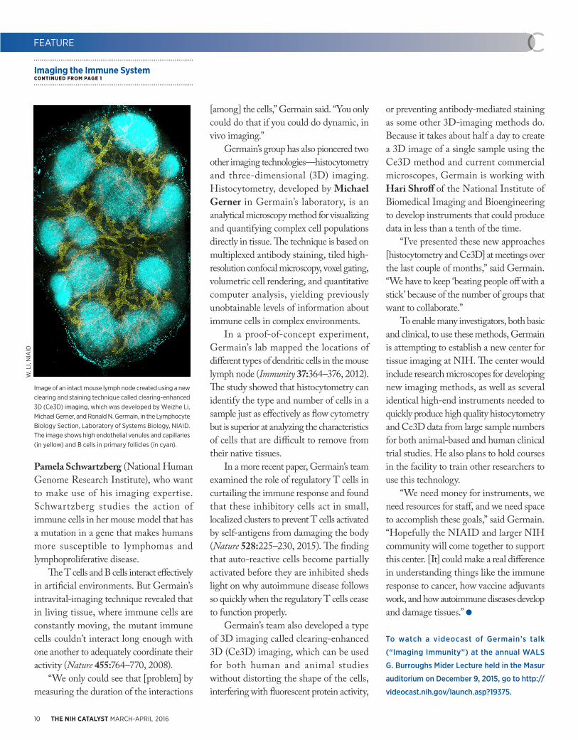
Image of an intact mouse lymph node created using a new clearing and staining technique called clearing-enhanced 3D (Ce3D) imaging, which was developed by Weizhe Li, Michael Gerner, and Ronald N. Germain, in the Lymphocyte Biology Section, Laboratory of Systems Biology, NIAID. The image shows high endothelial venules and capillaries (in yellow) and B cells in primary follicles (in cyan).
W. L
I, N
IAID

http://irp.nih.gov/catalyst 11
born in 1854 in Woodbridge, San Joaquin County, California. His great-grandfather was an orange rancher in southern California. His grandfather, a mechanical engineer, supplied materials used in the state’s oil fields. And Blakesley’s father was an electrical engineer who developed a distance-sensing radar system that enabled the Apollo space mission’s lunar modules to make soft landings on the moon.
“We had a workshop in our garage when I was growing up,” said Blakesley. “My father was always figuring something out, making new tools, or building electronic devices. I absorbed his desire to understand how things work and to build tools.”
As a child, Blakesley assembled model cars and radio-controlled planes for which he built his own controllers. By the time he reached high school, he had even built his own stereo system. A turning point came in the fall of 1963 when he read a long Los Angeles Times article about the 10th anniversary of the famous Francis Crick and James Watson discovery of the double-helical structure of DNA.
“Once I read that article, I wanted to know how genes worked,” Blakesley said. “It kick-started me and drove my education and career.”
He received his undergraduate degree in biochemistry from the University of California, Berkeley, and his Ph.D. in biochemistry at Michigan State University in East Lansing, Michigan. At Michigan, he learned to make his own reagents to study the kinetic and physical properties of polymerases.
Recruited by Bethesda Research LabsA career crossroads presented itself while he was working as a postdoctoral fellow at the University of Wisconsin at Madison. Blakesley was the only one in
the laboratory when a sales representative from BRL called and tried to sell him restriction enzymes.
“I told him that I make my own,” Blakesley recalled. The conversation eventually led to a job offer from BRL to oversee a research group that would develop an automated DNA-sequencing machine for medical diagnostics.
“That hooked me,” he said, adding that at the time, biochemists Frederick Sanger and Walter Gilbert (who shared the 1980 Nobel Prize in Chemistry with Paul Berg for their contributions to determining the base sequences of nucleic acids) thought that DNA sequencing might be important to medicine. “That was the challenge that got me interested in the company.”
BR L was f i r s t located in a 500-square-foot office and storage room in Gaithersburg, Maryland. Half of the space was used as a laboratory, and enzyme purifications were performed in a small beer cooler. “We often left the office door open because the air conditioner didn’t work well,” said Blakesley. “A huge dog from the next-door veterinarian clinic would often wander in to check our progress.”
It was an exciting time to direct new-product development for a start-up company. “We really listened to our customers, were as efficient as possible, and paid attention to detail. In the end, we delivered to them what they needed,” said Blakesley, who patented 10 products and procedures and introduced more than 150 products during his 23 years in private industry.
BRL grew quickly and eventually seemed to focus less on customers and more on profit. Increasingly frustrated, Blakesley contacted Green to ask whether he knew of any career opportunities. NISC, the nascent NIH organization that provided DNA-sequencing services
http://irp.nih.gov/catalyst 11
FEATURE
http://irp.nih.gov/catalyst 11
on a cost-recovery basis, needed someone with industry experience.
“Bob was one of the best hires I ever made,” said Green. The admiration is mutual: Blakesley credits Green for his unwavering support and for valuable introductions to those working in the large genome-sequencing centers around the world.
“ NISC benef ited a lot f rom collaborating with bigger groups that have successfully tackled big projects,” said Blakesley. “We were in almost constant contact with the large genome-sequencing centers at Washington University, the Broad Institute, and Baylor College of Medicine. When we ran into a problem, we called them up and asked how they solved it. I don’t think we could’ve gotten to where we are today without those interactions.”
The exchange has been a two-way street. Like a good team member, NISC shared its information with the other centers. “The real benefit to team science is that you can solve very complex problems by collaboration, by bringing in people from different disciplines,” said Blakesley. “I’m most proud of our collective successes at NISC. We have a fine group of individuals who work very hard and care about each other. I was sad to leave.”
Blakesley’s future plans revolve around his family. “I want to spend more time with my three kids and my six grandkids,” he said. “I’d like to spend time on genealogy, woodworking, gardening, and organizing the piles of 35-millimeter color slides and thousands of digital photos that have been stacking up.”
Whatever Blakesley’s direction, he will exceed all expectations just as he has at NISC, said Young. “He has really high standards for everything. It’s his nature.”
Blakesley CONTINUED FROM PAGE 3

12 THE NIH CATALYST MARCH-APRIL 2016
NEW: RESEARCH REPOSITORIES AND
PATIENT REGISTRIES (INCLUDES
BIOSPECIMENTS INTEREST GROUP)
In conjunction with the NIH Office of the Director and the Clini-cal Center, the Research Repositories and Patient Registries Scientific Inter-est Group (SIG) was established. With the launch of the Precision Medicine and Big Data to Knowledge (BD2K) initiatives, patient registries have been in the spotlight around the globe and recognized as an essential resource for accelerating research and making improvements in health care. The new SIG will facilitate discussion and the sharing of thoughts, knowledge, and data on a wide range of topics. Invited speakers will be from the NIH, from within the United States, and from other countries. The SIG can facilitate col-laboration not only within NIH but also with different organizations around the world. The existing Biospecimens Inter-est Group (BIG) is being merged into the new SIG and will be managed by Yaffa Rubinstein from NCATS. Bio-specimens members as well as others interested in signing up for the new SIG should register for the REPOSITO-RIES_AND_REGISTRIES-L LIST-SERV at https://list.nih.gov/cgi-bin/wa.exe?SUBED1=REPOSITORIES_AND_REGISTRIES-L&A=1. For more information, contact Yaffa Rubin-stein at [email protected].
NEW: MOBILE HEALTH
The mHealth SIG, established in September 2015, is a forum for the rapid exchange of ideas and information about the use of mobile technologies for assess-ment of or intervention in health-care matters. Open to NIH intramural inves-tigators at all levels, the mHealth SIG’s
goals are to enable members to network and solve problems, enhance intramu-ral access to new technology, promote mHealth research intramurally and extramurally, and prepare a framework for “path to approval” for new projects. The group meets monthly and maintains a LISTSERV. One advantage of getting involved now is that the priorities and practices are fairly open to input from new attendees. The next two meetings will be Wednesday March 23, 2016, and Wednesday April 27, 2016, both at 10:00 a.m. They will be held in Conference Room 2-3330 (Building 10). For more information, contact Kenzie Preston ([email protected]). Direc-tions to the conference room: From the north entrance of Building 10, go down the left corridor past the Au Bon Pain coffee shop; turn left at the “1 East Cor-ridor” sign; walk to the end of the hall-way; and take the “Southeast Elevators” to the second floor. The conference room is located in the glass enclosure next to the elevator lobby.
NEW: NONINVASIVE BRAIN STIMULATION
The purpose of the Noninvasive Brain Stimulation (NIBS) SIG is to provide a forum for the dissemination and discussion of scientif ic informa-tion among those using or interested in NIBS techniques and to help intramu-ral investigators deal with safety, regu-latory, and technical issues. Interested extramural personnel are also welcome. NIBS includes transcranial brain stimu-lation and neuromodulation techniques. The moderator is Eric Wassermann, a staff clinician in the National Institute of Neurological Disorders and Stroke (NINDS), a member of the NIH Neu-roscience Faculty, and an internationally recognized expert in NIBS. Although
the SIG will serve as a resource for any interested extramural investigators, its primary purpose will be support the rap-idly growing community of intramural entities interested in NIBS. The SIG will provide guidance, expertise, and oppor-tunities for collaboration and in so doing will lower the barriers for intramural labs and clinical groups that want to enter the NIBS f ield. Founding members who convened for a recent planning meet-ing included the PIs and staff of several labs in NINDS, the National Institute of Mental Health, the National Institute on Drug Abuse, and the National Center for Complementary and Integrative Health. Membership is expected to expand. To keep informed of meetings and activities, join the NIBS-L LISTSERV at https://list.nih.gov/cgi-bin/wa.exe?A0=nibs-l. For more information, contact Eric Was-sermann at [email protected].
NAME CHANGE: OPTOGENETICS SIG IS NOW
INNOVATIVE NEUROTECHNOLOGIES
The Optogenetics SIG, which began in 2013, has a new name to ref lect the broader scope of neurotechnological developments. The SIG, renamed Inno-vative Neurotechnologies, continues to meet monthly. To join the LIST-SERV, go to https://list.nih.gov/cgi-bin/wa.exe?A0=optogenetics. The SIG meets quarterly. For more information, contact Alexxai Kravitz ([email protected]).
THE SIG BEAT
NEWS FROM AND ABOUT THE SCIENTIFIC INTEREST GROUPS
MORE ABOUT SIGSNIH Scientific Interest Groups (SIGs) are
assemblies of scientists with common
research interests. For a complete list of
SIGs, go to:
http://www.nih.gov/research-training/
scientific-interest-groups

http://irp.nih.gov/catalyst 13
A mouth microbiome that’s out of whack can lead to serious health problems such as the chronic inflammatory disease periodontitis. Triggered by bacterial biof ilms, periodontal disease causes inflammation that damages gum tissue and can destroy the bone that supports the teeth. According to the CDC, over 47 percent of American adults over 30 have mild, moderate, or severe forms of the disease. If left untreated, periodontitis can lead to tooth loss and may contribute to other inf lammatory diseases such as diabetes and heart disease. More research is needed, however, to clarify the relationship between gum disease and health problems beyond the mouth.
National Institute of Dental and Craniofacial Research Clinical Investigator Niki Moutsopoulos is conducting research on periodontitis in order to understand its mechanisms and explore possible therapies. She described her work at a seminar hosted by the Inflammatory Disease Scientific Interest Group (SIG) on January 19, 2016.
To determine how immune defects may be associated with susceptibility to periodontal disease, Moutsopoulos recruit-ed healthy volunteers with and without periodontitis and people with the mono-genic immune defect leukocyte-adhesion deficiency (LAD-I). Her research team did clinical phenotyping, microbiome characterization, molecular profiling, and in vitro assays with human cells. She used periodontal probes to measure the loss of tooth-supporting structures and found that people with LAD-I had both increased bone loss and increased bacterial biomass on the surfaces of their teeth. Furthermore, the expression of the
protein integrin beta-2 (CD18) was inversely correlated with peri-odontitis severity, sug-gesting a link between defective neutrophil migration and the severity of LAD-I periodontitis.
The team a lso compared cytokine and chemokine gene expression in LAD-I pa t i ent s to t ha t of hea lthy people who had severe to mild periodontitis. I n t e r e s t i n g l y , t h e c y t o k i n e s interleukin-23 (IL-23) and IL-17 were induced in LAD-I periodontitis. Another finding was that the LAD-I microbiome stimulated an IL-17 immune response. It was also determined that abnormal neutrophil recruitment caused the upregulated IL-17 inflammatory response in the periodontal tissue and bone loss associated with bacterial overload. Finally, preliminary work using the drug ustekinumab (Stelara) to inhibit IL-23 is showing promise in patients.
Inf lammation is a complicated biological response to injury or harmful stimuli such as invading pathogens. Inflammatory responses can be protective when they are working properly, but their dysregulation or persistence can be destructive.
The Inflammatory Disease SIG aims to
bring together scientists to encourage
discussions and NIH-wide collaborations
that could potentially develop new treat-
ments for inflammatory diseases. The
SIG will host bimonthly seminars and
symposiums that focus on the basic and
translational characteristics of inflam-
mation. To join the LISTSERV (INFLAM-
DIS-L), visit https://list.nih.gov/cgi-bin/
wa.exe?SUBED1=INFLAM-DIS-L&A=1 or
contact Thomas A. Wynn at twynn@niaid.
nih.gov.
THE SIG BEAT
NEWS FROM AND ABOUT THE SCIENTIFIC INTEREST GROUPS
Periodontitis: A Microbial-Driven Inflammatory DiseaseNIDCR Clinical Investigator Niki Moutsopoulos Describes Her Work at a SIG SeminarBY HEBA DIAB, NHLBI
ALA
N H
OO
FRIN
G, N
IH M
EDIC
AL A
RTS
In periodontitis, an imbalanced microbiome triggers an exaggerated IL-17 immune response that leads to the activation of osteoclasts (cells that degrade bone) and bone loss. The overgrowth of bacterial biofilm stimulates antigen-presenting cells (APC) to upregulate the cytokine IL-23, which stimulates the development of T-helper (Th17) cells. Th17 cells produce the cytokine IL-17 and activate osteoclasts to resorb bone. IL-17 will also stimulate the recruitment of polymorphonuclear neutrophils (PMN) to the area, which will further amplify immunopathology in the lesion of periodontitis.

14 THE NIH CATALYST MARCH-APRIL 2016
Director Harold E. Varmus and NIH Clinical Center Director John I. Gallin. Gallin had recently been appointed to his position and was assessing the strengths of the organization.
The Clinical Center has always excelled at collaboration and investigation, with 18 NIH institutes conducting their intramural studies at the hospital at that time. Varmus and Gallin knew that the Clinical Center was mastering this part of the research portfolio. What they discussed were other ways the Clinical Center could make an impact on the broader research community, specifically focusing on the fact that as a part of its overarching mission, the Clinical Center offered training opportunities for physicians, research fellows, and other clinical staff.
Gallin and Varmus agreed that the Clinical Center served as a model for clinical research. With such a leading role, the world ’s largest hospital totally focused on clinical research was the perfect source for disseminating the best practices of clinical exploration. They determined that the best way to share this knowledge would be through a structured learning environment that drew upon the knowledge of the many talented biomedical scientists and clinical researchers working in the Clinical Center.
Gallin convened a group of experts from inside and outside the Clinical Center to establish the topics and curriculum that would create the foundation for “IPPCR.”
The “IPPCR” course was launched in the 1995–1996 academic year, with its first session held in September 1995 on the NIH campus. It included 25 participants.
With a premier course on clinical research established, the question was how to increase participation in the curriculum beyond the intramural program on NIH's
Bethesda, Maryland, campus. NIH clinician-scientists are only a small portion of the national research community. And only a limited number of people could fit into a room on campus to learn from subject-matter experts. There had to be a way to make the class grow.
The solution was one that was fitting for a research hospital familiar with implementing innovations: using available technology to share the educational content remotely.
Within three years of the educational program’s launch in 1995, it offered remote
access to NIH staff at the National Institute of Allergy and Infectious Diseases’ Rocky Mountain Lab in Hamilton, Montana, and the National Institute of Diabetes and Digestive and Kidney Diseases’ Epidemiology and Clinical Research Branch in Phoenix, Arizona.
Two years later, remote access was offered for Georgetown University (Washington, D.C.), the first non-NIH location to have access to “IPPCR.” In 2002, the f irst international remote access was provided through MAT and Asociados, based in Buenos Aries, Argentina. The international audience has been growing ever since.
Since the course’s inception, the number and content of the lectures have
increased, as has course enrollment. To date, more than 28,000 individuals have enrolled in the “IPPCR” course. For the current academic year, there are nearly 7,000 students registered, with 524 at the NIH Bethesda campus and 6,290 at 123 remote academic sites. Fifty-four of the remote sites are outside of the United States, with twenty-six nations participating as partners for the current course. (See sidebar for full details.)
An abbreviated, weeklong version of the course has also been taught abroad to bring the face-to-face learning experience
to clinicians overseas. Most recently, the course was taken to Cape Town, South Africa, in 2015 as a part of an agreement between NIH and the South African Medical Research Council. Other nations that have co-hosted the weeklong course with the NIH Clinical Center are China (Beijing and Chengdu), Russia, Nigeria, India, and Brazil.
In addition to teaching the principles of clinical research, the live international courses have also
focused on enhancing collaborations between scientists in biomedical and behavioral research at NIH and those in the host nations. In addition, a core principle with all of the international partners has been to try to enable the participating students to become teachers themselves and to share their new knowledge with colleagues, following a “train the trainer” model.
The textbook used for the course, Principles and Practice of Clinical Research, was first edited by Gallin and published in 2002. Second and third editions were published in 2007 and 2012 and were edited by Gallin and Frederick P. Ognibene, the Clinical Center’s deputy director for Educational Affairs and
FEATURE
The NIH Clinical Center, the world’s largest hospital focused on clinical research,
was the perfect source for disseminating the
best practices of clinical exploration.
Course CONTINUED FROM PAGE 1

http://irp.nih.gov/catalyst 15
Strategic Partnerships. As a testament to the international desire for clinical-research knowledge, the first edition of the text was translated into Japanese; the second edition into Chinese and Russian; and the third edition into Chinese and Japanese (in press). In addition, many academic institutions are using the textbook for other local instruction and programs in clinical research, in addition to “IPPCR.”
Recognizing that the clinical-research environment is constantly evolving, instructors have added topics to the class to ensure that its participants remain abreast of current areas of concern. Recent additions to the curriculum include management of clinical data and electronic health records; research on health disparities; and community-based research.
Since 2005, Ognibene has been “IPPCR” co-director with Gallin, and in 2013, Laura Lee Johnson, a biostatistician currently working at the FDA’s Center for Drug Evaluation and Research, joined as a third co-director.
In addition to “IPPCR,” the Clinical Center has two other courses in its core curriculum in clinical research. The “Principles of Clinical Pharmacology” course has been taught annually since 1998, and the “Ethical and Regulatory Aspects of Clinical Research” course since 1999. Both also use Web-based, long-distance learning techniques. For all three core curriculum courses, a cumulative total of 44,015 students have been enrolled since their inception.
“It has been incredibly gratifying to see this program grow from a concept into an international program,” said Gallin. “The hunger for this information, and the ability to draw upon internationally renowned experts, is a true testament to the influence and global reach of the Clinical Center.”
Year Number of Participants
Nations Involved Educational Delivery Method
1995–1996
25 United States (NIH Campus)
Face-to-face instruction
1998–1999
318 (1) United States (multiple locations)
Face-to-face instruction, NIH remote access
2000–2001
505 (2) United States (multiple locations)
Face-to-face instruction, U.S. remote access
2002–2003
653 United States, Argentina
Face-to-face instruction, U.S. and international remote access (3)
2009–2010
1,286 (4) 11 (including the United States)
Face-to-face instruction, online video
2015–present
6,814 27 (including the United States)
Face-to-face instruction, online video in real time as well as archived access
COURSE BENCHMARKS
1. First remote access to the course.
2. First time remote participation was higher than onsite participation in the course.
3. First international remote access to the course.
4. First time the course surpassed 1,000 participants at once.
Evolution of the “Introduction to the Principles and Practice of Clinical Research” Course
FEATURE
http://irp.nih.gov/catalyst 15

16 THE NIH CATALYST MARCH-APRIL 2016
CATHARINE BOSIO, PH.D., NIAID
Senior Investigator and Chief, Immunity to Pulmonary Pathogens Section, Laboratory of Bacteriology, Rocky Mountain Laboratories, National Institute of Allergy and Infectious DiseasesEducation: Washington State University,
Pullman, Wash. (B.Sc. in microbiology);
Colorado State University, Fort Collins,
Colo. (Ph.D. in microbiology)
Training: Postdoctoral fellowships at the
Food and Drug Administration Center
for Biologics Evaluation and Research
(Bethesda, Md.) and at the U.S. Army
Medical Research Institute for Infectious
Diseases (Frederick, Md.), studying innate
immunity to Mycobacterium tuberculosis,
Francisella tularensis, and Ebola and
Marburg viruses
Before coming to NIH: Assistant professor,
Department of Microbiology, Immunology,
and Pathology, Colorado State University
Came to NIH: In 2007
Selected professional activities: Editorial
board, Infection and Immunity; committee
and co-chair, American Society for
Microbiology Biodefense and Emerging
Diseases meeting
Outside interests: Running; reading; hiking;
cooking
Website: http://irp.nih.gov/pi/
catharine-bosio
Research interests: The focus of our research is to gain a better understanding of how aerosolized pathogens successfully infect and modulate the pulmonary environment to cause overt disease and death. Currently, our principal interest is the pathogenesis of aerosolized F. tularensis, the causative agent of pneumonic tularemia.
We are focused on uncovering the mechanisms by which F. tularensis modu-lates for innate and adaptive immunity. As an intracellular pathogen, F. tularensis is intimately associated with host cells. We have identified several major pathways by which the bacterium interferes with host-cell function, including accelerating decay of host mRNA, inhibiting transcription factors, and modulating host metabolism. F. tularensis can also affect the generation of effective adaptive responses. We have devel-oped several models to identify the specific cellular requirements for survival of tulare-mia and how the bacterium interferes with development of long-lived, antigen-specific, memory T cells. Identification of the micro-bial mechanisms and products embodied by F. tularensis that dampen mammalian immunity will aid in the development of novel vaccines and therapeutics for tulare-mia, as well as new therapies for unrelated diseases in which control of inflammation is required for survival.
HAROLD A. BURGESS, PH.D., NICHD
Senior Investigator and Head, Unit on Behavioral Neurogenetics, National Institute of Child Health and Human DevelopmentEducation: University of Melbourne,
Parkville, Victoria, Australia (B.S. in
biochemistry); the Weizmann Institute
of Science, Rehovot, Israel (Ph.D. in
developmental neuroscience)
Training: Postdoctoral training, University
of Pennsylvania (Philadelphia)
Came to NIH: In 2008
Selected professional activities: Academic
Editor, PLOS One
Outside interests: Geocaching with his
three kids; running; cultivating garlic;
reading detective fiction
Website: http://irp.nih.gov/pi/
harold-burgess
Research interests: My laboratory combines genetic and imaging techniques to study neural circuits required for sensory-guided behavior and motivational states. We use larval zebraf ish to understand the functional development of neuronal circuits that allow the larvae to choose the best responses to environmental stimuli. The zebrafish model is a great system because the larval brain has the same basic organization as the human brain, but it is, of course, much smaller, containing only around
COLLEAGUES
Recently Tenured
CATHARINE BOSIO, NIAID FRANCESCO DEMAYO, NIEHS BRUCE THOMAS HOPE, NIDAMONTSERRAT GARCÍA-CLOSAS, NCI-DCEG
HAROLD A. BURGESS, NICHD

http://irp.nih.gov/catalyst 17
100,000 neurons, instead of the 100 billion or so in humans. The transparency of the fish at larval stages means that we can observe the firing of individual neurons in real time.
Larval behavior is innate and varies little among individual fish so it’s relatively easy to apply computational tools to quickly assess the contribution of identified neurons to behavior. For instance, one major behavioral test that my laboratory uses is the startle response. Defects in the startle response are observed in many psychiatric disorders, and the brainstem regions that control startle responses are highly conserved in fish. By performing genetic screens in larval zebrafish, we identify genes and neurons that tightly control the threshold for startle responses that are also relevant to mammals. We also study light-seeking behavior in larvae. Remarkably, part of this behavior is actually controlled by light-sensitive neurons within the brain itself rather than through the retina. We think these neurons are part of a primitive control system for motivational state, allowing us to understand how the fish perform goal-directed actions.
FRANCESCO DEMAYO, PH.D., NIEHS
Senior Investigator and Deputy Chief, R eproduct ive and Developmental Biology Laboratory, National Institute of Environmental Health SciencesEducation: Cornell University, Ithaca, N.Y.
(B.S. in general studies); Michigan State
University, East Lansing, Mich. (M.S. and
Ph.D. in physiology)
Training: Postdoctoral training at Baylor
College of Medicine (Houston)
Before coming to NIH: Cullen-Duncan-
McAshan Endowed Chair in Cancer
Research and Professor of Molecular and
Cellular Biology and of Pediatrics, Baylor
College of Medicine
Came to NIH: In 2015
Selected professional activities: Co-editor-
in-chief of Biology of Reproduction
Outside interests: Bowling; bicycling;
enjoying opera
Website: http://1.usa.gov/1LgakhX
Research interests: I lead the Pregnancy and Female Reproduction Group, which studies the mechanisms involved from the implantation of an embryo in the uterus to the birth of a baby. To identify the proteins that regulate the female reproductive tract during these processes, we use genetically engineered mice, human cells, and transcription-factor analysis. These tools allow us to examine factors that allow proper embryo implantation, adequate uterine support for embryo development, and on-time delivery. The timing of lung development is also critical for embryonic maturation. Our group investigates what regulates lung physiology and homeostasis and how lung cancer develops.
Our major areas of research include understanding uterine biology, specifically, how several transcription factors regulate the window of uterine receptivity; determining what regulates differentiation of the stroma, one of the three layers of the uterus; examining the role of transcription factors in another layer of the uterus called the myometrium; and using mouse models to explore the development of lung cancer.
Specifically, our current projects include investigating how forkhead box protein O1 (FOXO1) and the progesterone receptor interact to regulate uterine receptivity for embryo implantation; establishing how WNK1 is involved in postimplantation support of the embryo; and how the progesterone receptor controls myometrial function during pregnancy and birth. We are also exploring the involvement of environmental factors in the development of squamous non-small-cell lung cancer.
http://irp.nih.gov/catalyst 17http://irp.nih.gov/catalyst 17
COLLEAGUES
MONTSERRAT GARCÍA-CLOSAS, M.D.,
DR.PH., M.P.H., NCI-DCEG
Senior Investigator and Deputy Director, Division of Cancer Epidemiology and Genetics, National Cancer InstituteEducation: University of Barcelona,
Barcelona, Spain (M.D.); Harvard School
of Public Health (M.P.H. in quantitative
methods; Dr.P.H. in epidemiology)
Training: Postdoctoral training at
NCI-DCEG
Came to NIH: In 1996 for training; became
a tenure-track investigator in 1999 and a
tenured senior investigator in 2007; left in
2008; returned in 2015
Before returning to NIH: Visiting scientist
at the Department of Oncology and
Strangeways Laboratory, University of
Cambridge University (Cambridge, U.K.)
in 2008–2010; professor of epidemiology
at Division of Genetic and Epidemiology of
the Institute of Cancer Research, University
of London (London) in 2010–2015
Selected professional activities: Editorial
board for Cancer Epidemiology, Biomarkers
& Prevention; senior editor for Molecular
and Genetic Epidemiology
Outside interests: Anything fun and
relaxing–yoga; hiking; visiting new places;
watching movies; photography; gardening;
you name it
Website: http://1.usa.gov/20XsqY4
Research interests: I conduct large epidemiological studies to investigate biomarkers of breast- and bladder-cancer risk in combination with other exposure information. In particular, I am interested in the molecular characterization of tumors to identify exposure signatures and subtype-specific causes of cancer; identif ication of biomarkers of risk, including genetic and epigenetic changes; integration of biomarkers and other risk factors into cancer-risk prediction models;
CONTINUED ON PAGE 18

18 THE NIH CATALYST MARCH-APRIL 2016
and evaluation of the utility of biomarkers in risk stratification to inform personal and public-health decisions in precision prevention.
The goal of my breast-cancer research is to facilitate the development and implementation of targeted prevention and screening strategies for different types of breast cancer. I am a co-leader of large-scale molecular-pathology studies within the international Breast Cancer Association Consortium. I have contributed to the development of one of the largest centralized collections of breast-tumor tissue with detailed data on risk factors and clinical outcomes. This resource has revealed important tumor markers that del ineate the et iolog ic heterogeneit y of breast cancer. At the University of London’s Institute of Cancer Research, I co-led the Breakthrough Generations Study, a large prospective cohort study of over 110,000 women in the United Kingdom aimed at evaluating risk factors for breast cancer. I am also an investigator in the ongoing international genotyping project OncoArray and am trying to identify inherited-susceptibility loci associated with specific subtypes of breast cancer.
In my bladder-cancer research, I am investigating the combined effects of genetic and environmental risk factors for this cancer. I have published the definitive study of the interaction between smoking history and the NAT2 genotype, which is one of the few consistent gene-environment interactions described to date. I contributed to the discovery of novel susceptibility loci for bladder cancer using genome-wide association studies, an area in which I continue to collaborate. In addition, I am engaged in epigenome-wide association studies of bladder cancer through NCI’s Prostate,
Lung, Colorectal and Ovarian Cancer Screening Trial (PLCO) and identifying additional risk biomarkers.
BRUCE THOMAS HOPE, PH.D., NIDA
Senior Investigator and Chief, Neuronal Ensembles in Drug Addiction, National Institute on Drug AbuseEducation: University of British Columbia,
Vancouver, B.C. (B.Sc. in biochemistry;
Ph.D. in neurological research)
Training: Postdoctoral training at the
Laboratory of Molecular Psychiatry, Yale
University, New Haven, Conn.
Before coming to NIH: Staff scientist at
New England Medical Center (Boston)
and assistant professor, Department of
Pharmacology, at Tufts University School
of Medicine (Boston); staff scientist at
Massachusetts General Hospital (Boston);
and instructor, Department of Psychiatry,
Harvard Medical School (Boston)
Came to NIH: In 1996 as a visiting scientist
in NINDS; then various positions in NIDA
(1998–2009); became tenure-track
investigator in NIDA in 2010
Selected professional activities: Associate
editor for the Journal of Neuroscience and
Synapse (2011–present); ad hoc reviewer
for almost 40 journals
Outside interests: Swimming; ballroom
dancing
Website: http://irp.nih.gov/pi/bruce-hope
Research interests: Use of drugs of abuse can cause learned associations to form between the drugs and the stimuli present in the drug-taking environment. With continued use, these stimuli can become cues that promote drug relapse. My group’s research is focused on figuring out how these memories are stored in the brain.
COLLEAGUES
We have identified sparsely distributed patterns of neurons in the brain called neuronal ensembles that are selectively activated by drug-related cues and are thought to encode the learned associations that mediate drug-seeking behavior. Drug-related cues activate specific genes such as c-fos within these neuronal ensembles and allow us to identify them in the brain. We exploit the c-fos promoter to turn on different transgenes in transgenic rats, allowing us to manipulate specific neuronal ensembles and assess their role in drug-related memories.
We also developed a f luorescence-activated cell-sorting procedure for purifying these activated ensembles and found unique molecular alterations within their cell bodies and synapses. We have developed novel c-fos-GFP transgenic rats that produce green fluorescent protein in activated neurons and found unique synaptic alterations using slice electrophysiology. Using a combination of novel viruses and transgenic rats developed in collaboration with a colleague, we continue to search and characterize drug-related memory engrams that promote drug relapse.
Recently Tenured CONTINUED FROM PAGE 17
RECENTLY TENURED? If you have been tenured in the past
few months, the NIH Catalyst will be in
touch with you soon to invite you to be
included on these pages. We will ask for
your CV and a recent photo, review your
website, and then draft an article for
your review and edits.

http://irp.nih.gov/catalyst 19
,CO-
2016 JSPS-NIH FORUM
Friday, March 11, 2016, 1:30-7:00 p.m.
(reception 5:30-7:00 p.m.)
Lawton-Childs International House (Stone
House, Building 16)
Register by March 4: https://goo.gl/kfMZ0O
Website: http://jspsusa.org/wp/info/
jsps-nih-forum/
The Japan Society for the Promotion of Sci-
ence (JSPS) is one of the largest research
funding agencies in Japan. Its Washington
office in cooperation with the Fogarty Inter-
national Center invites you to attend a forum
featuring special lectures. For information,
contact Yosuke Mukoyama at mukoyamay@
nhlbi.nih.gov.
Pi DAY CELEBRATION
Tuesday, March 14, 2016, 10:00 a.m.-4:30 p.m.
Lipsett Amphitheater, FAES Terrace (Bldg 10)
Website for details: https://datascience.nih.
gov/PiDay2016
A celebration of the irrational number Pi, 3.14.
NPR’S DIANE REHM
SPECIAL GUEST AT WALS CULTURAL LECTURE
Thursday, April 7, 2016, 3:00-4:00 p.m.
Masur Auditorium (Building 10)
The Wednesday Afternoon Lecture Series’
(WALS) Rall Cultural Lecture will feature
National Public Radio (NPR) talk-show host
Diane Rehm. On her program, the “Diane Rehm
Show,” she has interviewed many political and
cultural figures. She is also the author of two
autobiographies: Finding My Voice and Toward
Commitment: A Dialogue about Marriage, which
she co-wrote with her husband, John Rehm,
who died in 2014. She has a new book—On My
Own—about her struggle to reconstruct her life
after husband’s long, drawn-out death from
Parkinson disease. The event will be videocast
live (http://videocast.nih.gov).
UNDERSTANDING THE SEX BIAS IN DISEASE
April 18, 2016, 8:30 a.m.-4:30 p.m.
Lipsett Amphitheater (Building 10)
Register at: https://ncifrederick.cancer.gov/
events/SexBias/default.asp
Speakers in the NIH intramural program and
from academia will give talks that emphasize
the differences between sexes in autoimmu-
nity, infectious disease, cancer and inflamma-
tion. Individuals with disabilities who need rea-
sonable accommodation to participate in this
event should contact Howard Young (young-
[email protected] or 301-846-5743) or the
Federal Relay (800-877-8339) by April 4, 2016.
POSTBACCALAUREATE POSTER DAY 2016
Wed., April 20, 2016; 10:00 a.m-3:30 p.m.
Natcher Conference Center (Building 45)
More information: https://www.training.nih.
gov/postbac_poster_day.
The keynote address begins at 12:00 noon
and will be followed by the presentation of
Postbac Distinguished Mentoring Award to
NIH investigators selected by the postbacs.
Poster sessions will take place from 10:00 a.m.
to 12:00 noon and from 1:30 to 3:30 p.m.
SYMPOSIUM ON TRANSLATIONAL GENOMICS
WITH A SPECIAL FOCUS ON LIVER CANCER
March 17-18, 2016
Room 610 (Building 35)
Please register: https://ncifrederick.cancer.
gov/events/translationalgenomics2016/
For more information, contact Laura Hooper
STEPHEN E. STRAUS DISTINGUISHED
LECTURE
Tuesday, May 3, 2016, 10:00-11:00 a.m.
Masur Auditorium (Building 10)
Richard J. Davidson (University of Wisconsin-
Madison) talk entited “Change Your Brain by
Transforming Your Mind,” will provide an over-
view of his reserach on neural changes asso-
ciated with different forms of meditation. For
reasonable accommodation or other questions,
contact Prachi Patel (Prachi.patel@patelpa.
com or 301-275-4769).
BUILD YOUR CAREER; SHAPE YOUR FUTURE:
NIH CAREER SYMPOSIUM
Friday, May 6, 2016, 8:30 a.m.-5:00 p.m.
Natcher Conference Center (Building 45)
For more information: www.training.nih.gov
Fellows and graduate students can learn about
scientific career options and explore fac-
tors that lead to career success. There will be
over 20 breakout sessions highlighting career
opportunities for biomedical scientists; panel
sessions on academic, government, industry,
and non-profit career paths; and more than 80
speakers who will provide insights into their
careers.
CLINICAL AND TRANSLATIONAL RESEARCH
COURSE FOR PH.D. STUDENTS
July 11-22, 2016, NIH Bethesda campus
For more info and to apply: http://cc.nih.gov/
training/phdcourse
Application deadline: March 30, 2016
For more information, contact the NIH Clinical
Center Office of Clinical Research Training
and Medical Education ([email protected].
gov) or Terra Miller ([email protected] or
301-660-3519).
NEEDED: FAES EDUCATION COMMITTEE
CO-CHAIR
DEADLINE: March 21, 2016
The Foundation for Advanced Education in the
Sciences (FAES) is looking for a co-chair of its
Education Committee. For questions, contact
Christina Farias ([email protected] or
301-451-5973). If interested in the position, con-
tact Krisztina Miner ([email protected]).
VOLUNTEERS NEEDED
AT USA SCIENCE & ENGINEERING FESTIVAL
Saturday, April 16 (10:00 a.m.-6:00 p.m.)
Sunday, April 17 (10:00 a.m.-4:00 p.m.)
Walter E. Washington Convention Center
To volunteer and learn more about the event:
http://dpcpsi.nih.gov/SciFest
PFIZER’S CTI ACCEPTING PROPOSALS
For information: https://ncats.nih.gov/cti/
proposals
ANNOUNCEMENTS
http://irp.nih.gov/catalyst 19
Read more online at http://irp.nih.gov/
catalyst/v24i2/announcements.
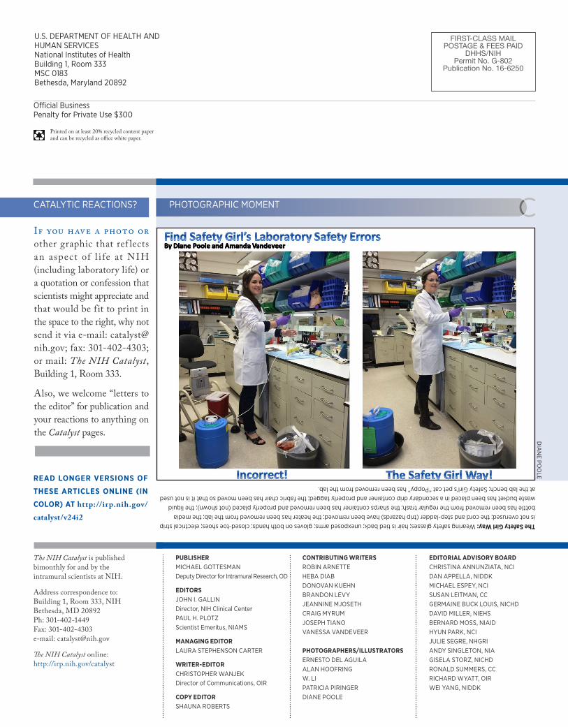
PHOTOGRAPHIC MOMENTThe Safety Girl Way: Wearing safety glasses; hair is tied back; unexposed arms; gloves on both hands; closed-toe shoes; electrical strip is not overused; the cord and step-ladder (trip hazards) have been removed; the heater has been removed from the lab; the media bottle has been removed from the regular trash; the sharps container has been removed and properly placed (not shown); the liquid waste bucket has been placed in a secondary drip container and properly tagged; the fabric chair has been moved so that it is not used at the lab bench; Safety Girl’s pet cat “Poppy” has been removed from the lab.
DIA
NE PO
OLE
CATALYTIC REACTIONS?
If you have a photo or other graphic that ref lects an aspect of l i fe at NIH (including laboratory life) or a quotation or confession that scientists might appreciate and that would be fit to print in the space to the right, why not send it via e-mail: [email protected]; fax: 301-402-4303; or mail: The NIH Catalyst, Building 1, Room 333.
Also, we welcome “letters to the editor” for publication and your reactions to anything on the Catalyst pages.
FIRST-CLASS MAILPOSTAGE & FEES PAID
DHHS/NIHPermit No. G-802
Publication No. 16-6250
Official BusinessPenalty for Private Use $300
U.S. DEPARTMENT OF HEALTH AND HUMAN SERVICESNational Institutes of HealthBuilding 1, Room 333MSC 0183Bethesda, Maryland 20892
Printed on at least 20% recycled content paper and can be recycled as office white paper.
PUBLISHERMICHAEL GOTTESMAN Deputy Director for Intramural Research, OD
EDITORS JOHN I. GALLIN Director, NIH Clinical Center PAUL H. PLOTZ Scientist Emeritus, NIAMS
MANAGING EDITOR LAURA STEPHENSON CARTER
WRITER-EDITORCHRISTOPHER WANJEK Director of Communications, OIR
COPY EDITORSHAUNA ROBERTS
CONTRIBUTING WRITERSROBIN ARNETTEHEBA DIABDONOVAN KUEHNBRANDON LEVYJEANNINE MJOSETHCRAIG MYRUMJOSEPH TIANOVANESSA VANDEVEER
PHOTOGRAPHERS/ILLUSTRATORS ERNESTO DEL AGUILAALAN HOOFRINGW. LIPATRICIA PIRINGERDIANE POOLE
EDITORIAL ADVISORY BOARDCHRISTINA ANNUNZIATA, NCIDAN APPELLA, NIDDKMICHAEL ESPEY, NCI SUSAN LEITMAN, CC GERMAINE BUCK LOUIS, NICHD DAVID MILLER, NIEHS BERNARD MOSS, NIAIDHYUN PARK, NCI JULIE SEGRE, NHGRI ANDY SINGLETON, NIA GISELA STORZ, NICHD RONALD SUMMERS, CCRICHARD WYATT, OIRWEI YANG, NIDDK
The NIH Catalyst is published bimonthly for and by the intramural scientists at NIH.
Address correspondence to: Building 1, Room 333, NIHBethesda, MD 20892Ph: 301-402-1449 Fax: 301-402-4303e-mail: [email protected]
The NIH Catalyst online: http://irp.nih.gov/catalyst
READ LONGER VERSIONS OF
THESE ARTICLES ONLINE (IN
COLOR) AT http://irp.nih.gov/catalyst/v24i2