vitamin c · 2018-06-27 · 4 vitamin c has an unquestionable role in health and disease, in...
TRANSCRIPT

1
CVitamin C
Indispensable Nutrient in Our Health and Its Therapeutic Potential in Cancer From Cellular Mechanisms to Clinical Applications
M. Waheed Roomi, Ph.D Aleksandra Niedzwiecki, Ph.D and Matthias Rath M.D.

2
Vitamin C Indispensable Nutrient in Our Health and Its Therapeutic Potential in Cancer: From Cellular Mechanisms to Clinical Applications
M.Waheed Roomi Ph.D. | Aleksandra Niedzwiecki Ph.D. | Matthias Rath M.D.
1st Edition
© 2018 Dr. Rath Health Foundation
Dr. Rath Research Institute, 1260 Memorex Drive, Santa Clara, CA 95050, USA
Distribution:
Dr. Rath Education Services B.V. Postbus 656, NL-6400 AR Heerlen
Tel.: 0031-457-111 222
Fax: 0031-457-111 229
Email: [email protected] [email protected] Internet: www.rath-eduserv.com
All rights reserved. Editor: Dr. Rath Health Foundation. Individual pages of this brochure can be copied for private and non-commercial purposes only. Any direct or indirect commercial use of this brochure or of its parts in any way is strictly forbidden without the written permission of the authors.

3
I. Vitamin C as an Essential Nutrient for Health and Life 5
II. Health Aspects of Vitamin C 14
III. Function and Physiological Role of Vitamin C 19
IV. Therapeutic Potential of Vitamin C in Cancer: 24
Scientific and Clinical Aspects
V. Combinations of Vitamin C with Other Natural Compounds 35
VI. Clinical Applications of Vitamin C in Cancer 39
References 50
Content

4
Vitamin C has an unquestionable role in health and disease, in particular in the treatment of cancers. This scientific review summarizes our knowledge of selected aspects of Vitamin C in relation to healthy functions in our body. It also outlines our current knowledge of its role in cancer, including understanding its mechanisms of action at the cellular and organismal levels, and clinical use of vitamin C in cancer patients. Some aspects on this topic were presented in our earlier publication “ Vitamin C in Health: Scientific focus on its anti-cancer efficacy, MW Roomi, N Shanker, A Niedzwiecki and M Rath, J of Cellular Medicine and Natural Health, June 15, 2016.”

5
I. Vitamin C as an Essential Nutrient for Health and Life
Discovery of Vitamin C The importance of vitamin C for human health can be traced back several centuries. This most versatile vitamin is produced internally in the body of most species in the animal world; however, humans, guinea pigs and some primates, have lost this ability and rely only on dietary sources for this crucial nutrient.
Its deficiency causes a disease called scurvy, which is life threatening and can be fatal. Scurvy is characterized by bleeding gums, fragile blood vessels, connective tissue dissolution, impaired wound healing and finally death [Norum KR, 2002; Carpenter KJ 1986].
The earliest documented case of scurvy was described by Hippocrates around 400 BC. This disease became rampant during the 16th century among sea voyageurs and sailors who were deprived of fresh fruits and vegetables in their diet during long expeditions. In 1753 in the “Treatise of the Scurvy” Dr. James Lind published the first clinical proof of effective therapy against scurvy [Lind J 1753] demonstrating that citrus fruit juice can cure this disease.
However, it was not until the 1900s, after establishment of the first guinea pig model of scurvy [Svirbely J 1932], that the antiscorbutic factor in citrus fruit was identified as “water-soluble vitamin C” or “ascorbic acid.” Ascorbic acid was first discovered in 1922. It was then isolated and its structure was identified by Hungarian scientist Albert Szent-Gyorgyi in 1928, and in 1933 he was awarded the Nobel Prize. During the same year, Dr. C. Haworth shared the Nobel Prize in chemistry for the synthesis of vitamin C. In 1933, Hoffmann-La Roche became the first pharmaceutical company responsible for the mass production of vitamin C.
Albert Szent-Gyorgyi

6
Chemical Structure of Vitamin C
Figure 1. Chemical structure of D-Glucose from which Vitamin C is synthe-sized and its reduced and oxidized forms ( L-Ascorbic Acid)
Natural vitamin C is also known as L-ascorbic acid and it is easily oxidized to dehydro ascorbic acid (Figure 1). It is a weak acid and structurally very similar to the glucose molecule. This molecule has two asymmetric carbon atoms that result in four stereoisomers: L- Ascorbic acid, D-Ascorbic Acid, D-iso ascorbic acid and L-iso Ascorbic Acid (Figure 2). Of the four isomers, only L-Ascorbic Acid, or vitamin C, is biologically active while the other three forms are inactive. All isomers could be separated as boron complexes on TLC plates. [Roomi 1998]
Vitamin C is a colorless, odorless, crystalline solid compound with a sharp acidic taste. Its chemical composition is C6H8O6, with a molecular weight of 176 and a melting point of 190-1920C. It is soluble in water but insol-uble in organic solvents. Vitamin C is easily oxidized by several agents including halogens and hydrogen peroxide. [Sauberlich HE 1994; Naidu KA 2003]
The Vitamin C molecule contains several reactive hydroxyl groups, especially at the 2- and 6- positions, allowing a variety of derivatives to be easily synthesized, 2-sulfate and 2- mono-phosphate. The compounds of vitamin C are much more stable than vitamin C itself, such as di- and tri-phosphate. Hence, they are used in feed formulations for shrimp, fish, guinea pigs and rhesus monkeys. L-ascorbyl 6-palmitate, a synthetic

7
lipophilic vitamin C derivative, is another effective and bio-available com pound used in foods and pharmaceuticals. [Sauberlich HE 1994; Naidu KA 2003]
It has been seen that both hydrophilic and lipophilic derivatives are cyto-toxic to a number of malignant cells.Natural Sources of Vitamin C and its Organ Distribution in Animals: Vitamin C is a ubiquitous compound necessary for sustaining life and it is considered one of the most versatile and vital dietary compounds [Sauber-lich HE 1994].
Vitamin C is found amply in the plant kingdom, often in large quantities that are distributed throughout the plant. It is widely available in fresh fruits and vegetables. Fruits such as oranges, lemons, grapes, strawberries, papaya, kiwi, cantaloupe, grapefruit, mango and honeydew melon are good sources of Vitamin C [Sauberlich HE 1994; Naidu KA 2003; Gaby SK 1991].
Vegetables rich in vitamin C include broccoli, Brussels sprouts, red and yellow peppers, tomatoes, cabbage, potatoes, sweet potatoes, cauliflower, snow peas and kale.
Figure 2. Stereoisomers of Vitamin C and their interconversion
8
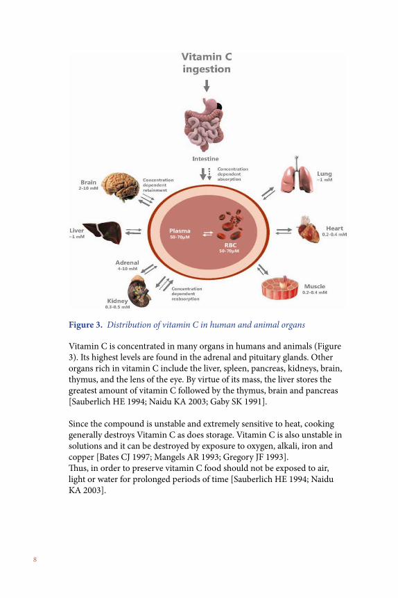
Vitamin C is concentrated in many organs in humans and animals (Figure 3). Its highest levels are found in the adrenal and pituitary glands. Other organs rich in vitamin C include the liver, spleen, pancreas, kidneys, brain, thymus, and the lens of the eye. By virtue of its mass, the liver stores the greatest amount of vitamin C followed by the thymus, brain and pancreas [Sauberlich HE 1994; Naidu KA 2003; Gaby SK 1991].
Since the compound is unstable and extremely sensitive to heat, cooking generally destroys Vitamin C as does storage. Vitamin C is also unstable in solutions and it can be destroyed by exposure to oxygen, alkali, iron and copper [Bates CJ 1997; Mangels AR 1993; Gregory JF 1993]. Thus, in order to preserve vitamin C food should not be exposed to air, light or water for prolonged periods of time [Sauberlich HE 1994; Naidu KA 2003].
Figure 3. Distribution of vitamin C in human and animal organs

9
Biosynthesis of Vitamin C
Animals: In most animals Vitamin C is synthesized from glucose accord-ing to biosynthetic pathway [Burns JJ 1959] presented in Figure 4. Higher animal species produce vitamin C via glucuronic acid from glucose. Accordingly, glucose is first converted into glucose 6-phosphate, which is then converted into uridine diphosphate glucose via uridine diphosphate glucuronic acid and then to glucuronic acid. This is further converted into D-glucuronolactone and later to gulonolactone under the influence of the enzyme L-gulonolactone reductase. Gulonolactone is then converted into 2-keto L-gulonolactone via the enzyme L-gulonolactone oxidase. In the last step, 2-keto L-gulonolactone is isomerized non-enzy-matically into L-ascorbic acid or Vitamin C.
In lower animals, such as amphibians, reptiles and birds, vitamin C is produced in the kidneys by the same pathway. Egg laying mammals and higher order birds usually synthesize vitamin C in the liver. Humans, some primates, guinea pigs, Indian fruit bats, some fish, birds and insects are unable to synthesize vitamin C due to a mutation in the gene coding for
Figure 4. Biosynthesis of Vitamin C in vertebrate

10
L-gulono gamma-lactone oxidase (GULO), the last enzyme in the biosyn-thetic pathway of vitamin C. As a result, humans and these animals have to obtain vitamin C through diet.
Plants: The biochemistry of vitamin C synthesis in plants is still not well understood. Most plants synthesize vitamin C from D-glucose, D-frustose, D-mannose, through several steps, latest of which include the formation of L- galactonolactone, and finally ascorbic acid. It is synthesized in the cytosol and released to the chloroplast apoplast and vacuole following a concentration gradient. In its function as an antioxidant, ascorbate in the chloroplast apoplast may be oxidized to dehydroascorbic acid, which can then be efficiently transported back into cytosol for regeneration.
Metabolism of Vitamin C
Vitamin C is metabolized in a series of biochemical steps in the liver and, to some extent, in the kidneys.
As shown in Figure 5, the principal pathway of vitamin C metabolism involves the loss of two electrons at 2- and 3- carbon position of ascor-bic acid to form dehydroascorbic acid, which then breaks down to give 2,3-diketgulonoic acid. A subsequent irreversible hydrolysis of 2,3-dike-togulonoic acid can lead to either decarboxylation to carbon dioxide and 5-C fragment to give xylose, xylonic acid, lyxose and lyxonic acid or to oxidation to oxalic acid and 4-C fragment, threonic acid.

11
Other metabolites beside those in the main pathway have also been identi-fied. Ascorbic acid-2-sulfate has been found in humans and 2-methyl ascor-bic acid has been found in rats. 2-O-methyl ascorbic acid is produced by the enzymatic methylation of vitamin C by catechol-O-methyl transferase.
Figure 5. Metabolism of vitamin C in humans

12
Intestinal Absorption
Dietary Vitamin C is absorbed mostly in the duodenum and proximal jejunum; however, a certain amount can also be taken up by the mucosal layer of the membrane in the mouth and gastric system. Although con-siderable controversy exists regarding the relationship between intake and uptake of Vitamin C, most studies have indicated that within its typical intake of up to 200 mg/day, 80-90% of Vitamin C is absorbed. The ab-sorption is carrier-dependent and it involves three mechanisms: intestinal absorption, tissue transport, and its reabsorption in the kidneys.
The gastrointestinal absorption of ascorbic acid occurs through an active transport process, as well as through passive diffusion. At low gastrointes-tinal concentrations of ascorbic acid, active transport predominates, while at higher gastrointestinal concentrations active transport becomes saturat-ed leaving passive diffusion. In theory, slowing down the rate of stomach emptying (e.g., by taking ascorbic acid with food or taking a slow-release form of ascorbic acid) should increase its absorption. While the bioavail-ability of ascorbic acid appears equivalent whether it is in the form of powder, chewable tablets, or non-chewable tablets, the bioavailability of ascorbic acid from slow-release preparations is less certain.
Intestinal absorption of Vitamin C at oral doses exceeding 1 gm is sig-nificantly decreased. Excess of Vitamin C above its physiological require-ments is either excreted by the kidneys or chemically degraded. High oral intake of vitamin C (10 gm per day ) can cause unpleasant osmotic effects in some individuals such as intestinal diarrhea or gastric irritation, which stop after decreasing its intake to a lower dose (see adverse effects).
wholelifebalance.com/digestion-101-small-intestine-absorption-meister/

13
Intracellular Transport of Vitamin C
In the majority of cells in the body there are two transport systems for vitamin C (Figure. 6) [Vera JC 1993; Wilson JX 2005; Agus DB 1997; Tsukaguchi H 1999].
In vitro studies have found vitamin C in its oxidized form - dehydroascor-bic acid (DHA) - enters the cell via nonspecific, low-affinity, high-capacity glucose transporters (GLUTs - hexose transporters) and is intracellularly reduced to L-ascorbic acid.
Reduced form of this vitamin (AA) is also directly transported inside the cells through another specific, high-affinity transporter - the low-capacity sodium-ascorbate transporters (SVCTs - sodium-ascorbate co-transporter).
In vivo studies have shown that L-ascorbic acid is utilized in cell metab-olism as an electron donor capable of donating one or two electrons. In the first step of oxidation, an ascorbyl radical is generated followed by the formation of dehydroascorbic acid in the second step. The ascorbyl radical does not accumulate in vivo, because two ascorbyl radicals dismutate into one ascorbic acid molecule and one dehydroascorbic acid molecule.
Figure 6. Cellular uptake of Vitamin C and its oxidized form, dehy-droascorbic acid (DHA) through different membrane transporters.
14
II. Health Aspects of Vitamin CVitamin C Deficiency - Scurvy
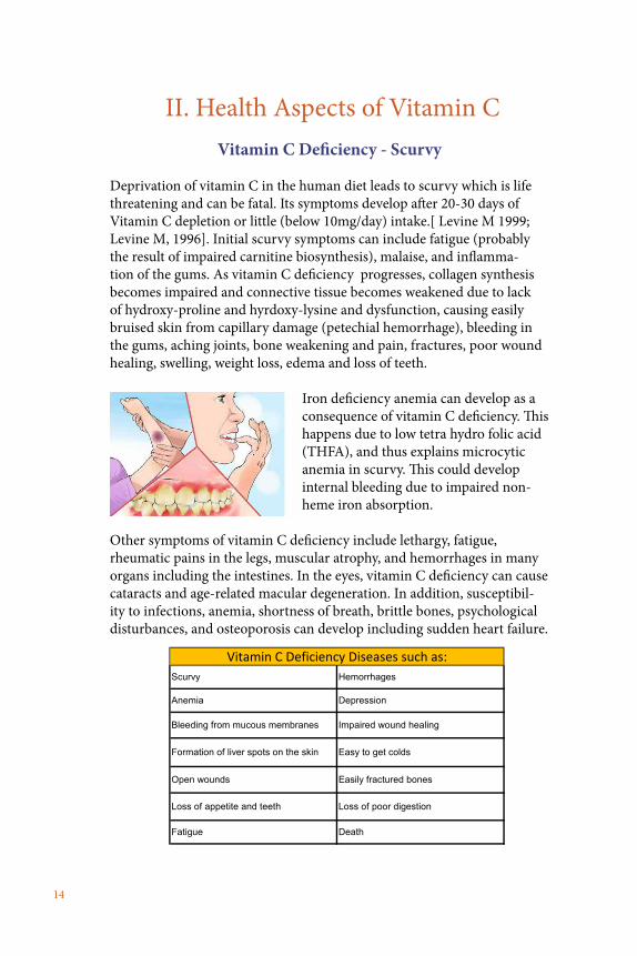
Deprivation of vitamin C in the human diet leads to scurvy which is life threatening and can be fatal. Its symptoms develop after 20-30 days of Vitamin C depletion or little (below 10mg/day) intake.[ Levine M 1999; Levine M, 1996]. Initial scurvy symptoms can include fatigue (probably the result of impaired carnitine biosynthesis), malaise, and inflamma-tion of the gums. As vitamin C deficiency progresses, collagen synthesis becomes impaired and connective tissue becomes weakened due to lack of hydroxy-proline and hyrdoxy-lysine and dysfunction, causing easily bruised skin from capillary damage (petechial hemorrhage), bleeding in the gums, aching joints, bone weakening and pain, fractures, poor wound healing, swelling, weight loss, edema and loss of teeth.
Iron deficiency anemia can develop as a consequence of vitamin C deficiency. This happens due to low tetra hydro folic acid (THFA), and thus explains microcytic anemia in scurvy. This could develop internal bleeding due to impaired non-heme iron absorption.
Other symptoms of vitamin C deficiency include lethargy, fatigue, rheumatic pains in the legs, muscular atrophy, and hemorrhages in many organs including the intestines. In the eyes, vitamin C deficiency can cause cataracts and age-related macular degeneration. In addition, susceptibil-ity to infections, anemia, shortness of breath, brittle bones, psychological disturbances, and osteoporosis can develop including sudden heart failure.
Fatigue Death
Vitamin C Deficiency Diseases such as:
Formation of liver spots on the skin Easy to get colds
Open wounds Easily fractured bones
Loss of appetite and teeth Loss of poor digestion
Scurvy Hemorrhages
Anemia Depression
Bleeding from mucous membranes Impaired wound healing

15
Today scurvy is considered rare and is mostly limited to the developing nations. However, a study conducted at the Centers for Disease Control and Prevention (USA) suggests that scurvy is still a serious, underappre-ciated problem even in the industrialized countries. Data on vitamin C blood levels collected from 2003 and 2004 indicate that 6 to 8 percent of the general population had scurvy-level deficiencies, with men on the higher end. Deficiency rates were greater for low-income people (10–17 percent), and highest among male smokers (18 percent) which is likely because smoking affects how the body absorbs vitamin C. These num-bers were actually an improvement over measures taken a decade earlier [Schleicher RL, 2009]. In addition, seniors are at a greater risk of develop-ing vitamin C deficiency due do decreased dietary intake, various ac-companying health problems, use of medications and impaired intestinal absorption.
In children, severe Vitamin C deficiency results in a condition known as Moeller-Barlow disease. It is seen in infants who are not breast fed and usually is manifested at approximately six months of age when maternally deprived stores of Vitamin C have been exhausted. Moeller-Barlow syndrome is charac-terized by widening of bone-cartilage boundaries, particularly of the rib cage, stressed epiphyseal car-tilage of the extremities, severe joint pain, anemia, and fever. Infants suffering from scurvy lie in the “pithed frog” position because movement is painful. The scorbutic infant usually lies on its back and makes little attempt to lift the leg or arm that hurts and both legs may be tender, as well as both arms. This symptom is usually the first sign of scurvy.
Vitamin C Intake levels: Recommended Dietary, Allowances (RDA) and Optimal Recommendations
The recommended dietary allowance (RDA) for Vitamin C was revised by the US Institute of Medicine (IOM) in 2000 from the previously recom-mended dose to 60 mg/day for men and women. The recommended intake for smokers is higher and was increased to 95 mg/day, because smokers are under increased oxidative stress from toxins contained in cigarette smoke and they generally have low blood levels of Vitamin C. The RDA is based on twice the amount of Vitamin C needed to prevent scurvy and on maintaining its neutrophil concentration resulting in minimal excretion of Vitamin C into urine.
16
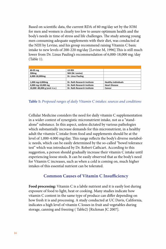
Based on scientific data, the current RDA of 60 mg/day set by the IOM for men and women is clearly too low to assure optimum health and the body’s needs in time of stress and life challenges. The study among young men consuming adequate supplements with their diet, was conducted at the NIH by Levine, and his group recommend raising Vitamin C basic intake to new levels of 200-220 mg/day [Levine M, 1996].This is still much lower from Dr. Linus Pauling’s recommendation of 6,000-18,000 mg /day (Table 1).
Recommended Intake/Day Source Condition 60-95 mg US RDI 200mg NIH (M. Levine) 6,000-18,000mg Dr. Linus Pauling 1,000 mg-4,000mg Dr. Rath Research Institute Healthy individuals 4,000 mg-10,000 mg Dr. Rath Research Institute Heart Disease 10,000 -80,000 g (oral +i.v.) Dr. Rath Research Institute Cancer
Table 1: Proposed ranges of daily Vitamin C intakes: sources and conditions
Cellular Medicine considers the need for daily vitamin C supplementation in a wider context of synergistic micronutrient intake, not as a “stand-alone” substance. In this aspect, unless dictated by various pathologies which substantially increase demands for this micronutrient, in a healthy adult the vitamin C intake from food and supplements should be at the level of 1,000-4.000 mg/day. This range reflects the body’s diverse metabol-ic needs, which can be easily determined by the so-called “bowel tolerance test” which was introduced by Dr. Robert Cathcart. According to this suggestion, a person should gradually increase their vitamin C intake until experiencing loose stools. It can be easily observed that as the body’s need for Vitamin C increases, such as when a cold is coming on, much higher intakes of this essential nutrient can be tolerated.
Common Causes of Vitamin C Insufficiency
Food processing: Vitamin C is a labile nutrient and it is easily lost during exposure of food to light, heat or cooking. Many studies indicate how vitamin C content in the same type of produce can differ depending on how fresh it is and processing. A study conducted at UC Davis, California, indicates a high level of vitamin C losses in fruit and vegetables during storage, canning and freezing ( Table2) [Rickman JC 2007].

17
Losses of vitamin C (% wet weight) in vegetables due to processing Canning Blanching and
Freezing Fresh Storage for 7 Days
20o 4o
Broccoli 84 30-55 56 0 Carrots 90 0-35 27 10 Green Beans 63 17-28 55 77 Green Peas 73-84 20-63 60 15 Spinach 62 50-61 100 75
Table 2: Examples of loss of vitamin C in vegetables due to food processing
Common Commercial Forms of Vitamin C
Ascorbic acid: Natural and synthetic L-ascorbic acid are chemically iden-tical, and there are no known differences in their biological activity. Two human studies indicated no clinically significant differences between the natural and synthetic L-ascorbic acid in terms of its bioavailabilty.
Mineral ascorbates: These are the salts of ascorbic acid and minerals and are less acidic, and therefore are considered “buffered.” Thus, mineral ascorbates are often recommended to people who experience gastroin-testinal problems (upset stomach or diarrhea) with plain ascorbic acid. When mineral salts of ascorbic acid are taken, both the ascorbic acid and the minerals appear to be well absorbed, so it is important to consider the dose of the mineral accompanying the ascorbic acid when taking large doses of mineral ascorbates. Mineral ascorbates are available in the follow-ing forms:
• Sodium ascorbate: 1,000 mg of sodium ascorbate generally contains 111 mg of sodium. Individuals following low-sodium diets (e.g., for high blood pressure) are generally advised to keep their total dietary sodium intake to less than 2,500 mg/day. Thus, megadoses of vitamin C in the form of sodium ascorbate could significantly increase sodium.
• Calcium ascorbate: Calcium ascorbate generally provides 90-110 mg of calcium with 890-910 mg of ascorbic acid per 1,000 mg of calcium ascorbate. Calcium in this form appears to be reason-ably well absorbed. The recommended dietary calcium intake for adults is 1,000 to 1,200 mg/day.
• Magnesium ascorbate: Magnesium ascorbate provides 77 mg of magnesium per 1,000 mg dose of magnesium ascorbate. The recommended dietary allowance for magnesium is 400-420 mg/day for adult men and 310-320 mg/day for adult women.

18
• Ester CTM: Ester-CTM contains mainly calcium ascorbate. In addition, it also contains small amounts of dehydroascorbic acid (oxidized ascorbic acid), calcium threonate, and trace levels of xy-lonate and lyxonate. The manufacturer states that the metabolites, especially threonate, increase the bioavailability of the vitamin C. However, a study in eight women and one man found no differ-ence between Ester-CTM® and commercially available ascorbic acid tablets with respect to the absorption and urinary excretion of vitamin C ( Johnston CS 1994). Another study published in 2008 in the Advances in Therapy also found no consistent differences in vitamin C plasma levels of subjects consuming ascorbic acid and Ester CTM. (Ester-CTM® should not be confused with ascorbyl palmitate, which is also marketed as “vitamin C ester.”)
Ascorbyl palmitate: Another commercial product of ascorbic acid is “Ascorbyl palmitate.” Ascorbyl palmitate has been shown to be better than ascorbic acid in terms of protecting red cell membranes and α-tocopher-ol (a fat-soluble antioxidant) from oxidation by free radicals. Further, it also has been shown to be more effective in mitigating collagen synthesis suppression by pharmaceutical drugs known as channel blockers, and also in restoring collagen synthesis and increasing the intracellular level of vitamin C [Ivanov V 2016].
Liposomal vitamin C: Vitamin C is also commercially available as liposo-mal vitamin C. Liposomes are bilayer (double-layer), liquid-filled bubbles made from phospholipids. Liposomes can overcome absorption barriers in biological membranes and have been used to deliver nutrients and drugs into the body and even into specific cells of the body. It appears that Lipo-somal vitamin C is predominately taken up by the lymphatic system in the gut, and not by portal circulation. Thus, there is no significant “one-pass” liver metabolism of vitamin C.
Ascorbyl palmitate

19
III. Function and Physiological Role of Vitamin C
Ascorbic acid and free radical damage: Since its discovery over 90 years ago, vitamin C has been associated with many health benefits from the common cold to cancer. In addition, it has been shown to be involved in a multitude of biochemical reactions occurring in the body, and for the growth and repair of tissues.
Oxidative stress caused by free radicals causes extensive damage to cells, and cellular components are free radicals which are highly reactive molecules containing a single electron. Free radicals are generated both during normal cellular metabolism as well as in pathological conditions. Free radical-induced damage to cellular proteins, fats and carbohydrate molecules, and their buildup over time contributes to the aging process as well as the development of many pathologies. The most known free radical compounds include superoxide, hydroxyl radicals, singlet oxygen, ozone, peroxynitrile, nitrogen dioxide and hypochlorus acid just to mention a few [Levine M 1996; Benedich A 1999].
Protection from free radicals is provided by intracellular defense mech-anisms (i.e., superoxide dismutase, catalase) and natural compounds with antioxidant properties that can scavenge the radicals. Vitamin C is considered the most effective, least toxic water soluble antioxidant. It is the predominant antioxidant in blood, tissues and in intracellular fluids [Frei B 1989; Frei B 1994]. It also recycles other antioxidants such as vitamin E and glutathione in the body. Due to its antioxidant function, Vitamin C helps protect several macromolecules such as DNA, lipids, proteins and

20
enzymes from free radical-induced damage. Consequently, it offers protec-tion against degenerative diseases such as cancer, aging, heart disease, and cataract formation.Ascorbic acid and collagen synthesis and structure: Besides its antiox-idant function vitamin C plays an indispensable role in the biosynthe-sis, maintenance and structure of collagen. Collagen is one of the main structural components of the blood vessels, skin, tendons, ligaments, bone, cartilage, heart valves and lens of the eye. Optimum collagen formation is essential in healing wounds and the assembly of the extracellular matrix. Vitamin C transcriptionally regulates synthesis of collagen and is also an essential co-factor of Lysyl and Prolyl hydroxylases, the enzymes that cata-lyze the formation of hydrogen bridges which link various collagen fibrils. Vitamin C affects the formation of connective tissue through its role in the synthesis of glycosaminoglycans, essential components of cartilage, bone and various types of connective tissue. Thus, wound healing and mainte-nance of tissue and organ structure and function largely depend on the availability of Vitamin C [Sauberlich HE 1994; Naidu KA 2003; Gaby SK 1991; Levine M 1996; Benedich A 1999].
Other metabolic effects: In addition, Vitamin C participates in peptide hormone synthesis, carnitine synthesis, and neurotransmitter functions such as norepinephrine synthesis, tyrosine metabolism, hypoxia-inducible factor, DNA methylation, and histone methylation. It also helps the body to absorb iron from non-heme sources, and helps keep metals such copper and iron in a reduced state, which is essential for many biological reac-tions in the human body.
Vitamin C regulates blood cholesterol levels as an inhibitor of HMGCoA reductase, a critical enzyme in cholesterol biosynthesis [Turley SD 1976;

21
Hemila H 1992]. In this aspect, vitamin C acts as a natural statin without the negative side effects of statins, a class of drugs highly promoted for lowering blood cholesterol levels. Low dietary and blood levels of vitamin C are also associated with the increased risk of heart disease.
Vitamin C is also an effective antimicrobial agent inhibiting viral and bacterial infections in the body, and it protects cells from radiation and stress. Vitamin C also interacts with several essential trace elements such as lead, cadmium, copper, nickel, vanadium, and selenium and reduces their toxicity. The re-duced forms of these trace elements are poorly absorbed and excreted rapidly. Vitamin C also enhances the utilization of low levels of selenium, increases tissue levels of manganese and stabilizes vitamin B12.Vitamin C has other protective effects against high blood pressure, diabetes, asthma and other allergic conditions, gout, infertility, schizophrenia, the com-mon cold, and depression [Naidu KA 2003; Gaby SK 1991; Levine M 1986].
Safety Aspects of Vitamin C
Vitamin C is a water-soluble substance and therefore it does not accumu-late in the body. Vitamin C in itself is not toxic. However, if ingested in extremely high doses it has only minor adverse effects such as diarrhea, nausea and other digestive disturbances that cease after decreasing the dose. These effects are due to the osmotic withdrawal of water from the in-testinal contents by the unabsorbed vitamin C in the gastrointestinal tract. A. Bendich’s critical analyses of the reports regarding possible adverse effects of vitamin C did not substantiate these concerns [Bendich A 1999; Diploc A 1995].
Vitamin C and Formation of Kidney Stones: Concerns have been raised that higher intakes of vitamin C can promote kidney stones in the form of calcium oxalate. It should be noted that oxalate is one of the endoge-nous metabolites of vitamin C. A thorough literature search by Goodwin and Tangum [Goodwin JS 1995] found no reliable source supporting this concern. On the contrary three case-controlled studies did not show clear association between ascorbate intake and excretion and kidney stone for-mation [Cowley DM 1987; Power C 1984; Felstrom B 1989].
The positive association between kidney stones and vitamin C reported in earlier studies by some may relate to using assays for urinary oxalates, which resulted in conversion of urinary ascorbic acid to oxalate during the storage and processing of the samples. A clinical study involving 29 people pre-dis-posed to kidney stone formation, and 19 age-matched non-stone formers

22
taking 2 g of vitamin C daily, showed higher secretion of oxalate in people pre-disposed to kidney stones, but not in the others [Chai W 2004]
However, in a large-scale Harvard Prospective Health Professional Fol-low-Up Study in 45,000 men, those groups in the highest quintile of vitamin C intake (> 15,000 mg/day) had a lower risk of kidney stones than the group in the lowest quintile [Curhan GC 1996].
Furthermore, a review of various studies led Gerster to conclude that the high intake of vitamin C does not increase the risk of calcium oxalate kidney stones [Gerster H 1997]. This was re-confirmed in the studies conducted in the cohort of over 85,000 women [Curhan GC 1999].
Safety of Megadoses of Vitamin C Delivered Intravenously
Intravenous delivery of vitamin C has been used in cancer and other human pathologies. It is worth mentioning that 115 g of Vitamin C ad-ministered daily by an intravenous route over 8 hours and 39 times in a period of 13 weeks did not produce any adverse effects in cancer patients (Riordan NH 1995].
Megadoses of vitamin C alone or in combination with other antioxidants have been used in several clinical studies without any toxic effects to the

23
patients (Moertel CG 1985; Cameron E 1991; Lockwood K 1994; Walker EM 2002; Drisco JA 2003]. Some people may experience bowel intoler-ance to high doses of oral vitamin C. In such cases, the intake of vitamin C should be gradually reduced until this effect stops.
Interactions of Vitamin C With Common Medical Drugs
Several drugs, such as contraceptive pills that contain estrogen, barbitu-rates and tetracyclines can reduce vitamin C levels.
In addition, regular intake of aspirin and nonsteroidal anti-inflammatory drugs (NSAIDs) can lower the amount of vitamin C in the body because they cause more of it to be lost in urine.
Both calcium channel blockers and sodium channel blockers can inhibit intracellular transport of vitamin C and cause impaired collagen synthesis in human vascular cells and fibroblasts. The inhibitory effect of these class of drugs can be mitigated by providing vitamin C, in particular, in the fat soluble form - ascorbyl palmitate [Ivanov V 2016].
Many commonly prescribed pain medications interfere with vitamin C as well as folic acid and iron. These nutrients are necessary to repair joint cartilage and reduce the chemical inflammation that leads to pain.
Taking aluminum-containing antacids (Maalox) in the presence of vitamin C can boost aluminum absorption and can therefore increase the risks of side effects from excess aluminum.
There have been rare reports of vitamin C interfering with the effective-ness of the blood thinning medication Warfarin (Coumadin). However, in recent follow-up studies no effect was found with doses of vitamin C up to 1,000 mg per day [University of Maryland medical center].

24
IV. Therapeutic Potential of Vitamin C in Cancer: Scientific and Clinical Aspects
Introduction to Cancer
Cancer is probably the most feared disease and the second leading cause of death after heart disease in the Western world. Occurrences of the disease continue to skyrocket, and it is estimated that 2.6 million new cancer cases will be diagnosed by 2050.
Chemicals, and radiation and viruses [Cairn J 1987; DeVita VT 1978; Pecorino L 2005] have been recognized as cancer causing agents in humans and in many animal species. Although there is great diversity in the nature of these agents, the resulting cellular response to them is the transformation of normal cells into cancer cells (Figure 7). This process of transformation primarily involves three distinct phases: initiation, promotion, and progression occurring over a period of several years to decades. Cancer initiation often requires sustained DNA damage induced by these agents, and it is considered to be one of the most important steps in cancer development. Although DNA repair enzymes correct most of the damage, some damaged cells may not be repaired. The mutations in the DNA accumulate over time thereby increasing the risk of cancer with each cell multiplication. It is therefore no surprise that rapidly dividing cells are more susceptible to carcinogens than slowly dividing cells. Finally, the stage of progression is reached when the uncontrolled multiplication of these cells forms a cell mass interfering with organ function, and the cells also acquire the ability to move into other tissues and metastasize. Based on the associated degree of spread, cancer development involves the following stages: (1) hyperplasia (increased number of cells and cell division), (2) invasion to the adjacent tissues, and (3) metastasis (cellular migration to distant organs [Hanahan D 2000].

25
Surgery, chemotherapy and radiation are currently considered standard cancer therapies [Pecorino L 2005]. Although these therapeutic approach-es can initially inhibit the tumor mass, they are ineffective in providing a cure. Moreover, these very toxic treatments indiscriminately attack all cells including healthy cells causing extensive cellular damage and cytotox-icity. This gives rise to new cancers and promotes development of drug resistance in cancer cells. Furthermore, these therapies are ineffective in curtailing cancers that have already metastasized.
Figure 7. Cancer causing agents and the stages of transforming normal cell to cancerous cells
There is an urgent need to develop effective non-toxic anti-cancer ap-proaches that include prevention of metastasis. In fact, approximately 80% of patients diagnosed with cancer seek alternate therapies which include antioxidants and other essential nutrients.The role of vitamin C as an anti-cancer agent was first suggested in 1952 when McCormick proposed its role as a chemotherapeutic agent [McCor-mick WJ 1952]. In recent decades it has been shown that vitamins, in par-ticular vitamin C used at high doses, can be effective as therapeutic agents that target several mechanisms associated with different pathologies. Among the different health applications of vitamin C, its role in relation to cancer has been most thoroughly investigated.
Specific Aspects of Vitamin C Metabolism in Cancer Cells
Cancer cells alter their metabolism in order to support their rapid prolif-eration and expansion across the body. In particular, compared to normal cells, tumor cells consume a large amount of glucose as an energy source but convert it into lactic acid even in the presence of oxygen (Warburg Effect).
DNA
Defectivecell
Cell multiplication Localizedtumor
MetastasisNormalcell
Chemicals
Radiation
Viruses
Repair
Damage
Precancerous cells Cancer
IN IT IAT ION PROMOTION CANCER METASTASISPROGRESSION

26
This important aspect of cancer cell metabolism relates to vitamin C’s structural similarity to glucose (Figure 1). Cancer cells which internalize large amounts of glucose compared to normal cells also have enhanced uptake of Vitamin C, which enters the cell through a common glucose transporter receptor (GLUT1). Therefore, if a large amount of vitamin C is provided to cancer cells it will be absorbed by glucose transporter recep-tors in excess compared to that absorbed by normal cells.
Once the vitamin C molecule is internalized by its receptors, it generates the superoxide ion in a reaction requiring the presence of iron and copper. This superoxide free radical is converted into hydrogen peroxide which is capable of inducing extensive cellular damage. This does not take place in normal cells, which convert the hydrogen peroxide into water and oxygen using the active anti-oxidant enzyme, catalase. In cancer cells, however, since the enzyme catalase is present in very small amounts the hydrogen peroxide is instead converted to even more reactive hydroxyl radicals in a reaction requiring iron and copper [Gonzales MJ 2014] (Figure 8). There-fore, an increase in the oxidative stress in malignant cells due to the higher intake of vitamin C consequently generates reactive oxygen species that cannot be eliminated and result in cancer cell death.
Further, since normal cells have protein-bound iron and no hydrogen per-oxide, vitamin C cannot increase oxidative stress in these cells. Actually, in normal cells the only effect of vitamin C is decreased oxidative stress. Additionally, in sufficient amounts vitamin C prevents the accumulation of hydrogen peroxide thus preventing the cells from oxidative damage and transformation into malignant cells.
Therefore, it can be inferred that due to the differences in cellular metabo-lism between cancer cells and normal cells, Vitamin C selectively inhibits the former while strengthening the latter [Gonzalez MJ 2014]. On the other hand, chemotherapy has adverse effects on both normal and cancer cells. In cancer cells, the increased state of intracellular oxidative stress is exacerbated by the additional pro-oxidant effects of chemotherapeutic agents causing cell death.

27
Figure 10. Differences in the mechanism of action of Vitamin C in normal versus tumor tissues (Gonzales MJ 2014) .
Possible Mechanism(s) By Which Vitamin C Inhibits Cancer Development:
Studies show that in a variety of cancer cell types vitamin C also elicits caspase-mediated apoptosis [Hong SW 2007; Kang JS 2003] and autopha-gy [Cullen JJ 2010].
In addition, upon treatment with Vitamin C the depletion of cellular ATP levels was found in many cancer models, which resulted in toxicity and cell death. With DNA damage and ATP depletion, a cellular signal in-volving AMP-activated protein kinase (AMPK) is activated which in turn results in tumor growth inhibition by cell cycle arrest, apoptosis, necrosis and autophagy.
Vitamin C also has been to seen exert inhibitory effects on hypoxia-induc-ible factor-1 (HIF-1) [Kawada H 2013], which regulates glucose metabo-lism and promotes oncogenic processes in cancer cells. NIH researchers demonstrate that it is abundant in untreated cancer and disappears upon vitamin C treatment.
28
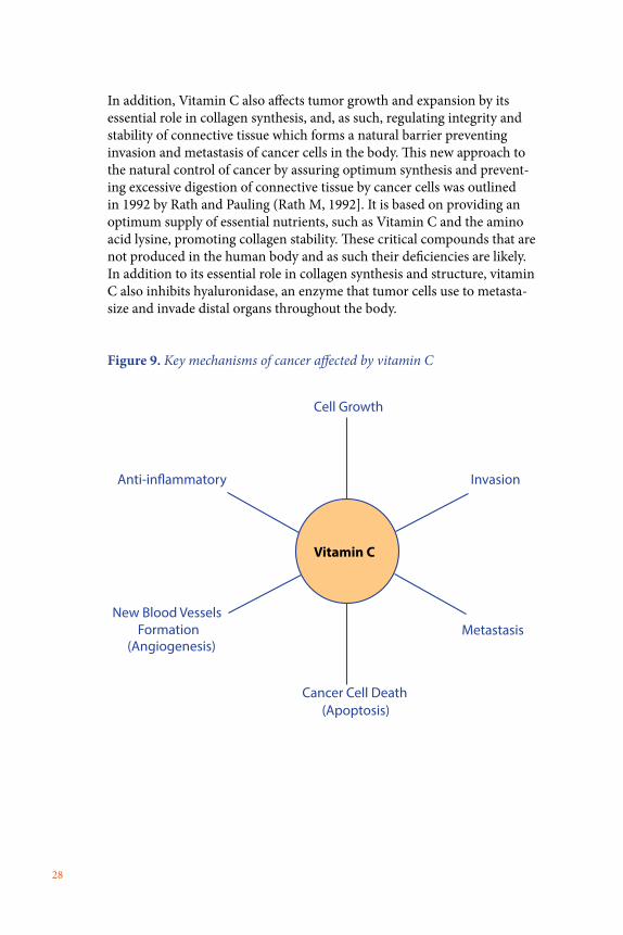
In addition, Vitamin C also affects tumor growth and expansion by its essential role in collagen synthesis, and, as such, regulating integrity and stability of connective tissue which forms a natural barrier preventing invasion and metastasis of cancer cells in the body. This new approach to the natural control of cancer by assuring optimum synthesis and prevent-ing excessive digestion of connective tissue by cancer cells was outlined in 1992 by Rath and Pauling (Rath M, 1992]. It is based on providing an optimum supply of essential nutrients, such as Vitamin C and the amino acid lysine, promoting collagen stability. These critical compounds that are not produced in the human body and as such their deficiencies are likely. In addition to its essential role in collagen synthesis and structure, vitamin C also inhibits hyaluronidase, an enzyme that tumor cells use to metasta-size and invade distal organs throughout the body.
Figure 9. Key mechanisms of cancer affected by vitamin C
Vitamin C
Cell Growth
Cancer Cell Death(Apoptosis)
(Angiogenesis)Metastasis
InvasionAnti-in�ammatory
New Blood Vessels Formation

29
Scientific Studies Supporting the Benefits of Vitamin C in Cancer
Several epidemiological, human, animal, and cell culture studies consis-tently and strongly suggest that vitamin C has a protective effect against cancer.
Epidemiological Studies. An extensive body of evidence accumulated over the years has shown that people consuming diets rich in fruits and vegetables are less likely to develop cancer than people who have lower intake of these foods [Block G 1991, Lee KW 2003, Enstrom EJ 1992]. Based on the evidence, the FDA allows the statement that “low fat diets rich in fruits and vegetables (foods that are low in fat and may contain dietary fiber, Vitamin A, or Vitamin C) may reduce the risk of some types of cancer, a disease associated with many factors.” The US Department of Agriculture and the NCI recommend the consumption of at least 5 servings of fruits and vegetables per day to prevent cancer [World Cancer research Fund 1997].
While there are many phytochemicals and micronutrients in fruits and vegetables that may have anticancer properties, vitamin C is likely to be one of the most effective anti-carcinogenic agents (Figure 9). It also has an effect on key mechanism of cancer in vitro and in vivo, such as cell growth, invasion, metastasis, apoptosis, angiogenesis and anti-inflammatory (Fig-ure 9). Its anti-carcinogenic properties can be attributed to its antioxidant properties such as preventing free radical damage to DNA, decreasing the formation of carcinogenic nitrosamines and mutagens, enhancing the immune system to fight cancer cells, accelerating the action of detoxifying

30
liver enzymes and blocking the toxic effects of carcinogens (e.g. polycyclic hydrocarbons, organochlorine pesticides, and heavy metals). In addition, through its essential role in collagen production vitamin C can hinder the spread of cancer cells in the body.
Several epidemiological studies have examined the role of vitamin C or vitamin C rich foods in cancer prevention, and the vast majority of them demonstrate a statistically significant protective effect. A number of stud-ies have investigated and demonstrated the preventive role of vitamin C in cancers of the oral cavity, gastric system, esophagus, colon, larynx and pancreas [Block G 1991; Lee KW 2003]. There is also substantial evidence of its protective effect against cancers of the breast and cervix. Several recent lung cancer studies suggest significant protective effects of vitamin C or of foods rich in vitamin C. Epidemiological findings have consistently established a correlation between a high intake of vitamin C (or foods rich in vitamin C), and a reduced risk of stomach cancer. This may be due to vitamin C blocking the formation of nitrosamine and other carcinogens in the stomach. A meta-analysis involving over 17,000 women clearly showed the benefits of vitamin C supplementation in reducing breast cancer mor-tality (Harris HR 2014].
Observational studies found a higher incidence of scurvy in cancer pa-tients [Fain O 1998] and presence of subclinical scurvy in patients with different malignancies [Krasner N 1974]. It has been shown that compared to healthy people cancer patients typically have significantly reduced levels of vitamin C in serum (Anthony HM 1982; Nunez C 1995; Gackowski D 2002]
The evidence documented by a prospective observational study (Shibata A 1992] and a retrospective observational study (Torun M 1995] support that increased consumption of vitamin C reduces the risk for cancer. The scientific evidence also indicates that increased consumption of vitamin C reduces the risk of death from cancer. The evidence documented by 4 prospective observational studies [Pandey DK 1995; Eichholzer M 1996; Eichholzer M 1999; Loria CM 2007] also supports this conclusion.
Animal Studies. Most of the beneficial effects of vitamin C in cancer observed in in vitro studies has been supported by in vivo testing as well [Gonzales MJ 2014; Cameron E 1979]. Studies by Dr. Linus Pauling and his colleagues have shown that high dietary intake of vitamin C decreases the incidence of and as well as delays the onset of malignant skin tumors in mice initiated by exposure to ultraviolet light [Tsao CS 1991], and as well delays the onset of spontaneous mammary tumors in mice [Pauling L 1991]. Vitamin C also has been shown to decrease the incidence of kidney

31
tumors generated by estradiol or diethylstilbesterol treatment in hamsters [Liehr JG 1991] by decreasing the formation of genotoxic metabolites. Other investigators have also found that vitamin C and its lipophilic derivatives, such as ascorbyl palmitate, are effective in preventing skin can-cer [Smart RC 1991]. Significant inhibition has also been observed with vitamin C treatment in the development of lung cancer in mice exposed to fiberglass dust [Cameron E 1979].
In a study by Levine, et al., following a regimen of daily pharmacological doses of ascorbate (4g/day) significantly decreased the growth rate of ovar-ian, pancreatic and glioblastoma tumors in mice [Chen Q 2008]. Higher concentrations of vitamin C also inhibited tumor growth in Balb/C mice implanted with sarcoma 180 cancer cell lines [Park S 2006]. The survival rate of cancer bearing mice in the group that received high doses of vita-min C increased by 20% compared to the control group.
In their pharmacokinetic studies in rats, Chen, et al., [Chen Q 2007] demonstrated that pharmacological doses of ascorbate administered parenterally can generate ascorbate radicals in extracellular space but not in blood. This study confirmed the therapeutic potential of pharmacologic doses of ascorbate in cancer and infections. A number of studies thereafter have shown that high doses of parenteral ascorbate inhibits the growth in mice of xenografts of a number of cancer cell lines such as glioblastoma, pancreatic cancer, breast cancer and neuroblastoma [Polireddy K 2017; Grasso C 2014; Chen Q, 2005, 2008].
Development of colon, kidney, and bladder cancers in animals can also be controlled by vitamin C. A study conducted by scientists at Johns Hopkins University showed that colon cancer cells carrying mutations in K-RAS or BRAF genes could specifically accumulate dehydroascorbic acid (DHA) and induce oxidative stress by depleting intracellular glutathione thereby

32
increasing the vulnerability of cancer cells to free radical damage and death. As expected, Apc/KrasG12D mutant mice exposed to high doses of Vitamin C developed fewer and smaller colon tumors compared to control mice [Yun J 2015].
Dietary ascorbate also decreased the number of melanoma xenograft tu-mors developing in GULO-/- mice which, like humans, cannot synthesize endogenous vitamin C. More importantly, the tumors were encapsulated by a layer of connective tissue rendering them less invasive (Figure 10). This was associated with a 71% reduction of tumor metastasis in vitamin C supplemented mice (Figure 11) [Cha J 2013].
Figure. 10. Effect of dietary ascorbate on growth of B16FO melanoma tu-mors in mice unable to synthesize vitamin C internally (Gulo KO). Repre-sentative for mice groups histology pictures of tumors. a — ascorbate-supple-mented mice showing a strong capsule around a tumor (pink); b — ascorbate depleted mice showing no tumor capsule and infiltrating cells
64% Lower
Figure. 11. Effect of dietary ascorbate on growth of B16FO melanoma tu-mors in Gulo KO mice (SEM bars)
In addition, vitamin C has beneficial effects on normal cells by decreasing the cytotoxic side effects of radiation and chemotherapeutic agents.

33
Cell Culture Studies. Much of the evidence for the anti-cancer efficacy of vitamin C comes from investigating the therapeutic potential of vitamin C and its derivatives directly on cancer cells (Gonzalez MJ 2014; Holman RA 1957; Leung Py 1993; Park CH 1980; Bishun NP 1978; Bordignon B 2013].
It has been reported that vitamin C is cytotoxic to some human cancers such as neuroblastoma, osteosarcoma and retinoblastoma. At concentra-tions ranging from 10 nM to 1 mM, vitamin C induces apoptosis in neuro-blastoma cells and in melanoma cells [Naidu KA 2003]. It also acts as a modulator of growth in mouse myeloma cells as seen in an in vitro colony forming assay and in human bone marrow cells. Vitamin C is also highly toxic to Ehrlich ascites carcinoma cells and 3T3 cells in culture [Gonzalez MJ 2014].
Also, vitamin C was cytotoxic to mouse lymphocytic leukemia cells, to cells from other mouse neoplasms, and to acute lymphoblastic leukemia human cell lines [Bourdignon B 2013; Park S 2004; Park S 2013]. Expo-sure of adult T-cell leukemia cells, both HTLV-1 dependent and indepen-dent, to vitamin C resulted in inhibition of cell proliferation, migration, expression of cancer promoting genes, induction of apoptosis and several other parameters of cancer development [Harakeh S 2007]. Recent studies conducted by this group ( Harakeh S et al. 2017) evaluated mechanistic as-pects of vitamin C in human adult T-cell leukemia virus Type1 (HTVL1) positive. The results demonstrated that anti-proliferative effects of vitamin C are mediated through NF-kB activation pathway by suppression of Tax expression. Ascorbate inhibited MMP-9, an important enzyme in cancer cells invasion and metastasis, by affecting its protein expression and enzy-matic activity, but not its mRNA levels.
Japanese scientists have demonstrated the anti-cancer effect of a vitamin C derivative, benzylidene ascorbate, against human tumors of the ovaries, stomach, pancreas, uterus, bile ducts, and lungs [Park S 2013]. Ben-zylidene ascorbate has also been shown to induce apoptosis, or cell death, in human myelogenous leukemic cell lines, rat hepatocellular carcinoma cells, and in an HIV-replicating human lymphoma cell line. Its cytotoxic effect has been related to its pro-oxidant activity and activation of tran-
Brest Cancer Cells in Culture

34
scriptional factor NF-kappa B [Block G 1991; Park S 2013].
Park, et al., [Park S 2004; Park S 2006; Park S 2013] and Chen [Chen Q 2005] studied the effect of vitamin C on a number of cancer and normal cell lines as summarized in Table 3.
Table 3. Effect of vitamin C on select human cancer and normal cell survival based on its estimated half maximal concentration values (IC50) (based on S. Park, Nutrients 2013; 5, 3496-3505).
Human Cell Lines Tested Range of IC50 (mM) Myeloid leukemia (HL60), Retinoic acid resistant Acute Promyelocytic Leukemia (NB4-R1), Histiocytic lymphoma (U937), Chronic myelogenous leukemia (K562)
0.3- 0.5
Acute Promyelocytic Leukemia (NB4, NB4R2), Myeloblast (KG1) 0.7- 1.0 Normal bone marrow, Breast cancer (MCF7, MB231), Lymphoma (LJP119) 1-10 Normal fibroblasts (CCD24SK), Normal lymphocyte, Normal monocyte, Normal breast cell (Hs587Bst), Human breast cancer (Hs587t), Ovarian cancer (OVCAR and SK-OV3)
10 - >20
IC50: the concentration of vitamin C which causes inhibition of cellular prolif-eration in 50% of the cells. The IC50 values are means ± standard deviations from triplicate experiments. *IC50 value was determined using H3 incorpora-tion proliferation assay for 24h.
Their results clearly demonstrate that vitamin C at differing concentrations may utilize varying mechanisms to target cancer cells. At concentrations of 0.25 mM-20 mM, Vitamin C induces a dose, time, and cancer type depen-dent cancer cell death by generating H2O2 and oxidative stress. Treatment of cells with higher doses of vitamin C resulted in increased intracellular GSH (glutathione) and GSH-S transferase activity that was accompanied by an uptake of cysteine thereby suggesting that high concentrations of vitamin C can modulate intracellular sulfur containing compounds such as glutathione and cysteine.
In addition, the study by Yun, et al., shed more light on the anti-cancer mechanism of vitamin C. The researchers used colon cancer cells with common BRAF and K-RAS mutations, which make unusually high levels of a protein that transports glucose across the cell membrane [Yun J 2015]. Since glucose and vitamin C share the same transporter, GLUT1, large doses of vitamin C raise the free radical levels in these mutated cells which, in turn, inactivate an enzyme Glyceraldehyde 3-phosphate de-hydrogenase (GAPDH) which is needed to metabolize glucose, thereby depriving these cells of energy. This results in cell death.

35
A number of vitamin C isomers and derivatives were synthesized and tested for their anti-cancer effects in vitro, and it was demonstrated that derivatives with substitutions at -2 or -6 and at both positions have a marked cytotoxic effect. For instance, Ascorbate 6-palmitate and 6-stea-rate, were found to be more potent inhibitors of murine leukemia cell pro-liferation compared to ascorbate 2-phosphate, and ascorbate 6-phosphate or ascorbate 6-sulphate, respectively [Roomi MW 1998, Roomi MW 1998, Roomi MW 1998]. Ascorbate 6- palmitate and ascorbate 6-stearate have also been shown to inhibit the proliferation of mouse gliomas, human gliomas, U-373 and T98G cells, and renal carcinomas. All of these were attributed to inhibition of cell proliferation, cell cycle arrest, and induction of apoptosis (Naidu KA 2003; Naidu AK 1993].
V. Combinations of Vitamin C with Other Natural Compounds.
In the majority of studies on the anti-cancer effects of vitamins and other micronutrients, these compounds were applied individually, however, several approaches also tested their combinations [Demeule M 2000; Mukhtar H 2000; Kawakami S 2001; Yoon SO 2001]. In most studies, vita-min C was combined with other antioxidant compounds such as:
Vitamin C with Selenium: Vitamin C in combination with selenium in-duced re-differentiation of cancer cells and inhibited their growth [Zheng QS 2002].
Vitamin C with Copper: A mixture of vitamin C and cupric sulfate ad-ministered orally significantly inhibited human mammary tumor growth in mice (Chen Q 2015; Tsao CS 1991; Chen Q 2008].

36
Vitamin C with Vitamin K3 (menadione): A combination of these vita-mins in 100:1 ratio (VC:VK) applied in vitro and in vivo resulted in tumor growth inhibition and increased lifespan in tumor-bearing mice [Calderon PB 2002]. Further studies indicated its efficacy in various types of cancers through autoschizic cell death among other mechanisms (Verrax J 2003]. It appears that this nutrient combination targets tumor cells by inflam-mation (Verrax J 2006]. Further work indicated the safety and benefits of these nutrients in prostate and bladder cancers (Tareen B 2008; McGuire K 2013].
Vitamin C with Carotenoids, vitamin E, and retinoic acid: A study by Prasad, et al., indicated that individual compounds used at doses 50 ug/ml of vitamin C, 10 ug/ml carotenoids, 10 ug of alpha-tocopherol succinate, and 7.5 ug/ml of retinoic acid did not affect growth of melanoma cells. However, their combination reduced cell numbers by 56%. Increase of ascorbate level in this mixture to 100 ug/ml resulted in further reduction of melanoma growth to 13% (Prasad KN 1994].
Vitamin C in synergy with multiple nutrients: A different approach is represented by Cellular Medicine, in which the anti-cancer efficacy of vita-min C is enhanced by complexing it with a select group of natural com-pounds (Table 4). In this aspect, vitamin C is a synergistically cooperating team member supporting several anti-cancer mechanisms, in particular the formation and stability of the biological barrier around cancer cells (collagen) through optimizing the synthesis of the extracellular matrix and inhibiting MMPs and plasmin, the key enzymes involved in its proteolytic degradation and associated with cancer expansion. This novel anti-can-cer target mechanism is especially critical in metastasis by hindering the spread and invasion of cancer cells to other organs. In addition, this synergistic combination which includes vitamin C, lysine, proline, green tea extract, and other micronutrients, is effective in inhibiting oxidative stress, inflammation, and angiogenesis, and triggers cellular mechanisms promoting cell apoptosis among other anti-cancer effects. Since the targeted mechanisms are universal to all types of cancer malignancy, the micronutrient combination (NM) used in different doses was effective in inhibiting these mechanisms in over 50 human cancer cell types selected on the basis of organ malignancies (carcinomas, sarcomas and leukemia) (Table 5) [Niedzwiecki A 2010].

37
Table 4: Composition of the nutrient synergy mixture (NM)
Components of NM Concentrations of Individual Nutrients in 1,000 mcg/ml
NM Solution Ascorbate 900 mcM Lysine 1,100 mcM Proline 1,100 mcM Arginine 500 mcM N-Acetyl-Cysteine 250 mcM EGCG (from green tea) 150 mcM Selenium 85 mcM Copper 7 mcM Manganese 4 mcM
By introducing the element of synergy in the interaction between various micronutrients, we could obtain better anti-cancer efficacy using lower doses of individual micronutrients than when applied individually( Figure 12) . Moreover, proper selection of nutrients for precise biological process-es allowed achievement of pleiotropic effects by targeting several relevant cancer mechanisms at the same time, such as cancer cell proliferation, invasion, metastasis and angiogenesis.
Figure 12. Inhibition of breast cancer cells growth by individual nutrients and their combinations (Kale 2010)
0
10
20
30
40
50
60
70
80
90
100
kontrola Witamina C EGCG Lizyna +Prolina
Witamina C+ EGCG
Lizyna +Prolina +
EGCG
Lizyna +Prolina +EGCG +
Witamina C
tez inneskladniki
Brea
st c
ance
r cel
l gro
wth
inhi
bitio
n (%
)
Lysine + Proline + EGCG + Vitamin C + other
Lysine + Proline + EGCG + Vitamin C
Lysine + Proline + EGCG
Vitamin C + EGCG
Lysine + Proline
EGCG Vitamin C Control

38
The in vitro findings of NM were also confirmed by in vivo studies, in both cancer prevention and therapeutic aspects. NM inhibited growth of cancer cells implanted as xenografts in athymic mice. It also inhibited can-cer development induced by chemicals, such as N-methyl N-Nitrosourea (MNU), in mammary tumors of rats, urethane induced lung cancer in mice and dimethyl ben-anthracene (DMBA) induced skin cancer in mice [[Niedzwiecki A 2010]. Tumor bearing mice exposed to NM exhibited decreased growth of tumors. More importantly, the majority (90%) of the tumors in NM exposed mice were non-malignant [Kale A 2010]
Table 5: Inhibition of extracellular matrix invasion of several human cancer cell lines by different concentrations of NM
NM concentrations needed by different types of cancer cells to achieve 100% inhibition of tissue invasion
100 mcg/ml 500 mcg/ml 1,000 mcg/ml Breast (MDA_MB 231) Cervical cancer (CCL2) Bladder cancer (T24) Breast(MCF-7+ estradiol) Lung carcinoma (A548) Cervical cancer (DoTc2451) Osteosarcoma (MNNG/U2OS) Pancreas (MIA PACA2) Fibrosarcoma (HT1080) Prostate (LNCaP) Ovarian cancer (SKOV3 Testis (NT2/DT) Prostate (PC3) Colon (HCT116) Renal Carcinoma (786-0) Synovial Carcinoma
In vivo studies in a mouse model confirmed that NM inhibited metastasis of B16FO melanoma cells into the lungs [Roomi MW 2006], and from the spleen into the liver [Roomi MW 2008], and from the testicles to the lungs [Roomi MW 2012]. The inhibition of invasion by NM can be attributed to its inhibitory effect on the synthesis and activity of MMPs, the enzymes responsible for digestion of the extracellular matrix. MMPs are elevated in several types of human cancers and are associated with a poor prog-nosis. Thus by inhibiting the MMP activity, NM affects multiplication of cancer cells, and also the spread of cancer (Figure 9) [Roomi MW, Bhanap B 2015]. In addition, NM was also effective in inducing cell death in all tested cancer cell types [Roomi MW, Shanker N 2015] (Figure 10).

39
VI. Clinical Applications of Vitamin C in Cancer
Many conventional physicians, in particular oncologists, are often skep-tical about the use of vitamin C or other antioxidants in cancer patients, citing lack of clinical evidence. Compared to the investment pouring into biotechnology and pharmacotherapy as the most profitable business approaches, there is hardly financial interest in natural health approaches in general, and in therapeutic application of vitamin C in particular. Most published clinical information on the efficacy of Vitamin C in cancer patients has been derived from the studies, which vary in design, timing of observation/intervention, intervention protocols, stages of malignan-cy and anti-cancer regimens as well as doses and methods of vitamin C delivery. These differences often result in different outcomes, which the opponents of this new approach use as a reason for questioning the thera-peutic value of vitamin C.
However, with a lack of significant therapeutic success of conventional approaches there has steadily been growing interest among both patients and therapists in using vitamin C in cancer treatment which now provides more and more evidence on its therapeutic potential (Carr AC 2014, Pa-dayalty SJ 2006, Stephensom CM 2013, Mikirova N, 2012, Monti DA 2012, Welsh JL 2013, Takahashi H 2012)
A recently published systematic review of human interventional and observational studies by Fritz, et al., [Fritz H 2014] forms a solid basis of support for further studies on the benefits of IV vitamin C in various phases of cancer.
40
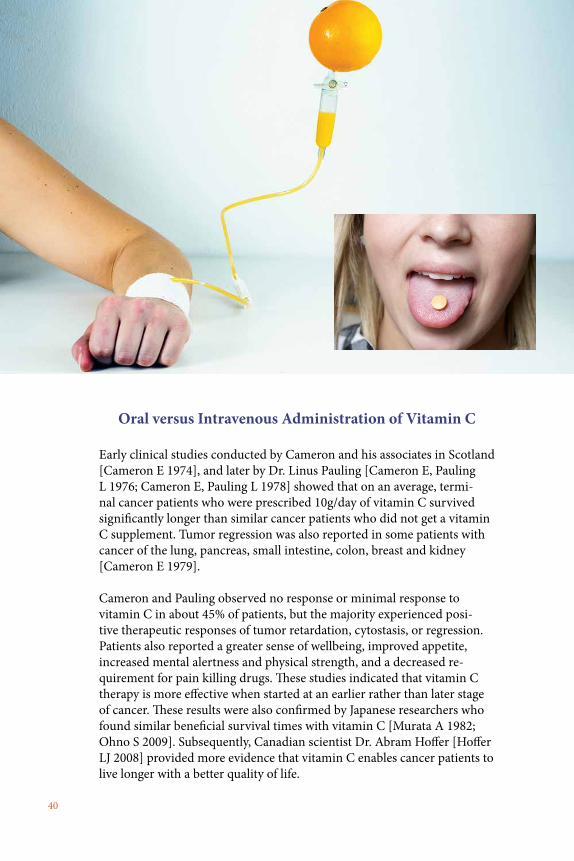
Oral versus Intravenous Administration of Vitamin C
Early clinical studies conducted by Cameron and his associates in Scotland [Cameron E 1974], and later by Dr. Linus Pauling [Cameron E, Pauling L 1976; Cameron E, Pauling L 1978] showed that on an average, termi-nal cancer patients who were prescribed 10g/day of vitamin C survived significantly longer than similar cancer patients who did not get a vitamin C supplement. Tumor regression was also reported in some patients with cancer of the lung, pancreas, small intestine, colon, breast and kidney [Cameron E 1979].
Cameron and Pauling observed no response or minimal response to vitamin C in about 45% of patients, but the majority experienced posi-tive therapeutic responses of tumor retardation, cytostasis, or regression. Patients also reported a greater sense of wellbeing, improved appetite, increased mental alertness and physical strength, and a decreased re-quirement for pain killing drugs. These studies indicated that vitamin C therapy is more effective when started at an earlier rather than later stage of cancer. These results were also confirmed by Japanese researchers who found similar beneficial survival times with vitamin C [Murata A 1982; Ohno S 2009]. Subsequently, Canadian scientist Dr. Abram Hoffer [Hoffer LJ 2008] provided more evidence that vitamin C enables cancer patients to live longer with a better quality of life.

41
However, Cameron’s and Pauling’s work on vitamin C suffered a setback from subsequent studies, including a randomized, double-blind, place-bo-controlled clinical trial by Dr. Charles Moertel of the Mayo Clinic [Moertel CG 1985]. However, these studies differed in the modes of ad-ministration of Vitamin C. In the Pauling study vitamin C was given both intravenous and orally, while in the Moertel study vitamin C was given only orally thereby limiting the achieving of higher vitamin C plasma con-centrations and, as such, the anti-cancer efficacy of vitamin C.
The subsequent pharmacokinetic analysis with oral, and oral plus intrave-nous administration of Vitamin C by Levine [Chen Q 2005; Chen Q 2008; Chen Q 2015], Padayatty and Riordan [Padayatty SJ 2006; Riordan NH 1995; Riordan NH 1996; Riordan 2000] suitably explained the conflicting findings observed in humans. Oral intake of vitamin C, even at a very large dose, can raise plasma vitamin C concentrations to a maximum of only 200 μm/L; whereas an intravenous administration can raise plasma concentrations to as high as 26,000 μm/L. Vitamin C concentrations of this magnitude can selectively kill tumor cells in vitro without any adverse effect on normal human cells. When 0.25-0.5 g/kg vitamin C was injected into rats intravenously or intraperitoneally, it produced an ascorbate con-centration of 60-100 times higher in blood and tissue than with the same oral dose.
Table 6: Peak plasma concentrations after oral and intravenous administra-tion of Vitamin C and its cytotoxic effect in normal and tumor cells (Modifed after: S. Ohno eta la., Anticancer Research 2009; 29;809-816 )
Vitamin C intake/daily
Route of administration
Attainable concentrations in plasma
Level causing cytotoxicity in: Tumor cells Normal cells
Dietary Oral <0.1 mM 1-3 g Oral 0.15 – 0.2 mM 10-100g Intravenous 5 – 15 mM 1-10 mM At least >20 mM
A study conducted by Padayatty, et al., in 17 healthy hospitalized vol-unteers showed that peak plasma vitamin C concentrations were higher after administration of intravenous doses than after administration of oral doses (P < 0.001), and the difference increased with an increase in the dose. Vitamin C at a dose of 1.25 g administered orally produced mean (+/-sd) peak plasma concentrations of 134.8 +/- 20.6 micromol/L com-pared with 885 +/- 201.2 micromol/L for intravenous administration. For the maximum tolerated oral dose of 3 g every 4 hours, pharmacokinetic modeling predicted peak plasma vitamin C concentrations of 220 micro-mol/L and 13 400 micromol/L for a 50-g intravenous dose. Peak predicted urine concentrations of vitamin C from intravenous administration were 140-times higher than those from maximum oral doses. Oral vitamin C
42
produces plasma concentrations that are tightly controlled. Only intrave-nous administration of vitamin C produces high enough plasma concen-trations that might have antitumor activity [Padayatti SJ 2004].
Vitamin C and Advanced Stages of Cancer
The effects of vitamin C on the health-related quality of life of cancer patients, was prospectively studied in 39 terminal cancer patients. All patients were given an intravenous administration of 10 g vitamin C twice with a 3-day interval and an oral intake of 4 g vitamin C daily for a week. In the global health/quality of life scale, health score improved from 36+/-18 to 55+/-16 after administration of vitamin C (p = 0.001). In functional scale, the patients reported significantly higher scores for physical, role, emotional, and cognitive function after administration of vitamin C (p<0.05). In symptom scale, the patients reported significantly lower scores for fatigue, nausea/vomiting, pain, and appetite loss after administration of vitamin C (p < 0.005). The other function and symptom scales were not significantly changed after administration of vitamin C. In terminal cancer patients, the quality of life is as important as cure and the authors concluded that vitamin C is considered a safe and effective therapy to improve the quality of life of terminal cancer patients [Yeom CH 2007].
Patients with advanced cancer or hematologic malignancy were assigned to sequential cohorts infused with 0.4, 0.6, 0.9 and 1.5 g ascorbic acid/kg body weight three times weekly. Adverse events and toxicity were min-imal at all dose levels. No patient had an objective anticancer response. High-dose IV ascorbic acid was well tolerated but failed to demonstrate anticancer activity when administered to patients with previously treated advanced malignancies. The promise of this approach may lie in combina-tion with cytotoxic or other redox-active molecules. [Hoffer LJ 2008]
This phase I clinical trial evaluated the safety, tolerability, and pharmaco-kinetics of high dose intravenous (IV) ascorbic acid as a monotherapy in patients with advanced solid tumors refractory to standard therapy. Five cohorts of three patients received I.V. ascorbic acid administered at 1 g/min for 4 consecutive days/week for 4 weeks, starting at 30 g/m² in the first cohort. For subsequent cohorts, the dose was increased by 20 g/m² until a maximum tolerated dose was found. Ascorbic acid was eliminated by simple first-order kinetics. Half-life and clearance values were similar for all patients of all cohorts (2.0 ± 0.6 h, 21 ± 5 dL/h m², respectively). C(max) and AUC values increased proportionately with a dose between 0 and 70 g/m², but appeared to reach maximal values at 70 g/m² (49 mM and 220 h mM, respectively). Doses of 70, 90, and 110 g/m² maintained

43
levels at or above 10-20 mM for 5-6 h. All doses were well tolerated. No patient demonstrated an objective antitumor response. Ascorbic acid administered IV at 1 g/min for 4 consecutive days/week for 4 weeks produced up to 49 mM ascorbic acid in the blood of the patients and was well tolerated. The recommended dose for future studies is 70-80 g/m² [Stephenson CM 2013].
Overall, it was reported that intravenous vitamin C used by practitioners of complementary and alternate medicine (CAM) has no serious side effects in patients. Out of 11,233 patients that received vitamin C in 2006 and 8,876 in 2008, only 59 reported experiencing lethargy or fatigue (Pa-dayatty SJ 2010].
Vitamin C as An Adjunct to Standard Cancer Treatments
Currently most types of cancer have standard protocols that in addition to surgery, include chemotherapy and radiation. These modalities are asso-ciated with severe and often debilitating side effects during the treatment and trigger many new health problems in cancer survivors. While many patients subjugate to fear of following non-conventional approaches there is an increasing interest in using vitamin C as an adjunct to alleviate severe side effects of conventional methods.
In vitro and in vivo studies from several groups have demonstrated the synergistic effect of pharmacologic ascorbate doses when combined with common chemotherapeutic drugs such as gemcitabine, paclitaxel and carboplatin [Kurbacher CM 1996]. In several studies, Riordan, et al.. [Riordan NH 1995; Riordan NH 1996; Riordan 2000] clearly indicated the benefits of vitamin C intake when used with chemotherapy. Prasad showed that high doses of vitamin C as well as other antioxidants could protect healthy cells during chemotherapy treatment [Prasad KN 1994]. He further states that cancer cells unlike healthy cells cannot regulate vitamin C uptake, which results in their death. This study was further supported by the findings from UCLA and MD Anderson Cancer Center advocating that antioxidants should be taken during cancer therapy [Moss RW 2006, MItra R].
Adjunct to Gemcitabine and Erlotinib: In a Phase I study, 14 subjects with metastatic stage IV pancreatic cancer were recruited to receive an eight-week cycle of intravenous ascorbic acid (three infusions per week), using a dose escalation design of 50, 75 and 100g, along with standard treatment of gemcitabine and erlotinib. Of the 14 recruited subjects en-rolled, nine completed the study (three in each dosage tier). Tumor mass
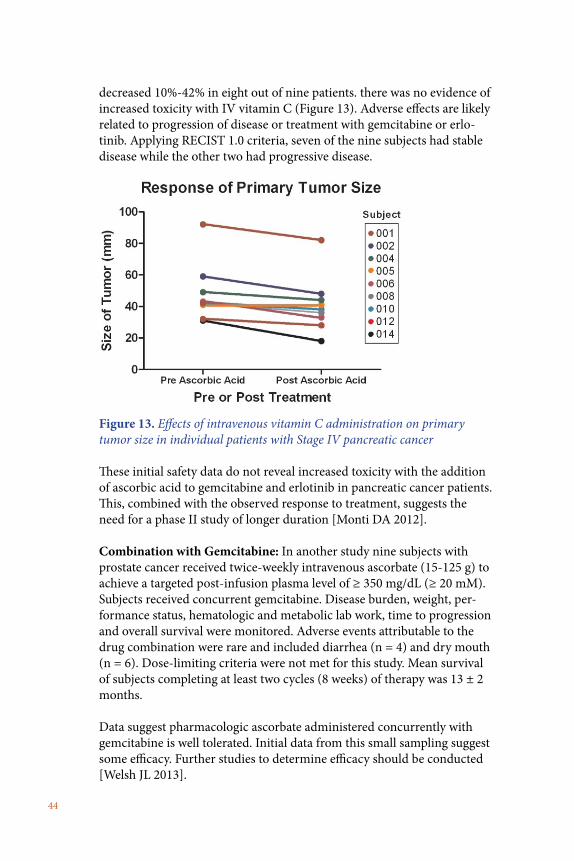
44
decreased 10%-42% in eight out of nine patients. there was no evidence of increased toxicity with IV vitamin C (Figure 13). Adverse effects are likely related to progression of disease or treatment with gemcitabine or erlo-tinib. Applying RECIST 1.0 criteria, seven of the nine subjects had stable disease while the other two had progressive disease.
Figure 13. Effects of intravenous vitamin C administration on primary tumor size in individual patients with Stage IV pancreatic cancer
These initial safety data do not reveal increased toxicity with the addition of ascorbic acid to gemcitabine and erlotinib in pancreatic cancer patients. This, combined with the observed response to treatment, suggests the need for a phase II study of longer duration [Monti DA 2012].
Combination with Gemcitabine: In another study nine subjects with prostate cancer received twice-weekly intravenous ascorbate (15-125 g) to achieve a targeted post-infusion plasma level of ≥ 350 mg/dL (≥ 20 mM). Subjects received concurrent gemcitabine. Disease burden, weight, per-formance status, hematologic and metabolic lab work, time to progression and overall survival were monitored. Adverse events attributable to the drug combination were rare and included diarrhea (n = 4) and dry mouth (n = 6). Dose-limiting criteria were not met for this study. Mean survival of subjects completing at least two cycles (8 weeks) of therapy was 13 ± 2 months.
Data suggest pharmacologic ascorbate administered concurrently with gemcitabine is well tolerated. Initial data from this small sampling suggest some efficacy. Further studies to determine efficacy should be conducted [Welsh JL 2013].

45
Combination with Carboplatin and Paclitaxel: In 2014, a phase I/IIA clinical trial evaluated the toxicities of combining IV ascorbate with carboplatin and paclitaxel in stage III /IV ovarian cancer. Twenty-seven patients were randomly assigned to receive either chemotherapy alone or chemotherapy and IV vitamin C concurrently. Chemotherapy was given for 6 months, and IV vitamin C was given for 12 months. The addition of IV vitamin C was associated with reduced chemotherapy-related toxicities [Ma Y 2014]. Other studies showed reduced chemotherapy-related toxicities of IV ascorbate in patients treated with carboplatin and paclitaxel in stage II/IV ovarian cancer [Ma Y 2014].
Combination with standard therapies for breast cancer: In an epidemi-ological multicenter cohort study, which included 15 gynecologists and general practitioners representatively distributed in Germany, the data from 125 breast cancer patients in UICC stages IIa to IIIb were selected for the study. A total of 53 of these patients were treated with IV vitamin C (supplied as Pascorbin® 7.5 g) in addition to standard tumor therapy for at least 4 weeks (study group) and 72 were treated without the additional therapy (control group). Standard therapies were: epirubicin/cyclophos-phamide(56%),cyclophosphamide/methotrexate/fluorouracil (20%) and fluorouracil/epirubicin/cyclophosphamide (15%). In the study IV vitamin C administration significantly reduced the side effects of chemotherapy, in particular, nausea, loss of appetite, fatigue, depression, sleep disorders, dizziness and hemorrhagic diathesis. After adjustment for age and baseline conditions (intensity score before adjuvant therapy, chemotherapy, radio-therapy), the overall intensity score of symptoms during adjuvant therapy and aftercare was nearly twice as high in the control group compared to the study group. No side effects of the IV vitamin C administration were documented [Vollbracht C 2011].

46
Combination of vitamin C, vitamin E and beta carotene with conventional therapy.
In a clinical study [Pathac AK 2002], patients receiving 6,1000 mg of ascorbic acid, 1,050mg of d-alpha tocopherol succinate, 60 mg of beta carotene and a trace mineral mixture along with chemotherapy benefited as evaluated by the number of patients in whom cancer did not progress, the number of survivors over a one-year period, and partial response of the patients.
At the same cancer conference Walker, et al., [Walker EM 2002] report-ed results from a randomized pilot trial (Phase I/II) in Stage 0-III breast cancer patients receiving radiation therapy and a high-dose of multiple antioxidant oral preparations containing 8g of vitamin C (as calcium ascorbate), 800 IU of vitamin E (alpha tocopheryl succinate), and 60 mg of natural beta carotene divided into two doses. Out of 25 patients receiving radiation alone, two developed new cancers, while no new cancers were detected in 22 patients receiving radiation and antioxidants.

47
Consideration in administration of vitamin C at high doses:
Although high doses of vitamin C are generally safe in the majority of people there are some aspects to be considered before administering vita-min C treatment.
Iron overload: Since administration of vitamin C enhances iron absorp-tion, iron overload must be ruled out in patients with hemochromatosis, thalassemia and sideroblastic anemia. The most common side effects in these patients are abdomnal pain, loose stools and vitamin B12 deficiency.
Glucose-6-Phospahte Dehydrogenase (G6PD) deficiency: Patients with G6PD deficiency (an enzyme used to maintain integrity of the red blood cell membranes) were found to be susceptible to hemolysis following a very high dose of vitamin C administration. Luckily, G6PD deficiency is relatively uncommon. It is an X-linked disorder prevalently affecting people of African, Asian, and Mediterranean descent. It is more frequent in men. G6PD deficiency is polymorphic, with more than 400 variants spread over malaria endemic regions.
Sodium fluctuations: Intravenous injection of vitamin C may also create a transient high load of sodium, which may lead to fluid overload in patients with congestive heart failure, renal insufficiency or renal failure. Vitamin C in mega-doses may result in diminution of uric acid excretion which may in turn precipitate an acute gouty arthritis.
Abrupt discontinuation of high doses of vitamin C: In some individuals this may cause “rebound scurvy.” However, this is rare.
Some analytical tests: It should be taken into consideration that large dos-es of Vitamin C may interfere with some stool tests for occult blood (false positive). Vitamin C can make a urine test for sugar unreliable because it reduces the copper in the clinical test and Benedict solution giving false positive results.
48
Other beneficial effects of Vitamin C in cancer
Interest in the chemo-preventive functions of vitamin C and other antioxi-dants [Sauberlich HE 1994; Naidu KA 2003; Gonzalez MJ 2014] has grown considerably in recent years since conventional therapies such as che-motherapy and radiation therapy have been unable to provide a cure for cancer. These conventional approaches are ineffective in treating cancers diagnosed at a late stage when metastasis has already occurred. Even more, cellular damage that results from these modalities leads to new cancers, infections, severe anemia, impaired immunity, and bleeding.
Improve quality of life: On the other hand, Takahashi, et al., have proven that a high dose of intra-venous vitamin C improves quality of life in cancer patients. They followed 60 newly diagnosed cancer patients who received intravenous vitamin C as an adjuvant cancer therapy and evaluated their response based on stan-dard QOL questionnaire. Reporting 46% and 60% improvement at 2 and 4 weeks respectively, without any adverse reactions, the researchers strongly suggest intravenous vitamin C (IVC) for advanced cancer cases [Taka-hashi 2012]. Carr, et al., further elaborate on this potential mechanism to explain the quality of life improvements in cancer patients after IVC. Radiation and chemotherapy contribute to treatment-related fatigue,and vitamin C via the antioxidant action decreases fatigue by reducing oxidant mediated toxicity of cancer treatment [Carr 2014].
Anti-inflammatory: Inflammation also plays a strong role in cancer-related fatigue thereby decreasing the patient’s quality of life. Anti-inflammatory effect of IVC was studied in 45 cancer patients (prostate, breast, bladder, pancreas, lung, thyroid, skin and lymphoma). The results showed that a high dose of IVC reduces markers of inflammation such as C-reactive protein (CRP), tumor necrosis factor (TNF), interferons and various interleukins [Mikirova N 2012].
Survival and quality of life: High dose IVC ranging from 25-100 grams/day has also been studied in cancer patients of various stages. The results showed they survived beyond individual prognostic estimate, had improved quality of life and improved tolerance to conventional treatments [Raymond YC 2016].
IVC has been reported to be effective as palliative treatment as well. Thir-ty-nine cancer patients with extensive bone metastasis and severe pain, and who were declared radiotherapy-resistant, were studied retrospective-

49
ly. Fifteen of those patients received 2.5 grams IVC, 15 patients received chemotherapy, and 9 were assigned to the control group. Four patients in IVC group and one in the chemotherapy group reported improved per-formance. A median 50% pain reduction and 10-month survival time was observed in the IVC group versus the median survival in the chemothera-py and the control group was two months [Günes-Bayir A 2015].
Taking into account the overall benefits of vitamin C and its tolerance when administered in high doses, there is an urgent need to apply it in clinical practice.
Acknowledgements: The authors are thankful to Professor D.S.R. Sarma for his valuable comments and suggestions, Dr. Bilwa Bhanap and Cathy Flowers for proofreading, and Elijah Artman for graphic design.
50
References:• Agus DB, Gambler SS, Pardridge WM, Spielholz C, Baselga J, Vera JC, Golde DW. Vitamin C
crosses the blood-brain barrier in the oxidized form through the glucose transporters. J Clin Invest. 1997; 100 (11): 2842-2848
• Anthony HM, Schorach CJ, Severe hypovitaminosis C in lung cancer patients. The Utilization of vitamin C in surgical repair and lymphocyte related host resistance. Br J Cancer 1982; 46, 354-367
Bates CJ. Bioavailability of vitamin C. Eur J Clin Nutr. 1997; 51 Suppl 1: 28-33• Bendich A. Antioxidant micronutrients and immune responses. In: Bendich A, Chandra RK,
ed. Micronutrients and immune functions. Ann N Y Acad Sci. 1999; 587: 168-80• Bishun NP, Basu TK, Metcalfe S, Williams DC. The effect of ascorbic acid (vitamin C) on two
tumor cell lines in culture. Oncology. 1978; 35: 160-162• Block G. Vitamin C and cancer prevention: the epidemiologic evidence. Am J Clin Nutr. 1991;
53: 270S-282S• Bordignon B, Chiron J, Fontes M. Ascorbic acid derivatives as a new class of antiproliferative
molecules. Cancer Lett. 2013; 338(2): 317-327• Burns JJ. Biosynthesis of L-ascorbic acid: basic defect in scurvy. Am J Med. 1959; 26: 740• Cairn J. The origin of human cancer. Nature. 1987. 289: 353-357• Calderon PB, Cadrobbi J, Marques C, Hong-Ngoc N, Jamison JM, Gilloteaux J, Summers JL,
Taper HS, Potential therapeutic application of the association of vitamin C and K3 in cancer treatment, Curr Med Chem., 2002; 9(24), 2271-2285
• Cameron E, Campbell A, Innovation versus quality control: An ‘unpublishable” clinical trial of supplemental ascorbate in incurable cancer, Medical Hypotheses 1991; 36, 185-189
• Cameron E, Campbell A. The orthomolecular treatment of cancer. Clinical trial of high dose ascorbic acid supplements in advanced human cancer. Chem Biol Interact. 1974; 9 (4): 285-315
• Cameron E, Pauling E, Leibovitz B. Ascorbic acid and cancer: a review. Cancer Res. 1979; 39: 663-681
• Cameron E, Pauling L. Supplemental ascorbate in supportive treatment of cancer: Prolongation of survival times in terminal human cancer. Proc Natl Acad Sci USA. 1976; 72(10):m3685-3689.
Cameron E, Pauling L. Supplemental ascorbate in supportive treatment of cancer: Reevaluation of prolongation of survival times in terminal human cancer. Proc Natl Acad Sci USA. 1978; 75(9): 4538-4542.
• Carpenter KJ. The history of scurvy and vitamin C. Cambridge, Cambridge Univ. Press. 8: pp VIII, 288; 1986
• Carr AC, Vissers MC, Cook JC. The effect of intravenous vitamin C in cancer and chemothera-py related to fatigue and quality of life. Front Oncol. 2014, 4, 1-7
• Cha J, Roomi MW, Kalinovsky T, Niezwiecki A, Rath M. Ascorbate supplementation inhibits growth and metastasis of B16F0 melanoma and 4T1 breast cancer cells in vitamin C-deficient mice. Int J Oncol. 2013; 42 (1): 55-64
• Chai W, Liebman M, Kynast-Gale S, Massey L, Oxalate absorption and oxalate synthesis from ascorbate in calcium oxalate stone formers and non-stone formers, Am J Kidney Dis 2004; 44, 1060-1069
• Chen Q, Espey MG, Sun AY, Lee JH, Krishna MC, Shacter E, Choyke PL, Pooput C, Kirk KL, Buettner GR, Levine M, Ascorbate in pharmacologic concentrations selectively generates ascorbate radical and hydrogen peroxide in extracellular fluid in vivo, Proc Natl Acad Sci U S A. 2007 May 22; 104(21): 8749–8754
• Chen Q, Espey MG, Krishna MC, et al. Pharmacologic ascorbic acid concentrations selectively kill cancer cells: action as pro-drug to deliver hydrogen peroxide to tissues. Proc Natl Acad Sci USA. 2005; 102(38): 13604-13609
• Chen Q, Espey MG, Sun AY, Pooput C, Kirk KL, Krishna MC, Khosh DB, Drisko J, Levine M. Pharmacologic doses of ascorbate act as a prooxidant and decrease growth of aggressive tumor xenografts in mice. Proc Natl Acad Sci USA. 2008; 105 (32): 11105-11109
• Chen Q, Polireddy K, Chen P, Dong R. The unpaved journey of vitamin C in cancer treatment. Can J Physiol Pharmacol. 2015; 93 (12): 1055-63
• Cowley DM, McWhinney BC, Brown JM, Chalmer AH. Chemical factors important to calcium

51
nephrolithiasis: Evidence for impaired hydroxcarboxylic acid absorption causing hyperoxal-uria. Clin Chem. 1987; 33, 243-247
• Cullen JJ. Ascorbate induces autophagy in pancreatic cancer. Autophagy. 2010; 6(3): 421-422• Curhan GC, Willet WS, Rimm EB, Stampher MJ, A prospective study of vitamin B6 and C and
the risk of kidney stones in men. J Urol 1996; 155, 1848- 1851• Curhan GC, Willet WS, Rimm EB, Stampher MJ, Intake of vitamin B6 and C and the risk of
kidney stones in women, J Am Soc Nephrol 1999; 10(4), 840-845• Demeule M, Brossard M, Page M, Gingras D, Beliveau R. Matrix metalloproteinase inhibition
by green tea catechins. Biochem Biophy Acta. 2000; 1478 (1): 51-60• DeVita V.T. The evolution of therapeutic research in cancer. N Engl J Med. 1978; 298 (16):
907-10• Diploc A, Safety of antioxidant vitamins and beta-carotene, Am J Clin Nutr 1995; 62,
1510s-1516s • Drisco JA, Chapman, J , Hunter VJ, The use of antioxidants with first-line chemotherapoy in
two cases of ovarian cancer, J Am Coll Nutr 2003; 22, 118-123• Eichholzer M, Stähelin HB, Gey KF, Lüdin E, Bernasconi F. Prediction of male cancer mortality
by plasma levels of interacting vitamins: 17-year follow-up of the prospective Basel study. Int J Cancer 1996;66:145-150
• Eichholzer M, Stähelin HB, Lüdin E, Bernasconi F. Smoking, plasma vitamins C, E, retinol, and carotene, and fatal prostate cancer: Seventeen-year follow-up of the prospective Basel study. Prostate 1999;38:189-198
• Enstrom EJ, Kanim LE, Klein MA. Vitamin C intake and mortality among a sample of the United States population. Epidemiol. 1992; 3: 194-202.
• Fain O, Mathieu E, Thomas M (1998) Scurvy in patients with cancer Br Med J 316, 1661-1662• Fellstrom B, Denielson BG, Karlstrom B et al. Br J Urol. 1989; 63(6), 575-780.• Frei B, England L, Ames BN. Ascorbate is an outstanding antioxidant in human blood plasma.
Proc Natl Acad Sci USA. 1989; 86: 6377-6381• Frei B. Reactive oxygen species and antioxidant vitamins: Mechanism of action. Am J Med.
1994; 97: 5S-13S• Fritz H, Flower G, Weeks L, et al. Intravenous vitamin C and cancer: A systematic review.
Integrative Cancer Therapies. 2014; 13, 280-300• Gaby SK, Singh VN. Vitamin C. Vitamin intake and health. A Scientific Review. New York. NY:
Marcel Dekker; 1991: 105-108• Gackowski D, Banaszkiewicz Z, Rozalski R et al, Persistent oxidative stress in colorectal carci-
noma patients , Int J Cancer 2002; 101, 395-397) • Gerster H, No contribution of ascorbic acid to renal calcium oxalate stones.Ann.Nutr.Metab
1997; 41, 269-282• Gonzalez MJ, Miranda-Massari JR. Springer briefs in Cancer Research: New Insights on Vita-
min C and Cancer. New York: NY Springer; 2014• Goodwin JS, Tangum MR , Battling quackery. Attitudes about micronutrient supplements in
American academic medicine , 1995, Arch Intern Med 158; 2187-2191)• Grasso C, Fabre MS, Collis SV, Castro ML, Cameron S. Field, Nanette Schleich, Melanie J. Mc-
Connell, and Patries M. Herst, Pharmacological Doses of Daily Ascorbate Protect Tumors from Radiation Damage after a Single Dose of Radiation in an Intracranial Mouse Glioma Model, Front Oncol. 2014; 4: 356
• Gregory JF. Ascorbic acid bioavailability in food and supplements. Nutr Rev. 1993; 51: 301-303• Hanahan D, Weinberger RA. Cell. The hallmarks of cancer. 2000; 100 (1): 57-70• Harakeh S, Diab-Assaf M, Khalife JC, Abu-el-Ardat KA, Baydoun E, Niedzwiecki A, El-Sabban
ME, Rath M.Ascorbic acid induces apoptosis in adult T-cell leukemia. Anticancer Res. 2007 Jan-Feb;27(1A):289-98
• Harakeh S, Khalife J, Baydoun E, et al. Effect of ascorbic acid on Tax, NF-kB and MMP-9 in human T-cell lymphocytes. Anti-cancer Agent in Medicinal Chem. 2017, July 25( Epub ahead of print PMIS 28745235).
• Harri Hemilä, Vitamin C and Plasma Cholesterol, Critical Reviews in Food Science and Nutri-tion, 32(l):33-57;1992
• Harris HR, Orsini N, Wolk A (2014) Vitamin C and survival among women with breast cancer: a meta-analysis, Eur. J Cancer 50, 1223-1231

52
• Hoffer LJ, Levine M, Assouline S, Melnychuk D, Padayatty SJ, Rosadiuk K, Rousseau C, Robi-taille L, Miller WH Jr; Phase I clinical trial of I.V. ascorbic acid in advanced malignancy. Ann Oncol. 2008 19(11):1969-74
• Holman RA. A method of destroying a malignant rat tumor in vivo. Nature. 1957; 179: 1033• Hong SW, Jin DH, Hahm ES, et al. Ascorbate (vitamin C) induces cell death through the apop-
tosis-inducing factor in human breast cancer cells. Oncol Rep. 2007; 18(4): 811-815• Ivanov V, Ivanova S, Kalinovsky T, Niedzwiecki A, Rath M. Inhibition of collagen synthesis by
select calcium and sodium channel blockers can be mitigated by ascorbic acid and ascorbyl palmitate. Am J Cardiovasc Dis. 2016 May 18;6(2):26-35. eCollection 2016.)
• Johnston CS & Luo B. Comparison of the absorption and excretion of three commercially available sources of vitamin C. Journal of the American Dietetic Association. 1994; volume 94: pages 779-781
• Kale A, Gawande S, Kotwal S, Netke S, Roomi MW, Ivanov V, Niedzwiecki A, Rath M. A com-bination of green tea extract, specific nutrient mixture and quercetin: An effective intervention treatment for the regression of N-methyl-N-nitrosourea (MNU)-induced mammary tumors in Wistar rats. Oncol Lett. 2010 Mar;1(2):313-317. Epub 2010 Mar 1
• Kang JS, Cho D, Kim Y, et al. L-ascorbic acid (vitamin C) induces the apoptosis of B16 murine melanoma cells via a caspase-8-independent pathway. Cancer Immunol Immunother. 2003; 52 (11): 693-698
• Kawada H, Kaneko M, Sawanobori M, Uno T, Matsuzawa H, Nakamura Y, Matsushita H, Ando K; High concentrations of L-ascorbic acid specifically inhibit the growth of human leuke-mic cells via downregulation of HIF-1⍺ PLOS One. 2013; 8(4): e62717.
• Kawakami S, Kageyama Y, Fujii Y, Kihara K, Oshima H. Inhibitory effect of N-acetylcysteine on invasion and MMP-9 production of T24 human bladder cancer cells. Anticancer Res. 2001; 21 (1A): 213-219
• Krasner N, Dymock IW (1974), Ascorbic acid deficiency in malignant diseases: a clinical and biochemical study, Br J Cancer 30, 142-145
• Kurbacher CM, Wagner U, Kolster B, Andreotti PE, Krebs D, Bruckner HW, Ascorbic acid (vi-tamin C) improves the antineoplastic activity of doxorubicin, cisplatin and paclitaxel in human breast carcinoma cells in vitro , Cancer Letters 1996;103, 183-189
• Lee KW, Lee HJ, Surh YJ, Lee CY. Vitamin C and cancer chemoprevention: reappraisal. Am J Clin Nutr. 2003; 78: 1074-1078
• Leung PY, Miyashita K, Young M, Tsao CS. Cytotoxic effect of ascorbate and its derivatives on cultured malignant and nonmalignant cell lines. Anticancer Res. 1993; 13: 47-80
• Levine M, Conry-Cantilena C, Wang Y, Welch RW, Washko PW, Dhariwal KR, et al. Vitamin C pharmacokinetics in healthy volunteers: evidence for a recommended dietary allowance. Proc Natl Acad Sci U S A 1996;93:3704-9.)
• Levine M, Rumsey SC, Daruwala R, Park JB, Wang Y. Criteria and recommendations for vita-min C intake. JAMA 1999;281:1415-23
• Levine M. New concepts in the biology and biochemistry of ascorbic acid. New Engl J Med. 1986; 31: 892-902
• Liehr JG. Vitamin C reduces the incidence and severity of renal tumors induced by estradiol or diethylstibesterol. Am J Clin Nutr. 1991; 54 (6 Suppl): 1256S-1260S
• Lind J. A treatise of scurvy. Edinburgh: Sands, Murray & Cochran; 1753• Lockwood K, Moesgaard S, Hanoika T, Folkers K, Apparent partial remission of breast cancer
in high-risk patients supplemented with nutritional antioxidants, essential fatty acids and Coenzyme Q10, Mol Aspects Med 1994; 15S:S231-240
• Loria CM, Klag MJ, Caulfield LE, Whelton PK. Vitamin C status and mortality in US adults. Am J Clin Nutr 2000;72:139-145)
• Ma Y, Chapman J, Levine M, Polireddy K, Drisko J, Chen Q , High-dose parenteral ascorbate enhanced chemosensitivity of ovarian cancer and reduced toxicity of chemotherapy. Sci Transl Med. 2014; 5;6(222)
• Mangels AR, Block G, Frey CM, Patterson BH, Taylor PR, Norkus EP. The bioavailability to human of ascorbic acid from orange juice and cooked broccoli is similar to that of synthetic ascorbic acid. J Nutr. 1993; 123: 1054-1061
• McCormick WJ. Ascorbic acid as a chemotherapeutic agent. Arch Pediat. 1952; 69: 151-155• McGuire K, Jamison JM, Gilloteaux J, Summers JL,Vitamin C and K3 combination causes en-

53
hanced anticancer activity against RT-4 bladder cancer cells, J Cancer Sci Ther 2013; 5, 325-333• Mikirova N, Casciari J, Roger S, et al, Effect of high-dose intravenous vitamin C on inflamma-
tion in cancer patients. J Tranl Med. 2012, Sep 11, doi:10.1186/1479-10-189.• Mitra R; Antioxidant Supplements & Cancer; http://www.mdanderson.org/depart-
ments/andersonnet/print.cfm?displayPrint=1&id=becd1d25-f75d-4c9aa0807b8349b-44f50&method=displayfull&PrintPage=1)
• Moertel CG, Fleming TR, Creagan ET, Robin J, O’Connell MJ, Ames MM. High dose vitamin C versus placebo in the treatment of patients with advanced cancer who have had no prior chemotherapy. A randomized double–blind comparison. N Engl J Med. 1985; 312 (3): 137-141
• Monti DA, Mitchell E, Bazzan AJ, Littman S, Zabrecky G, Yeo CJ, Pillai MV, Newberg AB, Deshmukh S, Levine M. Phase I evaluation of intravenous ascorbic acid in combination with gemcitabine and erlotinib in patients with metastatic pancreatic cancer PLoS One. 2012;7(1):e29794.
• Moss RW. Should patients undergoing chemotherapy and radiotherapy be prescribed antioxi-dants? Integr Cancer Ther. 2006 Mar;5(1):63-82.
• Mukhtar H, Ahmad N. Tea polyphenols: prevention of cancer and optimizing health. Am J Clin Nutr. 2000; 71 (6 Suppl): 1698s-1702s
• Murata A, Morishige F, Yamaguchi H. Prolongation of survival times of terminal cancer patients by administration of large doses of ascorbate. Int J Vitam Nutr Res Suppl. 1982; 23: 101-113
• Naidu AK, Wiranowska M, Kori SH, Prockop LD, Kulkarni AP. Inhibition of human glioma cell proliferation and glutathione-S-transferase by ascorbyl esters and interferon. Anticancer Res. 1993; 13 (5A): 1469-1475
• Naidu KA. Vitamin C in human health and disease is still a mystery? An overview. Nutr J. 2003; 2:7, 1-10
• Niedzwiecki A, Roomi MW, Kalinovsky T, Rath M. Micronutrient synergy—a new tool in effective control of metastasis and other key mechanisms of cancer. Cancer Metastasis Rev. 2010; 29 (3): 529-542
• Norum KR, Grav HJ. Axel Holst and Theodor Frolich–pioneers in the combat of scurvy. Tidss-kr Nor Laegeforen, 2002; 122(17): 1686-1687
• Nunez C, Ortiz de Apocada Y, Ruiz A, Ascorbic acid in the plasma and blood cells of women with breast cancer. The effect of consumption of food with elevated content of this vitamin, Nutr Hosp 1995; 10, 368-372
• Ohno S, Ohno Y, Suzuki N, Soma G, Inoue M. High-dose vitamin C (ascorbic acid) therapy in the treatment of patients with advanced cancer. AntiCancer Res. 2009; 29(3): 809-815
• Padayatty SJ, Riordan HD, Hewitt SM, Katz A., Hoffer LJ, Levine M. Intravenously adminis-tered vitamin C as cancer therapy: three cases. CMAJ. 2006; 174: 937-942
• Padayatty SJ, Sun AY, Chen Q, Espey MG, Grislo J, Levine M, Vitamin C: Intravenous use by complementary and alternative medicine practitioners and adverse effects, PLoS One 2010, 5; e11411
• Padayatty SJ, Sun H, Wang Y, Riordan HD, Hewitt SM, Katz A, Wesley RA, Levine M. Vitamin C pharmacokinetics: implications for oral and intravenous use. Ann Intern Med. 2004; 140(7):533-7
• Pandey DK, Shekelle R, Selwyn BJ, Tangney C, Stamler J. Dietary vitamin C and β-carotene and risk of death in middle-aged men. The Western Electric Study. Am J Epidemiol 1995;142:1269-1278
• Park CH, Amare M, Savin MA, Hoogstraten B. Growth suppression of human leukemia cells in vitro by L-ascorbic acid. Cancer Res. 1980; 40: 1062-1065
• Park S, Han SS, Park CH, et al. L-ascorbic acid induces apoptosis in acute myeloid leukemia cells via hydrogen peroxide-mediated mechanisms. Int J Biochem Cell Biol. 2004; 36: 2180-2195
• Park S, Lee J, Yeom C.H. A proteomic approach to the identification of early molecular targets changed by L-ascorbic acid in NB4 human leukemia cell. Cell Biochem. 2006; 99: 1628-1641
• Park S. The effect of high concentrations of vitamin C on cancer cells. Nutrient. 2013; 5: 3496-3505
• Pathak AK, Singh N, Khanna N, et al. Potentiation of the paclitaxel and carboplatin by antioxi-dant mixture on human lung h520 cell. J Am Coll Nut. 2002; 21(5), 416-421.

54
• Pauling L, Effect of vitamin C on incidence of spontaneous mammary tumors and UV-induced skin tumors in mice, Am J Clin Nutr 1991; 54(6 Suppl(, 1252S-1255S
• Pecorino L. Molecular Biology of Cancer: Mechanisms, Targets and Therapeutics. Oxford University Press; 2005
• Polireddy K ,Ruochen D, Gregory R, Jun Yu, Ping Chen, Stephen Williamson, Pierre-Christian Violet, Ziyan Pessetto, Andrew K. Godwin, Fang Fan, Mark Levine, Jeanne A. Drisko & Qi Chen, High Dose Parenteral Ascorbate Inhibited Pancreatic Cancer Growth and Metastasis: Mechanisms and a Phase I/IIa study. Scientific Reports 2017: 7, Article number: 17188
• Power C, Baker DJ, Nelson M, Winter PD, Diet and renal stones: A case control study, Br J Urol 1984; 56; 456-459
• Prasad KN, Hernandez C, Edwards PJ et al. Modification of the effect of tamoxifen, cisplatin, DTIC and interferon alpha 2b on human melanoma cells in culture by mixture of vitamins , Nutr Cancer 1994; 22, 223- 245
• Rath M, Pauling L. Plasmin-induced proteolysis and therole of apoprotein(a), lysine, and synthetic lysine analogs. J Orthomol Med.1992;7:17-23
• Raymond YCF, Glenda CSL, Meng LK. Effect of high doses od vitamin C on cancer patients in Singapore: Nine cases. Integrative Cancer Therapies. 2016; 15, 197-204.
• Rickman JC, DB Barret, CM Bruhn, Nutritional comparison of fresh, frozen and canned fruits and vegetables. Part 1. Vitamins C and B and phenolic compounds, J Sci Food Agric 2007; 87, 930-944)
• Riordan NH, Jackson JA, Riordan HD. Intravenous vitamin C in a terminal cancer patient. J Ortho Med. 1996; 11 (2): 80-82
• Riordan NH, Riordan HD, Casciari JP. Clinical and experimental experiences with intravenous vitamin C. J Ortho Med. 2000, 15 (4): 201-213
• Riordan NH, Riordan HD, Meng X, Li Y, Jackson JA. Intravenous ascorbate as a tumor cytotox-ic chemotherapeutic agent. Med Hypotheses. 1995; 44 (3): 207-213
• Roomi MW, Bhanap B, Niedzwiecki A, Rath M. (2015) Suppression of matrix metalloprotein-ases -2 and -9 in various human cancer cell lines by a nutrient mixture. J Oncobiomarkers. 2015; 2 (2): 1-17
• Roomi MW, House D, Eckert-Maksić M, Maksić ZB, Tsao CS. Growth suppression of malig-nant leukemia cell line in vitro by ascorbic acid (vitamin C) and its derivatives. Cancer Lett. 1998 Jan 9;122(1-2):93-9
• Roomi MW, House D, Tsao CS. Cytotoxic effect of substitution at 2-, 6-, and 2,6-positions in ascorbic acid on malignant cell line. Cancer Biochem Biophys. 1998 Nov;16(4):295-300
• Roomi MW, Kalinovsky T, Roomi NW, Ivanov V, Rath M, Niedzwiecki A. Suppression of growth and hepatic metastasis of murine B16FO melanoma cells by a novel nutrient mixture. Oncol Rep. 2008; 20 (4): 809-817
• Roomi MW, Kalinovsky T., Roomi NW, Niedzwiecki A, Rath M. Suppression of metastasis of intratesticular inoculation of B16FO melanoma cells by a novel nutrient mixture in male athymic nude mice, Exp Ther Med. 2012, 4 (5): 775-780
• Roomi MW, Roomi N, Ivanov V, Kalinovsky T, Niedzwiecki A, Rath M. Inhibition of pulmo-nary metastasis of melanoma B16FO cells in C57BL/6 mice by a nutrient mixture consisting of ascorbic acid, lysine, proline , arginine and green tea extract. Exp Lung Res. 2006; 32 (10): 517-530
• Roomi MW, Shanker N, Niedzwiecki A, Rath M. (2015) Induction of apoptosis in the human prostate cancer cell line DU-145 by a novel micronutrient formulation. OJApo. 2015; 4: 11-21
• Roomi MW, Tsao CS. Thin-Layer Chromatographic Separation of Isomers of Ascorbic Acid and Dehydroascorbic Acid as Sodium Borate Complexes on Silica Gel and Cellulose Plates. J. Agric. Food Chem., 1998, 46 (4), pp 1406–1409
• Sauberlich HE. Pharmacology of Vitamin C. Annu Rev Nutr. 1994; 14: 371-391• Schleicher RL, Carroll MD, Ford ES, Lacher DA, Serum vitamin C and the prevalence of vita-
min C deficiency in the United States: 2003–2004 National Health and Nutrition Examination Survey (NHANES), Am J Clin Nutr 2009; 90, 1252-1263
• Shibata A, Paganini-Hill A, Ross RK, Henderson BE. Intake of vegetables, fruits, β-carotene, vitamin C and vitamin supplements and cancer incidence among the elderly: A prospective study. Br J Cancer 1992;66:673-679)

55
• Smart RC, Crawford CL, Effect of ascorbic acid and its synthetic lipophilic altewrnative ascor-byl palmitate on phorbolester-induced skin tumor promotion in mice, Am J Clin Nutr, 1991; 54 (suppl), 1266S-1273S
• Stephenson CM, Levin RD, Spector T, Lis CG. Phase I clinical trial to evaluate the safety, toler-ability, and pharmacokinetics of high-dose intravenous ascorbic acid in patients with advanced cancer. Cancer Chemother Pharmacol. 2013 Jul;72(1):139-46.
• Svirbely J, Szent-Gyorgyi A. Hexuronic acid as antiscorbutic factor. 1932; 129: 576 -576.• Takahashi H, Mizuno H, Yanagiasawa A. A high dose intravenous vitamin C improve quality of
life in cancer patients. Personalized Medicine Universe. 2012; 2(1), 49-53• Tareen B, Summers JL, Jamison JM. Neal DR, McGuire K eta la , A 12 week open label phase I/
II study using apatone for the treatment of prostate cancer patients who failed standard thera-py, Int J Med Sci 2008; 5, 62-67,
• Torun M, Yardim S, Gönenç A, Sargin H, Menevşe A, Símşek B. Serum β-carotene, vita-min E, vitamin C and malondialdehyde levels in several types of cancer. J Clin Pharm Ther 1995;20:259-263
• Tsao CS. Inhibiting effect of ascorbic acid on the growth of human mammary tumor xeno-grafts. Am J Clin Nutr. 1991; 54 (6 Suppl): 1274S-1280S
• Tsukaguchi H, Tokui T, Mackenzie B, Berger UV, Chen XZ, Wang Y, Brubaker RF, Hediger MA. A family of mammalian Na+-dependent L-ascorbic acid transporters.Nature. 1999; 399 (6731): 70-75
• Turley SD, West CE, And BJ Horton. The Role Of Ascorbic Acid In The Regulation Of Cholesterol Metabolism And In The Pathogenesis Of Atherosclerosis Atherosclerosis, 24 ;1976; L-18 Review
• University of Maryland Medical Center- (http://www.umm.edu/health/medical/altmed/supple-ment-interaction/possible-interactions-with-vitamin-c-ascorbic-acid)
• Vera JC, Rivas CI, Fitchburg J, Golde DW. Mammalian facilitative hexose transporters mediate the transport of dehydroascorbic acid. Nature. 1993; 364: 79-82
• Verrax J, Cadrobbi J, Delvaux M, Jamison JM, Gilloteaux J, Summers JL, Taper HS Buc Cal-deron P, ,The association of vitamin C and K3 kills cancer cells mainly by autoschizis, a novel form of cell death. Basis for their potential use as coadjuvants in cancer therapy, Eur J Med Chem 2003; 38(5), 451-457
• Verrax J, Stockkis J, Tisson A, Taper HS, Calderon PB, Oxidative strss by ascorbate/menadi-one association kills K562 human chronic myelogenous leukemia cells and inhibits its tumor growth in nude mice, Biochem Pharmacol 2006; 72, 671-680
• Vollbracht C, Schneider B, Leendert V, Weiss G, Auerbach L, Beuth J Intravenous vitamin C administration improves quality of life in breast cancer patients during chemo-/radiotherapy and aftercare: results of a retrospective, multicentre, epidemiological cohort study in Germany. In Vivo. 2011;25(6):983-90
• Walker EM, Ross D, Pegg J et al, Nutritional and high dose anti-oxidant interventions during radiation therapy for cancer of the breast, Int Conf Nutr Cancer 2002, Montevideo (Urugway) Abstract 27
• Welsh JL, Wagner BA, van’t Erve TJ, Zehr PS, Berg DJ, Halfdanarson TR, Yee NS, Bodeker KL, Du J, Roberts LJ 2nd, Drisko J, Levine M, Buettner GR, Pharmacological ascorbate with gem-citabine for the control of metastatic and node-positive pancreatic cancer (PACMAN): results from a phase I clinical trial. Cancer Chemother Pharmacol. 2013; ;71(3):765-75
• Wilson JX. Regulation of vitamin C transport. Annu Rev Nutr. 2005; 25: 105-125• World Cancer Research Fund/American Institute for Cancer Research. Food, nutrition and
prevention of cancer: a global perspective. (1997) Washington, DC. AICR• Yeom CH, Jung GC, Song KJ Changes of terminal cancer patients’ health-related quality of life
after high dose vitamin C administration, J Korean Med Sci. 2007; 22(1):7-11• Yoon SO, Kim MM, Chung AS. Inhibitory effect of selenite on invasion of HT-1080 tumor
cells. J Biol Chem. 2001; 276 (23): 20085-20092• Yun J, Mullark E. Lu, C. et al. Vitamin C selectively kills KRAS and BRAF mutant colorectal
cancer cells by targeting GAPDH. Science. 2015. 350; 1391-1396• Zheng QS, Sun XL, Wand CH, Redifferentiation of human gastric cancer cells induced by
ascorbic acid and sodium selenite, Biomed. Envir. Science 2002; 15, 223-232

56
© Dr. Rath Research Institute, 2018 Santa Clara, CA 95050
Matthias Rath, M.D.Dr. Rath is a world-renowned physician and scientist, who is known for his pioneering research in natural and cellular health. He is the founder of the scientific concept of Cellular Medicine - the systematic introduction into clinical medicine of the biochemical knowledge of the role of micronutrients as biocatalysts in a multitude of metabolic reactions at the cellular level.
Aleksandra Niedzwiecki, Ph.D.Currently the Director of Research at the Dr. Rath Research Institute, Dr. Niedzwiecki is a leading biomedical researcher in the development of nutrient synergy approaches in various aspects of health and disease. Her work in the areas of cardiovascular health and cancer has won her recognition for her research into the biochemical link between disease and nutrients.
M. Waheed Roomi, Ph.D.; DABTDr. Roomi earned a doctorate degree in Biochemical Toxicology from the University of Surrey, England, and is a Fellow of American College of Nutrition. He is certified by the American Board of Toxicology (DABT) and American College of Nutrition (CNS). Dr. Roomi worked at the Linus Pauling Institute in Palo Alto, California for five years before joining the Dr. Rath Research Institute as a Senior Researcher in 2000.
www.drrathresearch.org
RSA: 13112
Disclaimer:This booklet is not intended as a substitute for the medical advice of a physician. The reader should regularly consult a physician in matters relating to his or her health and particularly in respect to any symptoms that may require diagnosis or medical attention.
Dr. Rath Research InstituteThe Dr. Rath Institute in Cellular Medicine is located in the Silicon Valley, in California. The Institute is staffed with experts handpicked from fields of medicine, biochemistry, and nutrition. Here, world-class scientists conduct innovative
research utilizing the principle of nutrient synergy, and investigate the role of nutrients in preventing and treating a host of diseases.
Researchers at the Dr. Rath Research Institute are developing new scientific concepts based on Dr. Rath’s discoveries in heart disease, cancer, infectious disease, and other diseases. Their scientific work has been published in various media around the world.