vitellogenesis during the ovarian development in...
TRANSCRIPT
International Journal of Aquatic Science ISSN: 2008-8019 Vol 3, No 2, 2012
Vitellogenesis during the ovarian development in freshwater
female prawn Macrobrachium rosenbergii (De Man)
Peranandam Revathi1*, Palanisamy Iyapparaj2, Natesan Munuswamy3, Muthukalingan Krishnan1
1) Unit of Insect Molecular Biology, Department of Environmental Biotechnology, Bharathidasan University, Trichy- 620
024, Tamil nadu, India
2) CAS in Marine Biology, Faculty of Marine Sciences, Annamalai University, Parangipettai – 608 502, Tamil nadu,
India
3) Unit of Aquaculture and Cryobiology, Department of Zoology, University of Madras, Guindy Campus, Chennai- 600
025, Tamil nadu, India
Abstract: In the present investigation, vitellogenesis during ovarian development in the freshwater
prawn Macrobrachium rosenbergii was assessed. Ovarian development was classified into five stages
based on size, colour and texture of ovary. Interestingly, histological results clearly indicate that the
oocyte development gradually increased from stages I to V based on the yolk material accumulation.
Besides, the biochemical changes associated with ovarian development was also analyzed. On the other
hand, vitellogenin (Vg) and vitellin (Vt) content during ovarian development in female prawn was
quantified as a measure of reproductive activity.
Key Words: Macrobrachium rosenbergii, Ovarian development, Reproductive activity, Biomarker,
Vitellogenesis
Introduction In crustaceans, substantial quantities of
yolk accumulation within the developing
oocytes serve to meet the basic requirement of
embryonic and larval development (Adiyodi and
Subramonium, 1983). During maturation, the
ovary exhibits size and colour changes those
are macroscopically visible through the
transparent carapace. These changes are due
to the deposition of yolk material in the
oocytes, which results in a rapid increase in
oocyte diameter (Sagi et al., 1995; Tsukimura,
2001) and colour changes due to the
carotenoid components with specific colour
changes each being related to a new
maturation stage (Arculeo et al., 1995). The
main constituents of yolk are protein and lipid
Revathi et al., (2012) Vitellogenesis during the ovarian development in freshwater …
Int. J. Aqu. Sci; 3(2): 13-27, 2012 14
content; vitellin is the major yolk protein that
accumulates with in the ovary during
vitellogenesis (Chen et al., 1999).
Lipoglycoprotein and vitellin are the major
components of yolk and vitellogenin is a protein
that reacts immunologically to the antiserum
prepared against the purified vitellin from the
hemolymph of vitellogenic females. This FSP,
known as vitellogenin (Vg) is reported in all
organisms studied so far (Dehn et al., 1983;
Fyfee and OʼConnor, 1974; Susuki, 1987). In
decapods, the transformation of vitellogenin to
vitellin revealed that a vitellin subunit highest in
molecular mass disappeared during embryo-
genesis (Chang and Bradley, 1983). During
these processes, vitellogenin and vitellin are
modified through cleavage, glycosylation,
lipidation and phosphorylation (Raikhel and
Dhadialla, 1992). They serve as storage protein
providing amino acids, carbohydrates, lipid and
phosphates to the developing embryo (Byrne
and Gruber, 1989). Determination of oocyte
diameter with histological tools provides basic
information on classification of ovarian
development (Peixoto et al., 2005; Revathi,
2010).
Synthesis, secretion and processing of
vitellogenin differ among phyla (Chen et al.,
1997). Hemolymph vitellogenin concentration is
a good indicator of the onset of vitellogenesis
during early maturation, rapidly increasing until
reaching a plateau in mid-maturing females.
After being internalized into the ovary by a
receptor mediated endocytotic process, they
undergo proteolytic processing to give rise to
major yolk protein, namely vitellin which is
considered to be phosphorylated glycoprotein
(Fyffe and OʼConnor, 1974). In general,
vitellogenin is synthesized by extra ovarian
tissues like liver in vertebrates (Byrne and
Gruber, 1989) and fat body in insects. In
decapod crustaceans, the hepatopancreas
(Eastman- Reks and Fingerman, 1985; Khayat et
al., 1994; Lui and OʼConnor, 1976) and
subepidermal adipose tissues (Rani and
Subramoniam, 1997) have been reported as
vitellogenin synthetic sites. Quantification of Vg
and Vt are required for the investigation of the
dynamics during vitellogenesis. Earlier studies
have often relied on oocyte size or ovarian
weight (Anikumar and Adiyodi, 1980).
Studies related to ovarian cycle associated
with vitellogenesis as well as biochemical
changes in freshwater female prawn Macrobr-
achium rosenbergii is scarce. Hence, the present
study has been under taken to document the
ovarian development in female M. rosenbergii
with reference to morphological, histological,
biochemical changes and vitellogenesis.
Material and methods Collection and maintenance of prawn
Freshwater prawn, Macrobrachium rosenber-
gii were collected from the Aqua Nova hatchery
in Kannathur, Chennai, South India. The
collected prawns were brought to the laboratory
Revathi et al., (2012) Vitellogenesis during the ovarian development in freshwater …
Int. J. Aqu. Sci; 3(2): 13-27, 2012 15
in a plastic cover with aerated habitat water and
were transferred into plastic tanks with sufficient
aeration. The water was changed daily and they
were fed ad libitum with commercial pelletized
food. They were maintained in the laboratory for
2-3 weeks for acclimatization.
Experimental design
Five months old female prawn, weighing 16
± 2 gm were taken for experimental studies. A
total of 50 prawn were used for this study and
divided into 5 groups each with 10 prawns.
Ovarian developmental stages
Ovarian developmental stages were
determined according to the type, size and
frequency of the germinal cells (Chaves and
Magalhaes, 1993; Htun-Han, 1978; Martins et
al., 2007; Okumura and Aida, 2000).
Gonado Somatic Index and Hepato Somatic
Index
The prawns were weighed, gonads removed
and the weight of the gonads were recorded.
The Gonado Somatic Index (GSI) and Hepato
Somatic Index (HSI) were calculated following
the procedure outlined by Zhang et al. (2007).
Oocyte diameter
Oocyte diameter was measured using an
ocular micrometer calibrated with a stage
micrometer fitted in a light microscope (Labex,
India). For each prawn, the diameters of as
many as 30 oocytes were measured and mean
oocyte diameter was calculated. The stage of
oocyte development was characterized based on
the maximum number of oocytes confined to a
particular stage. Photomicrographs of various
stages of oocyte development were taken using
a Leica 2500 microscope (Germany).
Histology
For histological examinations, the ovary was
dissected from different ovarian stages of
prawns. The isolated ovarian samples were fixed
in Bouinʼs fixative for 24 h and washed with
distilled water. The samples were dehydrated
with different grades of an alcohol series and
processed by routine procedure. Sections of 6-8
µm thickness were taken and stained with
haematoxyline and eosin. The stained sections
were mounted using DPX and photomicrographs
of varying magnifications were taken using a
Leica 2500 microscope.
Biochemical analysis Protein
Various reproductive tissue samples were
taken from different ovarian stages of prawns
and used for protein estimation. The samples
such as hemolymph (100 µl), ovary and
hepatopancreas (100 mg) were taken individual-
lly, homogenized in 10% Trichloroacetic acid
(TCA) and centrifuged for 10 min at 9000 Xg at
4 ºC. The supernatant, diluted with 0.15 M NaCl,
was used to measure the protein concentration.
Revathi et al., (2012) Vitellogenesis during the ovarian development in freshwater …
Int. J. Aqu. Sci; 3(2): 13-27, 2012 16
For each sample, the soluble protein
concentration was determined spectro-
photometrically at 595nm by Coomassie brilliant
blue G–250 method described by Bradford
(1976). Bovine Serum Albumin (BSA) was used
as a standard.
Lipid
The total lipid content was analyzed using
the Vanillin–Phosphoric acid method according to
Folch et al. (1957). Hundred milligram of wet
tissue of each sample was taken and
homogenized with 0.5 ml of chlorof-
orm:methanol (2:1) and 0.5 ml of 0.9 % NaCl
was added and kept in a separating funnel at
room temperature for 12 h. The lower phase
was collected, 0.5 ml of Conc. H2SO4 was added,
heated in boiling water for 10 min, cooled to
room temperature and then 1ml of phosphoric
vanillin solution (13 mMol/l vanillin in 14 Mol/L
phosphoric acid) was mixed immediately and
held at room temperature for 30 min. The optical
density was measured at 547 nm. Cholesterol
was used as a standard.
Isolation of vitellogenin and vitellin
Vitellogenin and vitellin were isolated from
the hepatopancreas, hemolymph and ovaries of
prawn M. rosenbergii following the method of
Tsukimura et al. (2000). The reproductive
tissues were homogenized in homogenization
buffer (containing 0.1 M NaCl, 0.05 M Tris, 1mM
ethylene diamine tetra acetic acid and 0.1 %
Tween 20 with 10 mg/ml PMSF; pH 7.8) using
an ice cold glass homogenizer. The homogenate
was centrifuged at 4000 Xg for 5 min at 4 ºC.
The resultant supernatant was again centrifuged
at 20,000 Xg for 20 min at 4 ºC. To the
supernatant, saturated ammonium sulphate
(SAS) was added to produce 25 % SAS solution.
After incubation for 1 h at 4 ºC, the solution was
centrifuged at 20,000 Xg for 10 min at 4 ºC. The
supernatant was collected and SAS was added to
produce 40 %, 50 % and 60 % SAS solution
sequentially. The pellets of 60 % SAS solution
was suspended in appropriate volume of homo-
genization buffer and dialyzed thrice at 4 ºC for
12 h each against homogenization buffer. The
isolated vitellogenin and vitellin were stored at -
20 ºC for further analysis.
Enzyme linked immunosorbent assay
Hundred milligrams of ovary, hepatopancreas
and hemolymph (100 µl) samples were taken
from different ovarian stages of prawns. Tissues
were homogenized with phosphate buffer and
centrifuged at 13000 Xg for 10 min at 10 ºC, to
remove cellular debris. The supernatant was
collected and then coated on the 96-well plates
for overnight at 4 ºC. Then after three washing
with washings buffer, the wells were blocked
with 200 µl of blocking buffer and incubated at
37 ºC for 1 h. Washing was followed by the
addition of 100 µl of primary antibody (anti Vg at
1:2000), for 3 h at 37 ºC. After three times
washing, the wells were coated with 100 µl of
Revathi et al., (2012) Vitellogenesis during the ovarian development in freshwater …
Int. J. Aqu. Sci; 3(2): 13-27, 2012 17
secondary-antibody enzyme conjugate (Anti
rabbit IgG-Alkaline phosphatase) at 1:500
dilutions for 1h at 37ºC. Incubation was
terminated by washing and wells were filled with
100 µl of substrate solution (1mg pNPP- paranit-
rophenyl phosphate/ml of substrate buffer).The
reaction was stopped with the stop buffer after
the required colour development was attained.
Absorbance at 405 nm was measured in an
automated ELISA plate reader (Titertek
Multiscan Plus, MK II, Denmark).
Results In the present study the reproductive activity
of the adult freshwater female prawn M.
rosenbergii was assessed based on the
morphological variation of ovary, Gonado
Somatic Index and Hepato Somatic Index,
oocyte diameter, cellular level changes in ovary
and quantification of female specific protein at
different ovarian stages.
Morphological observation of the ovary
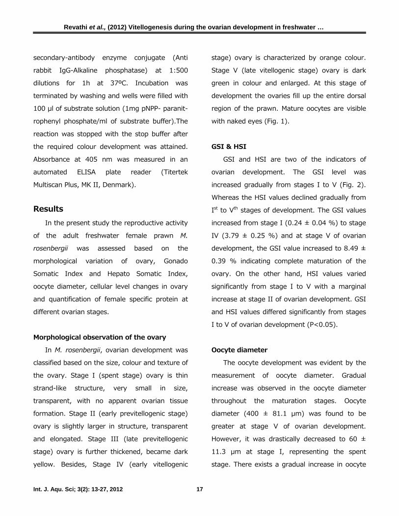
In M. rosenbergii, ovarian development was
classified based on the size, colour and texture of
the ovary. Stage I (spent stage) ovary is thin
strand-like structure, very small in size,
transparent, with no apparent ovarian tissue
formation. Stage II (early previtellogenic stage)
ovary is slightly larger in structure, transparent
and elongated. Stage III (late previtellogenic
stage) ovary is further thickened, became dark
yellow. Besides, Stage IV (early vitellogenic
stage) ovary is characterized by orange colour.
Stage V (late vitellogenic stage) ovary is dark
green in colour and enlarged. At this stage of
development the ovaries fill up the entire dorsal
region of the prawn. Mature oocytes are visible
with naked eyes (Fig. 1).
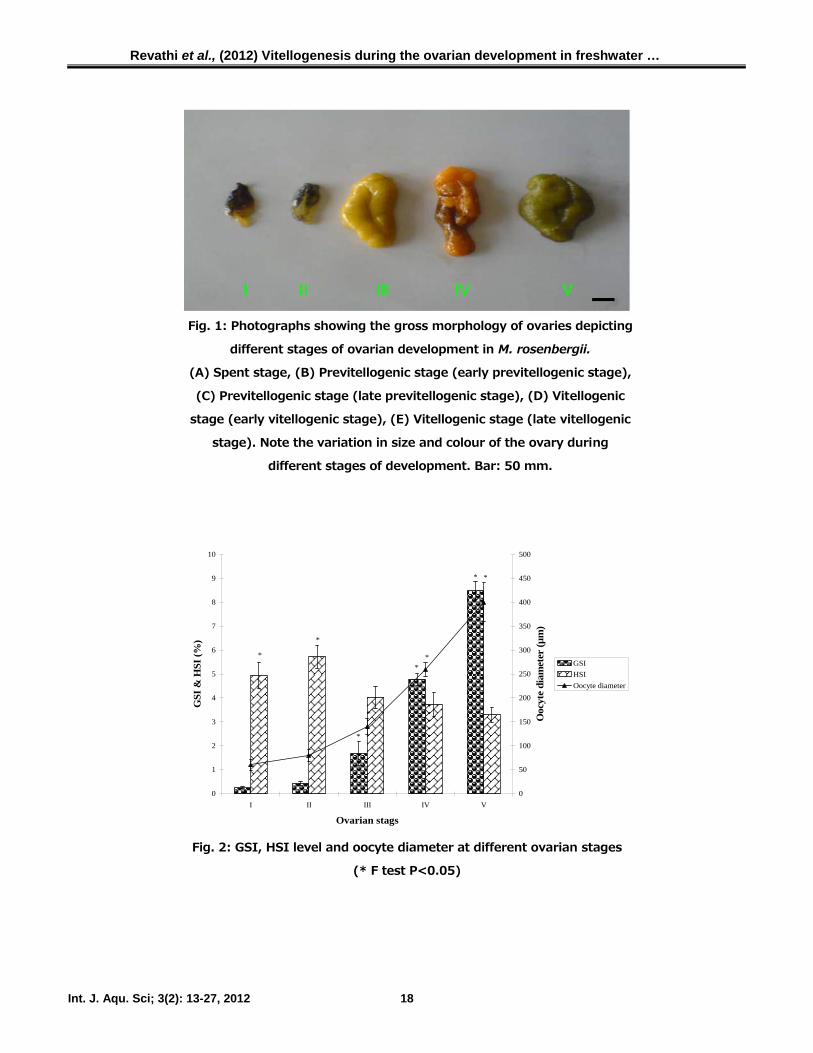
GSI & HSI
GSI and HSI are two of the indicators of
ovarian development. The GSI level was
increased gradually from stages I to V (Fig. 2).
Whereas the HSI values declined gradually from
Ist to Vth stages of development. The GSI values
increased from stage I (0.24 ± 0.04 %) to stage
IV (3.79 ± 0.25 %) and at stage V of ovarian
development, the GSI value increased to 8.49 ±
0.39 % indicating complete maturation of the
ovary. On the other hand, HSI values varied
significantly from stage I to V with a marginal
increase at stage II of ovarian development. GSI
and HSI values differed significantly from stages
I to V of ovarian development (P<0.05).
Oocyte diameter
The oocyte development was evident by the
measurement of oocyte diameter. Gradual
increase was observed in the oocyte diameter
throughout the maturation stages. Oocyte
diameter (400 ± 81.1 µm) was found to be
greater at stage V of ovarian development.
However, it was drastically decreased to 60 ±
11.3 µm at stage I, representing the spent
stage. There exists a gradual increase in oocyte
Revathi et al., (2012) Vitellogenesis during the ovarian development in freshwater …
Int. J. Aqu. Sci; 3(2): 13-27, 2012 18
Fig. 1: Photographs showing the gross morphology of ovaries depicting
different stages of ovarian development in M. rosenbergii.
(A) Spent stage, (B) Previtellogenic stage (early previtellogenic stage),
(C) Previtellogenic stage (late previtellogenic stage), (D) Vitellogenic
stage (early vitellogenic stage), (E) Vitellogenic stage (late vitellogenic
stage). Note the variation in size and colour of the ovary during
different stages of development. Bar: 50 mm.
*
*
*
*
*
*
*
*
0
1
2
3
4
5
6
7
8
9
10
I II III IV V
Ovarian stags
GSI
& H
SI (%
)
0
50
100
150
200
250
300
350
400
450
500
Ooc
yte
diam
eter
(µm
)
GSIHSIOocyte diameter
Fig. 2: GSI, HSI level and oocyte diameter at different ovarian stages
(* F test P<0.05)
Revathi et al., (2012) Vitellogenesis during the ovarian development in freshwater …
Int. J. Aqu. Sci; 3(2): 13-27, 2012 19
diameter from stage I to V which reflects oocyte
growth leading to vitellogenesis in the final stage
of ovarian development V (Fig. 2). Statistical
analysis revealed that the variation of oocyte
growth during various stages in ovarian
development is significant (P<0.05).
Histological variations in the ovary
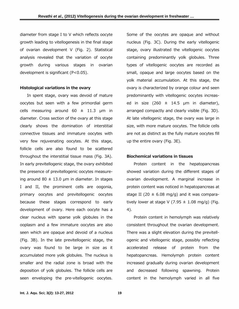
In spent stage, ovary was devoid of mature
oocytes but seen with a few primordial germ
cells measuring around 60 ± 11.3 µm in
diameter. Cross section of the ovary at this stage
clearly shows the domination of interstitial
connective tissues and immature oocytes with
very few rejuvenating oocytes. At this stage,
follicle cells are also found to be scattered
throughout the interstitial tissue mass (Fig. 3A).
In early previtellogenic stage, the ovary exhibited
the presence of previtellogenic oocytes measure-
ing around 80 ± 13.0 µm in diameter. In stages
I and II, the prominent cells are oogonia,
primary oocytes and previtellogenic oocytes
because these stages correspond to early
development of ovary. Here each oocyte has a
clear nucleus with sparse yolk globules in the
ooplasm and a few immature oocytes are also
seen which are opaque and devoid of a nucleus
(Fig. 3B). In the late previtellogenic stage, the
ovary was found to be large in size as it
accumulated more yolk globules. The nucleus is
smaller and the radial zone is broad with the
deposition of yolk globules. The follicle cells are
seen enveloping the pre-vitellogenic oocytes.
Some of the oocytes are opaque and without
nucleus (Fig. 3C). During the early vitellogenic
stage, ovary illustrated the vitellogenic oocytes
containing predominantly yolk globules. Three
types of vitellogenic oocytes are recorded as
small, opaque and large oocytes based on the
yolk material accumulation. At this stage, the
ovary is characterized by orange colour and seen
predominantly with vitellogenic oocytes increas-
ed in size (260 ± 14.5 µm in diameter),
arranged compactly and clearly visible (Fig. 3D).
At late vitellogenic stage, the ovary was large in
size, with more mature oocytes. The follicle cells
are not as distinct as the fully mature oocytes fill
up the entire ovary (Fig. 3E).
Biochemical variations in tissues
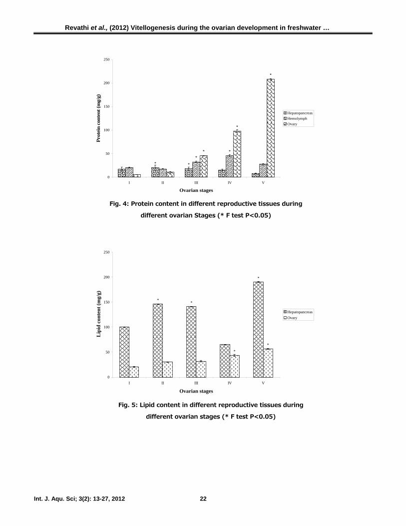
Protein content in the hepatopancreas
showed variation during the different stages of
ovarian development. A marginal increase in
protein content was noticed in hepatopancreas at
stage II (20 ± 6.08 mg/g) and it was compara-
tively lower at stage V (7.95 ± 1.08 mg/g) (Fig.
4).
Protein content in hemolymph was relatively
consistent throughout the ovarian development.
There was a slight elevation during the previtell-
ogenic and vitellogenic stage, possibly reflecting
accelerated release of protein from the
hepatopancreas. Hemolymph protein content
increased gradually during ovarian development
and decreased following spawning. Protein
content in the hemolymph varied in all five
Revathi et al., (2012) Vitellogenesis during the ovarian development in freshwater …
Int. J. Aqu. Sci; 3(2): 13-27, 2012 20
Fig. 3: Cross- section of ovary of different ovarian stages of prawns.
(A) Stage I ovary showing the presence of rejuvenating oocytes (RO) and immature
oocytes (IO). (B) Stage II ovary showing zone of proliferation (ZP), immature
oocytes (IO) and follicle cells (FC). (C) IIIrd stage ovary showing developing oocytes
(DO) with sparse yolk globules in the ooplasm, clear nucleus (N) and follicle cells
(FC). (D) Stage IV ovary showing vitellogenic oocytes with distinct ooplasm (OP)
filled yolk globules (Yg). Note oocytes are enveloped by a row of follicle cells (FC).
(E) Stage V ovary showing the vitellogenic oocyte (VO), oocytes are enveloped by a
row of follicle cells (FC). Note the accumulation of yolk globules (Yg) in the ooplasm
(OP) and prominent nucleus (N). Bar: 50 µm.
N
Revathi et al., (2012) Vitellogenesis during the ovarian development in freshwater …
Int. J. Aqu. Sci; 3(2): 13-27, 2012 21
stages and ranged from 20.16 ± 0.97 mg/ml to
27.32 ± 1.28 mg/ml.
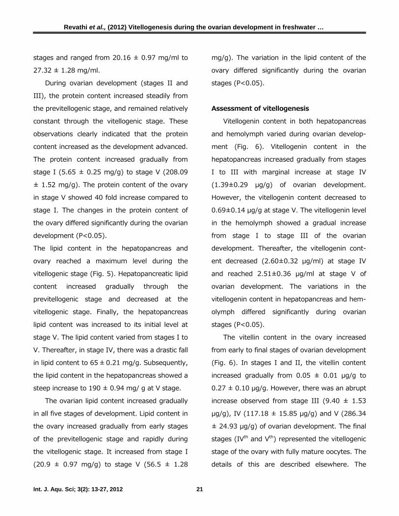
During ovarian development (stages II and
III), the protein content increased steadily from
the previtellogenic stage, and remained relatively
constant through the vitellogenic stage. These
observations clearly indicated that the protein
content increased as the development advanced.
The protein content increased gradually from
stage I (5.65 ± 0.25 mg/g) to stage V (208.09
± 1.52 mg/g). The protein content of the ovary
in stage V showed 40 fold increase compared to
stage I. The changes in the protein content of
the ovary differed significantly during the ovarian
development (P<0.05).
The lipid content in the hepatopancreas and
ovary reached a maximum level during the
vitellogenic stage (Fig. 5). Hepatopancreatic lipid
content increased gradually through the
previtellogenic stage and decreased at the
vitellogenic stage. Finally, the hepatopancreas
lipid content was increased to its initial level at
stage V. The lipid content varied from stages I to
V. Thereafter, in stage IV, there was a drastic fall
in lipid content to 65 ± 0.21 mg/g. Subsequently,
the lipid content in the hepatopancreas showed a
steep increase to 190 ± 0.94 mg/ g at V stage.
The ovarian lipid content increased gradually
in all five stages of development. Lipid content in
the ovary increased gradually from early stages
of the previtellogenic stage and rapidly during
the vitellogenic stage. It increased from stage I
(20.9 ± 0.97 mg/g) to stage V (56.5 ± 1.28
mg/g). The variation in the lipid content of the
ovary differed significantly during the ovarian
stages (P<0.05).
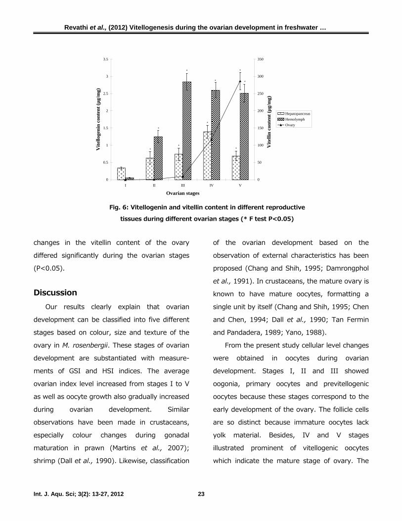
Assessment of vitellogenesis
Vitellogenin content in both hepatopancreas
and hemolymph varied during ovarian develop-
ment (Fig. 6). Vitellogenin content in the
hepatopancreas increased gradually from stages
I to III with marginal increase at stage IV
(1.39±0.29 µg/g) of ovarian development.
However, the vitellogenin content decreased to
0.69±0.14 µg/g at stage V. The vitellogenin level
in the hemolymph showed a gradual increase
from stage I to stage III of the ovarian
development. Thereafter, the vitellogenin cont-
ent decreased (2.60±0.32 µg/ml) at stage IV
and reached 2.51±0.36 µg/ml at stage V of
ovarian development. The variations in the
vitellogenin content in hepatopancreas and hem-
olymph differed significantly during ovarian
stages (P<0.05).
The vitellin content in the ovary increased
from early to final stages of ovarian development
(Fig. 6). In stages I and II, the vitellin content
increased gradually from 0.05 ± 0.01 µg/g to
0.27 ± 0.10 µg/g. However, there was an abrupt
increase observed from stage III (9.40 ± 1.53
µg/g), IV (117.18 ± 15.85 µg/g) and V (286.34
± 24.93 µg/g) of ovarian development. The final
stages (IVth and Vth) represented the vitellogenic
stage of the ovary with fully mature oocytes. The
details of this are described elsewhere. The
Revathi et al., (2012) Vitellogenesis during the ovarian development in freshwater …
Int. J. Aqu. Sci; 3(2): 13-27, 2012 22
**
**
*
*
*
0
50
100
150
200
250
I II III IV V
Ovarian stages
Prot
ein
cont
ent (
mg/
g)
HepatopancreasHemolymphOvary
Fig. 4: Protein content in different reproductive tissues during
different ovarian Stages (* F test P<0.05)
*
**
**
0
50
100
150
200
250
I II III IV V
Ovarian stages
Lip
id c
onte
nt (m
g/g)
HepatopancreasOvary
Fig. 5: Lipid content in different reproductive tissues during
different ovarian stages (* F test P<0.05)
Revathi et al., (2012) Vitellogenesis during the ovarian development in freshwater …
Int. J. Aqu. Sci; 3(2): 13-27, 2012 23
**
*
*
*
**
*
*
*
0
0.5
1
1.5
2
2.5
3
3.5
I II III IV V
Ovarian stages
Vite
lloge
nin
cont
ent (
µg/m
g)
0
50
100
150
200
250
300
350
Vite
llin
cont
ent (
µg/m
g)
HepatopancreasHemolymphOvary
Fig. 6: Vitellogenin and vitellin content in different reproductive
tissues during different ovarian stages (* F test P<0.05)
changes in the vitellin content of the ovary
differed significantly during the ovarian stages
(P<0.05).
Discussion Our results clearly explain that ovarian
development can be classified into five different
stages based on colour, size and texture of the
ovary in M. rosenbergii. These stages of ovarian
development are substantiated with measure-
ments of GSI and HSI indices. The average
ovarian index level increased from stages I to V
as well as oocyte growth also gradually increased
during ovarian development. Similar
observations have been made in crustaceans,
especially colour changes during gonadal
maturation in prawn (Martins et al., 2007);
shrimp (Dall et al., 1990). Likewise, classification
of the ovarian development based on the
observation of external characteristics has been
proposed (Chang and Shih, 1995; Damrongphol
et al., 1991). In crustaceans, the mature ovary is
known to have mature oocytes, formatting a
single unit by itself (Chang and Shih, 1995; Chen
and Chen, 1994; Dall et al., 1990; Tan Fermin
and Pandadera, 1989; Yano, 1988).
From the present study cellular level changes
were obtained in oocytes during ovarian
development. Stages I, II and III showed
oogonia, primary oocytes and previtellogenic
oocytes because these stages correspond to the
early development of the ovary. The follicle cells
are so distinct because immature oocytes lack
yolk material. Besides, IV and V stages
illustrated prominent of vitellogenic oocytes
which indicate the mature stage of ovary. The
Revathi et al., (2012) Vitellogenesis during the ovarian development in freshwater …
Int. J. Aqu. Sci; 3(2): 13-27, 2012 24
follicle cells are not so distinct because fully
mature oocytes fill up the entire ovary. Similar
observation has been reported in crustaceans,
oocyte cellular changes during gonadal
maturation in prawn (Martins et al., 2007).
Developing oocytes have a uniform structural
unit with the wall made up of thin layers of
follicle cells in Penaeus monodon (Tan Fermin
and Pandadera, 1989) and the deep sea shrimp
Aristaeo morpha (Kao et al., 1999). Follicle cells,
surrounding mature oocytes, are visible during
the initial vitellogenesis (stage III), as mentioned
by other authors (Chang and Shih, 1995). In
stages IV and V, follicle cells do not appear to be
enlarged, although this fact may result from the
stretching of its cytoplasm due to a substantial
increase of its cytoplasm (Van Herp and Payen,
1991).
The present results clearly explain that the
biochemical contents in tested reproductive
tissues varied during ovarian development. The
ovarian vitellin content and total protein content
were closely associated in the ovarian develop-
mental stages. Besides, lipid content also
fluctuated in the tested tissues during the
ovarian development. The oocyte development
correlated to the hemolymph vitellogenin content
in M. rosenbergii. In agreement with the present
results, Chang and Shih (1995) reported the
accumulation of vitellin content in the ovary from
stages I to V. The transfer in protein and lipid
contents from hepatopancreas to ovary, through
hemolymph in Crangon crangon supports the
hypothesis that organic reserves stored in the
hepatopancreas are transported to the ovary
through hemolymph during gonadal maturation.
Vitellogenesis involves the synthesis of numerous
components in the oocytes of crustaceans (Krol
et al., 1992). Protein and lipid contents
synthesized into more complex molecules
variously called vitellogenin (Croisille et al.,
1974), lipovitellin (Paulus and Laufer, 1982) and
high-density lipoproteins (Lee and Puppione,
1988). They originate from ingested food either
directly or after storage in the hepatopancreas
and must be transported via the hemolymph as
lipoproteins (Allen, 1972).
Our results clearly indicated that the
appearance of vitellogenin in immature female
hemolymph prior to gonadal development indica-
tes that an extra ovarian site may be involved in
vitellogenin synthesis. Increased hemolymph
vitellogenin levels at early vitellogenic stage
could also attribute vitellogenin synthesis and
release from the extra ovarian site. Vitellogenin
content in the hemolymph increased gradually to
reach a peak at stage III and declined at stage
IV in M .rosenbergii. The vitellogenin content
increased from 0.05 ± 0.01 µg/ml at the
beginning of the reproductive cycle to a
maximum level of 2.84 ± 0.35 µg/ml in stage III
and then declined sharply before spawning in M.
rosenbergii. A similar pattern has been reported
in M. nipponense (Vg range 1-9 mg/ml)
(Okumura et al., 1993) and H. americanus (Vg
range 0-12 mg/ml) (Byard and David, 1984).
Revathi et al., (2012) Vitellogenesis during the ovarian development in freshwater …
Int. J. Aqu. Sci; 3(2): 13-27, 2012 25
Similar results are reported from several
crustacean species, with a substantial increase of
vitellogenin content in the hemolymph during
vitellogenesis (Lee, 1991; Okumura et al., 1993;
Quackenbush, 1989; Vafopoulou and Steel,
1995). Although the site of vitellogenin synthesis
is still controversial, some evidence indicates that
the hepatopancreas is one of the possible sites
(Castille and Lawrence, 1989; Paulus and Laufer,
1987). Increase in carbohydrate, protein and
lipid content of the hepatopancreas during
gonadal maturation is an indicator of the extent
of glycogen and lipoprotein synthesis (Castille
and Lawrence, 1989; Quackenbush, 1989).
However, vitellin has been shown to be
synthesized in the ovary during stages of ovarian
maturity in invertebrates (Paulus and Laufer,
1987; Quackenbush, 1989).
This study demonstrated that the
classification of ovarian development based to
the colour, size and texture of the ovary and the
identification of the extra ovarian synthesis site.
On the other hand, it was evidenced that
biochemical constituents are also closely
associated with the ovarian development.
Vitellogenesis as a biomarker of female
reproductive activity, which indicate that the
vitellin accumulation gradually increased in
oocytes during ovarian development. Besides,
the vitellogenin content fluctuated in different
reproductive tissues during the ovarian
development in M. rosenbergii.
Acknowledgements Financial assistance from UGC- Dr. D. S.
Kothari Post Doctoral Fellowship to Dr. P. Revathi
is gratefully acknowledged. Thanks are due to
Mr. G.S. Samarasam for providing a hatchery
facility for the experiments.
Reference Allen W.V. (1972) Lipid transport in the Dungenes crab,
Cancer magister. Comp. Biochem. Physiol., 43: 193-207.
Adiyadi Subramonium T. (1983) Arthropoda-Crustacea, In:
Reproductive biology of invertebrates - Oogenesis,
Oviposition and Oosorption. K.G. Adiyodi and R.G. Adiyodi
eds., John Wiley and Sons, New York. pp. 443-495.
Anilkumar G. and Adiyodi K.G. (1980) Ovarian growth,
induced by eyestalk ablation during the prebreeding
season, is not normal in the crab, Paratelphusa
hydrodromous. Inter. J. Invert. Repro. Develop., 2: 95-
105.
Arculeo M.G. Payen A.G., Cuttita Galioto T. and Riggio S.
(1995) A survey of ovarian maturation in a population of
Aristeus antennatus (Crustacea: Decapoda). Ani. Biol. 4,
13-18.
Bradford M.M. (1976) A rapid and sensitive method for the
qualification of microgram quantities of protein utilizing the
principle of protein-dye binding. Anal. Biochem., 72: 248-
254.
Byard H.E. and David E. (1984) The relationship between
molting, reproduction and a hemolymph female-specific
protein in the lobster, Homarus americanus. Comp.
Biochem. Physiol ., 77: 749-757.
Byrne B.M. Gruber M. and AB E. (1989) The evolution of
egg yolk proteins. Prog. Biophys. Molec. Biol., 53: 33-69.
Castille F.L. and Lawrence A.L. (1989) Relationship
between maturation and biochemical composition of the
gonads and digestive glands of the shrimps Penaeus
aztecus and Penaeus setiferus (L). J. Crust. Biol., 9: 202-
211.
Revathi et al., (2012) Vitellogenesis during the ovarian development in freshwater …
Int. J. Aqu. Sci; 3(2): 13-27, 2012 26
Chang H.H. and Bradley J.T. (1983) Vitellogenin synthesis
and secretion in ovariectomized crickets. Comp. Biochem.
Physiol., 75B: 733-737
Chang C.F. and Shih T.W. (1995) Reproductive cycle of
ovarian development and vitellogenin profiles in the
freshwater prawn Macrobrachium rosenbergii. Invert.
Reprod. Dev., 27: 11-20.
Chaves P.T.C. and Magalhaes C. (1993) The oocytes
development of Macrobrachium amazonicum (Heller,
1862), a freshwater shrimp of the Amazon Region
(Crustacea: Deacapods; Palaemonidae). Acta. Amazonica.,
23: 17-23.
Chen C.C. and Chen S.N. (1994) Vitellogenesis in the giant
tiger shrimp Penaeus monodon. Comp. Biochem. Physiol.,
107: 453-460.
Chen J.S. Sappington T.W. and Raikhel A.S. (1997)
Extensive sequence conservation among insect, nematode,
and vertebrate vitellogenin reveals ancient common
ancestry. J. Mol. Evol., 44: 440–451.
Chen Y.N. Tseng D.Y. Ho P.Y. and Kuo C.M. (1999) Site of
vitellogenin synthesis determined from a cDNA encoding a
vitellogenin fragment in the freshwater giant prawn,
Macrobrachium rosenbergii. Mol. Repro. Develop., 54:
215-222.
Croisille Y. Junera H. Meusy J.J. and Charniaux-cotton H.
(1974) The female specific protein (vitellogenin) in
crustaceans with particular reference to Orchestia
gammarella (Amphipoda). Amer. Zool., 14: 1219-1228.
Dall W. Hill B.J. Rothlisberg P.C. and Sharples D.J. (1990)
The biology of the Penaeidae. In: Advances in Marine
Biology, Blaxter, J.H.S and Southward, A.J. (Eds.,).
Academic press, New York, p: 489.
Damrongphol P. Eangchuan N. and Poolsanguan N. (1991)
Spawning cycle and oocyte maturation in laboratory-
maintained giant freshwater prawn (Macrobrachium
rosenbergii). Aquaculture, 95: 347-357.
Dehn P.F. Aiken D.E. and Waddy S. (1983) Aspects of
vitellogenesis in the lobster Homarus americanus.
Canadian Techn. Rep. Fish. Aquat. Sci., 1161: 1–24.
Eastman-Reks S.B. and Fingerman M. (1985) In vitro
synthesis of vitellin by the ovary of the fiddler crab, Uca
pugilator. J. Exp. Zool., 233: 111–116.
Folch J. Lee M. and Bloane-Stanley M. (1957) A simple
method for the isolation and purification to total from
animal tissues. J. Biol. Chem. 266: 497–509.
Fyffe W.E. and OʼConnor J.D. (1974) Characterization and
quantification of a crustacean lipovitellin. Comp. Biochem.
Physiol., 48B: 389–399.
Htun-Han M. (1978) The reproductive biology of the dab
Limanda limanda (L) in the North Sea: Seasonal changes
in ovary. J. Fish. Bio., 13: 119-123.
Kao H.C. Chan T.Y. and Yu H.P. (1999) Aspects of
enrolment of CHH cell activity and hemolymph glucose
levels in crayfish. Biol. Bull., 175: 137-143.
Khayat M. Shenker O. Funkenstein B. Tom M. Lubzens E.
and Tietz A. (1994) Fat transport in the penaeid shrimp
Penaeus semisulcatus (de Haan). Isr. J. Aqua., 46 (1): 22–
32.
Krol R.M. Hawkins W.E. and Overstreet R.M. (1992)
Reproductive components. In: Microscopical Analysis of
Invertebrates. Harrison, F.W and Humes, A.G (Eds.).
Wiley-Liss, New York, pp: 295–343.
Lee R.F. and Puppione D.L. (1988) Lipoprotein I and II
from the hemolymph of the blue crab Callinectes sapidus:
Lipoprotein II associated with vitellogenesis. J. Exp. Zool.,
218: 278–289.
Lee R.F. (1991) Lipoproteins from the hemolymph and
ovaries of marine invertebrates. In: Advances in
comparative and environmental physiology, R. Gilles (Ed.).
Springer Verlag, Heidelberg, Germany, 7, pp: 187-207.
Lui C.W. and OʼConnor J.D. (1976) Biosynthesis of
crustacean lipovitellin. The incorporation of labeled amino
acids into the purified lipovitellin of the crab Pachigrapsus
crassipes. J. Exp. Zool., 199: 105–108.
Martins J. Karina R.T. Rangel-Figueiredo R. and Coimbra J.
(2007) Reproductive cycle, ovarian development, and
vertebrate-type steroids profile in the freshwater prawn
Macrobrachium rosenbergii. J. Crust. Biol., 27(2): 220-
228.
Okumura T. Han CH. Suzuki Y. Aida K. and Hanyu I.
(1993) Changes in haemolymph vitellogenin and
ectysteroid levels during the reproductive and non-
Revathi et al., (2012) Vitellogenesis during the ovarian development in freshwater …
Int. J. Aqu. Sci; 3(2): 13-27, 2012 27
reproductive moult cycles in the fresh water prawn,
Macrobrachium nipponense. Zool., Sci. 9: 37-45.
Okumura T. and Aida K. (2000) Hemolymph vitellogenin
levels and ovarian development during the reproductive
and non-reproductive molt cycles in the giant freshwater
prawn Macrobrachium rosenbergii. Fish. Sci., 66, 678-685.
Paulus J.E. and Laufer H. (1982) Vitellogenesis in the
hepatopancreas and ovaries of Carcinus maenas. Biol.
Bull., 163: 375–376.
Paulus J.E. and Laufer H. (1987) Vitellogenesis in
hepatopancreas of Carcinus maenas and Libinia
emarginata. Int. J. Invert. Reprod. Dev., 11: 29-44.
Peixoto S. Cavalli R.O. and Asielesky W. (2005) Recent
developments on broodstock maturation and reproduction
of Farfantepenaeus paulensis. Bra. Arch. Biol. Technol.,
48(6): 997-1006.
Quackenbush L.S. (1989) Vitellogenesis in the shrimp
Penaeus vannamei: In vitro studies of the isolated
hepatopancreas and ovary. Comp. Biochem. Physiol. 94B
(2) : 253-261.
Raikhel A.S. and Dhadialla T.S. (1992) Accumulation of
yolk proteins in insect oocytes. Annu. Rev. Entomol., 37,
217-251.
Rani Subramoniam T. (1997) Vitellogenesis is the mud-
crab Scylla serrata – An in vivo isopode study. J. Crust.
Biol., 17(4) : 659-665.
Revathi P. (2010) Studies on the endocrine disruptor and
its impact on the reproductive physiology of the freshwater
prawn Macrobrachium rosenbergii (De Man). Ph. D thesis,
University of Madras, Chennai, Tamil Nadu, India.
Sagi A. Soroka E. Chomsky O. Calderon J. and Milner Y.
(1995) Ovarian protein synthesis in the prawn
Macrobrachium rosenbergii: does ovarian vitellin synthesis
exist? Invert. Repro. Develp., 27: 41-47.
Suzuki S. (1987) Vitellins and vitelligenins of the terrestrial
isopod, Armadillidium vulgare. Biol. Bull. 173: 345-354.
Tan Fermin J.D. and Pandadera R.A. (1989) Ovarian
maturation stages of the wild giant tiger prawn, Penaeus
monodon. Aquaculture, 77: 229-242.
Tsukimura B. Bender J.S. and Linder C.J. (2000)
Developmental aspects of gonadal regulation in the
ridgeback shrimp, Sicyonia ingentis. Comp. Biochem.
Physiol., 127A: 215-224.
Tsukimura B. (2001) Crustacean vitellogenesis: its role in
oocyte development. Amer. Zool., 41: 465- 476.
Van Herp F. and Payen G.G. (1991) Crustacean
neuroendocrinology: perspectives for the control of
reproduction in aquacultural systems. Bull. Inst. Zool., 16:
513-539.
Vafopoulou, X. and Steel C.G.H. (1995) Vitellogenesis in
the terrestrial isopod, Oniscus asellus (L): Characterization
of vitellin and vitellogenesis and changes in their synthesis
throughout the intermolt cycle. Invert. Reprod. Dev., 28:
87-95.
Yano I. (1988) Oocyte development in the kuruma prawn,
Penaeus japonicus. Mar. Biol., 99: 547-553. Zhang I.L., Zuo Z.H., Chen Y.X., Zhao Y., Hu S. and Wang
C.G. (2007) Effect of tributyltin on the development of
ovary in female cuvier (Sebastiscus marmoratus).
Aquacult. Toxicol., 83: 174-179.