vivasens 09 2010 englisch - dentalcompare.com
TRANSCRIPT
Scientific Documentation
Scientific Documentation VivaSens® Page 2 of 26
Table of contents
1. Introduction......................................................................................................................3
1.1 The hydrodynamic theory of pain ............................................................................................3
1.2 How does VivaSens work?........................................................................................................4
2. Technical data..................................................................................................................6
3. In-vitro investigations......................................................................................................7
3.1 Sealing of dentin tubuli..............................................................................................................7
3.1.1 Electron microscopy............................................................................................................7
3.1.2 Confocal laser scanning microscopy ..................................................................................8
3.1.3 The reduction of dentin permeability...................................................................................9
3.2 Compatibility of VivaSens with restorative materials ..........................................................11
3.2.1 Effect of VivaSens on dentin bond strength using bovine molars ....................................12
3.2.2 Effect of VivaSens on dentin bond strength using human molars ....................................13
3.2.3 Effect of VivaSens on marginal leakage of amalgam restorations ...................................14
3.3 Use of VivaSens in combination with bleaching treatments...............................................14
3.3.1 Influence of desensitizers on the outcome of bleaching treatments.................................14
3.3.2 Protection from dentin dehydration due to bleaching treatment .......................................15
4. Clinical investigations ...................................................................................................17
4.1 Reduction in dentin sensitivity ...............................................................................................17
4.1.1 Dr. Arnd Peschke, Internal Clinic, R&D Ivoclar Vivadent, Schaan, Liechtenstein............17
4.1.2 Prof. Dr. Andrej Kielbassa, University of Berlin, Germany ...............................................18
4.1.3 Prof. Dr. Steven Duke, Indiana University, Indianapolis, USA .........................................18
4.1.4 Dr. Lenka Roubalikova, Brno, Czech Republic.................................................................19
4.1.5 Dr. Tijen Pamir, Ege University, Izmir, Turkey..................................................................20
4.1.6 Reduction in dentin sensitivity in bleaching treatments ....................................................20
4.1.7 Influence on root dentin demineralization .........................................................................21
5. Toxicology......................................................................................................................23
5.1 Introduction ..............................................................................................................................23
5.2 Toxicity of components ...........................................................................................................23
5.3 Mutagenicity .............................................................................................................................23
5.4 Irritation and sensitization ......................................................................................................23
5.5 Conclusion................................................................................................................................24
5.6 Literature on toxicology ..........................................................................................................24
6. Literature ........................................................................................................................24

Scientific Documentation VivaSens® Page 3 of 26
1. Introduction
Dentin hypersensitivity has been defined as a “short, sharp pain arising from exposed dentin in response to stimuli, typically thermal, evaporative, tactile, osmotic or chemical and which cannot be ascribed to any other form of dental defect or pathology” (Holland et al. 1997). The pain usually subsides soon after the stimulus has disappeared. It is therefore important not to confuse dentin hypersensitivity with persistent tooth ache, which is usually related to a pathological state of the tooth structure. (For reviews see Borodowski et al. 2003; Pashley 1994; Markowitz 1993)
Several studies have reported that between 5–57% of the adult population suffer from hypersensitivity in one way or another (Dababneh et al. 1999). Hypersensitive dentin is a problem in the daily life of patients and for the dentist during dental treatment. Many patients are familiar with the unpleasant sting experienced when the dentist suddenly applies cold water or air to the teeth during treatment. Hypersensitive dentin is also common after the placement of new direct or indirect restorations. In daily life, hypersensitivity may occur whilst consuming cold drinks, eating ice cream or chocolate, when rinsing the mouth after cleaning the teeth, or whilst inhaling cold air through the mouth.
1.1 The hydrodynamic theory of pain
Several theories have been proposed to explain the mechanism of dentin sensitivity and therefore of dentin hypersensitivity (Dowell et al. 1983). Of these, the most widely accepted is the hydrodynamic theory of sensitivity (Brännström et al. 1967). This theory postulates that rapid bi-directional shifts of the fluids within the dentin tubuli, following the application of a stimulus, result in the activation of the tooth’s sensory nerves. Essentially, certain stimuli create a pressure change across the dentin, which can excite individual intradental nerves. Studies performed in vivo revealed that the response of the pulpal nerves was proportional to the pressure and therefore the rate of fluid flow.
Interestingly, stimuli, such as cold, which cause fluid to flow away from the pulp, produce more rapid and greater pulp-nerve responses than those, such as heat, which cause an inward flow. This helps explain the tendency for a more rapid and severe response to cold stimuli compared to the slow dull response to heat. The exact mechanism by which the fluid flow stimulates pulpal nerves is not known with any certainty, however from animal experiments a mechano-receptor response is suggested (Matthews et al. 1994). The pressure change across dentin distorts the pain receptors at the pulp dentin border. This would be similar to the activation of touch sensitive nerves around hair follicles due to light pressure on the protruding hair.
It should be mentioned, however, that not all exposed dentin is sensitive. In a study to determine differences between sensitive and non-sensitive teeth, Absi et al. reported that non-sensitive teeth were unresponsive to any stimuli and had very few exposed tubuli (Absi et al. 1987). In contrast, sensitive teeth had much greater numbers of open tubuli per unit area (8 times as many tubuli at the root surface as non-sensitive teeth). Similarly, the average diameter of tubuli in sensitive teeth was almost 2 times greater than that of tubuli in non-sensitive teeth (0.83 µm vs. 0.4 µm). According to Poiseuille’s law, which states that fluid flow is proportional to the fourth power of the radius, diameter differences alone would indicate that the fluid flow in tubuli of hypersensitive teeth should be 16 (i.e. 24) times greater than that of fluid in non-sensitive teeth. Combining the increased number of open tubuli with increased diameter in sensitive teeth, it can be postulated that the fluid flow in sensitive teeth is approximately 100 times greater than in non-sensitive teeth.
A scanning electron microscope study, based on replica models of hypersensitive and non-sensitive dentin, showed that in hypersensitive dentin the smear layer was thinner, differed in structure and was probably under-calcified compared to non-sensitive dentin (Rimondini et al. 1995). These findings appear consistent with the hydrodynamic theory. The greater
Scientific Documentation VivaSens® Page 4 of 26
Pain
number of open and wider tubuli at the dentin surface would enhance fluid permeability through dentin and as such increase the possibility for stimulus transmission and subsequent pain response.
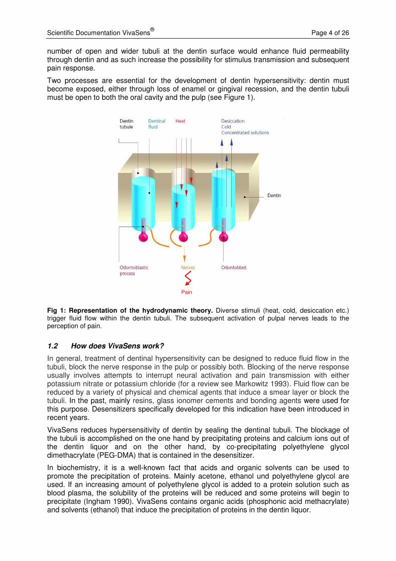
Two processes are essential for the development of dentin hypersensitivity: dentin must become exposed, either through loss of enamel or gingival recession, and the dentin tubuli must be open to both the oral cavity and the pulp (see Figure 1).
Fig 1: Representation of the hydrodynamic theory. Diverse stimuli (heat, cold, desiccation etc.) trigger fluid flow within the dentin tubuli. The subsequent activation of pulpal nerves leads to the perception of pain.
1.2 How does VivaSens work?
In general, treatment of dentinal hypersensitivity can be designed to reduce fluid flow in the tubuli, block the nerve response in the pulp or possibly both. Blocking of the nerve response usually involves attempts to interrupt neural activation and pain transmission with either potassium nitrate or potassium chloride (for a review see Markowitz 1993). Fluid flow can be reduced by a variety of physical and chemical agents that induce a smear layer or block the tubuli. In the past, mainly resins, glass ionomer cements and bonding agents were used for this purpose. Desensitizers specifically developed for this indication have been introduced in recent years.
VivaSens reduces hypersensitivity of dentin by sealing the dentinal tubuli. The blockage of the tubuli is accomplished on the one hand by precipitating proteins and calcium ions out of the dentin liquor and on the other hand, by co-precipitating polyethylene glycol dimethacrylate (PEG-DMA) that is contained in the desensitizer.
In biochemistry, it is a well-known fact that acids and organic solvents can be used to promote the precipitation of proteins. Mainly acetone, ethanol und polyethylene glycol are used. If an increasing amount of polyethylene glycol is added to a protein solution such as blood plasma, the solubility of the proteins will be reduced and some proteins will begin to precipitate (Ingham 1990). VivaSens contains organic acids (phosphonic acid methacrylate) and solvents (ethanol) that induce the precipitation of proteins in the dentin liquor.

Scientific Documentation VivaSens® Page 5 of 26
A second mode of action is the acid induced formation of salts. The dentinal fluid is rich in calcium ions. The phosponic acid methacrylate contained in VivaSens forms Ca-salts of low solubility, thus forming precipitates in the tubuli. The second acid component of the desensitizer, methacrylate modified polyacrylic acid, is a complex builder that leads to an additional formation of salts. The potassium ions of the fluoride component act supportively in the precipitation of the salts.
Finally, surface blockage of the tubuli is achieved, directly after the application of VivaSens as a hydroxypropyl cellulose film is built up. This film transiently seals the dentin tubuli, prevents dentinal fluid flow, and thus stops stimulation of nerves and the subsequent perception of pain (see Figure 2).
Fig 2: Working principle of VivaSens. Application of the desensitizer VivaSens to exposed dentinal surfaces blocks dentin tubuli and hence reduces fluid flow and tooth hypersensitivity.
Pain
Scientific Documentation VivaSens® Page 6 of 26
2. Technical data
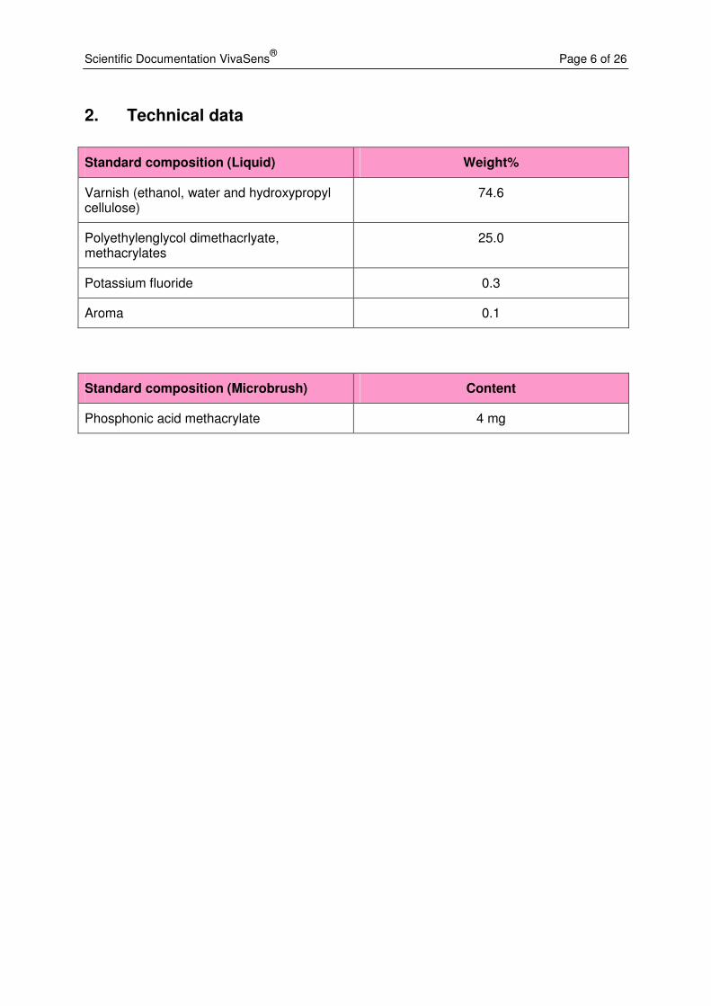
Standard composition (Liquid) Weight%
Varnish (ethanol, water and hydroxypropyl cellulose)
74.6
Polyethylenglycol dimethacrlyate, methacrylates
25.0
Potassium fluoride 0.3
Aroma 0.1
Standard composition (Microbrush) Content
Phosphonic acid methacrylate 4 mg

Scientific Documentation VivaSens® Page 7 of 26
3. In-vitro investigations
The primary action of VivaSens in hypersensitivity treatment is the clogging of the dentin tubuli via precipitating soluble components of the dentin liquor (e.g. proteins, Ca2+ ions). The sealing effect is enhanced by co-precipitating PEG-derivatives contained in VivaSens. In-vitro tests have been conducted to investigate the penetration depth of VivaSens into dentin tubuli and the formation of plugs/seals. The ability of VivaSens to reduce dentin permeability was also investigated. Moreover, the compatibility of VivaSens with restorative materials was examined, as VivaSens may also be used as a cavity liner. Dentin hypersensitivity occurs often after bleaching treatments; thus the effect of VivaSens on bleaching treatments was studied as well.
3.1 Sealing of dentin tubuli
3.1.1 Electron microscopy
Aim: The formation of precipitates within the dentinal tubuli after the application of VivaSens was investigated using electron microscopy.
Investigator: Ivoclar Vivadent AG, Research and Development, Schaan, Liechtenstein
Method: Bovine teeth were used in this study. A flat dentin surface was prepared on the occlusal side of each tooth using SiC paper (grit 120/1000). After cleaning and drying, the teeth were etched with Email Preparator and VivaSens was applied for 30 seconds. The teeth were dried extensively and analyzed by electron microscopy.
VivaSens Untreated
Fig. 3: Blocking of dentin tubuli. Treatment with VivaSens (left side) leads to a visible clogging of the tubuli, whereas the tubuli in the untreated control dentin are still open (right side). A & B: surface view; C & D: longitudinal view. Scanning microscopy images (SEM).
D C
B A
Scientific Documentation VivaSens® Page 8 of 26
Results: Figure 3 shows the SEM pictures of treated (A and C) and untreated (B and D) samples. Both the surface view (upper row) and the longitudinal view (lower row) of the tubuli demonstrate the clogging of the tubuli in the samples treated with VivaSens.
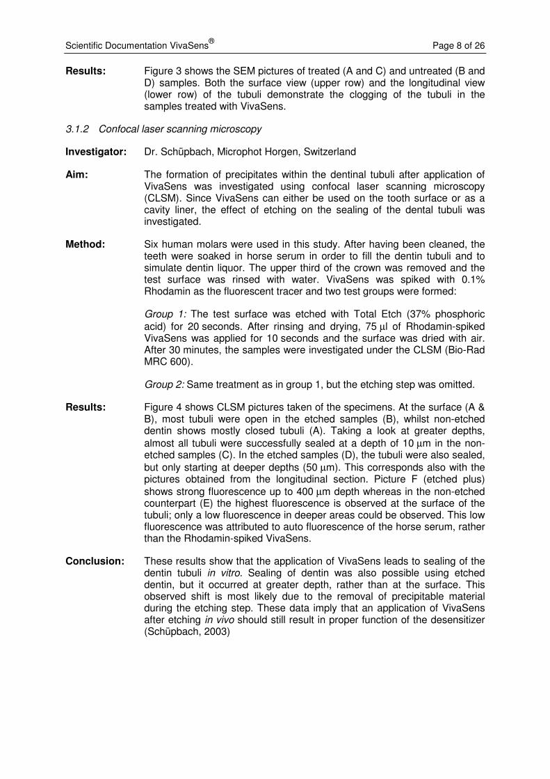
3.1.2 Confocal laser scanning microscopy
Investigator: Dr. Schüpbach, Microphot Horgen, Switzerland
Aim: The formation of precipitates within the dentinal tubuli after application of VivaSens was investigated using confocal laser scanning microscopy (CLSM). Since VivaSens can either be used on the tooth surface or as a cavity liner, the effect of etching on the sealing of the dental tubuli was investigated.
Method: Six human molars were used in this study. After having been cleaned, the teeth were soaked in horse serum in order to fill the dentin tubuli and to simulate dentin liquor. The upper third of the crown was removed and the test surface was rinsed with water. VivaSens was spiked with 0.1% Rhodamin as the fluorescent tracer and two test groups were formed:
Group 1: The test surface was etched with Total Etch (37% phosphoric acid) for 20 seconds. After rinsing and drying, 75 µl of Rhodamin-spiked VivaSens was applied for 10 seconds and the surface was dried with air. After 30 minutes, the samples were investigated under the CLSM (Bio-Rad MRC 600).
Group 2: Same treatment as in group 1, but the etching step was omitted.
Results: Figure 4 shows CLSM pictures taken of the specimens. At the surface (A & B), most tubuli were open in the etched samples (B), whilst non-etched dentin shows mostly closed tubuli (A). Taking a look at greater depths, almost all tubuli were successfully sealed at a depth of 10 µm in the non-etched samples (C). In the etched samples (D), the tubuli were also sealed, but only starting at deeper depths (50 µm). This corresponds also with the pictures obtained from the longitudinal section. Picture F (etched plus) shows strong fluorescence up to 400 µm depth whereas in the non-etched counterpart (E) the highest fluorescence is observed at the surface of the tubuli; only a low fluorescence in deeper areas could be observed. This low fluorescence was attributed to auto fluorescence of the horse serum, rather than the Rhodamin-spiked VivaSens.
Conclusion: These results show that the application of VivaSens leads to sealing of the dentin tubuli in vitro. Sealing of dentin was also possible using etched dentin, but it occurred at greater depth, rather than at the surface. This observed shift is most likely due to the removal of precipitable material during the etching step. These data imply that an application of VivaSens after etching in vivo should still result in proper function of the desensitizer (Schüpbach, 2003)
Scientific Documentation VivaSens® Page 9 of 26
Without etching Etched
Fig. 4: VivaSens penetration on etched and non-etched dentin. Dentin samples were etched with phosphoric acid (left column) or not etched (right column). VivaSens spiked with the fluorescent dye rhodamine was applied to the samples. Analysis was performed using confocal laser scanning microscopy Confocal laser scanning microscopy images (CLSM). A, B: surface; C: 10 µm, D: 50 µm, E & F: longitudinal view. VivaSens penetrates both etched and un-etched samples; blocking starts only at greater depths in etched samples.
3.1.3 The reduction of dentin permeability
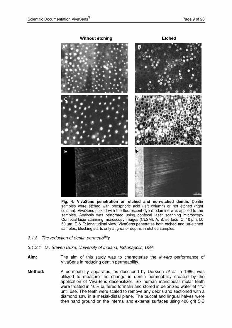
3.1.3.1 Dr. Steven Duke, University of Indiana, Indianapolis, USA
Aim: The aim of this study was to characterize the in-vitro performance of VivaSens in reducing dentin permeability.
Method: A permeability apparatus, as described by Derkson et al. in 1986, was utilized to measure the change in dentin permeability created by the application of VivaSens desensitizer. Six human mandibular molar teeth were treated in 10% buffered formalin and stored in deionized water at 4°C until use. The teeth were scaled to remove any debris and sectioned with a diamond saw in a mesial-distal plane. The buccal and lingual halves were then hand ground on the internal and external surfaces using 400 grit SiC
C
A
D
F E
B
Scientific Documentation VivaSens® Page 10 of 26
0
20
40
60
80
100
120
Before AfterPe
rme
ab
ilit
y (
arb
itra
ry u
nit
s)
paper. The roots were removed and twelve 1.5 - 2.0 mm thick slabs consisting of dentin and occlusal enamel were thus obtained. The slabs were immersed in deionized water and placed in an ultrasonic cleaner for 12 minutes. A 17% EDTA solution was placed on each surface to remove the smear layer. The slabs were then secured on the acrylic block of the test apparatus (Dental Ventures of America, Anaheim Hills, CA). The test was run using a 0.2% fluorescein dye solution and a nitrogen tank providing 10 psi to the fluid reservoir. An ultraviolet light and 2-fold magnification were used to determine the movement of a bubble in the fluid, indicative of fluid permeability through the dentin slab. The distance over which the bubble moved in 1 minute of time was recorded. This was treated as the control. The exposed surface of the slab was then treated by gently rubbing VivaSens onto the surface for 10 seconds. The test was run again and the movement of the bubble recorded and a percent reduction noted for each specimen.
Fig. 5: Reduction of dentin permeability after application of VivaSens. Permeability of dentinal slabs was measured using a method according to Derkson et al. Application of VivaSens lead to a significant reduction in permeability.
Results: The mean percentage reduction for the group (n = 12) was 69 ± 19 (Fig. 5). The data failed the normality test, thus a Wilcoxon Signed Rank test was performed. The observed reduction in permeability was significant (p < 0.001).
Conclusion: These data show that VivaSens is able to substantially reduce dentin permeability in vitro (Duke 2002).
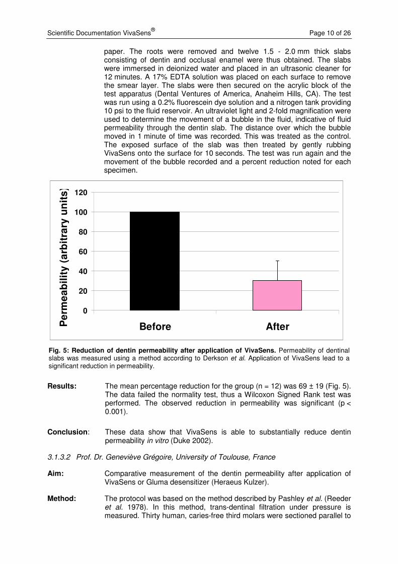
3.1.3.2 Prof. Dr. Geneviève Grégoire, University of Toulouse, France
Aim: Comparative measurement of the dentin permeability after application of VivaSens or Gluma desensitizer (Heraeus Kulzer).
Method: The protocol was based on the method described by Pashley et al. (Reeder et al. 1978). In this method, trans-dentinal filtration under pressure is measured. Thirty human, caries-free third molars were sectioned parallel to
Scientific Documentation VivaSens® Page 11 of 26
the occlusal surface above the pulp cavity. A 1 mm disc of dentin was prepared from each tooth. The resulting 30 dentin discs were randomly allocated to 3 groups (n=10 for each group). The discs were then glued onto a polycarbonate disc with a central perforation. The initial hydraulic conductance was determined as a reference for the subsequent measurements, which are expressed as percentage of the initial hydraulic conductance. Thus, each sample served as its own control. After reference measurements, the desensitizers were applied according to the instructions for use: VivaSens was applied to dry dentin and rubbed in for 10 seconds. The desensitizer was uniformely spread over the surface, dried and light cured for 20 seconds. Gluma desensitizer was applied, allowed to react for 60 seconds and dried and light cured for 20 seconds. Measurements were recorded every 30 seconds for 15 minutes; means and standard deviations were calculated for each sample and analyzed statistically (ANOVA, a posteriori tests).
Results: VivaSens reduced the dentin permeability by 29.78%, Gluma by 20.44% (see Figure 6). Thus, VivaSens provided a stronger sealing efficacy than Gluma (Grégoire 2003).
0
5
10
15
20
25
30
35
40
VivaSens Gluma
Re
du
cti
on
in
de
nti
n p
erm
ea
bilit
y [
%]
Fig. 6: Reduction of dentin permeability after application of desensitizers. Permeability of dentin discs was measured using a method described by Pashley et al. Shown is the relative reduction in dentin permeability before and after application of desensitizers. VivaSens lead to a higher reduction in permeability than Gluma.
3.2 Compatibility of VivaSens with restorative materials
Since VivaSens may be used as a cavity liner in conjunction with direct, temporary and permanent restoratives, the compatibility of VivaSens with such materials was investigated. Areas investigated included unintended adherence of temporaries and the retention of permanent restoratives. The dentin bond strength was evaluated on bovine teeth at Ivoclar
Scientific Documentation VivaSens® Page 12 of 26
Vivadent and in two independent studies on human molars at the universities of Indiana (USA) and Erlangen (Germany).
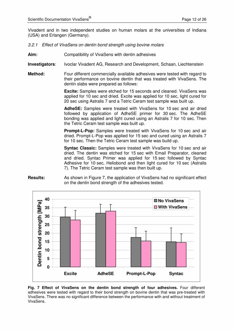
3.2.1 Effect of VivaSens on dentin bond strength using bovine molars
Aim: Compatibility of VivaSens with dentin adhesives
Investigators: Ivoclar Vivadent AG, Research and Development, Schaan, Liechtenstein
Method: Four different commercially available adhesives were tested with regard to their performance on bovine dentin that was treated with VivaSens. The dentin slabs were prepared as follows:
Excite: Samples were etched for 15 seconds and cleaned. VivaSens was applied for 10 sec and dried. Excite was applied for 10 sec, light cured for 20 sec using Astralis 7 and a Tetric Ceram test sample was built up.
AdheSE: Samples were treated with VivaSens for 10 sec and air dried followed by application of AdheSE primer for 30 sec. The AdheSE bonding was applied and light cured using an Astralis 7 for 10 sec. Then the Tetric Ceram test sample was built up.
Prompt-L-Pop: Samples were treated with VivaSens for 10 sec and air dried. Prompt-L-Pop was applied for 15 sec and cured using an Astralis 7 for 10 sec. Then the Tetric Ceram test sample was build up.
Syntac Classic: Samples were treated with VivaSens for 10 sec and air dried. The dentin was etched for 15 sec with Email Preparator, cleaned and dried. Syntac Primer was applied for 15 sec followed by Syntac Adhesive for 10 sec, Heliobond and then light cured for 10 sec (Astralis 7). The Tetric Ceram test sample was then built up.
Results: As shown in Figure 7, the application of VivaSens had no significant effect on the dentin bond strength of the adhesives tested.
0
5
10
15
20
25
30
35
40
Excite AdheSE Prompt-L-Pop Syntac
Den
tin
bo
nd
str
en
gth
[M
Pa] No VivaSens
With VivaSens
Fig. 7 Effect of VivaSens on the dentin bond strength of four adhesives. Four different adhesives were tested with regard to their bond strength on bovine dentin that was pre-treated with VivaSens. There was no significant difference between the performance with and without treatment of VivaSens.
Scientific Documentation VivaSens® Page 13 of 26
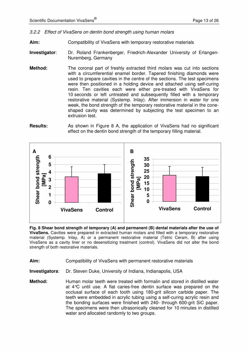
3.2.2 Effect of VivaSens on dentin bond strength using human molars
Aim: Compatibility of VivaSens with temporary restorative materials
Investigator: Dr. Roland Frankenberger, Friedrich-Alexander University of Erlangen-Nuremberg, Germany
Method: The coronal part of freshly extracted third molars was cut into sections with a circumferential enamel border. Tapered finishing diamonds were used to prepare cavities in the centre of the sections. The test specimens were then positioned in a holding device and attached using self-curing resin. Ten cavities each were either pre-treated with VivaSens for 10 seconds or left untreated and subsequently filled with a temporary restorative material (Systemp. Inlay). After immersion in water for one week, the bond strength of the temporary restorative material in the cone-shaped cavity was determined by subjecting the test specimen to an extrusion test.
Results: As shown in Figure 8 A, the application of VivaSens had no significant effect on the dentin bond strength of the temporary filling material.
0
1
2
3
4
5
6
VivaSens Control
Sh
ea
r b
on
d s
tre
ng
th
[MP
a]
0
5
10
15
20
25
30
35
VivaSens Control
Sh
ea
r b
on
d s
tre
ng
th
[MP
a]
Fig. 8 Shear bond strength of temporary (A) and permanent (B) dental materials after the use of VivaSens. Cavities were prepared in extracted human molars and filled with a temporary restorative material (Systemp. Inlay, A) or a permanent restorative material (Tetric Ceram, B) after using VivaSens as a cavity liner or no desensitizing treatment (control). VivaSens did not alter the bond strength of both restorative materials.
Aim: Compatibility of VivaSens with permanent restorative materials
Investigators: Dr. Steven Duke, University of Indiana, Indianapolis, USA
Method: Human molar teeth were treated with formalin and stored in distilled water at 4°C until use. A flat caries-free dentin surface was prepared on the occlusal surface of each tooth using 180-grit silicon carbide paper. The teeth were embedded in acrylic tubing using a self-curing acrylic resin and the bonding surfaces were finished with 240- through 600-grit SiC paper. The specimens were then ultrasonically cleaned for 10 minutes in distilled water and allocated randomly to two groups.
B A
Scientific Documentation VivaSens® Page 14 of 26
The first group was etched with Total Etch for 15 seconds. The surface was thoroughly cleaned and a generous amount of Excite light-cure adhesive was applied and cured. Each specimen was placed into a compression jig and Tetric Ceram was placed to form cylinders of 2 mm in height and 3.8 mm in diameter.
The second group was subjected to the same procedures as group 1 except that after etching with Total Etch, rinsing and drying, a 10-second application of VivaSens was carried out. This layer was air-dried before the application of Excite light-cure adhesive.
Specimens were stored at 36°C for one week. They were then thermocycled for 2500 cycles between 4°C and 48°C with a dwell time of 30 seconds and a transit time of 10 seconds. Specimens were then returned to 36°C distilled water until testing. Shear bond strength was determined using a stainless steel ring with a knife-edge along the inner circumference in an Instron Universal Testing machine. Fifteen teeth were used for each test group.
Results: As shown in Figure 8 B, the application of VivaSens had no significant effect on the dentin bond strength of the permanent filling material (p =0.771).
3.2.3 Effect of VivaSens on marginal leakage of amalgam restorations
Aim: To compare the marginal leakage of Class II amalgam restorations after lining of the cavities with either VivaSens, a self-etching adhesive (Clearfil S3 Bond, Kuraray Dental) or a copal varnish (Copalite, Copalite Dental Products).
Investigators: Dr. M. Ghavamnasiri, M. Alavi, S. Alavi, Mashad University of Medical Sciences, Iran
Method: Class II cavities were prepared in 56 freshly extracted human premolars with a proximal box on the mesial and distal surfaces of each tooth. The cavities were lined with one of the following products: VivaSens, Clearfil S3 Bond, Copalite or no lining treatment. Spherical high copper amalgam was hand-condensed into each preparation and specimens were then subjected to thermo-cycling. To evaluate microleakage by microscopy, specimens were stained and sectioned. Microleakage scores were calculated and statistically analyzed using the Kruskal Wallis and the Mann-Whitney tests.
Results: Lining with VivaSens resulted in significantly less microleakage of amalgam restorations than lining with the adhesive Clearfil S3 Bond or Copalite (Ghavamnasiri et al. 2007).
3.3 Use of VivaSens in combination with bleaching treatments
3.3.1 Influence of desensitizers on the outcome of bleaching treatments
Aim: This study examined the outcome of bleaching treatments after the application of desensitizers.
Investigators: H. Betke, P. Revas, C. Werner, T. Attin, University of Göttingen, Germany
Method: Enamel-dentin specimens were prepared from freshly extracted bovine incisors and subjected to different bleaching treatments: Opalescence
Scientific Documentation VivaSens® Page 15 of 26
Quick (35% carbamide peroxide), Opalescence Xtra boost (38% hydrogen peroxide) (both Ultradent), Illuminé (15% carbamide peroxide, Dentsply) or VivaStyle 16% (Ivoclar Vivadent) with or without prior application of desensitizers (Seal&Protect, Dentsply DeTrey; Bifluorid, Voco; VivaSens, Ivoclar Vivadent). Moreover, bleaching in the presence of desensitizers was performed either directly after application of the sealer or after a one-week period with simulated oral hygiene procedures (brushing simulation and storage in artificial saliva in between brushing cycles). The outcome of the bleaching treatment was determined using the ShadeEyeNCC colorimeter (Shofu Dental Corporation) by measuring the CIELab coordinates and calculating the colour differences. Statistical analysis was performed by Kruskal-Wallis-ANOVA- and Mann-Whitney-U-tests.
Results: In general, the application of VivaSens did not reduce the performance of the bleaching treatment, neither when used directly prior to bleaching nor after application followed by one week of simulated oral hygiene. Only the combination of VivaSens and bleaching with Illuminé after oral-hygiene simulation resulted in a slightely reduced bleaching performance. In contrast, the use of VivaSens prior to bleaching with Opalescence Quick and VivaStyle lead to an enhanced bleaching effect when compared to the application of Seal&Protect prior to bleaching.
Conclusion: Application of VivaSens directly prior to the bleaching treatment has no negative effect on the outcome of tooth bleaching. Some bleaching gels perform even better after the use of VivaSens (Betke et al. 2004, Betke et al. 2005).
3.3.2 Protection from dentin dehydration due to bleaching treatment
Aim: This study investigated the dehydration of dentin due to bleaching treatment and the possible protective effect of dentin desensitizers against dehydration.
Investigators: H. Betke, E. Kahler, A. Reitz, G. Hartmann, A. Lennon, T. Attin, University of Göttingen, Germany
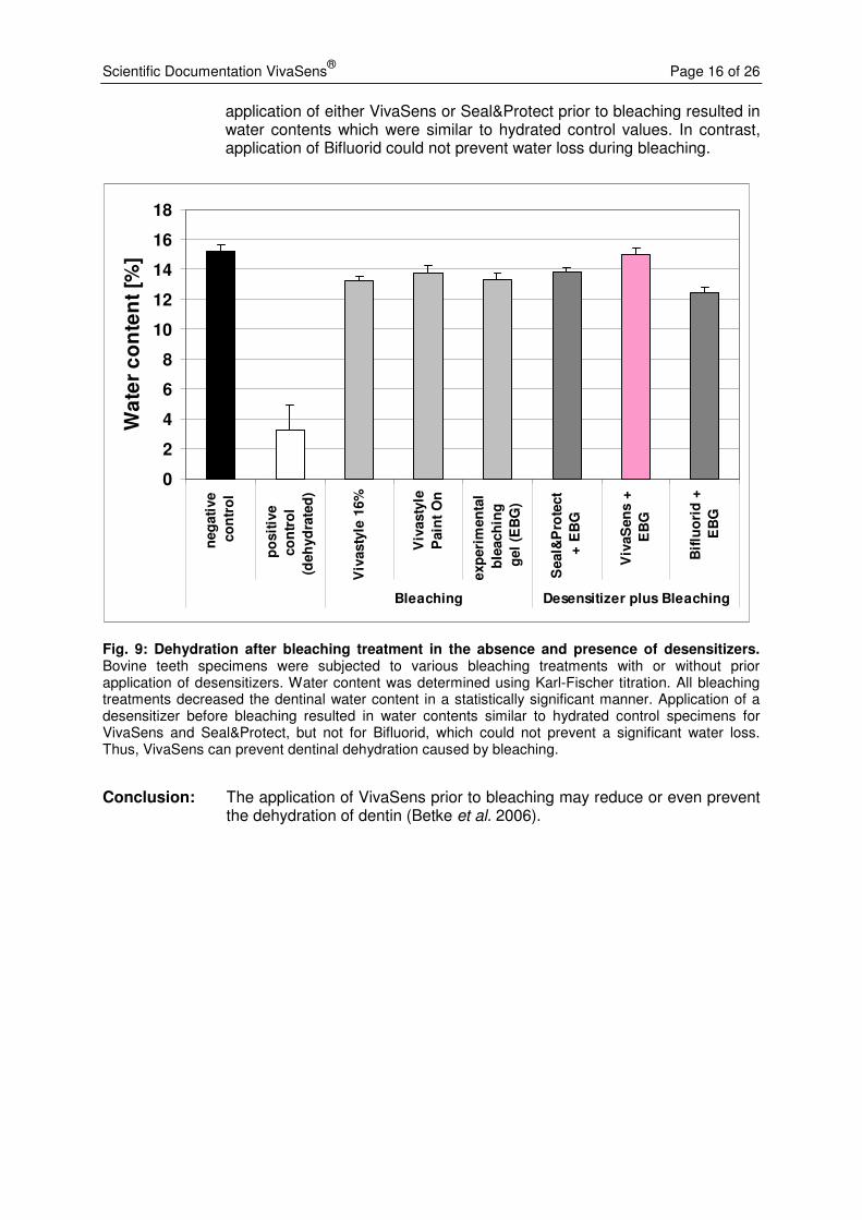
Method: Fifty-five standardized cylindrical dentin specimens were prepared from freshly extracted bovine incisors under constant water irrigation and conditioned at room temperature in a hygrophor for 14 days. Two control groups were established (each n=5), with Group A: complete dehydration (positive control) and Group B: no treatment, storage in hygrophor (negative control). Another three groups with 5 specimens each served as bleaching controls and were treated with three different bleaching agents: VivaStyle 16%; Ivoclar Vivadent; VivaStyle Paint On (6% carbamide peroxide), Ivoclar Vivadent; or an experimental glycerine-based bleaching gel with 20% carbamide peroxide. The other samples (n=10) were treated with desensitizers (Seal&Protect, Dentsply DeTrey; Bifluorid, Voco; VivaSens, Ivoclar Vivadent). From each sealed group, 5 specimens were subjected to bleaching treatment with the experimental bleaching gel. Bleaching was for 2 hours/day for the gels and 20 min/day for VivaStyle Paint On for 7 days. After the respective treatments, the water content of the specimens was determined using the analytical method of Karl-Fischer titration.
Results: All bleaching treatments reduced the dentinal water content in a statistically significant manner (see Figure 9) from 15% to approximately 13%. The
Scientific Documentation VivaSens® Page 16 of 26
application of either VivaSens or Seal&Protect prior to bleaching resulted in water contents which were similar to hydrated control values. In contrast, application of Bifluorid could not prevent water loss during bleaching.
0
2
4
6
8
10
12
14
16
18n
eg
ati
ve
co
ntr
ol
po
sit
ive
co
ntr
ol
(deh
yd
rate
d)
Viv
asty
le 1
6%
Viv
asty
le
Pain
t O
n
exp
eri
men
tal
ble
ach
ing
gel
(EB
G)
Seal&
Pro
tect
+ E
BG
Viv
aS
en
s +
EB
G
Bif
luo
rid
+
EB
G
Bleaching Desensitizer plus Bleaching
Wa
ter
co
nte
nt
[%]
Fig. 9: Dehydration after bleaching treatment in the absence and presence of desensitizers. Bovine teeth specimens were subjected to various bleaching treatments with or without prior application of desensitizers. Water content was determined using Karl-Fischer titration. All bleaching treatments decreased the dentinal water content in a statistically significant manner. Application of a desensitizer before bleaching resulted in water contents similar to hydrated control specimens for VivaSens and Seal&Protect, but not for Bifluorid, which could not prevent a significant water loss. Thus, VivaSens can prevent dentinal dehydration caused by bleaching.
Conclusion: The application of VivaSens prior to bleaching may reduce or even prevent the dehydration of dentin (Betke et al. 2006).
Scientific Documentation VivaSens® Page 17 of 26
4. Clinical investigations
VivaSens has been investigated in different clinical studies. In all studies the patients reported reduced dentin sensitivity, thus confirming the desensitizing effect of VivaSens. In exceptional cases, a complete loss of sensitivity was reported. In none of the studies adverse events were recorded. Furthermore, the acceptance of VivaSens by the patients was high.
4.1 Reduction in dentin sensitivity
4.1.1 Dr. Arnd Peschke, Internal Clinic, R&D Ivoclar Vivadent, Schaan, Liechtenstein
Aim: This pilot study aimed at evaluating the efficacy of VivaSens as dentin desensitizer in adults.
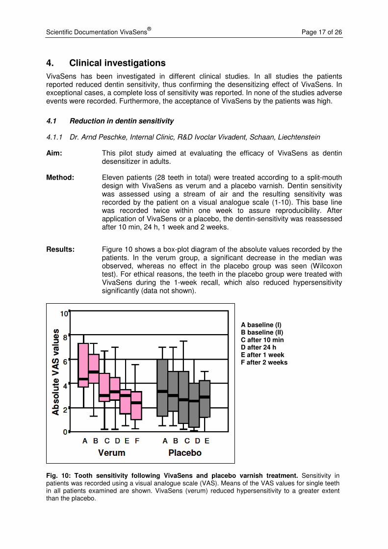
Method: Eleven patients (28 teeth in total) were treated according to a split-mouth design with VivaSens as verum and a placebo varnish. Dentin sensitivity was assessed using a stream of air and the resulting sensitivity was recorded by the patient on a visual analogue scale (1-10). This base line was recorded twice within one week to assure reproducibility. After application of VivaSens or a placebo, the dentin-sensitivity was reassessed after 10 min, 24 h, 1 week and 2 weeks.
Results: Figure 10 shows a box-plot diagram of the absolute values recorded by the patients. In the verum group, a significant decrease in the median was observed, whereas no effect in the placebo group was seen (Wilcoxon test). For ethical reasons, the teeth in the placebo group were treated with VivaSens during the 1-week recall, which also reduced hypersensitivity significantly (data not shown).
A baseline (I) B baseline (II) C after 10 min D after 24 h E after 1 week F after 2 weeks
Fig. 10: Tooth sensitivity following VivaSens and placebo varnish treatment. Sensitivity in patients was recorded using a visual analogue scale (VAS). Means of the VAS values for single teeth in all patients examined are shown. VivaSens (verum) reduced hypersensitivity to a greater extent than the placebo.
Scientific Documentation VivaSens® Page 18 of 26
Conclusion: Application of VivaSens reduced the dentin-sensitivity in the patients tested significantly. However, only in a few cases a complete loss of hypersensitivity was observed. Due to the small number of treated patients and the limited period of the study, no statement on the long-term effect of VivaSens can be made. During the course of this study, no side effects were detected and the patients rated the taste of VivaSens as neutral.
4.1.2 Prof. Dr. Andrej Kielbassa, University of Berlin, Germany
Aim: The purpose of this clinical trial was to evaluate the efficacy of VivaSens as dentin desensitizer in adults.
Method: This randomized, clinical, double-blind short term trial involved 88 patients. A randomly selected tooth was subjected to a thermal stimulus using a cold air stream and the resulting sensitivity was recorded by the patient on a visual analogue scale. Per patient, one tooth was treated with VivaSens, another tooth with water as placebo (split-mouth design).
Results: One week after application of VivaSens, 90% of the patients reported a reduction in sensitivity by an average of 26 points on the scale of 1 to 100. This corresponds to a reduction of 50% and was statistically significant. In the placebo group however, the reduction was also 26 points. At the 6-month recall, the reduction of sensitivity in the verum group was 16.5% compared to 25% in the placebo group. There was no statistically significant difference between the two treatment groups. However, high placebo effects are a common phenomenon in pain studies (Zanter et al. 2006).
4.1.3 Prof. Dr. Steven Duke, Indiana University, Indianapolis, USA
Aim: The purpose of this study was to evaluate the efficacy of VivaSens as a dentin desensitizer in adults.
Method: This prospective double-blind randomized clinical trial used VivaSens as verum and a placebo formulation as a control. The study was double-blinded with neither the evaluator nor the subject aware of which procedure was performed.
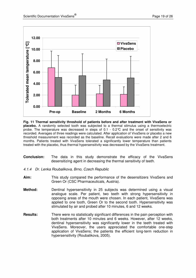
Fifty subjects with cervical hypersensitivity were randomly assigned to one of two equal groups (treatment and control). A randomly selected tooth was subjected to a thermal stimulus using a thermoelectric probe to establish a preoperative threshold measurement. The probe was held against the tooth and the temperature was decreased in steps of 0.1 - 0.2°C. The first onset of sensitivity was recorded as the pre-operative temperature. This procedure was performed three times and the average reading was calculated as the mean threshold measurement. After application of VivaSens or a placebo, a new threshold measurement was recorded as the baseline. Recall evaluations were made after 2 and 6 months.
Results: As shown in Figure 11, all 25 patients in the verum group tolerated a lower temperature following the application of VivaSens. In contrast to the placebo, VivaSens was significantly more effective (p<0.001). In the placebo group, the value after application (baseline) was not significantly different from the pre-operative mean threshold (p>0.001). At the 2-months and 6-months recalls, all patients from the verum group continued to demonstrate decreased sensitivity. VivaSens remained significantly more effective (p <0.001) than the placebo.
Scientific Documentation VivaSens® Page 19 of 26
0.00
2.00
4.00
6.00
8.00
10.00
12.00
Pre-op Baseline 2 Months 6 Months
To
lera
ted
me
an
te
mp
era
ture
[°C
]
VivaSens
Placebo
Fig. 11 Thermal sensitivity threshold of patients before and after treatment with VivaSens or placebo. A randomly selected tooth was subjected to a thermal stimulus using a thermoelectric probe. The temperature was decreased in steps of 0.1 - 0.2°C and the onset of sensitivity was recorded. Averages of three readings were calculated. After application of VivaSens or placebo a new threshold measurement was recorded as the baseline. Recall evaluations were made after 2 and 6 months. Patients treated with VivaSens tolerated a significantly lower temperature than patients treated with the placebo, thus thermal hypersensitivity was decreased by the VivaSens treatment.
Conclusion: The data in this study demonstrate the efficacy of the VivaSens desensitizing agent in decreasing the thermal sensitivity of teeth.
4.1.4 Dr. Lenka Roubalikova, Brno, Czech Republic
Aim: This study compared the performance of the desensitizers VivaSens and Green Or (CSC Pharmaceuticals, Austria).
Method: Dentinal hypersensitivity in 25 subjects was determined using a visual analogue scale. Per patient, two teeth with strong hypersensitivity in opposing areas of the mouth were chosen. In each patient, VivaSens was applied to one tooth, Green Or to the second tooth. Hypersensitivity was stimulated by air and probed after 10 minutes, 6 and 12 weeks.
Results: There were no statistically significant differences in the pain perception with both treatments after 10 minutes and 6 weeks. However, after 12 weeks, dentinal hypersensitivity was significantly lower in the teeth treated with VivaSens. Moreover, the users appreciated the comfortable one-step application of VivaSens; the patients the efficient long-term reduction in hypersensitivity (Roubalikova, 2005).
Scientific Documentation VivaSens® Page 20 of 26
4.1.5 Dr. Tijen Pamir, Ege University, Izmir, Turkey
Aim: This study compared the performance of the desensitizers VivaSens, Seal&Protect (Dentsply DeTrey GmbH, Konstanz, Germany) and BisBlock (BISCO, Schaumburg, IL, USA) to a placebo treatment (distilled water).
Method: In this double-blind, randomized, placebo-controlled trial, 60 patients with a history of sensitivity were included. Initial sensitivity levels were determined by a visual analogue scale. Sensitivity was triggered by evaporative (air-blast) and thermal stimuli. Per patient, one tooth with slight to moderate sensitivity was chosen and randomly assigned to a treatment or the placebo group. Patients were recalled after four weeks.
Results: All desensitizers (VivaSens, Seal&Protect, BisBlock) reduced sensitivity levels statistically significantly (p<0.05) when compared with baseline values. Efficacy of the different products was similar and statistically significantly different from the placebo treatment (p<0.05) (Pamir et al. 2007).
4.1.6 Reduction in dentin sensitivity in bleaching treatments
Investigators: Prof. Dr. T. Attin, Dr. D. Ziebolz, Dr. C. Hannig, University of Göttingen, Germany
Aim: The purpose of this study was to evaluate the clinical efficacy and safety of VivaSens in conjunction with a bleaching treatment using VivaStyle Paint On Plus (6% hydrogen peroxide).
Method: A total of 80 patients was randomly distributed in two groups (n=40 for each group). Group A received bleaching treatment without prior application of VivaSens, Group B used the bleaching agent after a single application of VivaSens. Tooth colour at baseline and after 10 days was assessed using a Vita shade guide. Sensitivity was triggered by a cold air stream, graded from 0 (no sensitivity) to 10 (high sensitivity), and assessed at baseline, after 7 days (end of bleaching treatment) and 10 days after the end of the bleaching treatment.
Patients (n=23) that showed hypersensitivity after the bleaching therapy were treated with VivaSens or a control treatment to evaluate the remediation of the acquired hypersensitivity.
Results: The use of VivaSens had no influence on the color change achieved by the bleaching treatment. Bleaching significantly increased tooth hypersensitivity compared to baseline values. In Group A, which performed bleaching without previous desensitizing treatment, 5 subjects reported increased hypersensitivity, whereas in Group B (bleaching after use of VivaSens) only 1 patient claimed elevated tooth hypersensitivity. Although there was a lower degree of hypersensitivity in the VivaSens group compared to the control group, these results were not statistically significantly different. This may be due to the high number of dropouts (n=13) (Ziebolz et al. 2008).
Scientific Documentation VivaSens® Page 21 of 26
0
0.5
1
1.5
2
2.5
3
3.5
0 1 2 3 4 5 6 7 8 9 10
Days
Pa
in u
nit
s [
0 t
o 1
0]
Placebo
VivaSens
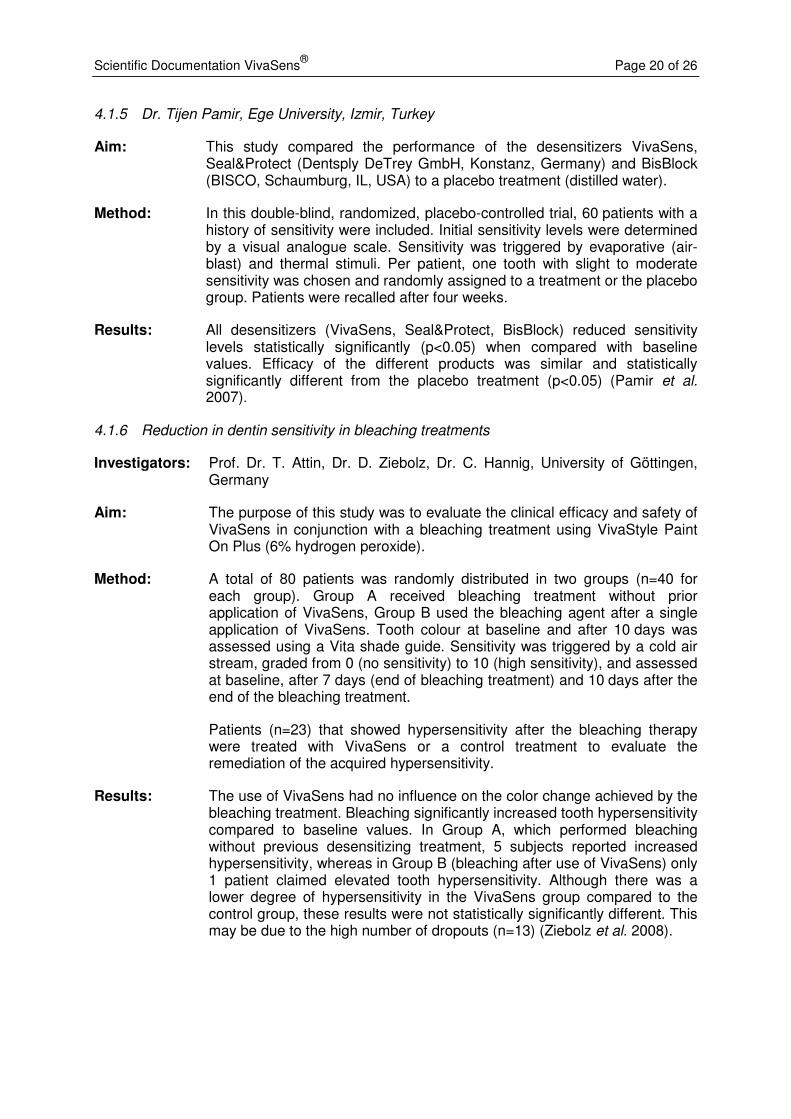
Fig. 12: Reduction in hypersensitivity after bleaching. Patients who had undergone bleaching treatment and claimed subsequent tooth hypersensitivity were treated with VivaSens or a placebo. (verum and placebo group were not statistically different at day 0). Pain was recorded by the patients in a pain diary. The group treated with VivaSens experienced a statistically significant decrease in hypersensitivity within the first 24 hours; hence, VivaSens provided immediate relief against bleaching-induced dental hypersensitivity, whereas pain subsided in the placebo group only after 3 days significantly.
Application of VivaSens in patients with hypersensitivities after the bleaching treatment led to a significant drop in sensitivity within the first 24 hours (p=0.012), whereas in the control group, sensitivity decreased only at day 3 after bleaching (see Figure 12).
Conclusion: The use of VivaSens prior to a bleaching treatment may have a favourable effect on hypersensitivity and does not interfere with the bleaching. Application of VivaSens after the treatment immediately reduces hypersensitivities acquired from the bleaching treatment.
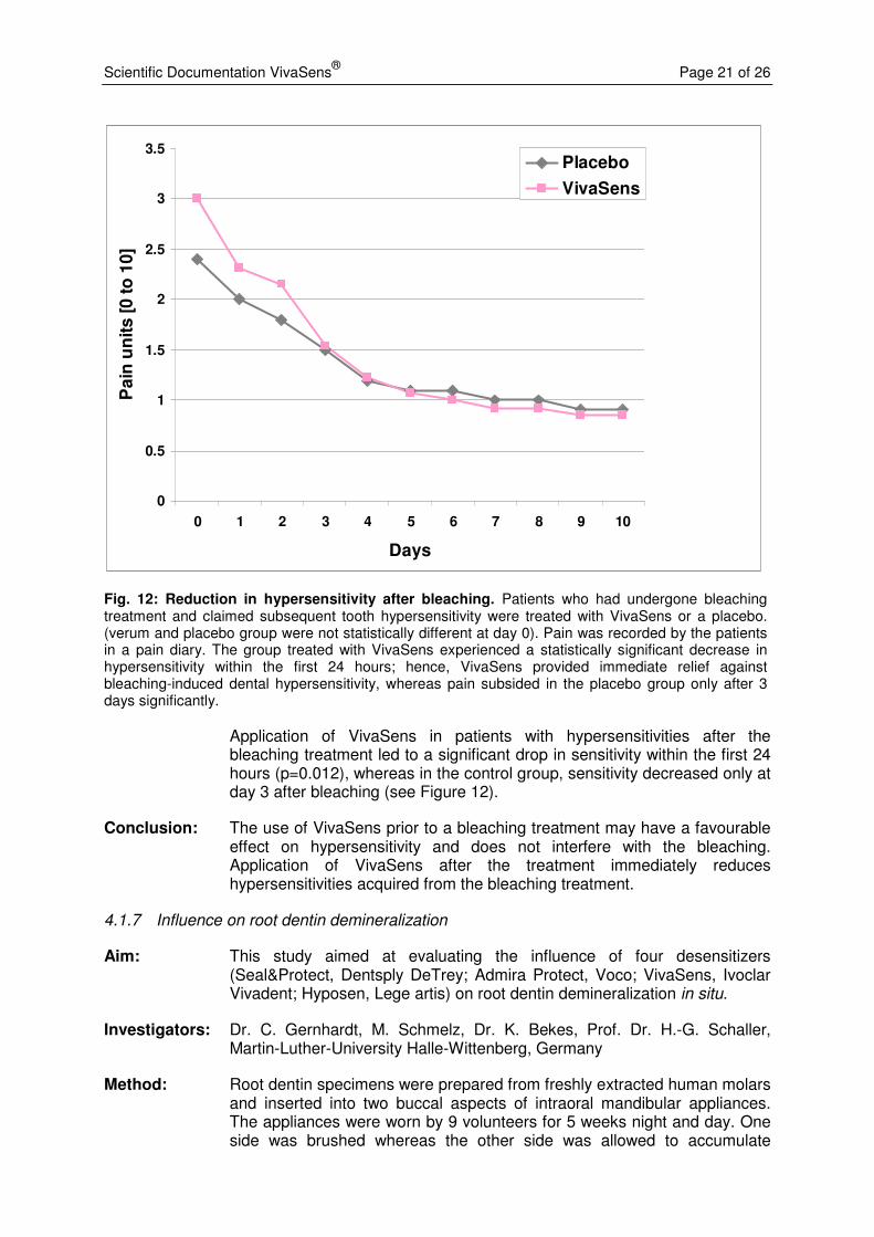
4.1.7 Influence on root dentin demineralization
Aim: This study aimed at evaluating the influence of four desensitizers (Seal&Protect, Dentsply DeTrey; Admira Protect, Voco; VivaSens, Ivoclar Vivadent; Hyposen, Lege artis) on root dentin demineralization in situ.
Investigators: Dr. C. Gernhardt, M. Schmelz, Dr. K. Bekes, Prof. Dr. H.-G. Schaller, Martin-Luther-University Halle-Wittenberg, Germany
Method: Root dentin specimens were prepared from freshly extracted human molars and inserted into two buccal aspects of intraoral mandibular appliances. The appliances were worn by 9 volunteers for 5 weeks night and day. One side was brushed whereas the other side was allowed to accumulate
Scientific Documentation VivaSens® Page 22 of 26
plaque. Fluoride was excluded from oral hygiene. After the in-situ period, two slabs were ground and the depth of demineralization was determined using a polarized light microscope. For each group means and standard deviations were calculated; statistical analysis was performed using ANOVA and Tukey’s test.
Results: The deepest lesion depths were found in control samples. Moreover, in unbrushed samples, lesion depths were in general deeper than in brushed root dentin specimens. Treatment with desensitizers decreased lesion depths in comparison to untreated control specimens (see Figure 13). The shallowest lesion depths were found in specimens treated with VivaSens and Admira Protect.
Conclusion: VivaSens effectively protected root dentin from demineralization (Gernhardt et al. 2006; Bekes et al. 2009).
0
10
20
30
40
50
60
Con
trol
Sea
l&Pro
tect
Adm
ira P
rote
ct
Viv
aSen
s
Hyp
osen
Me
an
le
sio
n d
ep
th [
µm
]
Fig. 13: Protection from demineralization by desensitizers. Root dentin specimens were worn in intraoral appliances by volunteers for 5 weeks night and day and treated with one of four different desensitizers (Seal&Protect, Admira Protect, VivaSens, Hyposen). No fluoride was used in oral hygiene. After the in-situ period, demineralization was determined by measuring the lesion depths of the specimens by polarized light microscopy. The shallowest lesion depths were found in specimens treated with VivaSens and Admira Protect. Thus, VivaSens is effective in protecting root dentin from demineralization.
Scientific Documentation VivaSens® Page 23 of 26
5. Toxicology
5.1 Introduction
VivaSens is indicated for treating hypersensitivity of exposed root dentin. It is applied with a purpose-designed brush. During a single application at one site, approximately 20-40 µl of desensitizer comes into contact with oral tissues. VivaSens contains methacrylate modified polyacrylic acid, polyethylene glycol dimethacrylate, phosphonic acid methacrylate, hydroxyl propyl cellulose, potassium fluoride, ethanol and water.
With the exception of phosphonic acid methacrylate, the desensitizer only contains components which have been used in dental materials for many years. New toxicological tests were therefore carried out only with this new component. Data regarding the other components were retrieved from earlier investigations or toxicological databases.
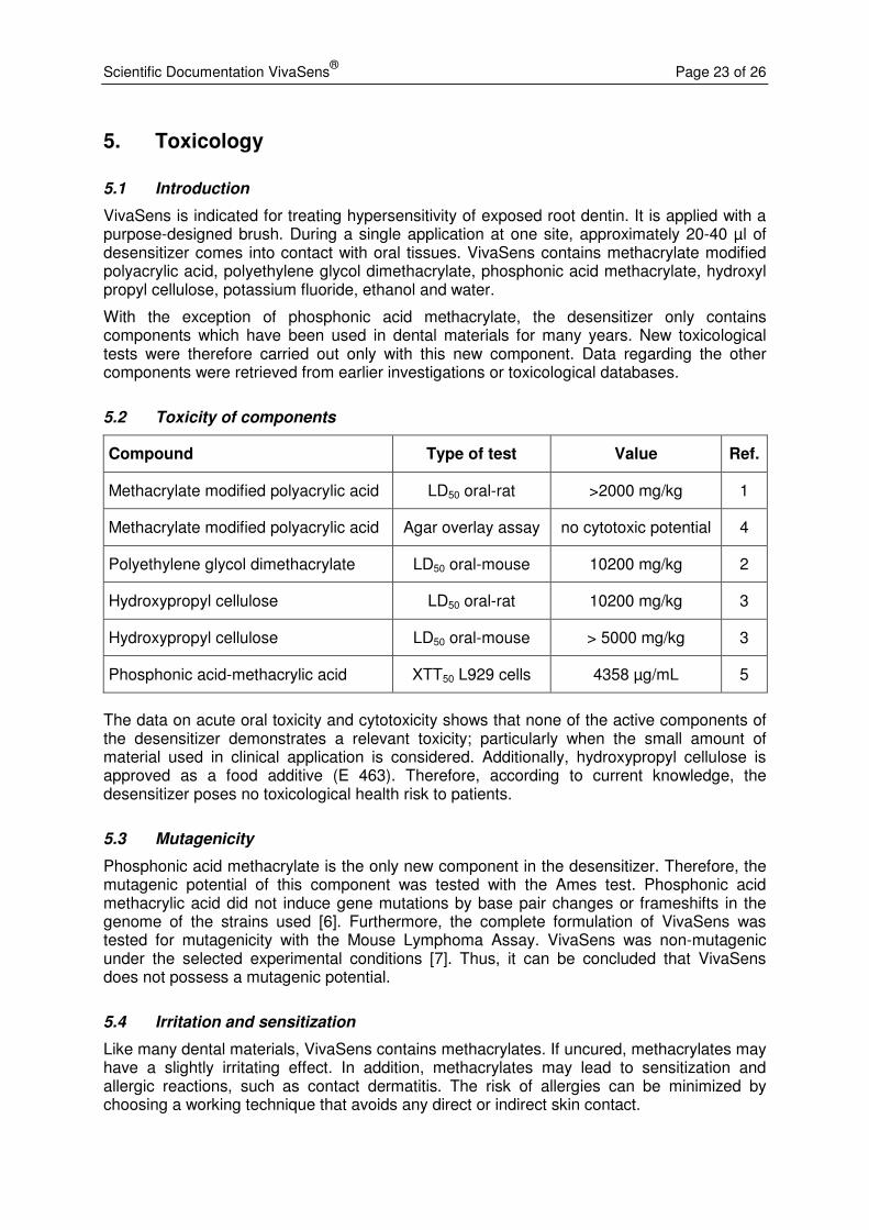
5.2 Toxicity of components
Compound Type of test Value Ref.
Methacrylate modified polyacrylic acid LD50 oral-rat >2000 mg/kg 1
Methacrylate modified polyacrylic acid Agar overlay assay no cytotoxic potential 4
Polyethylene glycol dimethacrylate LD50 oral-mouse 10200 mg/kg 2
Hydroxypropyl cellulose LD50 oral-rat 10200 mg/kg 3
Hydroxypropyl cellulose LD50 oral-mouse > 5000 mg/kg 3
Phosphonic acid-methacrylic acid XTT50 L929 cells 4358 µg/mL 5
The data on acute oral toxicity and cytotoxicity shows that none of the active components of the desensitizer demonstrates a relevant toxicity; particularly when the small amount of material used in clinical application is considered. Additionally, hydroxypropyl cellulose is approved as a food additive (E 463). Therefore, according to current knowledge, the desensitizer poses no toxicological health risk to patients.
5.3 Mutagenicity
Phosphonic acid methacrylate is the only new component in the desensitizer. Therefore, the mutagenic potential of this component was tested with the Ames test. Phosphonic acid methacrylic acid did not induce gene mutations by base pair changes or frameshifts in the genome of the strains used [6]. Furthermore, the complete formulation of VivaSens was tested for mutagenicity with the Mouse Lymphoma Assay. VivaSens was non-mutagenic under the selected experimental conditions [7]. Thus, it can be concluded that VivaSens does not possess a mutagenic potential.
5.4 Irritation and sensitization
Like many dental materials, VivaSens contains methacrylates. If uncured, methacrylates may have a slightly irritating effect. In addition, methacrylates may lead to sensitization and allergic reactions, such as contact dermatitis. The risk of allergies can be minimized by choosing a working technique that avoids any direct or indirect skin contact.
Scientific Documentation VivaSens® Page 24 of 26
5.5 Conclusion
According to current knowledge, the desensitizer VivaSens exhibits negligible acute oral toxicity and cytotoxicity and no mutagenicity. The irritating potential can be effectively minimized by a careful working technique. VivaSens can therefore be assumed to be safe for use when applied correctly.
5.6 Literature on toxicology
[1] Acute oral toxicity study in rats, RCC Report 384096, January 1995
[2] MSDS of polyethylene glycol dimethacryl-ate
[3] MSDS of hydroxypropyl cellulose.
[4] Cytotoxicity test in vitro: agar overlay assay. RCC Report 384107, March, 1995.
[5] Cytotoxicity assay in vitro: Evaluation of materials for medical devices (XTT-Test). RCC-CCR Report 697901, July 2001.
[6] Salmonella typhimurium reverse mutation assay. RCC-CCR Project 697902. October 2001.
[7] Cell mutation assay at the thymidine kinase locus (TL+/-) in mouse lymphoma cells with VivaSens, RCC-CCR Project 721200. February 2002.
6. Literature
Absi EG, Addy M, Adams D: Dentine hypersensitivity. A study of the patency of dentinal tubuli in sensitive and non-sensitive cervical dentine. J Clin Periodontol. 1987 May;14(5):280-4.
Bekes K, Schmelz M, et al. The influence of application of different desensitisers on root dentine demineralisation in situ. Int Dent J. 2009;59(3):121-6.
Betke H, Kahler E, et al. Influence of bleaching agents and desensitizing varnishes on the water content of dentin. Oper Dent. 2006;31(5):536-542.
Betke H., Revas P, et al. Einfluss der Anwendung von Desensibilisierungslacken auf die Wirkung verschiedener Bleichgele. DGZ:. 2004;38.
Betke H., Revas P, et al. Einfluss von Desensibilisierungslacken auf die Zahnaufhellung in der Bleichtherapie. Quintessenz 2005:56(6):589-597.
Brännström M, Linden LÅ, Åstrom A: The hydrodynamics of the dental tubule and of pulp fluid. A discussion of its significance in relation to dentinal sensitivity. Caries Res. 1967;1: 310-317.
Brodowski D, Imfeld T: Dentinüberempfindlichkeit - eine Übersicht. Schweiz Monatsschr Zahnmed. 2003;113(1):49-58.
Dababneh RH, Khouri AT, Addy M M: Dentine hypersensitivity – an enigma? A review of terminology, epidemiology, mechanisms, aetiology and management. Br Dent J. 1999;187(11): 606-611.
Derkson GD, Pashley DH, Derkson ME: Microleakage measurement of selected restorative materials: a new in vitro method. J Prosthet Dent. 1986 Oct;56(4):435-40.
Dowell P, Addy M: Dentine hypersensitivity - a review. Aetiology, symptoms and theories of pain production. J Clin Periodontol. 1983 Jul;10(4):341-50.
Duke ES, Platt JA: An in vitro evaluation of VivaSens desensitizer. Final study report. June, 2002.
Duke ES: Prospective placebo controlled clinical trial of a desensitizing agent. Study report, 6 month recall, June 2003.
Frankenberger R: Influence of VivaSens desensitizers on dentin bond strength of temporary and permanent restorative materials. Study report, University of Erlangen, 2002.
Gernhardt C, Bekes K, Schaller HG. Influence of Different Sealants on Root Dentin Demineralization in Situ. Int Poster J Dent Oral Med 2006;Vol 8(04): Poster 336.
Ghavamnasiri M, Alavi M, et al. Effect of a resin-based desensitizing agent and a self-etching dentin adhesive on marginal leakage of amalgam restorations. J Contemp Dent Pract. 2007;8(7):54-61.

Scientific Documentation VivaSens® Page 25 of 26
Grégoire G: Evaluation of the permeability of VivaSens in comparison with Systemp Desensitizer and Gluma desensitizer, Study report, December 2003.
Holland GR, Narhi MN, Addy M, Gangarosa L, Orchardson R: Guidelines for the design and conduct of clinical trials on dentine hypersensitivity. J Clin Periodontol. 1997;24(11):808-13.
Ingham KC: Precipitation of proteins with polyethylene glycol. Methods Enzymol. 1990;182:301-6.
Markowitz K: Tooth sensitivity: mechanisms and management. Compendium. 1993 Aug;14(8):1032, 1034 passim; quiz 1046.
Matthews B, Vongsavan N: Interactions between neural and hydrodynamic mechanisms in dentine and pulp. Arch Oral Biol. 1994;39 Suppl:87S-95S.
Pashley DH: Dentine permeability and its role in the pathobiology of dentine sensitivity. Arch Oral Biol. 1994;39 Suppl:73S-80S.
Pamir T, Dalgar H, et al. Clinical evaluation of three desensitizing agents in relieving dentin hypersensitivity. Oper Dent. 2007;32(6):544-548.
Reeder OW, Jr., Walton RE, et al. Dentin permeability: determinants of hydraulic conductance. J Dent Res. 1978;57(2):187-93.
Rimondini L, Baroni C, Carrassi A: Ultrastructure of hypersensitive and non-sensitive dentine. A study on replica models. J Clin Periodontol. 1995 Dec;22(12):899-902.
Roubalikova L, Wilhelm Z: VivaSens und Green Or im Vergleich. Dentalhygiene Journal 2005 (3):26-28.
Schüpbach P: Closing of dentinal tubuli by VivaSens. Microphot study report. March 2003.
Zantner C, Kielbassa AM: Clinical efficacy of a newly developed desensitizing varnish. Study report. 2003.
Zantner C, Popescu O, et al. Randomized clinical study on the efficacy of a new lacquer for dentine hypersensitivity. Schweiz Monatsschr Zahnmed. 2006;116(12):1232-1237.
Ziebolz D, Hannig C, et al. Influence of a desensitizing agent on efficacy of a paint-on bleaching agent. Am J Dent. 2008;21(2):77-82.
Scientific Documentation VivaSens® Page 26 of 26
This documentation contains an overview of internal and external scientific data (information). The documentation and the corresponding information have been prepared exclusively for in-house use and for the information of external partners of Ivoclar Vivadent AG. They are not intended for any other purpose. Although we assume that the information complies with the latest standard of technology, we did not check all of them and may thus not assume any responsibility for their accuracy, truthfulness, or reliability. Liability cannot be assumed for the use of this information, even if we obtain contrary information. The information is used at the sole risk of the reader. Information is made available 'as received' without explicit or implied warranty regarding suitability (without reservation) for any specific purpose. The information is made available free of charge. Ivoclar and its partners cannot be held accountable for any direct, indirect, immediate, or specific damage (including but not exclusively damage resulting from lost information, loss of use, or costs resulting from gathering comparable information), or for penal damages, which result from the use or failure to use this information, even if we or our representatives were informed about the possibility of such damage. Ivoclar Vivadent AG Research and Development Scientific Service Bendererstrasse 2 FL - 9494 Schaan Liechtenstein Content: Dr. Kathrin Fischer, Dr. Sandro Sbicego Edition: September 2010