voh. washington, novkmbkr 25, 1918 no. 8
TRANSCRIPT

Voh. XV WASHINGTON, D. C, NOVKMBKR 25, 1918 No. 8
A CONTRIBUTION TO THE BIOLOGY OF FRUIT-FLY PARASITES IN HAWAII
By C. E. PEMBERTON, Entomologist in Field Charge, and H. F. WIIXARD, Chief Fruit- Fly Quarantine Inspector, Mediterranean Fruit-Fly Investigations, Bureau of Ento- mology, United States Department of Agriculture
INTRODUCTION
With the termination of an intensive study of the Mediterranean fruit fly (Ceratitis capitata Wiedemann) in Hawaii in February, 1916, by the Bureau of Entomology, a general investigation of the biology, interrela- tion 1 and economic value of introduced parasites of this fruit fly was begun. The results of this investigation are herewith presented.
The natural enemies of the Mediterranean fruit fly now contributing toward its control in Hawaii are#three species of Opiinae—viz, Diachasma tryoni Cameron, O pins humilis Silvestri, and D. fullawayi Silvestti, one species of Bulophinae, Tetrastichus giffardianus Silvestri, and an ant, Pheidole megacephala Fabricius. Two other parasites occasionally reared from the fruit fly are Opius fletcheri Silvestri, normally a melon-fly para- site, and Pachycrepoideus dubius, a dung-fly parasite. At present para- sitism by these two parasites is not important, and may be more acci- dental than normal. NThe opiines and the eulophid are strictly larval parasites. The ant is important as a predacious enemy of the larvae and to a lesser extent of the pupae. The pteromalid Pachycrepoideus dubius attacks the pupa. There is striking similarity in habit, structure, and development among the opiines undçr discussion, and in view of this the species now most abundant in Hawaii, Diachasma tryoni, will be dealt with the most completely, to be followed by notes on the special features of the two other important opiine species together with comparisons with the species tryoni in sufficient detail to give a thorough conception of the biology of each.2
1 For previous studies on fruit-fly interrelations in Hawaii, see Pemberton and Willard (6). [Reference
is made by number (italic) to " literature cited," p. 465.] 2 For the original descriptions and history of the introductions of the important species above mentioned,
see BACK, H. A., and PEMBERTON, C. B. (5). For detailed records on the extent of parasitism in Hawaii by these species see Back and Pemberton
(7, 2) and Pemberton and Willard (5).
Journal of Agricultural Research, Vol. XV, No. 8 Washington, D. C. Nov. 25,1918 ah Key No. K-73
(419)

420 Journal of Agricultural Research vol. xv. No. s
DIACHASMA TRYONI
Diachasma tryoni Cameron was first observed in New South Wales in 1908 and was described in 1911. It was introduced into Hawaii from New South Wales by Silvestri in May, 1913. It soon became definitely established and by 1916 its importance as a parasite and its ready adaptation to Hawaiian conditions were demonstrated clearly. During 1917 it excelled the work of the other introduced parasites
DESCRIPTION AND UFEÎ HISTORY
EGG
The egg (fig. 1, 2) is cylindrical, translucent white, with smooth, glis- tening surface, drawn out at each end into a short, rounded protuberance
and when first deposited is surrounded ^^^^^ifM^^^Ê^r-^^ ky a thin, transparent membrane,
^i¿^*^;;--^^^ possibly the exochorion. This mem- Fig. 1.—Diachasma tryoni; Egg just laid; length braUC adheres to the COnf Ormity of
0■4âmm■ the egg but does not tightly inclose it. The egg is faintly concave ventrally and distinctly convex dorsally. The protuberance on the cephalic end is slightly broader and shorter than is that on the caudal end. At deposition the egg averages 0.45 mm. in length, including the enveloping membrane, its greatest width being about one-sixth of the length. When fully developed the egg averages 0.65 mm. in length and is about one-fourth as broad as long. During development the outer enveloping membrane is ruptured and entirely separated from the egg proper. At maturity each end of the egg stands out as an abrupt, broad tubercle. The embryo is then clearly revealed by transmitted light.
Although the eggs are deposited only slightly beneath the surface of the larval skin, they are invisible even under strong sunlight and magni- fication. The duration of the egg period can be determined only by dissections of host tissue at frequent intervals to locate the eggs and by the use of numbers
„ „ , - , Fig. 2.—Diachasma tryoni: Eggmature; length 0.65 mm. of well-parasitized larvae known to have been oviposited in for a short and definite period. The egg stage in Honolulu (Table I) was found to last from 54 to 73 hours, the variations depending upon fluctuations in temperature. As the average incubation period is about 2^4 days, the exact number of hours required for the development of eggs deposited in the morning is less than is that where eggs are deposited in the late afternoon. Eggs deposited in the morfiing hours occupy parts of three days and two nights before hatching, and thus develop under a somewhat higher average temperature than would obtain in the case of eggs deposited in the afternoon or early evening.

Nov. 25,1918 Biology of Fruit-Fly Parasites in Hawaii 421
These pass through parts of three nights and two days as opposed to three days and two nights and would thus be subjected to somewhat lower average temperatures. This will explain some of the misleading variations shown in Table I.
TABI,3 I.—Duration of the egg stage of Diachasma tryoni in Honolulu
Number of eggs
tmder ob- servation.
Eggs deposited. Eggs hatched. Average duration
of egg stage.
Mean tempera-
ture.
24 138 130
73
11::::: 47
Jan. 4, 11 a. m. to z p. m... Mar. 21, 11 a. m. to 1 p. m Apr. 16, 9 a. m. to 10 a. m.
May 31, 3 p. m. to 3. 30 p.m. July 6, 11 a. m. to 2 p. m.. Tulv 7.20. m
Jan. 7, 11 a. m. to 2 p. m... Mar. 24, 9 a. m. to 10 a. m.' Apr. 18, 10 p. m, to 11.30
p. m. June 3, 6 a. m. to 8 a. m... July 8, 6 p. m. to 10 p. m.. July 9, 9 p. m. to 11 p. m.. Oct. 7, 1 p. m. to 4 p. m...
Hours. 73 70 .02
64 56 56 54
0F. 71.O 73-2 74.2
74.0 75-9 76. 0 76.6 Oct. 5, 8 a. m. to 10 a. m. .
The fully developed egg is so swollen and the membrane so thin that actual emergence of the larva from the egg is sudden, and rather an ex- plosive process. Many eggs on hatching have been under observation. The egg membrane is suddenly ruptured longitudinally, probably by the movements of the larva within, and the larva floats out without ap- parent effort into the semiliquid medium of the host surrounding it. The egg may hatch while the host is still in the active, feeding, larval stage or it may hatch after the puparium is formed and the complete histolysis of larval tissue has taken place. No important histogenetic action occurs in a puparium containing a parasite egg or larva. The parasitized host larva feeds and develops to maturity even though heavily superparasitized, leaves the fruit normally, and forms a perfect pupa- rium in the usual manner. The complete histolysis of the larval tissues within the puparium then follows, but here all fly development ceases. Henceforth the content of the puparium is but a liquid mass containing the broken-down bits of larval tissue and the rapidly developing para- site larva. The death of the host then may be said properly to occur at the cessation of histolysis in the newly formed puparium. From over 3,000 parasitized puparia opened during 1916 and 1917 no single case was ever noted in which a perfect or even partially formed fly pupa occurred.
I^ARVA
The larva undergoes many interesting phases in the processes of transformation. When first hatched, it is about 0.85 mm. long. This is the most active period in the larval life. At this time it is so markedly different from the later instars that it appears to simulate the larval structure and habits of an ancestral type. It usually hatches while the host is still in the larval stage. The parasitic larva then lies in a well-

422 Journal of Agricultural Research Vol. XV, No. 8
organized body, wherein its food, which seems largely* the fat body of the host, is in a semisolid state, in part isolated into definitely compacted masses. The larva is lodged in an area which is well organized in mus- cular, digestive, nervous, and respiratory structures, all of which com- bine to interfere with its freedom of action. Special characters, not
appearing in the succeeding in- stars, are peculiarly adapted to this stage. The head is large, heavily chitinized and brownish, and bears a pair of sickle-like mandibles, with bases broadly separated and capable of wide movement and quick action. Above the mandibles and seem- ingly on the labrum is a pair of small, short antennal structures, which are frequently extended and withdrawn in a rapid, vibra- tory manner as the larva feeds and moves about. On the ce- phalic edge of the chitinized ven- tral portion of the head is borne a pair of pointed teeth, well sepa- rated and together forming a dis- tinct letter U with the basal con- necting line more or less straight. This lies below and directly be- tween the bases of the mandibles (fig. 3). Its shape affords the best character for differentiating the larva of this species from the newly hatched larva of Diachas- ma fullawayi or that of Opius humilis. A clearly defined, sim- ple trachea! system is present (fig. 4) and becomes filled with air shortly after the larva has hatched. No spiracles occur, but eight minute, oval swellings can
be seen along each main trachea! trunk in body segments 1 to 8. The larva lies strictly within the host, and the air which quickly fills the tracheae must be obtained by osmosis from the aerated liquid media surrounding it. The tracheae are filled with air before food has been taken, which shows that the air is not extracted internally from the ingested food. The digestive canal (fig. 5) is a simple, straight tube
Vvi
FIG. s.—Diachasmatryoni: Cast skin of first-instar larva, showing head characters of firät instar and serosal cel- lular mass still clinging to ventral surface, length 1 mm.

Nov. 25,1918 Biology of Fruit-Fly Parasites in Hawaii 423
with short, uarrow esophagus, large midintestine occupying the greater bulk of the body and closed caudally, and the short proctodaeum ter- minating with an apparently open, oval anus situated on the ventral surface of the third to the last body segment. The only food taken that is readily visible is the fat of the host. With the development of this instar the midintestine becomes gradually filled and swollen with globules of fat. Newly hatched larvae generally are found moving
. about in the fat body and have been dissected frequently from fly larvae with the mandibles closed into portions of the fat. Though the large, pointed man- dibles enable the larva to lacer- ate tissues other than the fat body, through some unknown influence the delicate vital or- gansof the host larva seem never to be injured, even in cases of superparasitism when six or eight newly hatched larvae may be cutting about with their mandibles, either in the sepa- ration of food or in the de- struction of one another. The first-instar larva moves about by contorting the body, and its movements are aided by grip- ping fresh tissues coming into contact with the mandibles coincident with the body move- ments. The brownish, chitin- ized head can be seen moving within the host larva when under strong light and fair en- largement. The larva is leg- less, but bears a pair of soft, short, saclike appendages on the ventral side of the body just back of the head (fig. 3,4). They are in- capable of movement and may be gill-like in their function. No tracheae can be seen leading into them even when examined under high magnifi- cation. Extending along the ventral surface of the body, from the back of the head to the tip of the abdomen, is a gelatinous mass of large cells. These are the serosal cells of the egg and adhere to the larva until it molts for the first time (fig. 3). Just before molting the larva becomes greatly engorged with food and has increased to about 1.2 mm. in length.
FIG. ^.—Diachasma iryoni: I^arva of first instar, lateral aspect, showing right main trachea! trunk with branches, and characteristic position and volume of egg serosal cells clinging to ventral surface. I^ength i mm.

424 Journal of Agricultural Research Vol. XV, No. 8
Fio. 5.—Diachasmatryoni: I,arva of first instar about to molt, lateral aspect showing food canal filled with fat globules and illustrating the beginning of the formation of the meconium. Length 1.5 mm.
The duration of the first-instar larva is dependent upon a curious circumstance. The larva never molts until the fruit-fly larva attempts to pupate. Thus, a small fly larva may be stung by the parasite while the larva is in a fruit of dry texture or hard pulp. Usually the larva will not mature or try to pupate until from 6 to 10 days later when in such a fruit. In this case the parasite egg hatches in the usual time (54 to 73 hours) and the parasitic larva remains in the first instar during the long 6- or 7-day period remaining until the host prepares to pupate.
Under ordinary cir-. cumstances the para- site stings a mature larva, which usually forms a puparium a few days later. Here the parasitic larva hatches about the time the host larva is fully developed and ready to attempt pu-
pation. The first instar in such cases lasts from 36 to 48 hours. The following specific cases were observed during 1917: Two small fruit-fly larvae, each stung by a female of Diachasma iryoni on June 14, still contained first-instar iryoni larvae on June 21. Thirty-one small fruit-fly larvae, stung by iryoni females on August 1, still contained first-instar iryoni larvae when opened on August 8. On August 12 four had formed into puparia by 9 a. m.1 They were opened on the same day at 4 p. m. and each was found to contain a freshly transformed second-instar iryoni larva. The other fly larvae did not show signs of pupating after several days. Four of them were opened on August 16 and each contained a first-instar iryoni larva. The re- maining four, still active, were opened August 18 and found to contain a well-developed iryoni larva in the first instar. In this series of examina- tions of the 12 parasitized fly larvae the first 4 produced iryoni larvae whose first instar lasted about 36 hours, the second 4 contained iryoni larvae whose first instar had already lasted about 6 days and the last 4 contained iryoni larvae whose first stage had already extended about 8 days. The extended time in the duration of the instar was in each case entirely controlled by the delay of the host in attempting to pupate.
FIG. 6.—Diachasma tryoni: Larva in second instar, dorsal aspect, show- ing general shape of body and food canal. Length 1.5 mm.
1A11 references to clock time refer to ''standard time."

Nov. as, 1918 Biology of Fruit-Fly Parasites in Hawaii 425
FIG. 7.—Diachasma tryoni; Mandible of second-instar larva, showing mandible of third instar pushing from within. Length 0.021 mm.
When the larva has molted to the second instar, the molted skin can be dissected easily from the fly puparium (fig. 3). In the second instar the larva is greatly changed (fig. 6). The head does not stand out strongly differentiated from the other body segments as in the preceding instar. It is soft, unchitinized, and without pro- nounced visible characters. The articulations of the 14 body segments can be clearly seen. The body is glabrous throughout. The mandibles (fig. 7) are soft and translucent and can be seen only with difficulty. The weight of a coverglass may easily crush them be- yond recognition. They are sharply pointed, short, and about as long as broad, averaging 0.021 mm. in length. Mandibles are not needed in this instar, as the food is composed entirely of fluids, minute glob- ules of fat, and possibly fragments of disintegrated tissue. As the de- velopment of the larva progresses the mandibles of the third instar may be seen distinctly pushing into ultimate position at the bases of the mandibles. The larva averages about 1.5 mm. in length in this stage.
One striking feature in the second instar is the total absence of tracheae, as careful ex- aminations of more than 100 second-instar larvae under the highest magnification and best light failed to reveal
any evidence of trachea! trunks or branches. In view of the presence of a well-marked respiratory system in the preceding instar, the absence, at this stage, of tracheae is of distinct interest. As the larva is now immersed in a thin liquid there would seem to be no need for tracheae. The digestive tube is filled with food, and, as in , the first instar, takes the form of the simple midintestine. The oily fat globules of the host which are ingested are conspicuous in this portion of the intestine. This intestine is closed caudally, although the short hind intestine may be seen leading up to it. In this instar the larva is very sluggish, and there is no need for action, considering the accessibility and character of the food. The duration of this stage has not been determined accurately. There is no wide variation in its length, however, such as occurs in the first and fourth instars. The average duration of the second larval instar is about 48 hours. There
FIG. 8.—Diachasma tryoni: Larva of the third instar, dorsal aspect. Length 2.9 mm.
FIG. g.—Diachasma tryoni; Mandible of third-instar larva, showing man- dible of fourth instar pushing from within. Length 0.035 mm.

426 Journal of Agricultural Research Vol. XV, No. 8
is little to distinguish the third (fig. 8) from the second larval instar, and it is even less pronounced in character The mandibles (fig. 9) are slightly heavier and are about 0.035 mirL i*1 length. Otherwise they are almost identical with those of the previous instar. The mandibles of the forming fourth instar can be seen pushing from within into the bases of the mandibles. There are no hard, darkened or chitinized parts in any portion of the body. As in the preceding instar, no traces of tra- cheae occur, but late in the development of this stage the strong, heavy trachea! trunks, branches, and stigmata of the succeeding instar may be seen organizing beneath the skin. The stigmata do not, at any period in the development of the third instar, become opened to the surface, as they are not a developed accessory of this stage, but belong strictly to the succeeding instar. The body is glabrous throughout. Late in the development of this stage the spiny cutícula of the succeeding
instar may be seen beneath the integument. No change has been noted in fhe digest- ive tract, other than that of a gradual increase in the volume of food ingested and the increased volume of waste matter accumulating in the closed midintestine (fig. 10). This stage averages about 2.4 mm. in length. It still lies immersed in the body
fluids of its host, although shortly before molting to the last instar a large part of the body is usually exposed in the hollow puparium. The average duration of the third larval instar will approximate 48 hours.
When the fourth instar is reached, the conditions surrounding the larva have undergone a great change. Much of the liquid and semiliquid contents of the host have been consumed, and within a short time little remains in the puparium but the parasitic larva. To meet this condi- tion, the parasitic larva is possessed from the first of a well-defined trachea! system. Nine large, open stigmata are borne on each side of the second to the eleventh body segments (fig. 11). No stigmata occur on the third segment, however, although a branch from the trachea! trunk on each side leads to points on the surface corresponding in posi- tion with the stigmata on the other body segments. A main trachea! trunk extends along each side of the body with a special branch to each stigma and to the dorsal and ventral portion of each body segment. A connecting branch joins the two main trunks posteriorly and anteriorly. With the exception of the head the entire body is closely covered with
FIG. 10.—Diachasma tryoni: Third-instar larva, lateral aspect, showing digestive canal. Length 3.1 mm.

Nov. 25,1918 Biology of Fruit-Fly Parasites in Hawaii 427
"short, sharp, curved spines with broad bases (fig. 12, 13). These spines are absent along the line of articulation between each segment. The body averages 3.1 mm. in length and about 1 mm. in width. The characters of the head are strongly developed, (fig. 14). A well-defined labrum, heavily pointed mandibles about 0.12 mm. long, with broad rounded bases and brownish chitinization at the tips and bases (fig. 15), distinct maxillae with short major and minor palpi, and a chiti- nized labium with palpi, can be dis- tinguished readily. The palpi are only short tubercles. Well-devel- oped, yellowish, tentorial ridges in the head support the mouth parts. The mandibles and tentorial struc- tures of the head are colorless at first. Some hours after the molt they assume the yellowish-brown color that so readily distinguishes this instar from the preceding. The head is about 0.5 mm. in width. As the remaining food consumed by this instar is liquid, the purpose of the well-developed mandibles, which may be vigorously moved, has not as yet been established. No change takes place in the alimentary canal upon the molt to this instar. The larva is very sluggish, though it may bend its body slowly from side to side. It usually lies with its head in the head end of the puparium. Of 76 parasitized fruit-fly puparia opened to determine this point, 64 contained mature Diachasma tryoni krvae in this position. In the remaining 12 the position was reversed. When in the first instar, the larva moves about with its head in no constant direction.
The duration of the mature larval stage is of unusual interest. In Hawaii it may extend from about 6 days to over a year. Larvae ordi-
!Fig. n.—Diachasma tryoni: Larva of fourth in- star, lateral aspect, showing complete right trachea! trunk with branches and stigmata. Length 3.1 nun.

428 Journal of Agricultural Research Vol. XV, No. 8
nariiy pupate within 8 or 9 days after the host pupatium is formed and the adult parasites emerge in from 5 to 8 days later. A certain proportion of the larvse reaching maturity each month of the year, however, pass into a dormant state and may remain 'in this condition for from a few weeks to several months, or occasionally a year. No doubt periods of long drouth and scarcity of host material in Australia, in the localities
Fig. i2.—Diachasma iryoni: Mature larva, lateral aspect, length 3.1 mm
where this species is probably native, are frequent, and these conditions may have necessitated such an adaptation. This parasite may thus persist under very unfavorable conditions. With its introduction into Hawaii this strong, inherent trait endures, even though climatic and host conditions are ideal for continuous reproduction throughout the year. The hibernating larvas look much the same as do other mature larvae, although the body is somewhat contracted, shortened, and slightly paler in color. During the entire period of dormancy the larva is capa- ble of slow movement. The dormant larvae are not necessarily under- nourished individuals. In fact, hibernation has been noted among in- dividuals possessed of large, fully nourished bodies. No great va- riation in the degree of hibernation has been noted to occur in fly larvae from any special variety of fruit. The number of males produced from hibernating material greatly exceeds the number of females. Between August, 1916, and July, 1917, 663 males to 118 females emerged from hibernating lots, ranging from 1 to 12 months old.
Parasitized fruit-fly puparia placed in dry glass vials or jars yield Ö. much lower percentage in hibernation than do puparia left in the soil
Fig. 13.—Diachasma tryoni; Greatly enlarged view of spines covering surface of body of mature larva. Length, o.oi mm.

Nov. 25> isns Biology of Fruit-Fly Parasites in Hawaii 429
Fig. 14.—Diachasmatryoni; Head of mature larva, dorso-cephalicview. Greatest width 0.50 mm.
under natural conditions. Thus, in March, 1917, a total of 2,725 French cherries, Eugenia uniflora, collected in Honolulu, yielded 1,213 puparia parasitized by either Diachasma tryoni or D, fullawayi. One-half of the puparia were placed in glass vials and the remainder in a screened box and cov- ered with yi inch of sand. Of the lot placed in vials 242, or 39.9 per cent, of the larvae of Diachasma spp. hibernated, and of those in the sand 543, or 89.4 per cent, en- tered hibernation. Again, in September, 1917, during a warm part of the year, a quantity of kamani nuts (Terminalia catappa) was collected in Honolulu and placed in a large screened box containing sand, and left in the open air. Of 785 parasitized fruit-fly puparia forming in the sand in this box 271 produced
living adults of D. tryoni in the usual time, while the re- maining 514, or 65.5 per cent, upon ex- amination late in October were found to contain living larvae of D. tryoni. A repetition of this experiment was started in November, 1917. Of 934 para- sitized fruit-fly pu- paria forming in the sand in the box from November 2 to 20, only 69 produced
adults of D. tryoni in the usual time. The remaining 865 puparia were opened in January, 1918, and all were found to contain living larvae of this species. This is an average hibernation of 92.6 per cent and repre-
rf* •¿r
Fig. 15.—Diachasma tryoni: Mandible of mature larva. length 0.12 mm.

430 Journal of Agricultural Research Vol. XV, No. 8
sents fairly natural conditions. Table II shows the extent of hibernation occuning in the laboratory among fruit-fly puparia that were collected during every month throughout the year. The greatest hibernation occurred during the winter months commencing in December and the least during the warmest months. As the data are secured from mate- rial held in the laboratory in glass, the degree of hibernation is lower than would obtain in the field normally, which is evidenced by the box experiments.
TABLE II.—Hibernation of Diachasma tryoni and D.fullawayia
Host puparia collected. Total Number of Number of Number of
Diachasma larvée going into hiber-
nation.
number of D. tryoni D. fullawayi parasitized emerging m emerging in puparia. nonnaltime. normal time.
302 193 10 99
1,816 791 83 942 774 367 120 287
1» 599 7Ó5 33I 503 iiOSS 882 I36 37 1,942 1,767 153 22
925 902 17 6 1,263 1,218 41 4 1,605 1,356 236 13 2,946 2,821 8l 44 1,960 1,558 374 28 2,069 1,692 285 92 1, no 744 189 183
Total percentage of hibernation.
1916. December
1917. January February March April May June July -. August September October November December
32.8
Si-9 37-1 31.5 3-5 1.1 0.6 «»•3 0.8
1-5 1.4 4.4
16. 4
a The ratio of hibernating larvae of Diachasma tryoni is greater than is that of D. fullawayi, but just how much greater as yet has not been determined.
The greatest emergences of adults from hibernating individuals occurs during the first seven months after the larva goes into hibernation.* From 781 larvae going into hibernation between August 1, 1916, and July 1, 1917, inclusive, 129, 119, 36, 67, 128, 147, 81, 27, 19, 16, 9, and 3 pupated and became adult during the period from the first to the twelfth month, respectively. As the greater number began their dormant period during the winter months and as the average duration of this period is from two to six months, it follows that the greatest emergence from hibernating individuals occurs in the spring and early summer months in Hawaii.
No doubt more than one factor enters into the cause of hibernation among the larvae of the two species of Diachasma. Cool temperatures seem the most important, as suggested in Table II. During August, September, and October of 1917 the total hibernation, as occurring in material placed in glass vials, was 0.8, 1.5, and 1.4 per cent, respectively, based upon records of 1,605, 2,946, and 1,960 parasitized puparia, respec- tively. The average mean temperature at which this material was kept during the three months was 78.7o, 74.7o, and 75o F., respectively.

Nov. 25,1918 Biology of Fruit-Fly Parasites in Hawaii 431
In the same months a quantity of pai'asitized, freshly formed fruit-fly puparia was placed in similar glass vials in a refrigerator running evenly
: from 60o to 64o during the entire duration of the experiment. From this material 384 adults of species of Diachasma emerged over a period some- what retarded but not greatly prolonged, while 814 puparia failed to produce anything and upon being opened on December 20 were found to contain living, hibernating larvae of this genus. This represents a hiberna- tion of 67.7 per cent of all the material parasi- tized by species of Dia- chasma that was placed in the refrigerator, and is striking when com- pared with the hiber- nation of slightly over 1 per cent among the larvae of this genus held at the same time at a temperature about 13 degrees higher. Another unknown cause for hibernation must exist, as mate- rial kept beneath sand or soil at any time of the year produces a greater degree of hiber- nation than does mate- rial held coincidently at nearly the same temperature but in dry glass vials exposed to light.
PUPA
F P ** 0*0 FIG. 16.—Diachasma fryoni: Alimentary canal removed from a mature to 4 mm. long by I.6 pupa, showing the position and shape of the meconium. Greatly
mm. wide and at first is eíÚB-T^a, pale white with reddish eyes. In a few days the adult colorations appear. At pupation the old larval skin is split from the head back- wards and slips back to the caudal tip of the pupa and is there immediately pushed forward by the tips of the anteamae of the male or ovipositor of the female as these parts are forced forward over the dorsum of the body. The exuvium then comes to rest as a yellowish, crumpled mass at the tip of the ovipositor* at a point generally over the pupal meta- thorax and extending, in part, back along the side of the ovipositor, or.

432 Journal of Agricultural Research Vol. XV, No. 8
in the case of the male, it lies at the tips of the antennae over the first two abdominal segments. When the adult parasite emerges, this exuvium is carried about for a short time on the antennae of the male or the ovipositor of the female. With the complex changes in the alimentary canal, accompanying the formation of the pupa, the unvoided waste and accumulated food which fills the larval midintestine is forced to occupy a greatly reduced space. In the pupa, then, the midintestine is found to be short, oval, and filled with a dark compressed pellet (fig. 16). This pellet is the meconium. No portion of it is voided at the time of pupation or during the pupal period. In the mature pupa this meconium occasionally may pass into the hind intestine just caudad of the urinary tubes, but it never passes from the pupa. The pupa stage, following a short prepupal period of from i to 2 days, may last from 6 to i o days. The duration of the pupa stage varies at any time of year.
The duration of the combined egg, larval, and pupal periods (Table III) is from iS^ days in midsummer to about 25 days in the coolest months. This represents the shortest period elapsing from egg to egg, for oviposition may occur on the day of emergence. This is a slightly shorter average period than obtains in the life of the fruit fly.
TABI,£ III.—Duration of the combined egg, larval, and pupal stages of Diachasma tryoni and Optus humilis
Diachasma tryoniA Opius humilis.
Number individ-
uals under obser-
vation.
Duration.
Mean tem- pera- tures.
Date.
Number individ-
uals under obser- vation.
Duration.
Mean tem- pera- tures.
Date. Extremes.
Aver- age
days. Extremes.
Aver- age
days.
1917 Tan 200
56 122
56 60
522 745 428
1,557 751 711 493
21-29 23-30 21-30
19.5-24. s 18. 5-25- 5 17- 5-24- 5
16-24 17-23 17-25
17-5-24-5 18. 5-27.5
20-28
24-5 25 23-5
21 20 20 18.5 18. 5 18.5 20 20.6 22.5
7o-5 70. 2 71.9
74.2 75-2 75-6 76.8 76.6 77-3 76.0 75-0 72.4
1916 Jan Feb Mar Apr May June July Aug Sept Oct Nov Dec
34 177 83
208 258 46 42
104 133 100 158 104
18.5-23-5 I7-5-2S-5 17- 5-23. 5
16-21 15-21
13-5-17-5 13- 5-16. 5 14*5-17.5 I4-5-I7.S
15-19 15.5-20. 5
18-23
30.5 20 19.5 17.9 17 15-5 14-5 15-5 15. S 16 17*5 X9
eF. 71.0
Feb......:. Mar
1916 Apr May June July Aug Sept Oct
72.6 73-2 74-» 75-2
76.8 76.6 77-3 76.0 75-0 72.4
Nov Dec
« This table does not include hibernating individuals.
ADULT
The adult extricates itself from the host puparium by actively gnaw- ing the part directly in contact with the head. In opening and closing the mandibles a transverse cut is made, usually in the third or fourth pupal segment and extending around about one-third of the circum-

Nov. 25,1918 Biology of Fruit-Fly Parasites in Hawaii 433
ference. In pressing and working the head through this cut the entire end of the puparium usually is broken off and the parasite quickly emerges (fig. 17). In a few moments the antennae, legs, and ovipositor straighten out. Immediately upon emerging the meconium is dis- charged. The period in the development of parasitic Hymenoptera when the meconium is discharged is interesting. With the opiines treated in this publication the meconium is never voided until the adult emerges. In the eulophid Tetrastichus giffardianus a major meconium is discharged just after the emergence of the adult and there is a barely perceptible discharge during the prepupal period. The pteromalid Pachycrepoideus dubius and the proctotrupid Galesus silvestrii void a large quantity of waste while in the prepupal stage and later, upon emergence, void an insignificant meconium.
Males nearly always commence emerging before the females and usually are all out while the females are still actively emerging. The period of greatest emergence of the males is from two to three days earlier than is that of the females. After leaving the puparium noth- ing can be found of the pupal skin. It is so ex- tremely thin as to be almost invisible in water beneath a cover glass. Before the adult emerges, however, the pupal skin can be torn from the pupa as a thin, transparent covering.
Copulation takes place most frequently during the first few days after emergence. In the laboratory mating has been repeatedly observed within 5 to 10 minutes after the adults have come out. The copulation period is short. It lasts from about 10 to about 60 seconds. The sex attraction is most strongly evident in the male. Practically all activity prior to and coincident with mating is on the part of the male. It becomes greatly excited when within 1 to 3 inches of the female and vibrates the wings rapidly and spasmodically. The male emits a strong, sweet odor. It is greatest in intensity in the presence of the female. No perceptible odor issues from the female. It never shows any great interest in the male and can readily repel it with the posterior pair of legs. In glass tubes the males make no distinction between mated and virgin females. Weak or injured females, unable to repel the male, may mate an indefinite number of times. A male may successfully copulate with different females more than once within a short period. A freshly emerged male was observed to mate with different females on August 24, 1917, at 10.20 a. m.,1 10.25 a- mM and 11.05 a- m* Normal
FIG. 17.—Diachasfna tryoni: Fruit fly puparium showing emergence hole made by adult parasite. Length 4 mm. Typical of exit hole made by D. fvllawayi and Optus humilis.
1 All references to clock time relate to "standard time."

434 Journal of Agricultural Research vol. xv, NO. S
females confined with males in large sterilizing tubes have been observed in copulation as many as four to seven times during a given hour. Mating is probably best secured with the parasites confined in large screened cages a foot or more in diameter. Large cages with plenty of light are cer- tainly superior to glass tubes for this purpose, although mating will occur in small shell vials. Under certain conditions males of Dia- chasma tryoni have mated with females of D, fullawayi. This may be brought about by confining many fresh males of tryoni with a few females of both tryoni and fullawayi. The progeny from two fullawayi females mated in this manner on June 29, 1916, were all males. Evi- dently these two species can not be crossed
Males are more abundant than are females. During 1916 and 1917, 16,845 males and 10,130 females were bred from fruit-fly material col- lected in the field. Experiments conducted in the laboratory show that mated females produce a varying proportion of male and female progeny. From 11 mated females emerging on July 6 and 7 and August 24, 1917, and immediately isolated into separate cages for oviposition until death, the following progeny were secured: No. 1, 13 males and 23 females; No. 2, 24 males andy females; No. 3, 19 males and 27 females; No. 4, 5 males and 9 females; No. 5, 7 males and 21 females; No. 6, 14 males and no females; No. 7, 4 males and no females; No. 8, 49 males and 10 females; No. 9, 16 males and 15 females; No. 10, 23 males and no females; No. 11, 15 males and 2 females. In another case 25 freshly emerged females were placed separately with males on July 13, 1917, and left until all had mated. From these a total of 189 males and 86 females was reared. Three females, known to have mated with vigorous males four, five, and seven times, respectively, were given opportunity to oviposit from August 9, 1917, until death from three to four weeks later. From these a total of 47 males and 24 females was reared. The female which had mated seven times produced 14 males and 4 females. Such data indicate the importance of other factors than mere successful mating in the determination of sex proportion.
Unmated »females of Diachasma tryoni as well as the other two opiine species treated herein are positively arrhenotokous. Ovipositing virgins during 1916 selected to prove this point consistently produced nothing but males.
Females may begin oviposition on the day of emergence, irrespective of whether they have mated or not. The ovaries are well filled with eggs, in a mature condition, at the time of emergence. Eggs are also present in varying degrees of development (fig. 19). A dissection of the ovaries of 24 females 24 hours old gave an average of 84 mature eggs per female. The greatest number of mature eggs found in a single day- old female was 125. Females which have been hibernating in the larva stage from 3 to 12 months are fertile, mate and reproduce, but are not

Nov. 25,1918 Biology of Fruit-Fly Parasites in Hawaii 435
so prolific as are females coming from nonhibemating larvae. The longer the larvae have undergone hibernation, the weaker the repro- ductive system when the female finally emerges. An examination of the ovaries of nine females during 1916 and 1917, originating from larvae hibernating from 3 to 12 months, gave an average of 37 mature eggs per female. Examination of the ovaries of two females originating as larvae that had hibernated 12 months disclosed only 9 and 13 eggs, respectively. Examination of the ovaries of three females maturing fronr larvae which had lain dormant for 10 months disclosed 31, 10, and 24 mature eggs, respectively.
OVIPOSITION
The maximum number of eggs usually are deposited during the first week of the life of the female. As many as 30 eggs have been deposited by a single female in a given day. A healthy female usually deposits from 5 to 9 eggs daily for the first week and only a few eggs daily thereafter. Death usually follows a few days after the cessation of egg-laying. The largest number of eggs deposited by a female in confinement was 148 (Table IV). This female lived only 12 days and died with 54 mature eggs in the ovaries. As noted later, ovipositing females do not live as long as do individuals given no opportunity to oviposit. Only one egg is deposited at a time. The total operation of laying a single egg requires from 15 to 45 seconds after the ovipositor has penetrated the fruit and located a host larva. A female may deposit an egg, rest a moment, and oviposit into the same or another larva immediately, but the ovipositor is completely re- moved after the placing of each egg. In ovipositing, the female moves about over the fruit, frequently pausing and moving in a circle over certain spots. The location of the larva, lying invisible in the fruit, appears to be wholly through a sense of touch. Judging from the actions of the female, all movements over the surface of the fruit indicate attempts to detect vi- brations on the surface due to movements of the larva beneath the skin. When a larva is located, the female elevates the abdomen to an angle of about 45 o and the ovipositor is brought to an almost vertical position with the tip resting against the fruit beneath the body. (PI. 32, A.) The parasite never enters a broken fruit or penetrates into exposed pulp containing larvae, all oviposition being entirely from the surface. Upon the insertion of the ovipositor the larva usually attempts to escape and frequently does. Barbs on the end of each of the two sharp, piercing ovipositor blades (fig. 18, c, d) probably enable the female to hold the larva in position, once the blades are fairly inserted. A third blade (fig. 18, 6), which enters with the two piercing blades, but which is not sharp or barbed, is specifically designed for conveying'the acid or poison from the acid and alkaline glands (fig. 19, a, 6). The head end of this blade bears numerous perforations which are the surface openings of minute branching channels leading from the large central, hollow part of
83817°—18 2

436 Journal of Agrictdtural Research Vol. XV, No. 8
this blade, through which the toxin flows during oviposition. A slight temporary paralysis of the larva seems to result after the ovipositor is fairly inserted, for little struggling ensues after the egg is extruded. The two other parts of the ovipositor, in addition to the piercing blades, consist of the outer pair of hollow sheaths (fig. 18, a, e), which do not pene- trate beyond the surface of the fruit during oviposition unless the larva attacked lies under a large break or hole in the fruit. Several hours after
K
l::.
I i \
B C D FiG. 18.—Diachasma tryoni: Parts of ovipositor: A, E, lateral sheaths; B, poison blade; C and D, pierdng
blades, showing characters of ends of each blade. Greatly enlarged.
a larva has been stung, a minute, oval, brownish spot develops on its body at the point where the ovipositor was inserted. This spot becomes a permanent scar and can be distinctly seen on the surface of the pupa- rium. The female exhibits no decided capacity for discerning parasitized from unparasitized larvae. Superparasitism is thus very common, al- though only one parasite ever develops in a superparasitized larva. This is owing to the cannibalistic habits of newly hatched larvae of Diachasma tryoni.

Nov. 25, i9î8 Biology of Fruit-Fly Parasites in Hawaii 437
FIG. 19.—Diachasma tryoni: Reproductive system of newly emerged female: A, alkaline gland; Bt poison reservoir with poison glands leading to it; C, spermatheca; D, ovaries, showing position and usual number of eggs and developing egg cells in newly emerged female. Greatly enlarged.

438 Journal of Agricultural Research Vol. XV, No. S
tABtE IV. —Daily rate pf oviposition ofDiachasma try ont, ICI?
Date of ovipttóition. Number of eggs deposited.
No. 1.. No. 2. No. 3. No. 4. No. 5. No. 6. No. 7. No. 8. No. 9.
May 18 16
""6 16
1
1
7 4
26 II
17
7
24 12
5 23
6
19 3
IS 18 20
7 30 14 4
18 Died
TQ 21 22
■8 27 23
7 1
20. . 21 22 27 0 2 A 7 2c; 26
Died 27 28 Died Died
Tune ç Died * ; ¿ ulv 10 3
5 10 8 9
13 14 10
9 1
4 10
4
J "V ^v * 20 2
17 13 9
21
S
21 22 . . 23 *o 2a. . . . , 2K
20 27 S
I 28 20
■30. . , I
Died 3I
AtlST. 2
Oct. 9 Died 10 2
6 I
.15 13 12
4 7 S 7 2
II 12
12 6 13
10 14 ic; 8 l6. 7
7 5 4 1
17 18 10 20. 2 Î 7
12 25 26 Died 27 2 i::.:::.:::::.: 6 30 1 31 1
Nov. 8 Died
Total 109 51 61 78 148 100 75 74 104
No. 1 to 5, in-elusive, emerged on May 13 and were given fruit-fly larvae in which to oviposit on May 18 and daily thereafter.
Nos. 6 and 7 emerged July 18 and Nos. 8 and 9 emerged on October 10. No. 1 died with 10 mature eggs in the ovaries. No. 2 died with 22 mature eggs in the ovaries. No. 3 died with o mature eggs in the ovaries. No. 4 died with 10 mature eggs in the ovaries. No. 5 died with 54 mature eggs in the ovaries. No. 6 died witli 5 mature eggs in the ovaries. No. 7 died with 38 mature eggs in the ovaries. No. 8 died with o rqature eggs in the ovaries. No. 9 died with 42 mature eggs in the ovaries.

Nov. 25,1918 Biology of Fruit-Fly Parasites in Hawaii 439
Although the mature fruit-fly larva is the stage most frequently at- tacked by the parasite, younger larvae are often sucessfully parasitized. On September 15, 1916, a microscopical examination of the contents of 107 fruit-fly larvae in the second instar, removed from fruit collected in Honolulu, gave interesting results. Twenty-four of thèse contained eggs or larvae of either Diachasma tryoni or Opius humilis.
The female of Diachasma tryoni oviposits in larvae in fruit after it has fallen to the ground and with equal facility attacks the larvae in the fruit before it falls. The heaviest parasitism, however, probably occurs while the fruit is still on the tree. In March and April, 1917, a quan- tity of fruit, infested with fruit-fly larvae known to be unparasitized, was placed on the ground beneath fruit trees and left for periods of 24 to 48 hours. It was then collected and placed in glass jars. From this fruit 208 individuals of D. tryoni were reared. A total parasitism by ZX tryoni of 27.1 per cent was secured during August, September, and October, 1917, from 1,435 fruit-fly puparia coming from fruit col- lected from the trees in Honolulu. From 15,907 fruit-fly puparia secured from fruit collected from the ground during the same months a para- sitism of 31.1 per cent by D. tryoni was obtained.
Of 83,304 fruit-fly larvae secured in Hawaii during 1916, 13.3 per cent were parasitized by Diachasma tryoni. During 1917, as determined from collections of 72,139 larvae, the parasitism by this species was 20.3 per cent.
Parasites confined in glass sterilizing tubes closed at one end, plugged with cotton, and kept continuously in partial darkness usually will remain alive and active for about two months.. After eight or ten weeks of con- finement the mortality is heavy. Only a few individuals have been kept alive beyond 80 days. Females not permitted to oviposit generally out- live the males. There is one record, however, of a male that lived for 125 days. Ovipositing females do not live in confinement much beyond 25 days even under the best of care. During May and June, 1917,98 females were confined in small screened boxes kept in partial darkness and given material in which to oviposit constantly. Of these, 87 lived from 12 to 18 days and only 11 lived from 20 to 24 days. These were fed daily with a thin solution of honey and water placed in minute drops upon fresh leaves. Brown sugar, diluted with water in a ratio of 1 part of sugar to 5 of water, is a satisfactory food, though the results of feeding with diluted honey are better. A small portion of crushed apple or other fruit is rel- ished by the parasites. Extract of beef added to the honey solution has been tried with unsatisfactory results. Concentrated honey or sugar solutions are also unsatisfactory. Parasites have been kept in a vigor- ous state longest when given honey diluted with 4 or 5 parts of water varied with a 3- or 4-hour period daily during which nothing but water is given.1 Without food and held in bright light, the majority of the adults of this species under observation died in from 50 to 60 hours after emergence.
1 For useful methods of confining parasites see PEMBBRTON and WmvAKD {4).

44° Journal of agricultural Research vol. xvt NO. S
OPIUS HUMILIS
The parasite Optus humilis was brought to Honolulu from West Africa by Sil ves tri in May, 1913. It was soon established in the Kona coffee district of the Island of Hawaii, owing to the liberation there of a few individuals in June, 1913. By October of the following year it was
found frequently parasitizing from 80 to 95 per cent of the larvae devel- oping in coffee in this district. Its general distribution and value were
FIG. 2o.-opius humiiis:: Egg freshly laid, proven in Honolulu several months liength 0.48 mm.
previously. As shown by data pub- lished elsewhere by the writers, the importance and effectiveness of this parasite soon became greatly curtailed through the restraint operated over it by the other introduced parasites.
DESCRIPTION AND IvlFE HISTORY
EGG
When first deposited (fig. 20), the egg is cylindrical, transparent, with smooth glistening surface, slightly curved and bluntly pointed at each end. The cepahalic end is less pointed than is the opposite end. It is 0.48 mm. long and is neatly one-fifth as wide as long. No tubercular protuberances are present at either end when first laid nor is the outer enveloping membrane present that surrounds the egg of Diachasma tryoni. When fully developed it is 0.85 mm. long and less than one- third as broad as long (fig. 21), and is hardly recognizable as the egg deposited two days previously. Each end is prolonged into a distinct tubercle, the caudal end being prolonged much more than the opposite end. As in D. tryoni, the egg is placed just beneath the surface of the larval skin but so far as to be invisible from the surface. The wound made on the larva by the oviposi- tor remains permanently as an oval, brown scar.
The duration of the egg stage ranges from about 45 hours in the summer months to 53 hours in the winter ^ 2i^pius humiHs: MatUfe egg ^^.g^. months. (For temperatures see Table III.) This is an average of about nine hours shorter than the similar period for Diachasma tryoni. Ten eggs deposited on March 24, 1917, at 10 a. m. hatched 48 hours later. One hundred and sixty-two eggs deposited on July 6,1917, between 12 m. and 2 p. m. hatched from 45 to 47 hours later. Forty-two eggs deposited on May 13, 1916, between 10 a. m. and 12 m. hatched 48 hours later. The differences in duration are due to va- riations in temperature. The hatching of the egg and the effect of the egg upon the development of the host are identical with that of D. tryoni.

Nov. 2-5,19x8 Biology of Fruit-Fly Parasites in Hawaii 441
FIG. 22.—Optus humilis: I^arva of first instar, lateral aspect, showing position and quantity of egg serosal cells clinging to ventral surface. Length 1.2 mm.
LARVA
The newly batched larva (fig. 22, 23,14) is almost exactly the same in size, structure, and habit as is the newly emerged larva of Diachasma tryoniy with the follow- ing exceptions: (1) The two pointed teeth situated at the middle of the cephalic edge of the chitinized ventral plate of the head are closer together than in tryoni and are joined basally to form a smoothly rounded let- ter "U"; (2) in tryoni the "U" formed by these two teeth is somewhat squarely
made (compare fig. 3 and 24) ; (3) the head is some- what smaller and squarer than is that of tryoni; (4) the ventral mass of serosal cells, retained after the hatching of the larva, is much smaller in volume, less conspicuous, and is often broken away from the larva before the latter molts to the second instar. The du- ration of this instar and the circumstances influencing the duration are almost identical with those of D. tryoni.
The characters, habits, internal development, and duration of the second and third larval instars are so similar to those of Diachasma tryoni as to need no special comment. With the molt to the fourth instar, however, some distinguishing characters are leadily seen. The mandibles are smaller than are those of Z>. tryoni, being 0.065 mm. long, more narrowly pointed, and wholly lacking in chitinization at the base (fig. 25). The dark chitinized ring at the base of the mandibles of the mature larva of D. tryoni immediately distinguishes it from larva of Opius hu- milis in the same instar. The period of the mature larva is short and does not extend much beyond five days. The larva never hibernates. As a large num- ber of larvae of species of Diachasma hibernate through- out the year, the absence of this trait in O. humilis renders it a more prolific parasite, in conjunction with its other characters, than is either of the two
species of Diachasma. There are four larval instars.
FIG. 23.~*Opius humilis: I^arva of first instar, dorsal aspect, showing head characters, com- plete trachea! system, and digestive canal, length 1.1 mm.

442 Journal of Agricultural Research Vol. XV, No. 8
PUPA
The pupa may be distinguished from the pupa of Diachasma tryoni by the short antennae and ovipositor sheath. The ovipositor sheath extends
beneath the body and up over the abdomen but the tips do not reach as far forward as the tho- rax. The pupa stage is slightly shorter than is that of D. tryoni.
Although the'egg, larva, and pupa stages are slightly shorter than are those of D. tryoni, the average period covering their combined devel- opment is consistently shorter by 3t o 4 days (Table III). During the summer months this combined period averages 15K days and in- creases to about 20X days in the winter. As eggs are deposited by the female on the day of emergence, the life cycle of this speoies covers a distinctly shorter average period than does that of its host.
ADULT
The emergence of the adult is similar to that of Diachasma tryoni. Males likewise precede the females by a day or more. The meconium is immediately discharged, as in D. tryoni. The pupal skin is extremely thin and difficult to see. The general habits of mating are identical with those of D. tryoni. The male emits a distinct but rather delicate sweet odor. No odor can be
FIG, U.<-OP¡»S humiiis: Molted detected on the female. The proportion of sexes skin of first-instar larva, showing is better equalized than is the case with D. tryoni. head chatacters. length i mm. -^ • ^J J_ , i r ¿- n i t During 1916 and 1917 a total of 6,128 males and
4,715 females was reared from material collected in the field. This is a percentage of 56.5 of males as compared with a percentage of 62.4 of males of D. tryoni secured over the same period.
In confinement no conditions could be obtained under which this species would reproduce as favorable a proportion of the sexes as occurs in the field. The best results were obtained with females con- fined in a large glass jar (9 by 15 inches). In one experiment 25 males and 25 fe- males were placed in such a jar imme- diately upon emergence, kept in strong light, and daily given material in which to oviposit. In three days' time mating and oviposition oc-
FIG. 25.—Optus humiiis: Mandible of mature larva. I*ength 0.065 mm.

Nov. as, 1918 Biology of 'Fruit-Fly Parasites in Hawaii 443
curred, resulting in the rearing of 114 males and 52 females. It has been found possible to rear reasonable quantities of females under various conditions of confinement. A large, well-lighted glass ,or screen cage is strongly recommended. Cages less than 9 or 10 inches in diameter, or even small glass tubes, can always be safely used for ovi- positing females, but satisfactory mating does not occur in small cages. From 100 males and 75 females placed, upon emergence, in a small cage having a diameter oí 2% inches and a length of 7 inches, a total of 558 males and 27 females was reared during their lifetime. This is an average result in rearing this species when the cage is small. It shows an abundant oviposition but little mating.
Unmated females reproduce parthenogenetically, the progeny being always males. These males have been proved fertile.
OVIPOSITION
Oviposition may commence upon the day of emergence. An average of 80 mature eggs occurs in the ovaries of the newly emerged female. The manner of egg deposition is similar to that described for Dia- chasma tryoni. Only one egg is deposited at a time. An average period of 17 seconds is required for the deposition of a single egg, after the ovipositor has penetrated the fruit and located a larva. This is based upon the timing of 31 separate egg depositions. The shortest time was 10 seconds and the longest 3 minutes and 15 seconds. The female attacks larvae in fruit on the ground as well as larvae in fruit on the tree, she may oviposit into fruit-fly larvae in the second instar, and selects no particular part of a larva in which to oviposit. The daily rate of oviposition is indicated in Table V. Female 6 therein is of unusual interest. A total of 255 eggs, deposited quite generally over a period of 20 days, indicates that this species oviposits probably over a longer period than does D. tryoni. The greatest number of eggs deposited in 24 hours was 34. It is of interest that most of the individuals shown in Table V died shortly after the. last egg was deposited. Female 6 was given, in addition to honey and water in the proportions of 1 part honey to 6 of water, a daily feeding of pure water. During the morning hours nothing but water was given, the honey being added in the after- noon. All of the parasites used in the oviposition records were confined in glass cylinders 6 inches long and 1 inch in diameter, open at both ends but protected with screened caps. Unparasitized fruit-fly larvae reared in the laboratory were used in obtaining the records. The larvae were daily placed in the fruits of Mimnsops elengi (Plate 32, A) and were removed daily thereafter and dissected, under magnification, for eggs of the parasite and replaced daily by others so that the experiment might be continued until the death of the females.

444 Journal of Agricultural Research Vol. XV, No. 8
TABLE V.—Daily rate of oviposition of Opius humilis. IQ16-17
Date of oviposition. Number of eggs deposited.
| No. 1. j No. 2. I No. 3. ] No. 4. ¡ No. 5. No. 6.
Oct. 10 1 Ó 3 17
! II 18 23 16
8 5 1
Died. .
11 12 , T-? 14. TC à.. :::::: :::::::::.:.. 17
18 20 2Ç
Nov. 1 Tulv 6 2
14 2
32 7 5
15 19
2 IO
7 1
1
1
14 8 4
1 2
10
5
1
5
3 S 7
8 0
10 11 4
»10 8 1
1
12 12 0 14 IÇ
16 28 9 4
11
5 18 6
n 17
18 10
4 4
5 12 12
5
20, 21 22 22 .... 3
5 6 4
24 1 3 2<
26 1
27 18 28 24
24 10
20 20 Died. Died. ! It 1 25
34 22
â ¿ Aug. 1
2 ;
2 ! 16 A 10 c 1 Died. 2
6 ; 13 7 Died. ! 8 6 Q, 6
10 8 II S
4 11
12 12 IC 9
Died. 22
Total 109 ; 142 no 100 32 | 255
No. 1 emerged on October lo, 1916. Nos. 2, 3, 4, and s emerged on July 6, 1917. No. 6 emerged on July 27, 1917. No. r died with 14 mature eggs in the ovaries. No. 2 died with 8 mature eggs in the ovaries. No. 4 died with 18 mature eggs in the ovaries. No. 6 died with 10 mature eggs in the ovaries. No examination was made of the ovaries of Nos. 3 and s at death.

Nov. 25,1918 Biology of Fruit-Fly Parasites in Hawaii 445
Superparasitism in the field is common and indiscriminate, no pref- erence for parasitized or unparasitized larvae being evident.
The ovipositor blades of this species, though hardly one-third as long as those of Diachasma iryoni, are otherwise almost identical in struc- ture, position, and number.
Of 83,304 fruit-fly larvse secured in Hawaii during 1916,17.2 per cent were parasitized by Optus humilis and during 1917, as determined from collections of 72,139 larvse, parasitism by this species was 12.7 per cent.
Individuals held in glass sterilizing tubes and kept in partial darkness lived Somewhat longer than did adults FlG- ^.—Diachasmafullawayi: Freshly deposited
of D, tryoni, when fed and held un- der the same conditions. Of 541 individuals confined with diluted honey and water as food, 18 females lived 100 days or more, two of these living until 125 days old.
DIACHASMA FUIXAWAYI
Diachasma fullawayi was brought to Honolulu by D. T. Fullaway in October, 1914. The material was secured in West Africa by D. T. Fullaway and J. C. Bridwell. It was originally found in West Africa by Silvestri in 1912.
DESCRIPTION AND UFE HISTORY
EGG
When first deposited, the egg (fig. 26) is about 0.33 mm. long and 0.066 mm. wide. At maturity (fig. 27) it is about 0.66 mm. long and 0.22 mm. wide. Other than being slightly broader than is the egg of D. tryoni, it is almost identical with the latter in development, shape,
color, size, duration of stage, and manner of hatching.
LARVA
The newly hatched larva has been found to differ from
FIG. 27.—Diachasma fullawayi: Mature egg. Length 0.66 mm, that of D. tryom in one no-
ticeable respect. The cephalic edge of the chitinized ventral portion of the head bears three pointed teeth instead of two, as in D. tryoni. The middle tooth is less than one-half as long as the other two (fig. 28). The color of the chitin in the head is a shade darker than in D. tryoni. Otherwise the two larvse are practically identical in appearance, movements, internal structure, feeding habits, and duration of the instar.

446 Journal of Agricultural Research Vol. XV, No. 8
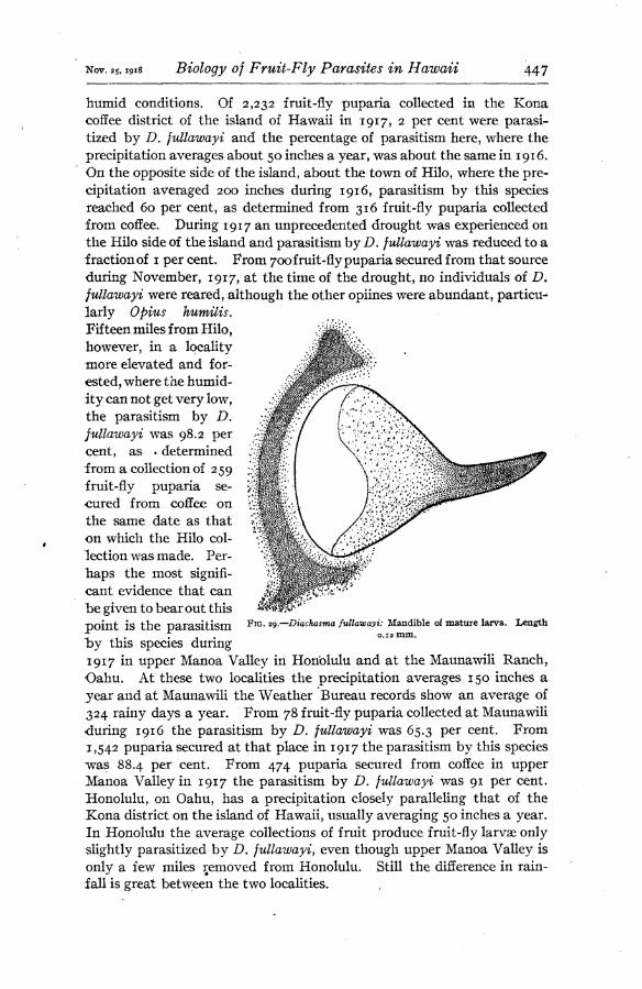
The larvae of the second and third instars have not been distinguished in any respect from those of Z). tryoni. The mature larva, which is the fourth instar, differs from the tryoni larva only in being faintly yellowish in color and in having a slightly darker chitinization of the mandibles (fig. 29). The mature larva of D. fullawayi also hibernates, but the per- centage of larvae hibernating appears to be less than occurs with D. tryoni; the period of hibernation also is shorter. The greatest amount
of adult emergence froïn hibernat- ing individuals occurs during the first three months after the larva ma- tures. From 68 larvae passing into a state of dormancy between August 1, i9i6,and July 31,1917, inclusive, 29, 16, 21, and 1 pupated and be- came adults during the period from the first to the fourth months, re- spectively. One individual hiber- nated for 8 months and 15 days. The greater proportion of the larvae under observation went into hiber- nation during the months of Novem- ber, December, January, February, and March, and the greatest period of adult emergence from hibernat- ing material was in March, April, and May. A much greater degree of hibernation occurs in fruit-fly puparia left in soil or sand than ob- tains in dry glass vials.
PUPA.
FIG. zS.—Diachasma fidlawayi: Cast skin of first- instar larva, showing head characters and egg serosal cells still clinging to ventral surface, length 1 mm.
The pupa may be distinguished from that of Z>. tryoni by the unu- sually long ovipositor sheath which
extends back over the body almost to the head. The duration of the pupa stage is the same as that of D. tryoni, as is also the duration of the combined egg, larva, and pupa stages.
ADULT
The behaviof of D. fullawayi ^fter general distribution in Hawaii is interesting. Localities having high humidity and precipitation have proven especially favorable for this species. At points where the aver- age humidity is low it has had evident difficulty in existing at all, even under very favorable host conditions. An investigation of this species in 1917, after wide distribution and thorough establishment, clearly indicated the particular capacity of this species for life under
Nov. 25,191* Biology of Fruit-Fly Parasites in Hawaii 447
humid conditions. Of 2,232 fruit-fly puparia collected in the Kona coffee district of the island of Hawaii in 1917, 2 per cent were parasi- tized by D. fullawayi and the percentage of parasitism here, where the precipitation averages about 50 inches a year, was about the same in 1916. On the opposite side of the island, about the town of Hilo, where the pre- cipitation averaged 200 inches during 1916, parasitism by this species reached 60 per cent, as determined from 316 fruit-fly puparia collected from coffee. During 1917 an unprecedented drought was experienced on the Hilo side of the island and parasitism by D. fullawayi was reduced to a fraction of 1 per cent. From 700 fruit-fly puparia secured from that source during November, 1917, at the time of the drought, no individuals of D. fullawayi were reared, although the other opiines were abundant, particu- larly Opius humilis. Fifteen miles from Hilo, however, in a locality more elevated and for- ested, where the humid- ity can not get very low, the parasitism by D. fullawayi was 98.2 per cent, as . determined from a collection of 259 fruit-fly puparia se- cured from coffee on the same date as that on which the Hilo col- lection was made. Per- haps the most signifi- cant evidence that can be given to bear out this point is the parasitism hy this species during 1917 in upper Manoa Valley in Honolulu and at the Maunawili Ranch, Oahu. At these two localities the precipitation averages 150 inches a year and at Maunawili the Weather Bureau records show an average of 324 rainy days a year. From 78 fruit-fly puparia collected at Maunawili during 1916 the parasitism by D. fullawayi was 65.3 per cent. From 1,542 puparia secured at that place in 1917 the parasitism by this species was 88.4 per cent. From 474 puparia secured from coffee in upper Manoa Valley in 1917 the parasitism by D. fullawayi was 91 per cent. Honolulu, on Oahu, has a precipitation closely paralleling that of the Kona district on the island of Hawaii, usually averaging 50 inches a year. In Honolulu the average collections of fruit produce fruit-fly larvae only slightly parasitized by D. fullawayi, even though upper Manoa Valley is only a few miles removed from Honolulu. Still the difference in rain- fall is great between the two localities.
FIG. w.—Diachasma fullawayi: Mandible of mature larva. 0.12 mm.
Length

448 Journal of Agricultural Research vol. xv. No. s
In addition to being especially adapted for propagation in wet areas, this parasite, for some unexplained reason, favors certain fruits harboring the host larvae. It is produced in Honolulu most frequently from fly puparia secured from the loquat (Eriobotrya japónica), bestill (Thevetia neriifolia), French cherry (Eugenia uniflora), coffee, and the fruits of Chrysophyllum monopyrenum. It will frequently attack larvae in these fruits to the almost total exclusion of heavily infested fruits of the kamani that may be growing close by. It particularly favors the loquat.
Emergence, mating, and oviposition are not different, so far as can be noted, from these habits as described for D. tryoni. No odor is emitted by the male or female. A meconium is discharged immediately after emergence. The females mav reproduce parthenogenetically, the progeny being always males.
Adults of this species have been kept alive longer than have any of the other parasites. Between December, 1916, and May, 1917, of 235 adults held in confinement in glass tubes and fed honey and water 43 lived over too days, 11 lived 120 days or over, 1 lived for 134 days, and 1 for 141 days. Most of the long-lived individuals were females. Three males lived for 120, 121, and 127 days, respectively. Without food and held in bright light the majority of the adults die in about 55 hours. One male has been kept alive for 103 hours and 2 females for 79 hours.
The proportion of sexes is more favorably in this species than in the case of either D. tryoni or Opius humilis. During 1916 and 1917 a total of 5,528 males and 5,566 females emerged from fruit-fly material collected in the field.
Of 83,304 fruit-fly larvae secured in Hawaii during 1916, 2.1 per cent were parasitized by D. fullawayi. During 1917, as determined from col- lections of 72,139 larvae, the parasitism by this species was 7.3 per cent.
TETRASTICHUS GIFFARDIANUS
This species was brought to Honolulu from West Africa by Fullaway in October, 1914. It was collected and colonized in Africa by Fullaway and Bridwell. It soon became largely propagated and widely distrib- uted in the Territory of Hawaii. Though its life cycle is short and its development as rapid as is that of the opiines, its importance as a para- site prior to 1917 was doubtful. During 1917, however, it had become better established and had increased very considerably the total para- sitism of the fruit fly about Honolulu in fruits having thick pulps, such as oranges, peaches, and guavas. Larvae in these fruits are not easily reached by braconid parasites. It is the most prolific of any of the intro- duced parasites of the fruit fly, and,* under favorable host and weather conditions, may multiply enormously in localized spots. Owing to the short life of the adult, the absence of a hibernating form, its small size, and seemingly poor powers of flight, it rapidly drops off in effectiveness as soon as host material becomes scarce within a short radius.

Nov. 25,1918 Biology of Fruit-Fly Parasites in Hawaii 449
DESCRIPTION AND LIFE) HISTORY
FlG. 30.—Tetrastichus çijfardianus: Egg newly deposited. Length 0.25 mm.
EGG
The newly deposited egg measures from 0.20 to 0.25 mm. in length, is less than a third as broad as long, is pale white, smooth, glistening, cylindrical, and broadly rounded at each end (fig. 30). The eggs are placed in clusters just beneath the larval integument. They are faintly visible from the surface as dark bodies if strong light is transmitted through the larva from below. The egg does not undergo much change in size as the development of the embryo progresses. The duration of the egg stage is from 2 to 2% days. Of 400 eggs deposited January 4,1917, at 11 a. m., all hatched in about 60 hours. Fruit-fly larvae containing eggs of this parasite develop to maturity and form a perfect puparium, in the same manner as do the other parasites under discussion. No fly pupa is formed after the histolysis of the larval tissues in the puparium, and
the egg may hatch either before or after the puparium is formed.
LARVA
There is nothing of unusual interest in the develop- ment of the larva, and only little external difference in the appearance of the instars, with the exception of the gradual increase in size. When first hatched, the larva is about 0.25 mm. long (fig. 31). The body is composed of 13 segments and head, the latter inconspicuous and not unlike the other body segments in general appear- ance. The mandibles are minute, short, curved, broad at the base, and well chitinized. With only slight modification they are constant in the succeeding instars. Most of the food ingested by this parasitic larva con- sists of fat bodies. The larva bears 9 pairs of small open spiracles on body segments 2 to 10, inclusive. The trachea! system is open and becomes air filled as soon as the larva hatches. In the first instar the trunks and branches are very faint and threadlike. The larva
can move only very slowly and sluggishly in all of its instars. The number of larval instars has not been determined, though three
forms have been distinguished. The mature larva may be distinguished readily by the large, heavy tracheae and stigmata. As 8 or 10 larvae nearly always develop in a single host individual, and the food is rap- idlv consumed, the necessity for well-developed tracheae is thus apparent
FIG. 31.—Tetrastichus „ qiffardianus: Newly
hatched larva. Length 0.25 mm.

450 Journal of Agricultural Research Vol. XV, No. 8
even within a few days after the larva hatches. No cannibalism occurs among the larvae of this parasite although from 20 to 30 individuals may be developing simultaneously. In such congested cases they nearly all mature, but result in dwarfed adults. No waste matter is passed by the larva, the midintestine being closed caudally during the entire period of larval development. The larva enters a short prepupal period, from 2 to 3 days in duration, before pupating. During this prepupal period a very small portion of the accumulated waste is voided. This would indicate that the mesenteron and proctodeum become connected
even before the pupa is actually formed. No hibernating form of the larva is known to occur.
The pupa is formed with its head facing the cephalic end of the puparium (fig. 32). In cases where 20 or 30 pupae are packed within the puparium, a few may lie with the position reversed. Of 200 puparia containing pupae of T. giffardianus, examined during August and September, 1917, 3 per cent of the pupae were lying with the head in this reversed position.
The duration of the combined egg, larva, and pupa stages is from 24 to 31 days in the cool months and lasts about 18 days dur- ing the warm months. In January, 1917, 243 adults emerged in from 2.4 to 31 days after the eggs were deposited, with an average period pf 28 days. In April, 1917, 894 adults emerged in from 19 to 26 days after the eggs
were deposited, with an average period of 21 days, and in July, 1917, 455 adults emerged in from 17 to 19 days after the eggs were deposited.
FIG. 32.—Tetrastickus giffardianus: Pupae in normal position and number in fruit-fly puparium opened to show contents. Greatly- enlarged.
ADULT
The adult emerges from the puparium by gnawing a small hole, more or less ragged and circular. Though the puparium may contain from 3 to 30 parasites, usually only one emergence hole is made. The typical position of the emergence opening is shown in figure 33. Occasionally two holes are made, and rarely three. They may be at either end of the puparium or between the extremities. More than one emergence hole usually results from the development of an excessive number of individuals in the puparium. In one rare case wherein 39 parasites emerged from a single puparium, three emergence holes were made. A distinct, thin, brownish pupal skin is left in the puparium after the adult has issued. Although wrinkled and twisted, the skin is an exact replica of the mature pupa. The parasites may twist and turn about

Nov. as, 1918 Biology of Fruit-Fly Parasites in Hawaii 451
in the puparium for many hours before an opening is cut through which they can escape. Males and females all appear to emerge at the same time, for as soon as a sufficiently large exit hole is made, the adult flies come out as quickly as possible, irrespective of sex. The males remain hovering about the puparia but the females immediately crawl away. The meconium, which is developed and retained in the mid- intestine by the larva and held there in the pupa stage, is voided by the adult immediately upon emergence.
Mating occurs as soon as the adults are out of the puparium. The entire process occupies only a few seconds. Females once mated have no difficulty in warding off the males. No mating occurs within the puparium even though the parasites may be actively moving about in it for several hours before they escape. This is proved from the fact that all females taken immediately after emergence and isolated produce parthenogenetic males. One male may mate with several females withiji a few hours. On September 8, 1916, one male was placed with 8 virgin females and left for four days. Each female was then placed in a separate vial and given opportu- nity to oviposit. All produced both male and female progeny. That the male is particularly capacitated for frequent mating is to be expected when the proportion of the sexes is considered, the females always greatly outnumbering the males. During 1916 and 1917 a total of 13,114 males and 47,804 females, or 78.5 per cent, ^¡rdilníT^- emerged from fruit-fly puparia collected in the field fly puparium
, , TT f i showing character- about Honolulu. istic emergence
Unmated females are always arrhenotokous. Prom hole made by adult the proportion of sexes secured from field collections, it parasi e' is shown that the smaller proportion of males readily gain access to and mate with the much larger proportion of females.
OVIPOSITION
Females may begin ovipositing as soon as they emerge, whether mated or not. The mature pupa has well-developed eggs in the ovaries. Four- teen newly emerged females when examined October 20, 1917, contained 73, 70, 72, 60, 73, 66, 71, 81, 65, 45, 58, 64, 61, and 52 mature eggs, re- spectively. The method of oviposition is best understood by an exam- ination of Plate 32, B, C, and D. The female enters the fruit wherever access can be gained through holes, decayed spots, or breaks on the surface. There is no evidence to indicate that the parasite bores into firm pulp or into the skin of the fruit. Once into the fruit, however, the female may become attached to a larva and be drawn through all manner of pulp and juice before her object has been attained. As soon as a larva is located, the ovipositor is quickly brought forward and beneath the
83817°—18 3

452 Journal of Agricultural Research vol. xv, No.8
body and inserted into the body of the larva. In this operation the body is reared up with the front p&ir of legs stretched at full length or often drawn free from the larva. Only one or two seconds are required for the insertion of the ovipositor. The barbs at the tips of the piercing blades now hold the female securely to the larva. By the time the ovi- positor is well inserted the female is standing straight up on the ovi- positor and balancing, often on only one posterior leg and the ovipositor or on the ovipositor alone, as shown in the photomicrographs (PL 32), after which the female moves very little. The legs and wings are held perfectly still, although the larva may frantically bore about in the pulp or escape entirely from the fruit, in an endeavor to dislodge the parasite. To all contortions the female parasite remains indifferent. When the larva is held in position beneath a microscope a rapid transference of liquid and eggs can be seen passing down the ovipositor. After the eggs are inserted the ovipositor is withdrawn and sheathed in from 2 to 3 seconds. The ovipositor is not removed until the female brings all feet down again in touch with the larva or anything that may be conveniently in reach. The female deposits an average of 8 eggs in a cluster, with each inser- tion of the ovipositor. Between September 4 and 21, 1916, 50 separate ovipositions were under careful observation. The time elapsing for each oviposition ranged from 9 to 60 seconds, with an average of 35.6 seconds. The number of eggs deposited at each oviposition ranged from 2 to 16 with an average of 8.6. This average is interesting as a labora- tory experiment, for it was found that from 3,527 adults of T. gifjardianus emerging from 412 puparia collected in the field during 1916 and 1917, the average number emerging from these puparia was 8.6 per puparium. It indicates that the parasite generally oviposits only once into the same larva and that the larva is not again stung by another individual, in the majority of cases. One curious point in the rearing of this species from field material should be emphasized. In every case under actual observation wherein a fly puparium produced several females of T. giffardianus, one male, and occasionally more than one, developed in the same puparium with them. In other words, each time a mated female places a cluster of 6 to 8 eggs or more in a larva, at least one of those eggs is destined to produce a male, for usually 1 male and 6 or 7 females ultimately emerge.
It has been assumed generally that this parasite works mostly about the ground. Certain evidence has been secured to show that it works a great deal above ground also. Of 94 fruit-fly larvae emerging from bes till fruits (Thevetia neriifolia) picked from trees on August 28, 1917, 18.1 per cent were parasitized by T.giftardianus. Of 192 larvae secured from the same host picked from the trees on August 16, 1917, 9.9 per cent were parasitized by 7\ gifjardianus. Forty-five larvae from the same kind of fruit picked from the trees on September 4, 1917, produced 13.3 per cent T. gifjardianus. Of 533 fruits of the kamani picked from trees

Nov. 25,191S Biology of Fruit-Fly Parasites in Hawaii 453
on October 8, 1917, 7.3 per cent were parasitized by T, giffardianus. Parasitism by T. giffardianus of larvae from these fruits, and other varieties picked from the ground on the same dates, was not appreciably higher than that just cited. »The individuals reared from larvae coming from kamani fruits picked from the trees indicate that the females may fly well up above the groun'd, for the lowest fruits of the kamani are usually from 10 to 12 feet above the ground. It is a high, wide-spreading tree.
Although the female oviposits into any portion of the host larva within reach, the majority of the eggs are placed in the posterior half of the body. This is because the larva buries its head into pulp or juice almost as soon as a female touches it, and, in her attempts to cling to the larva and insert the ovipositor, the attachment is not ordinarily made until she has been drawn back toward the posterior end. The female may oviposit into a larva without being actually in contact with it. This occurs when the ovipositor is forced through thin layers of pulp under which the female may detect the presence of a larva. The larva must be close to the female, however. A distinct, brownish, oval scar remains on the larva at the point at which the ovipositor has been inserted. This character is more clearly discernible after the larva has formed into a puparium.
The maximum number of eggs deposited by a given female in confine- ment was 104 (see Table VI). The greater number of eggs is deposited during the first five or six days after the female emerges. They are usually deposited in large lots at intervals 24 to 48 hours apart, rather than in small numbers frequently. The greatest number of eggs de- posited by a given female over a period of 24 hours was 62. No oviposit- ing female has been kept alive longer than 12 days. The great prolific* ness of this species is here seen from its habit of oviposition and in the proportion of sexes above mentioned. With an average life cycle of about 25 days, a deposition of from 90 to 100 eggs within the first five or six days of life, and the resulting progeny 75 per cent female, the multi- plication is seen to be very rapid, ^ruit-fly larvae in the second instar may be successfully parasitized.
Adults fed with honey and water and confined in glass tubes held in. partial darkness have been kept alive from two to three months without difficulty, provided no opportunity is given for oviposition. Of 274 adults emerging on September 3, 1917, one male lived for 61 days and one female for 69 days. Most of the males died between the ages of 3a and 50 days, while the majority of the females died between the ages of 40 and 60 days. Of 267 adults emerging on December 19, 1917, one male lived for 58 days and one female for 102 days. Nine of the females lived over 90 days. This long life was due to the mild temperatures of December, January, and February. Without food this parasite usually dies within 46 hours, although one individual has been held alive for 55 hours.

454 Journal of Agricultural Research Vol. XV, No. 8
TABLE VI.—Daily rate of oviposiiion of Tetrastichus giffardianus, IQIó-IJ
Number of eggs deposited.
Date of oviposi- tion. Female No.
1 2
53 6 7
3
34
4 S 6 7 8 9 10 II 12 13 14 15
October. 0 30
io 10 24 (a)
52 7
9 20
4
12
7 7
21
4
(a')
8
7 10
15
7 3
"s" 10 8 5
14 1
10 1
4
ii , 12 22
30 Si 23 12
13 14 IO 8 ^ It 10 20
&.:::::::::: ïS 9 12
(ß) 17 («) (fl) (a) x& (a) 20
\h (a)
21 * 23 (a) (a)
July. 27 2
53 24 24
6T * / 20 30 7 31 62 7
(a)
10
7
0
August. 1 25 2 4, (G) í 2 2
7 (a) 12 0
Total... 74 104 85 84 34 79 46 52 48 41 30 62 ¡ 62 67 95
a Died.
No. i to 6, inclusive, emerged on October 9, 1916. No. 7 to 11, inclusive, emerged on October 10, 1916. No. 12 to 15, inclusive, emerged on July 27, 1917.
PARASITISM OF THE MELON FLY BY FRUIT-FLY PARASITES
Though great quantities of melon flies, Bactrocera cucurbitae Coquillett, were reared in the laboratory from material collected about Honolulu during the years 1914, 1915, and 1916 after the fruit-fly parasites had become established, no parasites were reared from the material until September 4, 1917. On this date,* 7 normal adults of Tetrastichus gif- fardianus were reared from a single melon-fly puparium. This puparium was part of a quantity secured from cucumbers collected in Honolulu two weeks previously. Before proceeding further it should be stated that a natural parasite of the melon fly was introduced into Hawaii by Mr. D. T. Fullaway in May, 1916. It was well established by the latter

Nov. 25,1918 Biology of Fruit-Fly Parasites in Hawaii 455
part of 1917. The rearing of these 7 individuals of Tetrastichus from the melon-fly puparium was entirely owing to the parasitism of the host, while a larva, first by this natural parasite, Optus fletcheri Silvestri, and later by T. giffardianus. Proof of this is shown by the following results.
Prior to September, 1917, many attempts were made to rear the fruit- fly parasites from melon-fly puparia. No difficulty was experienced in inducing all of the species to oviposit in the larvae, but all parasitized individuals developed into noimal flies. The opiines were easily led to oviposit into melon-fly larvae by transferring these larvae from various vegetables to the fruits of Mimusops elengi, a fruit usually heavily in- fested with fiuit-fly larvae. There was no need for transferring the larvae for T. giffardianus, however, as it readily entered any of the hosts of the melon fly, such as cu- cumber , pumpkin, e t c., and quickly located and oviposited into the maggots. The in- teresting results of these artificial at- tempts to rearfruit-fly parasites from the melon fly may be briefly summarized as follows :
During March, 1917, through the handling of melon-fly larvae as described, a total of 154 eggs of Diachasma tryoni were deposited into 40 melon-fly larvae, 17 eggs of D. fullawayi were deposited into 8 larvae, 33 eggs of Opius humilis were placed in 8 larvae, and 232 eggs of Tetrastichus giffardianus were deposited into 19 larvae. All of these larvae pupated normally. Some wçre mature when stung while others were only about two-thirds developed. From two to five days after each was stung, it was dissected, being now in the pupa stage, and the parasite eggs were counted and carefully examined. None of the opiine eggs hatched and only a small proportion of those of Tetrastichus. In every case, without exception, the opiine eggs developed only a little and then became closely encysted in a mass of transparent cellular material (fig. 34). The egg is at first only thinly surrounded with this encysting sub- stance, but after two or three days it becomes densely inclosed, forming a homogeneous, ovoid body. The egg has then collapsed a little and has become brownish in color. It can be faintly distinguished as it lies in this body. The Tetrastichus eggs and some larvae that managed to hatch be- came similarly encysted. In no case did a Tetrastichus larva live long
FIG. 34.—Diachasma tryoni: Dead encysted egg removed from melon-fly pupa. lycngth of capsule 0.55 mm.

456 Journal of Agricultural Research Vol. XV, No. 8
enough to ingest a visible quantity of food. As the Tetrastichus deposits its eggs in clusters of 8 or 10, they become "encapsulated'' in such groups (fig-35).
The presence of the eggs of these parasites in this dead, inclosed state offers no impediment to the normal development of the melon-fly larva to the adult fly. The adult emerges and carries within its body, usually in the fat masses, these eggs singly or in groups, until death. Death does not seem to be hastened by the presence of the foreign bodies within the body. On May 24 and 25, 1917, 25 melon-fly larvae were stung by fe- males of Diachasma tryoni and eggs deposited in each. The larvae were ¿hen permitted to develop, pupate, and finally produce living adult flies.
These flies were placed in large jars and kept alive for several months. On June 4, 14, and 20, July 10, August 10, and Sep- tember 27, a few of these flies were killed and ex- amined internally for evi- dences of the parasite eggs originally deposited in the larvae. From 1 to 9 dis- tinct, brownish, encysted eggs of D. tryoni were lo- cated in some portion of the body of each of the 25 flies on the foregoing dates.
When, on September 4, 1917, as previously stated, 7 adults of Tetrastichus giffardianus were reared from a melon-fly puparium
secured from the field, after such contradictory experiments had been care- fully completed in March, renewed attempts were made to rear this species in the laboratory from the melon fly. The possible significance of the presence of the newly introduced melon-fly parasite, Opius fletcheri, was immediately recognized. As the host larva was powerless to resist the development of this parasite, it was assumed that a subsequent oviposition by T. giffardianus into larvae previously stung by 0. fletcheri might result in the rearing of adults of T. giffardianus. With this object in view, melon-fly larvae wefe first subjected to attack by O, fletcheri and then by T. giffardianus. As the rearings and examinations from then on showed in every case where both species had parasitized the larvae, the individuals of T. giffardianus developed and emerged as perfect parasites, and in every case in which only the latter oviposited
^IG- 3S-—Tetrastichus Qißardianus; Dead encysted cluster of eggs removed from melon-fly pupa. Greatest width of capsule 0.56 mm.

Nov. 25,1918 Biology of Fruit-Fly Parasites in Hawaii 457
into the larva, no parasite developed and the fly matured and carried in its body the encysted eggs of T. giffardianus. In those cases in which both parasites oviposited, the larvae of 0. fletcheri always died while very young, and all of the larvae of T. giffardianus survived. One case out of 201 occurred, however, wherein a female of O. fletcheri and 5 adults of T. giffardianus emerged from the same puparium. During September, October, and November, 1917, 3,092 adults of T. giffardianus were reared from 201 melon-fly puparia by the foregoing method of double para- sitism. After all of the adults of T. giffardianus had emerged, micro- scopic examinations were made of the contents of each of the 201 empty puparia. In every case the dead latva of O. fletcheri, usually one to a puparium, was found. This was proof absolute that every host indi- vidual had been stung not only by T. giffardianus but also by 0. fletcheri. In conjunction with this, 429 melon-fly larvae were stung by T. giffar- dianus alone. These larvœ all matured to flies, and each of the flies when dissected was found to contain clusters of encysted dead eggs or larvae of T. giffardianus in various parts of the body.
As the duration of the egg of T. giffardianus is shorter than is that of O. fletcheri, cases have been observed in which larvae of the former were developing normally before the egg of the latter had hatched. This indicates that the immunization of the melon-fly larva against parasitic development is destroyed at the time it receives the egg of 0. fletcheri.
This weakened resistance of the melon-fly larva to parasitic develop- ment is caused most probably by toxic substances injected into the larva by the female of O. fletcheri during the deposition of the egg. The reduced resistance of the larva is not caused by any mechanical injury or bacterial infection attending the insertion of the ovipositor. Larvae have been pricked with fine cactus spines or heavily stung by Diachasma tryom and then exposed to attack by T. giffardianus. In such cases the eggs of T. giffardianus invariably became encysted as usual and died. The eggs even became encysted in larvae that had been almost killed by running a cactus spine through the body.
Unusual interest has attended the results of exposing melon-fly larvae to the attack of 0. fletcheri, followed by the exposure of the infested melon-fly larvae to fruit-fly parasites other than T. giffardianus. In all such cases the fruit-fly parasite eggs failed to develop, becoming as tightly encysted as if the O. fletcheri egg had not been present. In other words, no fruit-fly parasites will develop in melon-fly larvae under any conditions, except in the case of the eulophid 7\ giffardianus, and this species will do so only when combined with an egg or larva of the natural melon-fly parasite, O. fletcheri.
O. fletcheri will develop normally in fruit-fly larvae. Laboratory experiments have proved this positively and it has been reared from fruit-fly puparia secured in the field in Hawaii on three separate occasions.

453 Journal of Agricultural Research Vol. XV, Ko. 8
The parasitism of unnatural hosts has been investigated by others. Of Americans, the work of Mr. P. H. Timberlake is of unusual interest.
He succeeded in inducing Limnerium validum Cresson, which is a natural parasite of the fall webworm (Hyphantria cunea Drury), to oviposit in caterpillars of the brown-tail moth (Euproctis chrysorrhoea Linnaeus), the gipsy
FIG.36.—Gaiesussüvestrn:T¿gg,zd3Ly moth (Porthetria dispar Linnaeus), and the old. Length 0.36 mm. rusty vaporer moth (Notolophus antiquus
Linnaeus). In all the larvae of L. validum failed to develop. Timber- lake (7) states that the larvae
fail to survive the protective reactions of the host, which are visibly manifested by an accumulation of active blood cells or amœbocytes around the larvae, the cast eggshells, and even the eggs themselves. The amœbocytes presumably attack the living eggs and larvae, or at least ultimately efface the latter entirely.
RELATION OF THE INTRODUCED PUPAL PARASITES TO THE ESTAB- LISHED LARVAL PARASITES OF THE FRUIT FLY
GALESUS SILVESTRII
In addition to the larval parasites brought into Hawaii to control the Mediterranean fruit fly, a proctotrupid, Galesus silvestrii Kieffer, was introduced by Silvestri in May, 1913. This is a pupal parasite. Though readily breeding in fruit- fly puparia in confinement, it has never been established in the open.
Experiments with this species in the laboratory have shown definitely that it may act either as a primary or a secondary parasite, and that its tendencies are suspiciously more those of a sec- ondary than of a primary parasite. Fruit-fly puparia containing developing larvae of any of the opiine parasites above described or of Tetra- stichus giffardianus, when exposed to attack by G. silvestrii, may produce adults of G. silvestrii. Extensive experiments with combinations of G. silvestrii and the larval parasites of the fruit fly have proved beyond doubt that a female of G. silvestrii will never place her egg loosely in a fruit- fly puparium if that puparium contains already the developing larva of some other parasite. In such cases she invariably feels abput with her ovipositor until she locates the parasitic larva, into which she oviposits. This egg may hatch and ultimately a normal adult of G. silvestrii may emerge from the fully developed parasitic larva and bite its way out. of the puparium.
FIG. 37.—Galesus silvestrii: Newly hatched larva. Length 1 mm.

Nov. 25,1918 Biology of Fruit-Fly Parasites in Hawaii 459
Before discussing the relation of G. silvestrii to the other fruit-fly para- sites, a brief summary of its life history will be given.
Under ordinary circumstances the Galesus female attaches herself to the fruit-fly puparium (PI. 32, E), inserts her ovipositor, and within 3 to 15 minutes places an egg into the fly pupa. The subelliptical egg (fig. 36) hatches in 2 to 3 days. The newly hatched larva (fig. 37) is strictly an in- ternal feeder. Its general structure is strik- ingly like that of the newly hatched opiine larva. The heavily chitinized head, bearing long, curved mandibles, and the soft segmented body strongly suggest a close relationship to the opiines as shown in figures 3, 4, 23, 24, and 28. No trachese occur in the first-instar larva. With the molt to the second instar (fig. 38), the chitinized head is lost and the body is simple, without strong characters, and very much re- sembles the second-instar larvae of the opiines. A third instar occurs, resembling the second. Whether or not there is an intermediate stage between the third and the last has not been determined. With the molt to the last larval instar a strong, well-defined: open trachea! sys- tem appears, with three pairs of large stig- mata. The stigmata are borne on the first three segments back of the head. Heavily chitinized, sharply pointed mandibles are pres- ent (fig. 39). No waste material is voided by the larva. The midintestine is filled with this waste and is closed caudally. Just before pu- pation, in the prepupal stage, the entire waste
accumulation is discharged. Into this meconium the ex- uvium of the mature larva is shed upon pupation. The pupa then lies with the caudal tip embedded in the voided meconial mass and the crumpled larval exuvium. The entire egg, larval, and pupal period averages, in Honolulu, from 25 to 32
days, depending upon the- temperature. The adult emerges usually by pushing off the dorsocephalic cap of the puparium (fig. 40). Mating and oviposition occur immediately upon emergence. The female flies
Fig. 39.—Galesus silves- trii: Mandible of ma- ture larva. length 0.085 mm.
Fig. 38.—Galesus silvestrii: Larva of second instar. Length 1.5 mm.

460 Journal of Agricultural Research vol. xv, NO. S
very little. It remains about the ground, entering crevices and crawling under various objects, apparently in search of fly puparia. In Hawaii it has been reared from the puparia of several muscid species, but it has never been reared from any material collected in the field. The female may reproduce parthenogenetically, the resulting progeny being males.
In the laboratory the female does not discriminate between parasitized and unparasitized fly puparia. She will insert her ovipositor into any puparium. If it contains only a developing fruit-fly pupa, the egg is deposited into the pupa. If the puparium contains a parasitic larva, which may be even one of the small Tetrastichus larvae, her ovipositor will search out this larva and an egg will be deposited into it. A Galesus adult ultimately may mature within this larva. It will, of course, be dwarfed. These small adults are fertile. Such a habit indicates a strong hyperparasitic inclination.
During August, 1917, a total of 195 fruit-fly larvae were parasitized in the laboratory by Diachasma tryoni or Opius humüis. These larvae
were then permitted to develop and to form into perfect puparia. The puparia were then immediately placed in cages containing females of G. sihestrii and left for 2 days. From 5 to 7 days later each puparium was opened and the contents
. .. _ examined. In every case the puparium Fig. 40.—Galesus stlvestru: Fruit-fly pupa- , ,.
rium showing characteristic emergence Contained a dead OpUUe latva Or OCCa- hole made by adult parasite. Greatly sionally a pupa, in each of which was a enlarged. . "' 1 .
rapidly developing, strong larva of G. stives- trii. This, then, was hyperparasitism pure and simple. To prove that these larvae of G. silvestrii ultimately would develop to normal adults, a fresh lot of heavily parasitized fruit-fly larvae was collected in the field. They were variously parasitized by D. tryoni, D. fullawayi, O. humüis, and Tetrastichus giffardianus, and some were unparasitizecf. The puparia thus formed from the larvae were placed with 75 adults of G. sihestrii on September 26 and left for 24 hours. The puparia, 277 in number, were then removed and each placed separately in a small vial to await emergence. From this material 94 adults of G. silvestrii emerged, with both sexes well represented. Each of the 94 puparia producing the adults of G. silves- trii was then carefully dissected, opened, and examined. In 68 of the puparia the empty shell of a mature D. tryoni larva was found, and in each shell the cast mandibles of the larva of G. silvestrii as well as the characteristic prepupal meconial discharge of G. silvestrii was dis- closed. In 3 puparia a similar larval shell of O. humilis was found, each with similar contents. In 1 puparium the shell remains of a mature D. fullawayi larva was found, and in it also were the larval mandi- bles and prepupal meconial discharge of G. silvestrii. In the remain- ing 22 puparia which had produced adults of G. silvestrii remains of the

Nov. 25,1918 Biology of Fruit-Fly Parasites in Hawaii 461
fruit-fly pupa alone occurred, within which were the larval remains of G. süvestrii. The remaining 183, puparia not having been parasitized by G. süvestrii, produced 52 adults of D. tryoni, 14 of 0. humilis, 1 of D. fidlawayi, 12 of T. giffardianus■, 3 fruit flies, and the balance dried up and produced nothing. Thus, in the 94 cases above cited, 72 occurred in which a normal adult of G. süvestrii emerged as a hyperparasite on an opiine parasite of the fruit fly. In 22 other cases it emerged as a normal primary parasite of the fruit fly.
As above mentioned, females of G, süvestrii will always oviposit into larvae of Tetrastichus giffardianus as they lie developing in the fruit-fly puparium. T. giffardianus is a small parasite, being hardly one-half as large as G. süvestrii.. Its larvae are thus very small to receive the com- paratively large egg of G. süvestrii. Larvae of T. giffardianus about 0.7 mm. long are oviposited into by G. silvestrii) and the egg, which is about 0.35 mm. long, hatches and the larva of G. süvestrii develops, pupates, and may ultimately produce a dwarfed adult about 1.5 mm. long. The immature larva of T. giffardianus receiving the egg of G. sü- vestrii is able to develop to maturity. It is killed, however, before it is able to pupate. Females of G. süvestrii have actually oviposited into larvae of T. giffardianus only 2 days old. In all of the experiments conducted to prove the hyperparasitic action of G. süvestrii over T. giffardianus, the eggs of the former were deposited into larvae of the latter not more than 4 days old, the majority being only 2 and 3 days old.
On August 28 and 29, 1917, a total of 209 mature fruit-fly larvae were exposed to attack by T. giffardianus. All of these larvae formed into puparia during the next two days. On August 31 these 209 puparia were placed in a jar containing about 100 females of G. süvestrii and left until September 2. On September 5, 6, and 7, 59 of the puparia were opened, examined, and the contents carefully recorded. Each puparium contained from 3 to 19 well-developed larvae of T. giffardianus, and in every case from 1 to 6 of these larvae were dead and contained an active larva of G. süvestrii in the first, second, or third instar. The remaining 150 puparia were saved for adult emergences and ultimately produced fruit flies, adults of both species, and in some cases both species from the same puparium.
Thus we have positive proof that this proctotrupid may be strongly hyperparasitic upon any of the established parasites of the fruit fly that are now contributing toward its control in Hawaii, and that it may act with equal freedom as a primary parasite of the fruit fly.
PACHYCRKPOIDEUS DUBIUS
A pteromalid, Pachycrepoideus dubius Ashmead, introduced from the Philippines by Mr. D. T. Pullaway in 1914 as a dung-fly parasite, has

462 Journal of Agricultural Research Vol. XV, No. &
been reared occasionally from fruit-fly puparia. It is not an important fruit-fly parasite, but the interesting relations that may exist between this and the other fruit-fly parasites has led to a laboratory study of this species, with results worth recording.
The female oviposits into the puparium, the egg being placed on the surface of the developing pupa within, and its larva develops on the pupa as an external feeder. Thus from the first it bears a well-developed trachea! system. The fly pupa is killed shortly after the parasitic larva hatches and the larva continues to feed upon the decomposing pupa. It feeds much in the sense of a scavenger. It does not consume all of the food in the puparium. The meconium is voided just before pupa- tion and the pupa lies partly buried in the decayed remains of the fly
pupa and its own meconium. The adult parasite emerges by cutting a small, circular hole in the puparium (fig. 41) and is immediately ready for mating and oviposition. Another minute meconium is discharged immediately upon emergence. The female may reproduce^ partheno- genetically with the resulting progeny all males. The entire period from deposition of egg to emergence of adult lasts only from 15 to 16 days.
As the larva of this parasite is an external feeder within the puparium from the first, and the larvae of the other parasites are all internal feeders, the relationship between them is interesting. In a certain sense this parasite has been proved a primary, a secondary, or a tertiary parasite. As a primary it develops on the fruit-fly pupa, as a sec-
/^S
acteristic emer- gence hole made by adult parasite. Greatly enlarged.
FIG. 41.—Pachycre- poideus dubius: Fruit-fly pupari-
^ert^lmeí ondary it develops on larvae or pupae of ~ Tetrastichus giffardianus, Optus humilis, Diachasma tryoni, D, fulla- wayiy or Galesus silvesirii as they occur as parasites in the fruit-fly puparium, and as a tertiary parasite it will of
course develop on a larva of G. silvesirii, which in turn has been feeding in the larvae of the above-mentioned opiines or T. giffardianus. In the laboratory, in large jars, this parasite indiscriminately oviposits into fruit-fly puparia to bring about all of the above conditions of parasitism and may be expected to act similarly in the field, when able to reach fruit-fly pu- paria. Fortunately it does not show any decided capacity for penetrat- ing soil in search of fly puparia and is probably more attracted to situations harboring the dung-feeding species. The data demonstrating the various capabilities of this species as a primary, secondary, or tertiary parasite may be summarized as follows :
During November 24 and 25, 1917, a total of 341 fruit-fly puparia were secured from kamani fruits collected about Honolulu. These puparia were variously parasitized by <9. humilis, D. tryoni, D. fullawayi, and T. giffardianus. On November 29 they were placed in a jar con-

Nov. as, 1918 Biology of Fruit-Fly Parasites in Hawaii 463
taining about 100 females of P. dubius and left until December 2. They were then removed and on December 7 and 8 dissected and carefully examined. Thirty-one contained a dead larva or pupa of D. tryoni on which was feeding a mature larva of P. dubius; 73 contained a dead larva or pupa of 0. humilis on which fed a larva of P. dubius; 2 con- tained a dead larva of D. fullawayi on which was a larva of P. dubius; 22 contained from 3 to 31 larvae of T. giffardianus, some of which were in each case dead and on which fed a larva of P. dubius; 30 contained decomposed fruit-fly pupae parasitized by P. dubius alone. Of the re- maining 183 puparia, 46 were unparasitized, 88 were parasitized by only 0. humilis, 31 by only D, tryoni, and 18 by only T. giffardianus.
Again, on October 23, 50 fruit-fly puparia containing developing larvae of O. humilis and JD. tryoni were exposed to attack by (3. silvestrii for one day. On October 30 they were placed in a jar containing females of P. dubius and left for two days. They were then opened and ex- amined on November 10 with the following interesting results: Thirty- five contained dead larvae of G. silvestrii lying in dead larvae of O. hu- milis or D. tryoni, on the whole of which was feeding in each case a larva of P. dubius.
The relation of the P. dubius to T. giffardianus is also of interest. Very frequently a puparium containing 15 or 20 larvae or pupae of T. giffardianus and exposed in the laboratory to attack by P. dubius will yield a normal adult of P. dubius and several normal adults of T. giffardianus. On November 23 and 24, 83 fruit-fly larvae were para- sitized by T. giffardianus in the laboratory. The 83 puparia forming from these were then placed with adults of P. dubius and left for three days. On December 8 the puparia were opened and examined. In 23 cases examined, from 4 to 20 dead larvae of T. giffardianus were found, and on these in each case was feeding a mature larva of P. dubius; in 30 puparia from 1 to 10 living pupae of T. giffardianus occurred together with from 3 to 20 dead larvae of that species upon which were feeding larvae of P. dubius, one in each instance; in the remaining 33 puparia from 7 to 19 individuals of T. giffardianus were developing in the ab- sence of individuals of P. dubius.
In conclusion, it should be borne in mind that G. silvestrii is not known to be established in Hawaii as yet, and that P. dubius probably only parasitizes a fraction of 1 per cent of the fruit-fly puparia in the field, but that the relations between these and the other früit-fly parasites, as detailed in the foregoing pages, can be expected as a natural sequence if they ultimately adapt themselves to Hawaiian conditions and become thoroughly established.

464 Journal of Agricultural Research vol. xv, NO. S
PHKIDOLE MEGACEPHAIvA AS A PREDACIOUS ENEMY OF THE FRUIT FLY
Although several species of ants are common in the Hawaiian Islands, the cosmopolitan antPheidole megacephala'Pahncins greatly outnumbers all others and is enormously abundant throughout the islands at low elevations. . It is quite probable that it checks and greatly limits the increase of several of these species. It is quickly drawn to any fresh or decaying animal matter and the soft body of the fruit-fly larva falls an easy prey to it. It has been generally assumed that this ant destroys large numbers of fruit-fly larvae throughout the year. Very little defi- nite data are on record, however, covering investigations on this rela- tionship. Though the following records are not extensive, they all point in one direction and are significant.
Four hundred and eighty-eight kamani fruits collected from a certain tree in Honolulu on December 3, 1916, were spread out under the tree on a large piece of canvas covered with 1 inch of sand and left fo^ 10 days. By this time practically all of the contained fruit-fly larvae had developed and pupated in the sand beneath the fruits or had been carried away by ants. The sand was then sifted and the fruit-fly puparia were counted. A total of 606 fruit-fly puparia were obtained. This was an average of 1.2 larvae per fruit. During November and December 969 more fruits were gathered from the same tree and taken to an ant-proof insectary to be held in boxes of sand for 10 days. From these 4,895 fruit-fly puparia were obtained, or an average of 5 larvae per fruit. Here four times as many larvae per fruit developed in the lot where the ants were excluded. From 1,301 fruits of the same kind col- lected from another tree in Honolulu in November, 1917, and similarly placed and held for 10 days on canvas under the tree, 3,742 fruit- fly puparia were ultimately obtained. From the same tree and col- lected at approximately the same date, 909 fruits were taken and brought to the ant-proof insectary. Fiom these 10,119 fly puparia were secured. Here again almost four times as many larvae per fruit were obtained from the fruits held away from the ants. On January 25, 1918, 664 loquats were collected from several trees in a certain yard in Hono- lulu and divided in half, one half going out on the canvas and the othet part to the insectary. From the lot on the canvas, after 10 days' exposure, 156 fruit-fly puparia were gathered, and from those in the insectary a total of 840. In this case the ant-proofed lot produced over five times as many puparia. On January 29, February 2, and February 6, 1918, an aggregate of 3,100 fruits of the black myrobalan was collected from one tree. This total was divided in half as soon as each lot had been collected, the two portions being handled as in the above cases. From the fruit held in the insectary 6,668 fly puparia were obtained, while only 4,385 puparia were ultimately secured from the half placed out on

Nov. 25,19x8 Biology of Fruit-Fly Parasites in Hawaii 465
the canvas. Here the lot held away from the ants was i}4 times as great as that exposed to the ants.
The foregoing should represent a very conservative estimate of the destruction of fruit-fly larvae by this ant. It shows consistently that from one-third to four-fifths of the larvae developing in all fruits in the field very probably never mature to adult flies.
LITERATURE CITED
(1) BACK, E. A., AND PEMBIîRTON, C E. 1915. PARASITISM AMONG THE I,ARV^ OF THE MEDITERRANEAN FRUIT FLY (C.
CAPITATA) IN HAWAH DURING 1914. In Rpt. Bd. Comrs. Agr. and For. Hawaii, [1912]/14, p. 153-161.
(2) 1916. PARASITISM AMONG THE lyARV^E OF THE MEDITERRANEAN FRUIT FLY (c,
CAPITATA) IN HAWAII DURING 1915. In Jour. Econ. Ent.} v. 9, no. 2, p. 30Ó-3II.
(3) 1918. THE MEDITERRANEAN FRUIT FLY IN HAWAII. U. S. Dept. AgT. Bui. 536,
II9 p., 21 pi., 24 fig.
(4) PEMBERTON, C. E., AND WILLARD, H. F. 1917. NEW PARASITIC CAGES. In Jour. Econ. Ent., v. 10, no. o, p. 525-527,
3 fig., 1 pi.
(5) 1918. FRUIT-FLY PARASITISM IN HAWAII DURING 191Ó. In Jour. Agr. Research;
v. 12, no. 2, p. 103-108.
(6) 1918. INTERRELATIONS OF FRUIT-FLY PARASITES IN HAWAII. In Jour. Agr.
Research, v. 12, no. 5, p. 285-295, 4 pi., 13 fig. (7) TlMBERLAKE, P. H.
1912. EXPERIMENTAL PARASITISM: A STUDY OF THE BIOLOGY OF LIMNERIUM VALIDUM (CRESSON). U. S. Dept. Agr. Bur. Ent. Bul. Tech. Ser. 19,
pt. 5, p. 71-92, fig. 32-41.

PLATE 32
Oviposition of fruit-fly parasites:
A.—Diachasma tryoni ovipositing into fruit-fly larva in fruit of Mimusops elengL B, C, B.—Tetrastichus giffardianus ovipositing into fruit-fly larva. E.—Galesiis silvestrii ovipositing into fruit-fly puparium.
(466)

Biology of Fruit-Fly Parasites in Hawai PLATE 32
Journal of Agricultural Researcl* Vol. XV, No. 8