vol 27, no 2, 1979
TRANSCRIPT
Acknowledgment
C a s e s for the clinicopathological conferences at Henry Ford Hospital are selected by the CPC Planning Committee and scheduled bimonthly. The goal is to provide an educational experience. The diagnosis does not have to be mysterious, nor is it intended to be critical ofthe responsible physicians. Members of the Committee are: Gerald Fine, MD, Chairman (Pathology), Mohammed Ansari, MD (Surgery), Philip J. Dorman, MD (Internal Medicine), Kenneth A. Greenawald, MD (Pathology), Dorothy M. Kahkonen, MD (Internal Medicine), Ratnakar Kini, MD (Pediatrics), and Julius M. Ohorodnik, MD (Pathology).
The Editorial Board of the Journal would like to thank the Committee for its cooperation in makingthis series possible.
113
Henry Ford Hosp Med Journal
Vol 27, No 2, 1979
Henry Ford Hospital Clinicopathological Conference
1. Acute Respiratory Failure in a 33-year-old Man with Granulomatous Lung Disease
Participants: Protoco l : Dr. George C. Bower, Department of Internal Med ic ine , Div is ion of Pulmonary Med ic ine
Discussant: Dr. W i l l i a m Conway, Department o f Internal Med ic ine , Div is ion of Pulmonary Med ic ine
Radiology: Dr. Michae l Engel, Department of Diagnostic Radiology
Pathology: Dr. Julius M . Ohorodn i k , Department of Pathology
Case Presentation
A 33-year-old black man was transferred to Henry Ford Hospital in August, 1975 from another hospital where he had been for ten days with a diagnosis of possible tuberculosis. Three weeks earlier, he had begun to cough and produce dark green sputum that was occasionally blood tinged. He denied chest pain, fever, chills, sweating, weight loss, or anorexia. He had not iced some breathlessness during the preceding year but this was not aggravated during the recent illness. He had not worked for a number of years, smoked one pack of cigarettesdaily, and drankone six-pack of beer daily. There was no history of TB exposure, but he stated that he had had penumonia three times in the past, the last time in October, 1974. He took phenytoin and phenobarbital for a seizure disorder.
On examination he did not appear ill. Blood pressure was 130/80, pulse was regular at 72 / min, and his respirations were normal. A 1.5 cm nodule was noted in the left parotid area. The chest exhibited good expansion. Diminished breath sounds and increased fremitus were noted in the area ofthe right middle lobe. A few rales were heard at both bases. The rest of the exam ination was unremarkable.
Laboratory examinations included a hemoglobin of 14.7cm, white blood cell count 4,500 with a normal differential, and normal platelet and reticulocyte counts. Urinalysis was normal, creatinine 1.0 mg/d l , calcium 9.2 mg/d l , and phosphate 4.4 mg/d l . LDH, SCOT, and bilirubin were normal. Alkaline phosphatase was 64 onits (normal 9-38). Serum albumin was 3.4 and globulin 3.9 gm/d l . Protein electrophoresis yielded normal values for globulin fractions. Urinary calcium was 121 mg/24 hours. Numerous sputum studies were negative for acid-fast bacilli on smear and culture. No pathogenic fungi were cultured. Complement fixation
tests for histoplasmosis, blastomycosis, and coccidioidomycosis were negative. PPD-T, SKSD, monilia, histoplasmin, and coc-cidioidin skin tests were negative.
A chest radiograph showed a diffuse infiltrative process in both lungs, most pronounced in the right upper lobe, and consolidation in the middle lobe and in partof the right lower lobe. There was a suggestion of an enlarged azygos node and the left hilum appeared generous. Laminograms revealed the middle lobe bronchus to be patent but narrowed. Liver/spleen scan showed homogenous uptake in the liver without hepatosplenomegaly. Spleen size appeared normal.
Bronchoscopy revealed a friable, diffuse cobblestone and granular appearance of the mucosa, with increased secretions. The right middle lobe bronchus was narrowed by a heaped-up process. Biopsies revealed noncaseating granulomatous inftammation consistent with sarcoidosis. Pulmonary function studies showed a forced vital capacity of 2.41 (predicted 5.43). FEV .̂o was 1.32 I (predicted 4.55). Maximum voluntary ventilation was 48 L/min (predicted 186). Following a bronchodilator, the FVC was 3.08, FEV,.o was 1.43, and M W was 54. Residual lung volume was 3.0 I (predicted 1.92), total lung capacity 5.41 (predicted 7.47), and diffusing capacity 21.7 (predicted 37). Seven-minute nitrogen washout yielded a value of 4.2%. Resting arterial blood gases showed a PO2 of 57 mm Hg, saturation 91%, PC02 26 mm Hg, pH 7.43. A-a gradient for oxygen was 50 mm Hg. During exercise, oxygen consumption was 1.58 1 /min , respiratory quotient 0.93, and ventilatory equivalent for oxygen was 33. V Q / V J rose from 0.41 at rest to 0.52 during exercise and the A a-gradient rose to 59. The P02 during exercise was 43, saturation 74%, PC02 42, and pH 7.32. The shunt fraction was calculated at 18.7%.
The patient was treated for one week with Keflex for presumptive infective bronchitis. No ocular lesions of sarcoidosis were found.
114
HFH Clinicopathological Conference
Sputum cytology was reported suspicious for malignant cells, but repeat studies were negative. He was discharged on prednisone 80 mgq.o.d., isoniazid, pyridoxine, phenytoin, and phenobarbital.
The patient did not return for any clinic appointments. His final hospital admission was six months later on February 14, 1976 when he came to the Emergency Room complaining of wheezing and marked increase in shortness of breath for one week. Sputum had increase in amount and was described as red for one week. He denied fever and chills, had not been taking medications, and had continued to smoke and drink heavily. He was in acute respiratory distress, was noted to have an injected pharynx, poor teeth, one small left axillary lymph node, diffuse rales throughout his chest, and scattered wheezes. Cardiac examination was unremarkable, blood pressure was 110/60, pulse 112, respirations 32 /min , and temperature 37°C. The remainder of the examination was also unremarkable. Hemoglobin on admission was 14.8, white blood cell count 28,200, and electrolytes, BUN, and creatinine were normal. An arterial blood study with the patient receiving2 l /min of oxygen by nasal specs showed a P02 of 33 mm Hg, saturation 61%, PC02 28 mm Hg, and pH 7.35. With a 60% rebreathing mask, P02 was 73, saturation 94%, PC02 was 29, and pH 7.36.
The patient was transferred to the medical intensive care unit. Sputum showed numerous gram-posi t ive cocc i in pairs and chains, some gram-negative bacilli, and a few large gram-positive cocci and tetrads. Sputum smears were negative for AFB. Chest x-ray showed marked reticular and nodular changes, espcially in the upper lung fields, normal heart size, possible right hilar enlargement, and some clearing of the right middle lobe infiltrate. Follow-up film showed increased perihilar infiltrates. He was treated with oxygen and was afebrile but continued to have tachycardia and a paradoxical pulse of about 26. P02s remained in the range of 50 to 60 mm Hg on high concentrations of oxygen, and PC02s ranged from 20 to 30 mm Hg. Electrocardiogram showed an incomplete right bundle branch block. Large doses of Solumedrol were administered along with IV aminophylline and IPPB with bronchodilators. By February 16 the respiratory rate was 40 /min , blood pressure 100/70, and heart rate 130/min. He appeared tired and in considerable distress. Sputum cultures were reported as showing rare staph aureus, numerous Neisseria, micrococci, and rare Hemophilus inftuenzae. Cephazolin was begun. An S3 gallop was heard. He was started on digoxin and furosemide and a Swan-Canz catheter was inserted; right artrial pressure was 12/4, right ventricle 76 /0 , pulmonary artery 76/12, and wedge pressure 11 mm Hg. Shortly thereafter, he became bradycardic and unresponsive. Resuscitation was instituted, and he vomited and aspirated. He was intubated, placed on a respirator and developed atrial flutter with a 2 to 1 block. Despite the use o fa transthoracic pacemaker, blood pressure could not be obtained and the patient died.
Clinical Discussion
Dr. Conway:
This young black man with a history of alcohol abuse, cigarette smoking, and seizure disorder was inftially hospitalized and found to have a diffuse granular bronchitis.
pulmonary infiltrates, and a combined obstructive and restrictive lung disease wfth hypoxemia. He was treated for sarcoidosis but was unreliable in follow-up. Five months later he presented to the hospital wfth diffuse pulmonary infiltrates and died.
The first procedure in any patient with a suspected diagnosis of sarcoidosis should be to verify the diagnosis, since a large number of diseases can mimic sarcoidosis histologically. Three criteria should be fulfil led: first, the histology should be compatible; second, theclinical presentation and course should be compatible; and finally, other causes of granulomatous disease should be excluded. This patient had compatible histology which was obtained from a bronchial and transbronchial biopsy, but before commenting on his clinical presentation, we should review the x-rays.
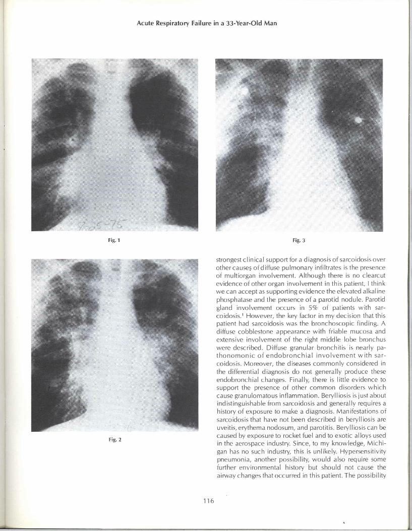
Dr. Engel:
The initial x-ray in August, 1975 (Figure 1) shows a somewhat hazy appearance inthe perihilar regions wfth densfties extending into the periphery. An infiltrate is present in the middle lobe, resulting in the so-called silhouette sign. There is a diffuse, somewhat linear to nodular pattern throughout both lungs wfth more patchy densities in the right upper lobe. Laminograms do not demonstrate definite adenopathy though there is suggestive fullness inthe perftracheal region. The intermediate bronchus is narrowed and the middle lobe is reduced in size. At the time of the final admission in February, 1976 (Figure2), bilateral infiltrates are presentwith patchy densities in the right upper lobe, more striking than on the original films. The lower lung fields are relatively clear. Just before the patient died, the pulmonary infiltrates progressed to a picture resembl ing the pattern of pulmonary edema but the heart was not enlarged (Figure 3).
Dr. Conway:
The bilateral reticulo-nodular characteristics ofthe infiltrate are consistent wfth sarcoidosis and are best classified as stage 3 disease. The atypical features include atelectasis and infiltration in the right middle lobe. When this occurs in sarcoidosis it is almost always due to extensive endobronchial disease with bronchial obstruction. Another atypical feature is the relative lack of symmetry in the parenchymal infiltrates, which can occur in around 5-13% ofcases, especially when the disease is evolving or regressing. When asymmetry occurs in the upper lobes, it suggests the possibility of tuberculosis. Despite these features, the x-rays are still compatible with sarcoidosis.
The clinical presentation was also compatible wfth sarcoidosis. The patient was young; 70% of individuals wfth sarcoid are under 40. He was black, and the disease is more common in blacks. Also, anergy was present, which occurs in a high percentage of patients wfth the disease. The
115
Acute Respiratory Failure in a 33-Year-Old Man
Fig.1 Fig. 3
Fig. 2
Strongest clinical support for a diagnosis of sarcoidosis over other causes of diffuse pulmonary infiltrates is the presence of multiorgan involvement. Although there is no clearcut evidence of other organ involvement in this patient, I think we can accept as supporting evidence the elevated alkaline phosphatase and the presence o fa parotid nodule. Parotid gland involvement occurs in 5% of patients with sarcoidosis.^ However, the key factor in my decision that this patient had sarcoidosis was the bronchoscopic finding. A diffuse cobblestone appearance with friable mucosa and extensive involvement of the right middle lobe bronchus were described. Diffuse granular bronchftis is nearly pa-thonomonic of endobronchia l involvement w i th sarcoidosis. Moreover, the diseases commonly considered in the differential diagnosis do not generally produce these endobronchial changes. Finally, there is little evidence to support the presence of other common disorders which cause granulomatous inflammation. Berylliosis is just about indistinguishable from sarcoidosis and generally requires a history of exposure to make a diagnosis. Manifestations of sarcoidosis that have not been described in berylliosis are uveftis, erythema nodosum, and parotftis. Berylliosis can be caused by exposure to rocket fuel and to exotic alloys used in the aerospace industry. Since, to my knowledge, Michigan has no such industry, this is unlikely. Hypersensitivity pneumonia, another possibility, would also require some further environmental history but should not cause the airway changes that occurred in this patient. The possibility
116

HFH Clinicopathological Conference
of a fungal or tuberculous infection was excluded by the number of specimens sent. If the endobronchial changes were misinterpreted and were, in fact, caused by an infectious agent, the cultures certainly should have been positive, as they are in a high percentage of patients with endobronchial tuberculosis. Cancer and lymphoma are sometimes considered in the differential diagnosis of sarcoidosis when hilar and paratracheal adenopathy are present, but these findings were not definite in this patient, ft is important to differentiate carcinoma from sarcoid when the histologic studies are obtained from a node that could possibly be draining a site of cancer. Since the biopsy here was obtained directly from bronchial and lung tissue, there should not be much reason to confuse it with a carcinomatous process.
The pulmonary function studies showed a moderate restrictive defect with severe airflow obstruction. Most published reports of lung function in sarcoidosis have stressed the occurrence of a restrictive defect. Recently, attention has been drawn to three types of airway disease. The first is small ainA'ays dysfunction, which can even occur in patients who have only bilateral hilar lymph adenopathy, i.e., stage 1 disease.^ Secondly, airway disease occurs in the terminal fibrotic stage of sarcoidosis. In a recent report of 16 patients with extensive fibrosis due to sarcoidosis and little or no smoking history, 75% had mild to severe airflow obstruction.^ The changes were attributed to the narrowing and distortion of the airways caused by bronchial and peribronchial fibrosis. Manifestations of this type of involvement include cough, sputum product ion, wheez ing, recurrent pneumonia, and atelectasis. Finally, a i r f low obstruction can occur in the presence of endobronchial infiltration by sarcoid granulomas. This is the type of involvement I believe our patient had. Because endobronchial involvement can occur in any stage ofthe disease, it has been suggested that bronchoscopy be performed and steroid therapy instftuted in patients with stage 1 disease who have airway symptoms." Our own experience is that about halfthe patients with stage 1 disease have histologic evidence of endobronchial disease.
At this point, we should ask what the natural course of disease could be in this patient. The prognosis in any case is unpredictable but, in general, two thirds recover completely with minimal residual lesions. Indications for a good prognosis include an acute onset, the presence of hilar adenopathy alone, onset w i th erythema nodosum, and an asymptomatic state. Poor prognostic signs include insidious onset with pulmonary parenchymal involvement, cutaneous or bone involvement, or involvement of more than three organ systems. In terms of the radiographic stages of the disease, individuals with stage 1 disease alone stand a greater chance for recovery than those with stage 3 disease
(parenchymal involvement alone).' Thus, this case clearly had a poor prognosis.
Finally, was sarcoidosis the only cause of death? Only 5% of patients with sarcoidosis eventually die ofthe disease, usually 3 to 20 years after diagnosis.' The most common cause of death is respiratory failure due to progressive and severe fibrosis. Although this patient did have evidence of respiratory failure, the radiographic picture is not that of end-stage fibrosis. Cor pulmonale may cause death, but ft usually occurs after a decade of disease and is associated with severe fibrosis. It is generally fatal within a year after the first episode of heart failure. However, this patient had no radiographic or electrocardiographic evidence of cor pulmonale, and the d isease was not present for the usual length of time required forthis complication. Hemorrhage dueto bronchiectasis or a fungus ball is also an occasional cause of death, but the severity of hemoptysis in this case was not sufficient to cause death. Unusual causes of death include renal failure due to hypercalcemia, hepatic failure due to granulomatous hepatitis, and myocardial involvement. While autopsy evidence of card iac involvement is present in 20%, the importance ofthis finding has been stressed only recently. Manifestations are arrhythmia, most commonly ventricular, heart block which is frequently manifested by syncope or sudden death, congestive heart failure due to extensive myocardial infiltration, and, much less often, papillary muscle dysfunction. The presence of myocardial involvement is felt to be incidental in one third of autopsy cases and is felt to be a significant abnormality or direct cause of death in about two thirds.'^ In one autopsy series of cardiac sarcoidosis, 75% of the patients who died, died suddenly,^ and the remainder were related to progressive heart failure. Others have shown that in myocardial sarcoidosis all sudden deaths were preceded by a cardiac rhythm disturbance. In fact, the presence of arrhythmias was felt to be the most predictive factor for determining whether myocardial sarcoidosis was present. Ninety percentof the sudden death victims in Roberts' series' had some prior manifestations of cardiac sarcoidosis, usually an arrhythmia or the presence of heart block. Although incomplete bundle branch block was described in our patient, I don't think there was significant cardiac involvement.
The most common nonsarcoid causes of death are infections, such as tuberculosisor bacterial pneumonia. The final illness in our patient was manifested by increased secret ions, wheezing, dyspnea, and hemoptysis. There was marked leukocytosis, severe hypoxemia with a large shunt, and increase in the pulmonary infiltrates developing over a short period of time. Sarcoidosis does not typically produce such an acute deterioration, and 1 believe that there must have been a superimposed process such as bacterial pneumonia. The large terminal paradoxical pulse was probably a
117

Acute Respiratory Failure in a 33-Year-Old Man
manifestation of severe airflow obstruction. Certainly, there was no indication of cardiac enlargement or pericardial effusion. The hemodynamic data suggest that pulmonary hypertension was present, probably on the basis of hypoxia combined with parenchymal lung disease ratherthan direct pulmonary vascular involvement with sarcoid granulomas.
What could have been the immediate cause of death in this patient? The development of an arrhythmia or heart block should have been picked up in the intensive care unit, and I doubt that this was present. A major vessel rupture is unlikely in this young patient. Pulmonary embolism should be considered but discarded because of his age, ambulatory state until two days before admission, and absenceof heart failure. Mural thrombi have been reported in sarcoidosis but usually require extensive myocardial involvement which is not 1 ikely in this case. The term inal event seems to have been the development of progressive hypoxemia. I believe we wi l l find extensive sarcoidosis involving the pulmonary parenchyma, bronchi, hilar lymph nodes, and possibly the pulmonary vasculature and liver. I suspect the presence of bacterial pneumonia as well.
Pathology Discussion
Dr. Ohorodnik:
At autopsy the lungs were twice their normal weight, diffusely fibrotic, and contained fresh emboli in the secondary branches of the arteries in both lower lobes and with early hemorrhagic infarction. The heart was slightly enlarged and the right side dilated. The superior vena cava near its entrance into the right atrium was thickened and involved by an inflammatory process. A review of the transbronchial biopsy done during life confirms the presence of noncaseating granulomas consisting of histiocytes and multinucleated giant cells wfth no necrosis (Figure 4). In some areas of the lung, extensive parenchymal alteration occurred. Considerable scarring and contraction was present, some of which extended to the pleural surface. Many of the granulomas were confluent. In the early or active stage one can see the lymphocytes at the periphery of the lesions, but in older lesions more fibrosis occurs and remnants of the granulomas are not specific. Microscopic examination of the smaller airways showed, in some, a
Fig. 4 Transbronchial biopsy. Noncaseating granuloma.
118
HFH Clinicopathological Conference
''*y
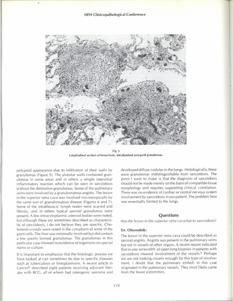
Fig. 5 Longitudinal section of bronchiole. Intraluminal polypoid granulomas.
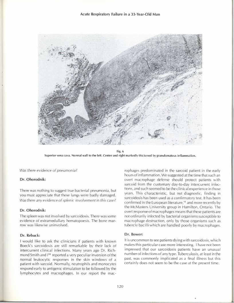
polypoid appearance due to infiltration of their walls by granulomas (Figure 5). The alveolar walls contained granulomas in some areas and in others a simple interstitial inflammatory reaction which can be seen in sarcoidosis without the distinctive granulomas. Some of the pulmonary veins were involved by a granulomatous angiitis. The lesion in the superior vena cava was involved microscopically by the same sort of granulomatous disease (Figures 6 and 7). Some of the intrathoracic lymph nodes were scarred and fibrotic, and in others typical sarcoid granulomas were present. A few intracytoplasmic asteroid bodies were noted, but although these are sometimes described as characteristic of sarcoidosis, I do not believe they are specific. Cholesterol crystals were noted in the cytoplasm of some ofthe giant cells. The liver was minimally involved but did contain a few poorly formed granulomas. The granulomas in this particular case showed no evidence of organisms on special stains or culture.
It is important to emphasize that the histologic process we have looked at can sometimes be due to specific diseases such as tuberculosis or histoplasmosis. A recent article in Cancer* described eight patients receiving adjuvant therapy with BCG, all of whom had osteogenic sarcoma and
developed diffuse nodules in the lungs. Histologically, these were granulomas indistinguishable from sarcoidosis. The point I want to make is that the diagnosis of sarcoidosis should not be made merely on the basisof compatible tissue morphology and requires supporting clinical correlation. There was no evidence of cardiac or central nervous system involvement by sarcoidosis in our patient. The problem here was essentially limited to the lungs.
Questions
Was the lesion in the superior vena cava due to sarcoidosis?
Dr. Ohorodnik:
The lesion in the superior vena cava could be described as sarcoid angiitis. Angiitis was present in the pulmonary veins but not in vessels ofother organs. A recent report indicated that in one series 69% of open lung biopsies in patients with sarcoidosis showed involvement of the vessels.̂ Perhaps we are not looking closely enough for this type of involvement. I doubt that the pulmonary emboli in this case originated in the pulmonary vessels. They most likely came from the lower extremities.
119
Acute Respiratory Failure in a 33-Year-Old Man
Fig. 6 Superior vena cava. Normal wall to the left. Center and right markedly thickened by granulomatous inflammation.
Was there evidence of pneumonia?
Dr. Ohorodnik:
There was nothing to suggest true bacterial pneumonia, but you must appreciate that these lungs were badly damaged. Was there any evidence of splenic involvement in this case?
Dr. Ohorodnik:
The spleen was not involved by sarcoidosis. There was some evidence of extramedullary hematopoesis. The bone marrow was likewise uninvolved.
Dr. Rebuck:
I would like to ask the clinicians if patients with known Boeck's sarcoidosis are still remarkable by their lack of intercurrent clinical infections. Many years ago Dr. Richmond Smith and V° reported a very peculiar inversion ofthe normal leukocytic responses in the skin windows of a patient wfth sarcoid. Normally, neutrophils and monocytes respond early to antigenic stimulation to be followed by the lymphocytes and macrophages. In our report the mac
rophages predominated in the sarcoid patient in the early hours of inflammation. We suggested at the time that such an overt macrophage defense should protect patients wfth sarcoid from the customary day-to-day intercurrent infections, and such seemed to be the d i n ical experience in those years. This characteristic, but not diagnostic, finding in sarcoidosis has been used as a confirmatory test, ft has been confirmed in the European literature," and more recently by the McMasters University group in Hamilton, Ontario. The overt response of macrophages means that these patients are not ordinarily infected by bacterial organisms susceptible to macrophage destruction, only by those organisms such as tubercle bacilli which are handled poorly by macrophages.
Dr. Bower:
It is uncommon to see patients dying with sarcoidosis, which makes this particular case more interesting. I have not been impressed that our sarcoidosis patients have an unusual numberof infectionsof any type. Tuberculosis, at least inthe past, was commonly implicated as a final illness but this certainly does not seem to be the case at the present time.
120

HFH Clinicopathological Conference
•'•y-y.
Fig. 7
Superior vena cava, subintimal. Granulomatous angiitis.
References
1. Mayock RL, Bertrand P, Morrison CE and Scott JH: Manifestations of sarcoidosis. Amer/ Med 35; 67-89, 1963.
2. Levinson RS, Metzger LF, Stanley NN, Kelsen SG, Altose MD , Cherniak NSand Brody JS; Airwayfunct ionof sarcoidosis.Amer/Med 62:51-59, 1977.
3. Miller A, Teirstein AS, jackler I, Chuang M and Siltzbach LE: Airway function in chronic pulmonary sarcoidosis wi th fibrosis. Amer Rev Resp Dis 109:179-189, 1974.
4. Dines DE, Stubbs SE and McDougall JC; Obstructive disease of the airways associated w i t h stage 1 sarcoidosis. Mayo C//n Proc 53:788-791, 1978.
5. Sones M and Israel HL: Course and prognosis of sarcoidosis. Amer / Med 60:84-93, 1960.
6. Silverman KJ, Hutchins GM and Bulkley BH; Cardiac sarcoid; A clinicopathologic study of 84 unselected patients with systemic sarcoidosis. Circulation 58:1204-1211, 1978.
7. Roberts WC, McAllister HA Jr and Ferrans VJ: Sarcoidosis of the heart. A clinicopathologic study of 35 necropsy patients (group I) and review of 78 previously described necropsy patients (group II). Amer / Med 63:86, 1977.
8. Au FC, Webber B and Rosenberg SA: Pulmonary granulomas induced by BCG. Cancer 41:2209-2214, 1978.
9. Yale R, Sanggiu M, Gourin Aand Lyons H: Granulomatous pulmonary angiitis in sarcoidosis. Arch Path Lab Med 101:170-174, 1977.
10. Rebuck JW, Smith RW and Margulis RR: ACTH and leukocytic performance in windows in man, in Proceedings of the Second ACTH Clinical Conference, Mote JR (ed). Philadelphia, Blakiston Co, 1951, vol 1, pp 460-467.
11. MIczoch F and Kohout J; Tissue reaction in sarcoidosis. A report on examinat ion using the cover-s l ip method . Acta Med Scand 425(suppl);25, 1964.
121