volume 1, chapter 5-1: ecophysiology of development: hormones
TRANSCRIPT

Glime, J. M. 2017. Ecophysiology of Development: Hormones. Chapt. 5-1. In: Glime, J. M. Bryophyte Ecology. Volume 1. 5-1-1 Physiological Ecology. Ebook sponsored by Michigan Technological University and the International Association of Bryologists. Last
updated 5 June 2020 and available at <http://digitalcommons.mtu.edu/bryophyte-ecology/>.
CHAPTER 5-1 ECOPHYSIOLOGY OF DEVELOPMENT:
HORMONES
TABLE OF CONTENTS
Introduction .................................................................................................................................................. 5-1-2 Developmental Adjustments ......................................................................................................................... 5-1-4 Life Cycle Importance .................................................................................................................................. 5-1-5 Growth Regulators........................................................................................................................................ 5-1-5 Auxins ................................................................................................................................................... 5-1-6 Cytokinins ............................................................................................................................................. 5-1-8 Factor H .............................................................................................................................................. 5-1-11 Gibberellins ......................................................................................................................................... 5-1-11 Abscisic Acid ...................................................................................................................................... 5-1-12 Lunularic Acid ..................................................................................................................................... 5-1-14 Ethylene .............................................................................................................................................. 5-1-16 Acetylcholine ...................................................................................................................................... 5-1-16 Cryptochromes .................................................................................................................................... 5-1-17 Summary .................................................................................................................................................... 5-1-17 Acknowledgments ...................................................................................................................................... 5-1-17 Literature Cited .......................................................................................................................................... 5-1-17

5-1-2 Chapter 5-1: Ecophysiology of Development: Hormones
CHAPTER 5-1 ECOPHYSIOLOGY OF DEVELOPMENT:
HORMONES
Figure 1. Funaria hygrometrica demonstrating the doughnut-shaped growth typical of cultures. Photo by Janice Glime.
Introduction
Although the field of development usually attracts scientists with very different interests from those of the ecologist, the two fields nevertheless have important overlaps that define the niche of the organism. It is the development and life cycle that permit the organism to time its life so that it can survive, from having water to grow, to dispersing its sperm and spores, to being dormant when the going is rough. Thus, it is appropriate for the ecologist to have some rudimentary understanding of the environmental controls on the physiological aspects of development and to understand the sorts of responses that might occur.
Bryophytes are limited in their occupancy of the world by a lack of lignin. This compound, providing strength and structure for the mighty sequoia, permits tracheophytes to attain heights unimaginable for the unlignified bryophyte. Height for most mosses standing alone is but a few centimeters, achieving greater heights when supported by their neighbors, the power of the clone! Yet some mosses, like Dawsonia (Figure 2), achieve heights exceeding 2 dm, with enough strength to maintain it alone.
Figure 2. Dawsonia superba, the tallest stand-alone moss. Photo by Janice Glime.

Chapter 5-1: Ecophysiology of Development: Hormones 5-1-3
In some cases, lignin-like compounds may add strength to the cellulose walls of the cells. But perhaps a new discovery may help in understanding how bryophytes maintain their strength. Extensins, previously known from tracheophytes, have just been found in mosses for the first time, in what else – Physcomitrella patens (Figure 3; Schipper et al. 2002). These glycoproteins, rich with hydroxyproline, comprise about 5-10% of the dry weight of most primary cell walls and serve to strengthen the walls (Taiz & Zieger 1991). Taiz and Zieger (1991) claim that tracheophyte fibers with a tensile strength similar to that of steel wire may gain their strength from the combination of both lignin and extensin. The importance of extensin to bryophyte strength remains to be demonstrated.
Figure 3. Physcomitrella patens in its natural habitat. Photo by Michael Lüth, with permission.
Bryophytes, with a very thin cuticle, if any, and leaves only one cell thick, easily lose water. Yet, there are about 15,000 species, more than any other group of plants besides flowering plants. How is it that they are able to survive in such harsh environments where they might completely dry out for months at a time? How do they live in places that never get any rainfall?
Then there is the problem of sexual reproduction, of transferring gametes from a male organ to a female organ when the male gamete, the sperm, requires water in order to swim! It seems that one of the best solutions was to produce gametes only when water was available, but that requires developing the gametangia well in advance of the fertilization event in order to be ready on time. Something has to trigger the plants to stop using all their energy for growth and put some of it into making gametangia. A method of receiving and responding to environmental signals was necessary.
Finally, these plants needed ways to get to new homes when theirs were being destroyed, whether by erosion, fire, or other unpredictable events. They needed reproductive structures that could travel in a medium of air and survive without water for a long period of time. Hence, they needed spores that did not swim and these needed a thick cover to prevent total desiccation.
All of these events had to be carefully controlled, timed to take advantage of seasons when water was available for fertilization and when dry air was available for spore dispersal. These "primitive" bryophytes have been successful at organizing their morphology, their
biochemistry, and their life cycles in a way best suited to their individual environments.
For these organisms to complete their life cycles, a coordinated set of developmental stages and environmental signals must exist. If this coordination is lacking, the plant may find itself in a life cycle stage that has requirements the environment is unable to supply. Unlike animals, the plant cannot move to a new habitat when the going gets rough. When the spore lands and germinates, a bryophyte must be able to develop its protonema, produce a leafy gametophore, develop archegonia and antheridia, achieve fertilization, develop a sporophyte with a capsule, and disperse its spores without changing its location.
As we have studied the taxonomy of bryophytes during the last two centuries, numerous examples of life cycle adaptations have become apparent through our descriptions of the genera and species that grow in a variety of habitats. It is obvious that many strategies exist, from the neotenous (having juvenile traits retained in adults) habit of Buxbaumia (Figure 4) to produce sporophytes without developing an upright gametophyte, to the highly developed gametophyte of Fissidens obscurus, where sporophytes are generally unknown. Some mosses readily form gametophores on nutrient-poor soil, such as the pioneer Funaria hygrometrica (Figure 1), whereas others such as Pylaisiella (Figure 25) seem to benefit from products of associated organisms (Spiess et al. 1971, 1972). Some rely predominantly on spores for dispersal, whereas others depend on abundant gemmae. Control of these life cycle differences depends on a complex evolutionary interaction with the environment to select the strategy that bests adapts the bryophyte to its particular set of circumstances.
Figure 4. Buxbaumia aphylla, demonstrating the neotenous development of reproductive structures and ultimately a sporophyte without the development of a leafy gametophyte. Photo by Janice Glime.
While our understanding of development has been progressing since the early descriptive work of Goebel (1930) and Lorch (1931), so has our understanding of moss ecology. During (1979) began to bridge the fields of development and ecology by his presentation of life cycle strategies. He has suggested that the ability to occupy a habitat is dependent upon life span, type of reproduction, time required for maturity, spore size, spore longevity, and growth form. Based on the review presented by Bopp (1981) and knowledge of the importance of growth hormones in regulating development in higher plants, it is
5-1-4 Chapter 5-1: Ecophysiology of Development: Hormones
possible now for us to consider the role of hormones during all stages of the life cycle. Reviews on developmental physiology by Bopp (1981), on biochemical constituents by Suire and Asakawa (1981), and recently the review on control of development by Christianson (2000a) begin to make it possible to evaluate environmental signals as they relate to known physiological responses that determine development.
Developmental Adjustments
Like some of the insects that can adjust their life cycle mid course, changing their developmental rates, at least some bryophytes likewise adjust their developmental periods based on seasonal and temperature effects. For example, Fontinalis squamosa (Figure 5) cultured in early May at 14º and 20°C required 18 days to germinate from tiny (10 µm), early season green spores. Capsules collected at the same time and stored at 10°C until late May provided spores that were larger (25 µm) and germinated under the same conditions in as few as 5 days (Glime & Knoop 1986). Capsules stored at 3°C until late May provided spores that generally failed to germinate, and those that did required a minimum of 15 days, failing to develop further.
In this case, spores shed prematurely apparently developed externally and took longer to germinate. Such adjustments suggest that under natural conditions at different latitudes the moss would have different responses, with the ones at colder temperatures being able to germinate more quickly when the critical temperature was reached, but at very cold temperatures, germination would generally not occur, thus protecting the protonema from potential freezing.
Figure 5. Fontinalis squamosa spore germinating. Photo by Janice Glime.
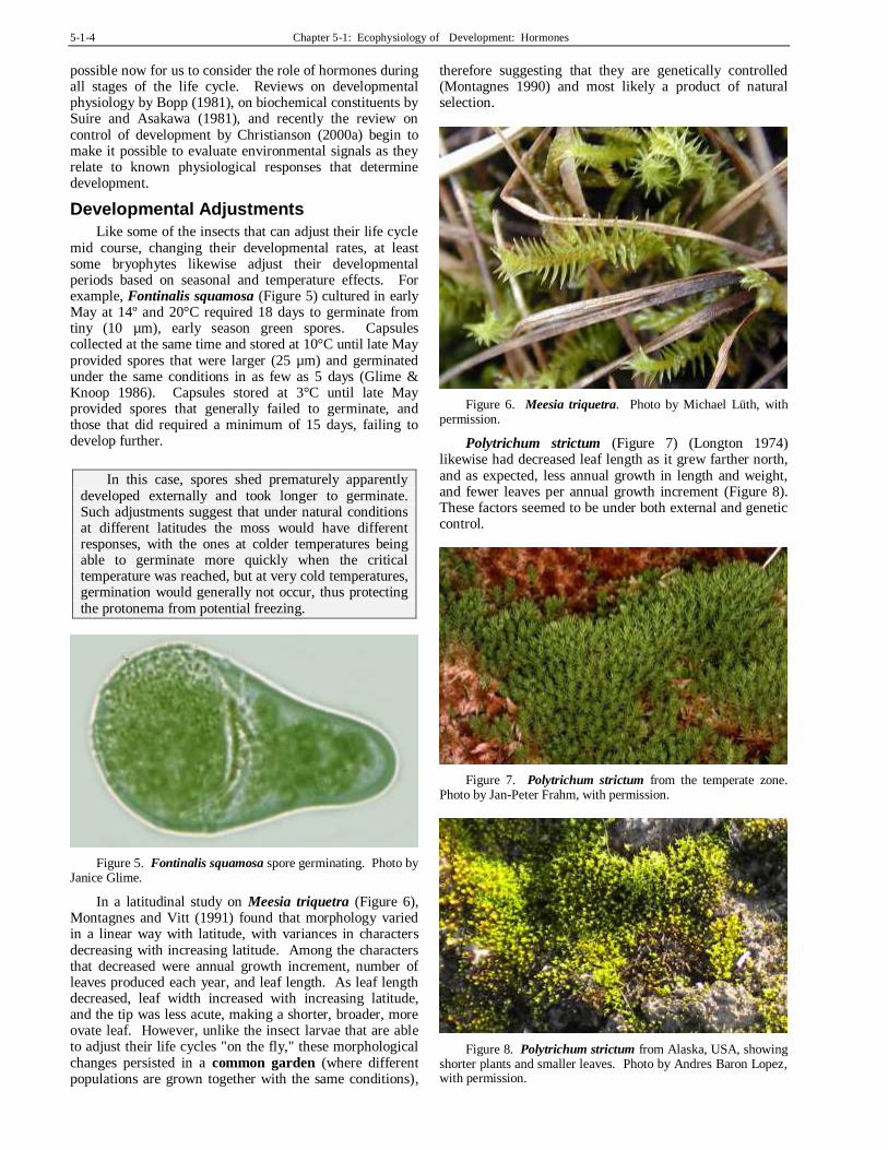
In a latitudinal study on Meesia triquetra (Figure 6), Montagnes and Vitt (1991) found that morphology varied in a linear way with latitude, with variances in characters decreasing with increasing latitude. Among the characters that decreased were annual growth increment, number of leaves produced each year, and leaf length. As leaf length decreased, leaf width increased with increasing latitude, and the tip was less acute, making a shorter, broader, more ovate leaf. However, unlike the insect larvae that are able to adjust their life cycles "on the fly," these morphological changes persisted in a common garden (where different populations are grown together with the same conditions),
therefore suggesting that they are genetically controlled (Montagnes 1990) and most likely a product of natural selection.
Figure 6. Meesia triquetra. Photo by Michael Lüth, with permission.
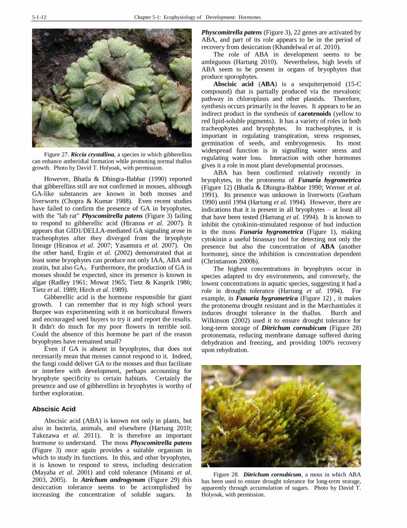
Polytrichum strictum (Figure 7) (Longton 1974) likewise had decreased leaf length as it grew farther north, and as expected, less annual growth in length and weight, and fewer leaves per annual growth increment (Figure 8). These factors seemed to be under both external and genetic control.
Figure 7. Polytrichum strictum from the temperate zone. Photo by Jan-Peter Frahm, with permission.
Figure 8. Polytrichum strictum from Alaska, USA, showing shorter plants and smaller leaves. Photo by Andres Baron Lopez, with permission.

Chapter 5-1: Ecophysiology of Development: Hormones 5-1-5
Hylocomium splendens (Figure 9) varies so much that subspecies and varieties have been named. On the west coast of Canada, it grows in wefts (loosely interwoven, often
ascending growth form), earning it the subspecies designation giganteum, and has the typical stair-step frond (Figure 10; Montagnes & Vitt 1991). North of the tree line, where it is designated var. obtusifolia, it lacks the stair-step character. The variety splendens is intermediate to these two taxa.
Figure 9. Hylocomium splendens in its typical weft form. Photo by Michael Lüth, with permission.
Figure 10. Hylocomium splendens showing stair-step growth form typical of the north temperate and boreal region. Photo by Janice Glime.
In summary, as demonstrated in Meesia, Polytrichum, and Hylocomium, increasing latitudes can select for mosses with shorter leaves, cause reduced annual growth, reduce the number of leaves produced per year, and change growth form and branching patterns. These differences can be under environmental or genetic control, or both.
Life Cycle Importance
Bryophyte life cycles have stimulated the curiosity of botanists for centuries. Their simple representation of two clearly visible generations makes them choice organisms for introducing the concept of a life cycle to students. Because of their ease of expressing genetic effects, bryophytes have provided the laboratory material for pioneering breakthrough research in several areas of genetics and molecular biology, permitting us to understand not only bryophyte development, but paving the way for understanding tracheophyte development as well (Reski 1998; Schumaker & Dietrich 1998; Christianson 2000b). The first sex (X & Y) chromosomes were found in
bryophytes, in Sphaerocarpos (Figure 11). The continuity of chromosomes during mitosis was elucidated in bryophytes. Discovery of non-Mendelian inheritance was first found in bryophytes. Furthermore, the haploid generation permits us to isolate gene mutations in order to determine their developmental roles.
Figure 11. Sphaerocarpos michelii. Photo by Michael Lüth, with permission.
The moss Physcomitrella patens (Figure 3) has become the experimental rival of Arabidopsis, Nicotiana, and Brassica. Its most recent advantage is in reverse genetics (genotype-driven technique in which genes are either knocked out or added to see the effect on phenotypic expression), enabling geneticists and physiologists to understand gene function by targetting specific genes. Because the moss is haploid, it is much easier to isolate a mutant gene and determine its function. As this new information becomes available, understanding the role of the environment in regulating gene function, and ultimately in influencing development, will become much clearer.
We should expect a variety of geographic differences in the life cycle as well as differences influenced by the weather in a given year in one location. To understand and predict these differences, we must first understand the developmental ecophysiology. This requires that we understand the functions of hormones.
Growth Regulators
Hormones, or growth regulators, were originally defined for animals as substances that are produced in one part of the organism and move to another where they carry out their action, in very small quantities. This definition works less well for plants, wherein ethylene always and others sometimes are produced in their final step at the site of action. But plant hormones differ from those of animals in other major ways as well. They have a much wider array of actions than the limited action ability of most animal hormones (Christianson 2000a). (Or do those animal folks just not understand their hormones as well as the botanists understand theirs?) Rather, in plants the hormones usually act in combinations that present a wide array of possible outcomes. In plants, as in animals, every aspect of development involves hormones. If hormones are within the organism, why should an ecologist even care to understand their nature and action? Hormones are often leaked into the environment by other

5-1-6 Chapter 5-1: Ecophysiology of Development: Hormones
organisms and those external sources may even be necessary to the development of the plants. Plants both excrete hormones and are affected by external hormones (Beutelmann & Bauer 1977). Bryophytes are no exception to these external regulators. This makes the role of the environment of far more importance than for most animal hormone functions (human contributions not withstanding). While the number of hormones known in plants is small ( Table 1), the importance of external hormones is poorly known, especially in bryophytes.
Consider for a moment what the bryophytes have been doing for their 400-million-year history. Limited in structure by their lack of lignin, they were not limited in any discernible way regarding their biochemical evolution. This has afforded them three times as long to perfect their development and biochemical adaptations compared to the Magnoliophyta (flowering plants) (Christianson 2000a). In fact, the very absence of large morphological adaptations has increased the selection pressures for cellular level biochemical ones (Christianson 2000a). Here we will examine what we do know about the hormones found in bryophytes.
Auxins
Auxins have long been known as plant growth hormones, and were conclusively demonstrated in bryophytes in 1985 (Law et al. 1985), but their mode of action is still not clearly understood. They are amino-acid based hormones, and through studies with Venus flytrap (Dionaea muscipula), we have discovered that they have a role in cell extension. This extension seems to be mediated by an efflux of H+ that accumulates between the cells, breakage of the calcium pectate bonds that glue cell walls together, and appearance of Ca++ inside the cells in the area of rapid growth. Concomitant with these events, the auxin IAA (indole-3-acetic acid) increases in the region of growth (in this case, the lower side of the midrib). Using the moss Funaria hygrometrica (Figure 12), Kapoor and Bhatla (1998) suggest that the influx of Ca++ to the cells may be induced by the IAA, although in this case it is in callose (complex, branched polysaccharide) synthesis that precedes the differentiation of chloronema (youngest part of protonema) to caulonema (part of protonema giving rise to leafy plants). IAA has a known role in this chloronema to caulonema transformation (Decker et al2006).
Table 1. Classes of growth regulators affecting bryophytes, their known presence in mosses and liverworts, and their known functions in that group.
Class Specific Regulator Presence Function
auxins IAA
mosses, liverworts
membrane transport (esp. Ca), cell elongation, protonema differentiation, stem elongation (promote at low, inhibit at high), rhizoid initiation, seta elongation, tropisms, apical dominance
cytokinins
zeatin mosses cell division, aging, bud initiation, archegonium initiation,
isopentenyladenine mosses gametophore production
Factor H? mosses inhibition of caulonema growth, bud initiation, gemma formation
analogs mosses, liverworts
promote thallus growth, slow aging, increase Ca in cell
gibberellins gibberellin-like
? development, promote growth, enhance antheridial development, decrease archegonial production
dormancy hormones
lunularic acid (LA) liverworts growth regulator, dormancy, drought tolerance, antiherbivory?
abscisic acid (ABA)
mosses hornworts?
drought tolerance, growth form, capsule stomatal closure, gametophore bud inhibition; controls cytokinin response
ethylene ethylene mosses liverworts
development, leaf morphology, epinasty, cell elongation, color changes, response to substrate, senescence, suppression of 3rd row of leaves in liverworts, increased number of antheridia, chloronema to caulonema, inhibits seta elongation, may control gametophore bud development
acetylcholine mosses,
liverworts? light response?; antiherbivory?; cellular regulation?
cryptochromes mosses,
liverworts? protonema branching, gametophore induction, development, auxin control, photoperiodic responses
While the Venus flytrap provides the advantages of knowing where and when the growth response will occur, the number of responses of a single plant is limited, and the response is extremely rapid, making it difficult to obtain large amounts of data. The moss system provides a slower response that can be controlled by the researcher through externally applied auxin. As a single-cell-thick response system (leaf or protonema, Figure 12), the moss offers strong advantages over leaves or buds of tracheophytes, where any externally applied auxin must slowly penetrate
the epidermis or other protective cells and substances. Because of these advantages, we are beginning to understand the role of IAA and calcium through the use of moss models.
Auxin activity seems to be an ancient character present when liverworts first emerged on land (Ishizaki et al. 2012). Ishizaki and coworkers demonstrated auxin activity at the bottom of gemma cups and junction of gametophyte and sporophyte in Marchantia polymorpha (Figure 13), suggesting its importance in actively dividing cells.

Chapter 5-1: Ecophysiology of Development: Hormones 5-1-7
Figure 12. Funaria hygrometrica protonema with differentiation into chloronema (perpendicular cross walls) and caulonema (diagonal cross walls). Photo by Janice Glime.
Figure 13. Marchantia polymorpha vegetative thallus with gemma cups. IAA moves basipetally (away from the tips) in this species. Photo by Janice Glime.
Our knowledge of the role of IAA in moss gametophores is still limited. We do know that the maximum concentrations are at the stem apex and base (Decker et al. 2006). The IAA seems to respond to changes in light quality, with red light retarding growth of protonemata but causing elongation of the gametophores, nevertheless making leaves shorter and narrower. Far red light enhances these responses (Bierfreund et al. 2003).
Thomas et al. (1983) demonstrated that IAA controlled seta elongation in the liverwort Pellia epiphylla (Figure 14). Although this and other studies provided indications of the presence of IAA in bryophytes, the first definitive HPLC (high-performance liquid chromatography) demonstration of its presence was published in 1985 by Law and coworkers in sterile culture of the liverwort Plagiochila asplenioides (Figure 15) subsp. arctica. The natural auxin is indole-3-acetic acid (IAA), which is produced in the stem and branch tips of higher plants, and among bryophytes the same apical production is indicated in Marchantia (Maravolo 1976; Gaal et al. 1982). Due to its polarity, IAA moves basipetally (toward the base), as demonstrated in Marchantia polymorpha (Figure 13) by Maravolo (1976, 1980), where it travels in the midrib. Its transport is inhibited by aging and ethylene.
Figure 14. Pellia epiphylla, a species in which IAA controls seta elongation. Photo by Malcolm Storey, through Creative Commons.
Figure 15. Plagiochila asplenioides, a liverwort in which the presence of IAA has been demonstrated. Photo by Dick Haaksma, with permission.
In mosses, we know that early development is triggered by the auxin IAA working with cytokinin (another hormone) and requiring light that acts through the mediation of phytochrome (pigment sensitive to photoperiod) and a blue light receptor (Reski 1998), possibly cryptochromes. Auxins respond to light and gravity and thus provide a means for plants to grow in the right direction relative to the Earth. Their mode of action is still controversial, despite extensive research into their movements within plants and plant responses.
IAA seems to be essential for normal stem elongation (Bidwell 1979). When researchers removed the tips of actively growing tracheophytes, growth stopped. If they applied IAA, growth continued. On the other hand, at least in flowering plants, removal of the stem apex can promote growth of the branches, which were heretofore inhibited by the IAA during its downward movement. Similar reactions seem to occur in at least some mosses, as exhibited by the innovations (new ascendant branches near the shoot tip; Figure 16) of mosses following gametangial senescence (i.e. loss of gametangial function with aging), but experimental evidence of the IAA connection in bryophytes seems to be lacking.

5-1-8 Chapter 5-1: Ecophysiology of Development: Hormones
Figure 16. Innovation (arrow) beneath senescing antheridial head of Philonotis caespitosa. Photo by Michael Lüth, with permission.
Auxins play major metabolic roles. IAA, in particular, seems to play a role in membrane transport; Lüttge and
coworkers (1972) demonstrated that IAA can enhance leaf uptake of potassium by Mnium from both KCl and K2SO4. Inhibition of IAA by TIBA (2,3,5-triiodobenzoic acid; polar auxin transport inhibitor) reduces starch accumulation at night and disrupts meristem polarity in the thallose liverwort Riella helicophylla (Figure 17) (Stange 1985). The role of IAA in cell extension is still unclear, but perhaps it again plays a metabolic role in the transport of substances across the cell membrane, particularly calcium, thus increasing the osmotic potential of the cell.
Figure 17. Riella helicophylla, a liverwort where polar auxin transport causes reduction in nighttime starch accumulation and disruption of meristem polarity. Photo from NACICCA, through Creative Commons.
Auxins promote stem elongation at low concentrations and inhibit it at high ones, presumably due to induction of
ethylene (Goodwin & Mercer 1983), and concentrations that promote growth in one part of a plant may inhibit it in another. In reviewing the body of literature on auxins in both non-tracheophytes and tracheophytes, Cooke and coworkers (2002) were surprised to find bryophytes exhibited most of the same physiological mechanisms for regulating IAA and for IAA-mediated responses as did the tracheophytes. These responses include tropisms, apical dominance, and rhizoid initiation. Both charophytes (the likely progenitors of bryophytes) and liverworts synthesize IAA via the tryptophan-independent pathway, regulating IAA levels through a balance between the rates of IAA biosynthesis and IAA degradation. All other land plants use the same pathway, but seem to have more precise spatial and temporal control through additional hydrolysis reactions. Although charophyte tips are apparently not sensitive to polar IAA transport inhibitors, both moss and liverwort gametophytes and moss sporophytes carry out polar transport, but sensitivity to the transport inhibitors differs within these groups.
The small quantities in which auxins are present in plants, combined with the small size of bryophytes, have made detection difficult. Their presence was indicated at least as early as 1963 when Cox and Westing demonstrated it in peat extracts. Despite its nanoconcentrations, Bhatla and Dhingra-Babbar (1990) report the presence of IAA in the protonemata of Funaria hygrometrica (Figure 12), Physcomitrella patens (Figure 3), and Polytrichastrum formosum (Figure 18), where it seems to be involved in differentiation. Many researchers (Cove et al. 2006; Von Schwartzenberg 2009) consider Physcomitrella patens to be a potential model system for study of this and other hormones because we now know its genome and can use gene knockout to determine the functions of the genes and ultimately the functions of the hormones.
Figure 18. Polytrichastrum formosum, a moss in which IAA seems to be important in differentiation. Photo by David T. Holyoak, with permission.
Cytokinins
Cytokinins are important in bud formation. Using Physcomitrella patens (Figure 3) as a model, we can observe that the apical cell of the protonema divides (Reutter et al. 1998). When bud development begins, some of the subapical cells produce three-faced apical cells. These are the buds that will develop into the gametophores

Chapter 5-1: Ecophysiology of Development: Hormones 5-1-9
(leafy shoots). Application of cytokinin enhances bud formation, but the buds often do not develop further. The moss P. patens produced isopentenyl-type cytokinins, whereas the zeatin-types produced by tracheophytes (non-bryophyte plants) were absent.
Cytokinins in bryophytes remained elusive until very recently because of their low concentrations. Cytokinins form another class of hormones that generally cause cell division (mitosis). Higher plants contain various endogenous cytokinins (produced within plant), such as zeatin, and scientists have identified many other compounds that act as cytokinins, such as kinetin and benzyl adenine. Unlike IAA, cytokinins travel to the tip of the protonema and accumulate there. Only two cytokinins (zeatin, isopentenyladenine) had been identified in bryophytes by 1979, both from protonemata (Cove et al. 1979, Gerhauser unpubl.). By 1990, there were indications that a third exists (Bhatla & Dhingra-Babbar 1990). Now we know that at least 20 of the 40 known cytokinins exist in the moss Physcomitrella patens (Figure 3), the most abundant of which are cis-Zeatin-riboside-O-glucoside, N6-(Δ2-isopentenyl)adenosine-5′-monophosphate (iPRMP), and trans-zeatin-riboside-O-glucoside as intracellular hormones (von Schwartzenberg et al. 2007).
The ability of cytokinins to affect Developmental changes in gametophores has been demonstrated experimentally. Chopra and Sood (1973) have shown that the cytokinin analog kinetin promotes growth of thalli in Riccia crystallina (Figure 27), but it also enhances archegonial formation. Vashistha (1987) likewise found that three different cytokinins applied to the liverwort Riccia frostii (Figure 19) stimulated vegetative growth and archegonial induction. Besides cell division, this hormone group can prevent or slow aging and cause changes in sex expression in higher plants (Kahn 1971). Cytokinins seem to cause the increase of calcium in the cell and together with calcium may cause an increase in ethylene. Magnesium ions seem to antagonize this calcium transport.
Figure 19. Riccia frostii, a liverwort that responds to cytokinin in the medium. Photo by Rosemary Taylor, with permission.
Mosses respond differently to different concentration levels of cytokinins (Reski & Abel 1985). Among protonemata, only the chloronemata respond to low cytokinin concentrations, At high concentrations, only the caulonemata responded by increased bud formation.
Hence, there is a specificity among cells in the concentrations to which they respond.
Reutter et al. (1998) were able to connect specific genes with their functions by using transgenic Physcomitrella patens (Figure 3). Using mutants that were unable to accomplish specific developmental tasks, they showed that cytokinins were able to supply the necessary signals for these events to occur (Figure 20).
In some cases, an outside source is needed to catalyze the production of cytokinins. For example, Agrobacterium tumefaciens (Figure 21) has the isopentenyl transferase gene that is needed to catalyze the first step in the biosynthesis of cytokinin (Decker et al. 2006). For some mosses, this bacterium is needed for development to go from the protonema to gametophore stage. Reutter et al. (1998) found that the moss Physcomitrella patens (Figure 3) responds differently to the same cytokinin when it is internal (endogenous) vs external (exogenous), and that most of both cytokinin and auxin is outside the moss (Reutter et al. 1998; Ralf Reski, pers. comm. 19 September 2013). Reutter et al. (1998) suggest that this external presence may permit translocation of the hormones in the bryophytes.
Figure 20. Physcomitrella patens hormonal contents. WT=wild type, PC22 = mutant defective in gametophore development and plastid division, P24=mutant that does not produce buds, ipt=gene of respective transgenic plant. Y axis is the immunoreactive IP, IPA, and IAA equivalent [pmol (gFW)-1] in 9-day-old plants in liquid culture. Note that hormone levels are elevated in all the ipt transgenics. Redrawn from Reutter et al. 1998.
Figure 21. Agrobacterium tumefaciens on a carrot, a species known to provide hormones to mosses in nature. Photo through Creative Commons.

5-1-10 Chapter 5-1: Ecophysiology of Development: Hormones
External application of cytokinins cause Physcomitrella patens (Figure 3) to develop abnormally, causing bud production without leafy gametophore development and becoming necrotic (Reutter et al. 1998). On the other hand, transgenic mutant mosses with the added bacterial ipt gene were able to develop normally with the internal production of cytokinins.
Cytokinins may have important roles in responding to the environment (Lorenz et al. 2003). For example, it seems to have a role in the change from juvenile tissue growth to sexual reproduction under high-energy conditions (exogenous carbohydrates or bright light). Thelander et al. (2005) found that high-energy conditions resulted in pronounced caulonema formation. Low energy conditions, resulting from low light, short days, or low temperatures, stimulate development of gametangia and subsequent development of sporophytes (Hohe et al. 2002).
The limited number of cell types, ability to regenerate from small fragments, and ease of cultivation of the entire life cycle in the laboratory makes bryophytes good experimental organisms for study of the functioning of cytokinins (von Schwartzenberg 2006). And the fully mapped genotype of Physcomitrella patens (Figure 3) provides us with an ideal study organism. Von Schwartzenberg et al. (2007) found that the nucleotide iPRMP is the most abundant extracellular cytokinin in Physcomitrella patens. By using cytokinin oxidase/ dehydrogenase (CKX)-overexpressing plants, von Schwartzenberg and coworkers observed reduced and retarded budding, absence of sexual reproduction, and abnormal protonema cells. Extracellular IP and IPR seem to be the primary cytokinins responsible for inducing buds in P. patens. Control of levels is undoubtedly important.
14C-labelled adenine has also shown up in cytokinin in the culture medium of Physcomitrella patens (Figure 3), indicating a similar role of adenine in production of cytokinin (Bhatla & Dhingra-Babbar 1990). A similar, perhaps same, substance in Bryum klinggraeffii (Figure 22) inhibits growth and stimulates gemma formation. Because it leaks into the medium, this substance could have interactive effects on other species of mosses and even control its own population size. More recently, Proust et al. (2011) found that strigolactones regulate the branching of protonemata in Physcomitrella patens and act as quorum sensors – a way of signalling that no more bryophytes should be added there. Hence, the strigolactones inhibit the growth of both that protonema and that of neighboring colonies.
Figure 22. Bryum klinggraeffii, a moss in which a cytokinin-like substance leaks into the environment and inhibits growth while promoting gemma formation. Photo by Michael Lüth, with permission.
Based on the work of Bopp (1963, 1968), Watson (1981) suggested that it could be the inhibitory properties of a hormone (Factor H – see below) that caused differing aggressive patterns among juvenile Polytrichum s.l. (Figure 7-Figure 8; Figure 18) species, thus affecting ultimate community structure. Perhaps more important is the effect of controlling simultaneous production of buds in the population so that they develop together and conserve moisture by creating a smooth surface. This same control would prevent them from over-shadowing one another, avoiding intra-specific light competition.
It seems that the moss need not produce its own cytokinin. Rather, it may serve as host to bacteria that produce this hormone. In Funaria hygrometrica (Figure 1), the bacterium Methylobacterium (Figure 23) is epiphytic on the moss, inhabiting leaf surfaces, especially in the grooves between adjacent leaf lamina cells (cells of the blade portion of the leaf, exclusive of costa) (Hornschuh et al. 2002). In the presence of these bacteria on agar cultures, the protonema produces buds just as it would in the presence of cytokinin, and the exudate also stimulates the growth of the protonemal filaments. Glime and Knoop (1986) suggested a similar relationship in Fontinalis squamosa (Figure 24), wherein the only protonemata cultures that produced buds on a mineral nutrient medium were the ones that became contaminated with bacteria and fungi.
Figure 23. Methylobacterium sp., a possible source of cytokinins for mosses, on sunflower stoma. Photo by Kutschera U., through Creative Commons.
Figure 24. Fontinalis squamosa protonema. Photo by Janice Glime.

Chapter 5-1: Ecophysiology of Development: Hormones 5-1-11
One aspect of the life cycle that will be discussed in other chapters is the production of asexual structures, a feature that is rare among tracheophytes (non-bryophyte plants). One example of this unique phenomenon is the discovery of protonemal gemmae in the aquatic moss Fontinalis antipyretica (Ares et al. 2014). In this species, where capsule production is relatively rare, vegetative shoots are important dispersal units. These dispersal units can come from detached cortical cells, margins and abaxial (away from the stem) surfaces of leaves, leaf laminae, and stems with leaves removed. Likewise, the protonema can continue growth from the filament or its rhizoids. But the discovery by Ares et al. is that these protonemata can also produce filamentous gemmae and spherical brood cells. These occur as the cultures age or dry out. Thus in nature they are produced as streams dry and water levels drop, providing a means of surviving these unfavorable periods. It is interesting that bacteria and fungi in the cultures (and in nature) seem to play a role in this development. but at this point in time we do not know what that biochemical interaction may be or how the drying of the environment may trigger the formation of propagules on the protonema.
One of the cytokinins that is effective on bryophytes is produced by the bacterium Agrobacterium (Figure 21). It appears that flowering plants lack the gene for this cytokinin, but evidence suggests that mosses may in fact possess it, and furthermore, Agrobacterium in the environment may supply it to some mosses. Addition of Agrobacterium tumefaciens (Figure 21) to the medium can stimulate the production of gametophores in Pylaisiella selwynii (Figure 25; Spiess et al. 1971), an epiphyte. The presence of this bacterium with the moss on tree bark suggests its possible role in the development of Pylaisiella selwynii in that habitat.
Figure 25. Pylaisiella selwynii growing on bark where it encounters the bacterium Agrobacterium tumefaciens, which most likely contributes to its production of gametophores there. Photo by Janice Glime.
Factor H
A possible cytokinin known as Factor H, an adenine derivative (Bhatla & Dhingra-Babbar 1990), has been known for much longer as a stimulant for increasing the number of gametophore buds (Klein 1967; Brandes & Kende 1968). Factor H has been isolated from the culture medium of Funaria hygrometrica (Figure 1) and from
tissue extracts of several other mosses (Bhatla & Dhingra-Babbar 1990). Its roles in inhibiting caulonema growth and promoting bud formation are clear, thus resembling the behavior of a cytokinin. Christianson (1998b) discovered that not all mosses have the same "Factor H."
Although the experiments mentioned above suggest that mosses respond to this hormone from other species, Ceratodon purpureus (Figure 26) is not affected by this substance from Funaria hygrometrica (Figure 1), nor is it able to affect the development of Funaria hygrometrica, but Ceratodon does exhibit interspecific regulation. Its growth substance does not pass through a dialysis membrane, whereas factor H does.
Figure 26. Ceratodon purpureus, a species that is not affected by "factor H" from neighboring species. Photo by Michael Lüth, with permission.
In 1980, Bopp determined that Factor H not only is not a cytokinin, it is not a cytokinin-like substance. But in 2013, Ralf Reski assured me it is most likely a cytokinin. Its identity seems still to be unknown. It does seem to carry out some of the functions we might attribute to a cytokinin.
The Factor H that has made medical news lately (Büttner-Mainik et al. 2011) should not be confused with the natural Factor H produced by bryophytes. The moss Physcomitrella patens (Figure 3), through recombinant DNA, is able to make the human complement regulatory serum protein Factor H – a substance that can assist in treatment of human diseases, including severe kidney and retinal disorders. It is a cheaper solution that does not involve the need for animals to manufacture the compound.
Gibberellins
Gibberellins (GA) are terpenoid-based hormones (Harborne 1982) that can stimulate stem elongation as well as cell division, depending on the species involved (Bidwell 1979). Gibberellins, unlike auxins, are non-polar and free to move about all over the plant. In studying Marchantia polymorpha (Figure 13) Melstrom and co-workers (1974) isolated three gibberellin-like substances from the thalli. Chopra and Sood (1973) found that gibberellins could enhance antheridial formation while promoting normal growth in the thallose liverwort Riccia crystallina (Figure 27). Chopra and Kumra (1986) later found that GA3 not only enhanced normal growth of Riccia gangetica, but also increased the production of antheridia while causing a decrease in archegonial production.
5-1-12 Chapter 5-1: Ecophysiology of Development: Hormones
Figure 27. Riccia crystallina, a species in which gibberellins can enhance antheridial formation while promoting normal thallus growth. Photo by David T. Holyoak, with permission.
However, Bhatla & Dhingra-Babbar (1990) reported that gibberellins still are not confirmed in mosses, although GA-like substances are known in both mosses and liverworts (Chopra & Kumar 1988). Even recent studies have failed to confirm the presence of GA in bryophytes, with the "lab rat" Physcomitrella patens (Figure 3) failing to respond to gibberellic acid (Hiranoa et al. 2007). It appears that GID1/DELLA-mediated GA signaling arose in tracheophytes after they diverged from the bryophyte lineage (Hiranoa et al. 2007; Yasamura et al. 2007). On the other hand, Ergün et al. (2002) demonstrated that at least some bryophytes can produce not only IAA, ABA and zeatin, but also GA3. Furthermore, the production of GA in mosses should be expected, since its presence is known in algae (Radley 1961; Mowat 1965; Tietz & Kasprik 1986; Tietz et al. 1989; Hirch et al. 1989).
Gibberellic acid is the hormone responsible for giant growth. I can remember that in my high school years Burpee was experimenting with it on horticultural flowers and encouraged seed buyers to try it and report the results. It didn't do much for my poor flowers in terrible soil. Could the absence of this hormone be part of the reason bryophytes have remained small?
Even if GA is absent in bryophytes, that does not necessarily mean that mosses cannot respond to it. Indeed, the fungi could deliver GA to the mosses and thus facilitate or interfere with development, perhaps accounting for bryophyte specificity to certain habitats. Certainly the presence and use of gibberellins in bryophytes is worthy of further exploration.
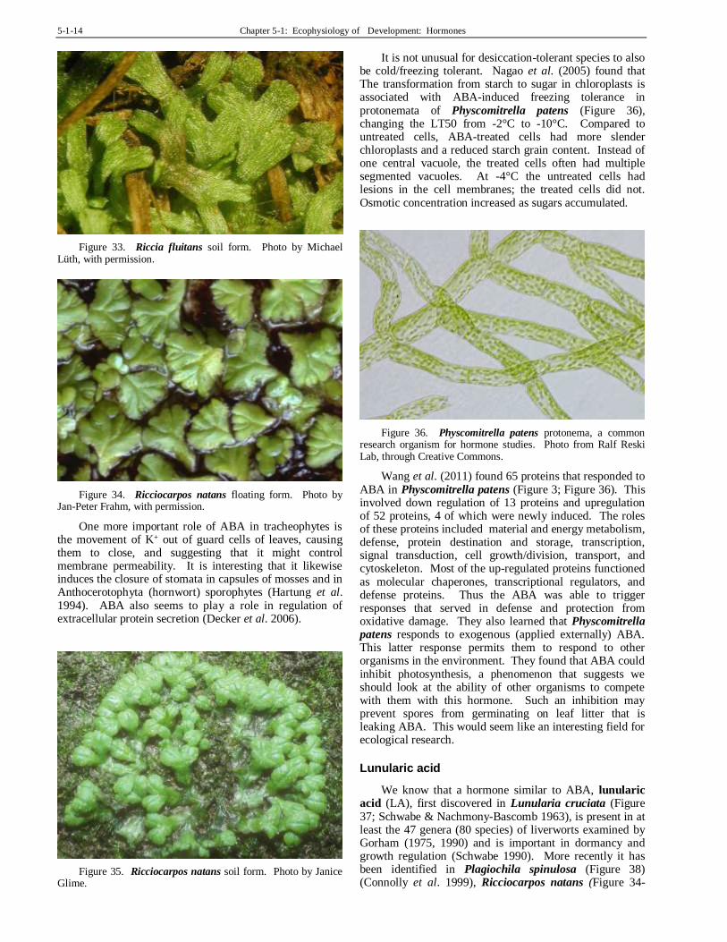
Abscisic Acid
Abscisic acid (ABA) is known not only in plants, but also in bacteria, animals, and elsewhere (Hartung 2010; Takezawa et al. 2011). It is therefore an important hormone to understand. The moss Physcomitrella patens (Figure 3) once again provides a suitable organism in which to study its functions. In this, and other bryophytes, it is known to respond to stress, including desiccation (Mayaba et al. 2001) and cold tolerance (Minami et al. 2003, 2005). In Atrichum androgynum (Figure 29) this desiccation tolerance seems to be accomplished by increasing the concentration of soluble sugars. In
Physcomitrella patens (Figure 3), 22 genes are activated by ABA, and part of its role appears to be in the period of recovery from desiccation (Khandelwal et al. 2010).
The role of ABA in development seems to be ambiguous (Hartung 2010). Nevertheless, high levels of ABA seem to be present in organs of bryophytes that produce sporophytes.
Abscisic acid (ABA) is a sesquiterpenoid (15-C compound) that is partially produced via the mevalonic pathway in chloroplasts and other plastids. Therefore, synthesis occurs primarily in the leaves. It appears to be an indirect product in the synthesis of carotenoids (yellow to red lipid-soluble pigments). It has a variety of roles in both tracheophytes and bryophytes. In tracheophytes, it is important in regulating transpiration, stress responses, germination of seeds, and embryogenesis. Its most widespread function is in signalling water stress and regulating water loss. Interaction with other hormones gives it a role in most plant developmental processes.
ABA has been confirmed relatively recently in bryophytes, in the protonema of Funaria hygrometrica (Figure 12) (Bhatla & Dhingra-Babbar 1990; Werner et al. 1991). Its presence was unknown in liverworts (Gorham 1990) until 1994 (Hartung et al. 1994). However, there are indications that it is present in all bryophytes – at least all that have been tested (Hartung et al. 1994). It is known to inhibit the cytokinin-stimulated response of bud induction in the moss Funaria hygrometrica (Figure 1), making cytokinin a useful bioassay tool for detecting not only the presence but also the concentration of ABA (another hormone), since the inhibition is concentration dependent (Christianson 2000b).
The highest concentrations in bryophytes occur in species adapted to dry environments, and conversely, the lowest concentrations in aquatic species, suggesting it had a role in drought tolerance (Hartung et al. 1994). For example, in Funaria hygrometrica (Figure 12) , it makes the protonema drought resistant and in the Marchantiales it induces drought tolerance in the thallus. Burch and Wilkinson (2002) used it to ensure drought tolerance for long-term storage of Ditrichum cornubicum (Figure 28) protonemata, reducing membrane damage suffered during dehydration and freezing, and providing 100% recovery upon rehydration.
Figure 28. Ditrichum cornubicum, a moss in which ABA has been used to ensure drought tolerance for long-term storage, apparently through accumulation of sugars. Photo by David T. Holyoak, with permission.

Chapter 5-1: Ecophysiology of Development: Hormones 5-1-13
The use of ABA for cryopreservation reduces both labor and loss of plant material in Ceratodon purpureus (Figure 26), Funaria hygrometrica (Figure 1), Physcomitrella patens (Figure 3), and Sphagnum spp. (Christianson 1998a). There are likewise genetic implications for its presence, with 11 expressed sequence tags matching up with tracheophyte stress response genes, "including responses which may involve ABA" (Machuka et al. 1999). In Atrichum androgynum (Figure 29), application of ABA prior to desiccation reduces membrane leakage (Beckett 1999). It appears that this drought tolerance mechanism may be similar to that in higher plants under stress, with ABA reducing membrane damage by reducing the changes in membrane lipids (Guschina et al. 2002). On the other hand, ABA does not endow all bryophytes with desiccation tolerance. Plagiochila (Figure 15) shows no response, and Marchantia polymorpha (Figure 13) requires both ABA and encapsulation in alginate (sticky gum) beads for successful cryopreservation (Pence 1998). Furthermore, in the desiccation-tolerant Syntrichia (Figure 30), desiccation tolerance is not under ABA control, despite a large number of desiccation-response genes (Oliver 1996).
But what is the role of ABA in development? Decker et al. (2006) found that under the influence of ABA the protonematal subapical cells differentiate into round, short cells (brachycytes) or tmema cells (short-lived abscission cells), the latter being nearly free of cytoplasm (Figure 31). Thus, ABA has a role in asexual reproduction of the protonema. We know that in Funaria hygrometrica (Figure 1), when the ABA is removed, these short, round cells (brachycytes) germinate and form new protonemata (Schnepf & Reinhard 1997). The role of ABA is at least in part that of restructuring the cell walls of the protonema (Schipper et al. 2002; Decker et al. 2006).
Figure 29 Atrichum androgynum, a moss in which membrane leakage is reduced by ABA application. Photo by Tom Thekathyil, with permission.
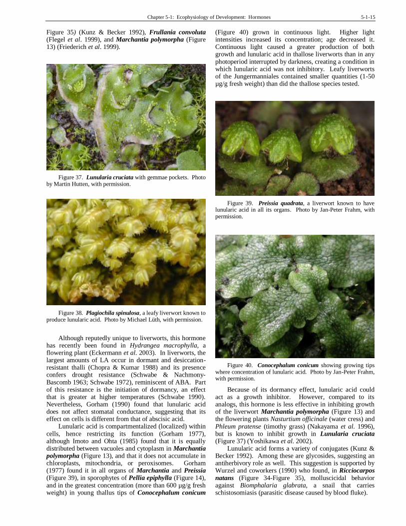
One interesting role of ABA is its ability to convert the aquatic (floating) forms of Riccia fluitans (Figure 32) and Ricciocarpos natans (Figure 34) into their terrestrial forms (Figure 33, Figure 35; Hartung et al. 1994). In Riccia fluitans, ABA causes changes in the gene expression that cause the nearly filamentous floating form to become the
broadly thallose soil form (Hellwege et al. 1996). This mechanism may be similar to that seen in the aquatic fern Marsilea quadrifolia in which ABA induces changes from aquatic to aerial leaf forms (Hsu et al. 2001).
Figure 30. Syntrichia ruraliformis on sand dunes at Harlech, Wales. This is a desiccation-tolerant moss whose tolerance is not controlled by ABA. Photo by Janice Glime.
Figure 31. Hormone pathways in the cell cycle for protonemata. Modified from Decker et al. 2006.
Figure 32. Riccia fluitans floating form. Photo by Janice Glime.
5-1-14 Chapter 5-1: Ecophysiology of Development: Hormones
Figure 33. Riccia fluitans soil form. Photo by Michael Lüth, with permission.
Figure 34. Ricciocarpos natans floating form. Photo by Jan-Peter Frahm, with permission.
One more important role of ABA in tracheophytes is the movement of K+ out of guard cells of leaves, causing them to close, and suggesting that it might control membrane permeability. It is interesting that it likewise induces the closure of stomata in capsules of mosses and in Anthocerotophyta (hornwort) sporophytes (Hartung et al. 1994). ABA also seems to play a role in regulation of extracellular protein secretion (Decker et al. 2006).
Figure 35. Ricciocarpos natans soil form. Photo by Janice Glime.
It is not unusual for desiccation-tolerant species to also be cold/freezing tolerant. Nagao et al. (2005) found that The transformation from starch to sugar in chloroplasts is associated with ABA-induced freezing tolerance in protonemata of Physcomitrella patens (Figure 36), changing the LT50 from -2°C to -10°C. Compared to untreated cells, ABA-treated cells had more slender chloroplasts and a reduced starch grain content. Instead of one central vacuole, the treated cells often had multiple segmented vacuoles. At -4°C the untreated cells had lesions in the cell membranes; the treated cells did not. Osmotic concentration increased as sugars accumulated.
Figure 36. Physcomitrella patens protonema, a common research organism for hormone studies. Photo from Ralf Reski Lab, through Creative Commons.
Wang et al. (2011) found 65 proteins that responded to ABA in Physcomitrella patens (Figure 3; Figure 36). This involved down regulation of 13 proteins and upregulation of 52 proteins, 4 of which were newly induced. The roles of these proteins included material and energy metabolism, defense, protein destination and storage, transcription, signal transduction, cell growth/division, transport, and cytoskeleton. Most of the up-regulated proteins functioned as molecular chaperones, transcriptional regulators, and defense proteins. Thus the ABA was able to trigger responses that served in defense and protection from oxidative damage. They also learned that Physcomitrella patens responds to exogenous (applied externally) ABA. This latter response permits them to respond to other organisms in the environment. They found that ABA could inhibit photosynthesis, a phenomenon that suggests we should look at the ability of other organisms to compete with them with this hormone. Such an inhibition may prevent spores from germinating on leaf litter that is leaking ABA. This would seem like an interesting field for ecological research.
Lunularic acid
We know that a hormone similar to ABA, lunularic acid (LA), first discovered in Lunularia cruciata (Figure 37; Schwabe & Nachmony-Bascomb 1963), is present in at least the 47 genera (80 species) of liverworts examined by Gorham (1975, 1990) and is important in dormancy and growth regulation (Schwabe 1990). More recently it has been identified in Plagiochila spinulosa (Figure 38) (Connolly et al. 1999), Ricciocarpos natans (Figure 34-
Chapter 5-1: Ecophysiology of Development: Hormones 5-1-15
Figure 35) (Kunz & Becker 1992), Frullania convoluta (Flegel et al. 1999), and Marchantia polymorpha (Figure 13) (Friederich et al. 1999).
Figure 37. Lunularia cruciata with gemmae pockets. Photo by Martin Hutten, with permission.
Figure 38. Plagiochila spinulosa, a leafy liverwort known to produce lunularic acid. Photo by Michael Lüth, with permission.
Although reputedly unique to liverworts, this hormone
has recently been found in Hydrangea macrophylla, a flowering plant (Eckermann et al. 2003). In liverworts, the largest amounts of LA occur in dormant and desiccation-resistant thalli (Chopra & Kumar 1988) and its presence confers drought resistance (Schwabe & Nachmony-Bascomb 1963; Schwabe 1972), reminiscent of ABA. Part of this resistance is the initiation of dormancy, an effect that is greater at higher temperatures (Schwabe 1990). Nevertheless, Gorham (1990) found that lunularic acid does not affect stomatal conductance, suggesting that its effect on cells is different from that of abscisic acid.
Lunularic acid is compartmentalized (localized) within cells, hence restricting its function (Gorham 1977), although Imoto and Ohta (1985) found that it is equally distributed between vacuoles and cytoplasm in Marchantia polymorpha (Figure 13), and that it does not accumulate in chloroplasts, mitochondria, or peroxisomes. Gorham (1977) found it in all organs of Marchantia and Preissia (Figure 39), in sporophytes of Pellia epiphylla (Figure 14), and in the greatest concentration (more than 600 µg/g fresh weight) in young thallus tips of Conocephalum conicum
(Figure 40) grown in continuous light. Higher light intensities increased its concentration; age decreased it. Continuous light caused a greater production of both growth and lunularic acid in thallose liverworts than in any photoperiod interrupted by darkness, creating a condition in which lunularic acid was not inhibitory. Leafy liverworts of the Jungermanniales contained smaller quantities (1-50 µg/g fresh weight) than did the thallose species tested.
Figure 39. Preissia quadrata, a liverwort known to have lunularic acid in all its organs. Photo by Jan-Peter Frahm, with permission.
Figure 40. Conocephalum conicum showing growing tips where concentration of lunularic acid. Photo by Jan-Peter Frahm, with permission.
Because of its dormancy effect, lunularic acid could act as a growth inhibitor. However, compared to its analogs, this hormone is less effective in inhibiting growth of the liverwort Marchantia polymorpha (Figure 13) and the flowering plants Nasturtium officinale (water cress) and Phleum pratense (timothy grass) (Nakayama et al. 1996), but is known to inhibit growth in Lunularia cruciata (Figure 37) (Yoshikawa et al. 2002).
Lunularic acid forms a variety of conjugates (Kunz & Becker 1992). Among these are glycosides, suggesting an antiherbivory role as well. This suggestion is supported by Wurzel and coworkers (1990) who found, in Ricciocarpos
natans (Figure 34-Figure 35), molluscicidal behavior against Biomphalaria glabrata, a snail that carries schistosomiasis (parasitic disease caused by blood fluke).

5-1-16 Chapter 5-1: Ecophysiology of Development: Hormones
Research on lunularic acid in this century is scarce, but we still have much to learn about its role in liverworts.
Ethylene
Ethylene (C2H4) is important in every step of the developmental process of higher plants (Abeles 1973), and has been demonstrated in both liverworts (Fredericq et al. 1977; Thomas et al. 1983) and mosses (Rohwer & Bopp 1985). It is known from the sporophyte of Pellia (Figure 14), especially during rapid seta elongation (Thomas et al. 1983) and from the thallus of Marchantia (Figure 13) (DeGreef et al. 1981). However, Stange and Osborne (1989) found that the liverwort Riella (Figure 17) appears to have a different pathway for ethylene synthesis from that of higher plants.
Ethylene is an unsaturated hydrocarbon synthesized in tracheophytes via the following pathway:
Methionine SAM ACC C2H4
IAA is possibly the catalyst for the conversion of SAM (S-adenosylmethionine) to ACC (1-aminocyclopropane-1-carboxylic acid) (Bradford & Yang 1980a), as suggested by the 10-fold increase in ethylene obtained when 10-6 IAA is supplied in the medium (Bhatla & Dhingra-Babbar 1990). O2 is required for the conversion of ACC to C2H4 (Bradford & Yang 1980b), suggesting that there might be interesting environmental responses for mosses that live part of their lives in water.
Although ethylene is a gaseous substance, it has been termed a growth hormone. It is important in senescence (aging) and its presence can cause epinasty (leaf and stem curling). In the aquatic moss Fontinalis squamosa, treatment with its precursor ACC causes color changes, wavy leaves, and curled tips (Figure 41), as well as inhibiting growth at high concentrations (Glime & Rohwer 1983). It is likely that these responses are actually to ethylene produced in response to the ACC application.
Figure 41. Left: Fontinalis squamosa grown with ACC, the precursor of ethylene, demonstrating the contorted leaves and curved tips. Right: Neckera pennata exhibiting undulate leaves that could prove to be the result of genetically controlled ethylene behavior. Photos by Janice Glime.
Ethylene production coincides with that of the change from chloronema to caulonema and is probably tied to the increase in auxins (Rohwer & Bopp 1985). We know that ethylene and IAA can work together in both bryophytes and higher plants (Mignone & Basile 2000). In bryophytes,
we know that an additive effect exists in at least some, for example Riella helicophylla (Figure 17), causing "super" cell elongation (Stange & Osborne 1988). Chopra and Sood (1973) demonstrated that ethrel, which produces ethylene in water, causes the production of more antheridia in Riccia crystallina (Figure 27).
IAA and ethylene often work in tandem, controlling each other's concentrations. For example, in Pellia epiphylla (Figure 14), IAA results in seta elongation, whereas ethylene inhibits it (Thomas et al. 1983). In the leafy liverworts, ethylene works together with auxin and certain arabinogalactan-proteins to suppress the third row of leaves by suppressing development of every third leaf primordium (Basile & Basile 1984; Mignone & Basile 2000). Mignone and Basile considered that ethylene played a suppression role in three processes. It is able to cause reductive development by causing failure in development of primordia to mature organs. It modulates the size and shapes of leaves. And it facilitates the change from diffuse growth to polar/apical growth. Nevertheless, ethylene remains largely a mystery.
The ACC pathway seems to work somewhat differently in bryophytes (Osborne et al. 1996). Lower plants seem unable to convert ACC to ethylene, nevertheless producing ethylene continuously. Although the Riella helicophylla (Figure 17) they studied seemed to take up the ACC easily, no ethylene gas was released. Nevertheless, in Fontinalis (Figure 41) ACC causes symptoms consistent with those expected from ethylene (Rohwer & Bopp 1985).
Acetylcholine
Acetylcholine – a compound better known for its role in nerve cells, has been conclusively shown in bacteria, protists, and mosses (Hartmann & Kilbinger 1974; Wessler et al. 1999), and more recently, in corn (Momonoki 1992). Interestingly, the original report (Hartmann & Kilbinger 1974) found it only in a hybrid of Funaria hygrometrica (Figure 1) and Physcomitrium pyriforme (Figure 3), whereas its hydrolyzing enzyme cholinesterase was not found in either (Fluck & Jaffe 1974). Later, however, Gupta et al. (2001) found cholinesterase in 30 out of 39 species of bryophytes tested, including five liverworts, with the highest activity in the moss Anoectangium bicolor.
In non-animal organisms, the production of acetylcholine (ACh) is always accompanied by cholinesterase activity, thus preventing it from behaving as a hormone (Wessler et al. 1999). Nevertheless, its activity and the activation of acetylcholine receptors can interfere with ion channels and key enzymes – the cellular signalling pathways. In this role, it appears to play a part in regulating such cellular functions as mitosis, cell differentiation, organization of the cytoskeleton, cell-to-cell contact, secretion, and absorption. Furthermore, it appears to contribute to the regulation of immune functions.
But the role of acetylcholine in bryophytes is still unclear (Bhatla & Dhingra-Babbar 1990). Light quality certainly affects its production in at least some bryophytes, with 56 times as much produced in red light as in red/far-red (Bhatla & Dhingra-Babbar 1990). The red/far-red response is indicative of regulation by phytochrome (pigment that measures day length), but researchers disagree on the mechanism. As a growth regulator, it could

Chapter 5-1: Ecophysiology of Development: Hormones 5-1-17
have an important role in habitat response and spore germination as a means of interpreting light quality.
In lactic acid bacteria, acetylcholine can be produced in response to osmotic stress (Kets et al. 1997). In a moss that is often desiccated by dust and other solutes on the surface, as well as being subjected to frequent desiccation due to weather, perhaps the acetylcholine might respond similarly.
Cryptochromes
Cryptochromes – This almost colorless yellow plant pigment has both enlightened and dumbfounded the plant physiologists since its discovery. Although we know that it responds to light and somehow signals to IAA in a way that affects plant development, its mechanism has remained elusive. Then entered the moss, of course the lab rat of all mosses, Physcomitrella patens (Figure 3). In 1999, Imaizumi and coworkers posted the identification of a cryptochrome homologue from this moss. Physcomitrella patens is more than just a convenient, small organism for testing things. It is unique. It is the only plant found thus far in which gene replacement is predictably reliable due to the high frequency of homologous recombination. In plain English, that means that instead of one chance in a million for a transplanted gene entering the genome, it is a predictable certainty.
Hence, to discover how cryptochromes function in plants, researchers (Imaizumi et al. 2002) created a moss [a strain of Physcomitrella patens (Figure 3)] with a defective genome, one that had disruptants for the two known genes for cryptochromes (CRY1 & CRY2). The moss could not make its cryptochromes. The results were rather astounding. They revealed that cryptochrome signals regulate induction of side branching of the protonema, gametophore induction, and development. Furthermore, disruption of these cryptochromes altered the induction of the auxin-inducible genes. Since these modified mosses were more sensitive to external auxin than their unmodified relatives in blue-light responses, it appears that the cryptochromes provide the signal to repress auxin signals that control plant development. This breakthrough in discovering the utility of Physcomitrella patens in delineating gene function could have astounding contributions to the entire field of plant physiology! In fact, it already does.
Summary
All aspects of development are influenced not only by the internal environment, but also by the external environment. These signals trigger responses in the bryophytes that permit them to survive and take advantage of the ever-changing conditions of their environment, from growth forms to drought resistance to dormancy.
These responses are typically mediated by hormones. Known bryophyte hormones include auxins (IAA) that regulate growth and gametangial production, cytokinins (isopentenyladenine, zeatin, and most likely Factor H) that regulate protonemal bud formation and branching, gibberellin-like compounds that inhibit cytokinin responses, lunularic acid and
ABA (abscisic acid) that regulate dormancy and drought resistance, and ethylene that controls antheridial production and triggers senescence; acetylcholine and cryptochromes (photo-receptive pigments) also play a role in controlling bryophyte growth and development. The modes of control of these growth regulators are poorly understood in bryophytes, although in most cases they seem to act similarly to their mode of action in tracheophytes.
Some hormones may be supplied exogenously, that is, supplied by other organisms in the environment such as bacteria and fungi. And some of the hormones may be moved from place to place in the bryophyte by external conduction.
Acknowledgments
Inspiration for these chapters on development evolved from discussions with Dr. Martin Bopp and especially with Dr. Gert Steen Mogensen. Several of the experiments were conducted at the Botanisches Institut, Universitat Heidelberg, Germany. I appreciate the many suggestions from a student's perspective by Medora Burke-Scoll. Linda Luster checked the literature citations, proofread, and checked for needed glossary entries. KT McConnell helped with clarity and suggested the minisummaries after some of the topics. Ralf Reski helped me sort out the two kinds of Factor H and provided me with references.
Literature Cited Abeles, F. B. 1973. Ethylene in Plant Biology. Academic Press,
New York.
Ares, A. A., Duckett, J. G., and Pressel, S. 2014. Asexual reproduction and protonemal development in vitro in Fontinalis antipyretica Hedw. J. Bryol. 36: 122-133.
Basile, D. V. and Basile, M. R. 1984. Probing the evolutionary history of bryophytes experimentally. J. Hattori Bot. Lab. 55: 173-185.
Beckett, R. P. 1999. Partial dehydration and ABA induce tolerance to desiccation-induced ion leakage in the moss Atrichum androgynum. S. Afr. J. Bot. 65: 212-217.
Beutelmann, P. and Bauer, L. 1977. Purification and identification of a cytokinin from moss callus cells. Planta 133: 215-217.
Bhatla, S. C. and Dhingra-Babbar, S. 1990. Growth regulating substances in mosses. In: Chopra, R. N. and Bhatla, S. C. (eds.). Bryophyte Development: Physiology and Biochemistry, CRC Press, Ann Arbor, pp. 79-101.
Bidwell, R. G. S. 1979. Plant Physiology (second edition). Macmillan Publishing Company, Inc., New York, 726 pp.
Bierfreund, N. M., Reski, R., and Decker, E. L. 2003. Use of an inducible reporter gene system for the analysis of auxin distribution in the moss Physcomitrella patens. Plant Cell Repts. 21: 1143-1152.
Bopp, M. 1963. Development of the protonema and bud formation in mosses. J. Linn. Soc. Bot. 58: 305-309.
Bopp, M. 1968. Control of differentiation in fern-allies and bryophytes. Ann. Rev. Plant Physiol. 19: 361-380.
Bopp, M. 1980. The hormonal regulation of morphogenesis in mosses. Proc. Life Sci. 1980: 351-361.
5-1-18 Chapter 5-1: Ecophysiology of Development: Hormones
Bopp, M. 1981. Entwicklungsphysiologie der Moose. In: Schultze-Motel, W. (ed.). Advances in Bryology. Vol. 1. J. Cramer, Vaduz, pp. 11-77.
Bradford, K. J. and Yang, S. F. 1980a. Xylem transport of 1-aminocyclopropane-1-carboxylic acid, an ethylene precursor, in waterlogged tomato plants. Plant Physiol. 65: 322-326.
Bradford, K. J. and Yang, S. F. 1980b. Stress-induced ethylene production in the ethylene- requiring tomato mutant diageotropica. Plant Physiol. 65: 327-330.
Brandes, H. and Kende, H. 1968. Studies on cytokinin-controlled bud formation in moss protonemata. Plant Physiol. 43: 827-837.
Burch, J. and Wilkinson, T. 2002. Cryopreservation of protonemata of Ditrichum cornubicum (Paton) comparing the effectiveness of four cryoprotectant pretreatments. Cryo-Letters 23: 197-208.
Büttner-Mainik, A., Parsons, J., Jérome, H., Hartmann, A., Lamer, S., Schaaf, A., Schlosser, A., Zipfel, P. F., Reski, R., and Decker, E. L. 2011. Production of biologically active recombinant human factor H in Physcomitrella. Plant Biotechnol. J. 9: 373-383.
Chopra, R. N. and Kumar, P. K. 1988. Biology of Bryophytes. Wiley, New York, 350 pp.
Chopra, R. N. and Kumra, S. 1986. Hormonal regulation of growth and gametangial formation in Riccia gangetica Ahmad. Beitr. Biol. Pflanzen 61: 99-115.
Chopra, R. N. and Sood, S. 1973. In vitro studies in Marchantiales. I. Effects of some carbohydrates, agar, pH, light, and growth regulators on the growth and sexuality in Riccia crystallina. Phytomorphology 23: 230-244.
Christianson, M. L. 1998a. A simple protocol for cryopreservation of mosses. Bryologist 101: 32-35.
Christianson, M. L. 1998b. The mosses Funaria hygrometrica and Ceratodon purpureus use different molecules to regulate growth of adjacent protonema. Amer. J. Bot. (Abstr. Suppl.) 85: 7.
Christianson, M. L. 2000a. Control of morphogenesis in bryophytes. In: Shaw, J. A. and Goffinet, B. Bryophyte Biology. Cambridge University Press, Cambridge, UK, pp. 199-224.
Christianson, M. L. 2000b. ABA prevents the second cytokinin-mediated event during the induction of shoot buds in the moss Funaria hygrometrica. Amer. J. Bot. 87: 1540-1545.
Connolly, J. D., Rycroft, D. S., Srivastava, D. L., Cole, W. J., Ifeadike, P., Kimbu, S. F., Singh, J., Hughes, M., Thom, C., Gerhard, U., Organ, A. J., Smith, R. J., and Harrison, L. J. 1999. Aromatic compounds from the liverwort Plagiochila spinulosa. Phytochemistry 50: 1159-1165.
Cooke, T. J., Poli, D., Sztein, A. E., and Cohen, J. D. 2002. Evolutionary patterns in auxin action. Plant Molec. Biol. 49: 319-338.
Cove, D. J., Ashton, N. W., Featherstone, D. R., and Wang, T. L. 1979. The use of mutant strains in the study of hormone action and metabolism in the moss Physcomitrella patens. Proceedings of the Fourth John Innes Symposium: 231-241.
Cove, D., Bezanilla, M., Harries, P., and Quatrano, R. 2006. Mosses as model systems for the study of metabolism and development. Ann. Rev. Plant Biol. 57: 497-520.
Cox, R. L. and Westing, A. H. 1963. The effect of peat-moss extracts on seed germination. Proc. Indiana Acad. Sci. 73: 113-115.
Decker, E. L., Frank, W., Sarnighausen, E., and Reski, R. 2006. Moss systems biology en route: Phytohormones in Physcomitrella development. Plant Biol. 8: 397-406.
DeGreef, J. A., DeProft, M., Veroustraete, F., and Fredericq, H. 1981. Case studies of ethylene release in higher and lower plant systems. In: Jeffcoat, B. (ed.). Aspects and Prospects of Plant Growth Regulators. Wesses, Oxfordshire, pp. 9-18.
During, H. J. 1979. Life strategies of bryophytes: A preliminary review. Lindbergia 5: 2-18.
Eckermann, C., Schröder, G., Eckermann, S., Strack, D., Schmidt, J., Schneider, B., and Schröder, J. 2003. Stilbenecarboxylate biosynthesis: A new function in the family of chalcone synthase-related proteins. Phytochemistry 62: 271-286.
Ergün, N., Topcuoğlu, Ş. F., and Yildiz, A. 2002. Auxin (indole-3-acetic acid), gibberellic acid (GA3), abscisic acid (ABA) and cytokinin (zeatin) production by some species of mosses and lichens. Turk. J. Bot. 26: 13-18.
Flegel, M., Adam, K. P., and Becker, H. 1999. Sesquiterpene lactones and bisbibenzyl derivatives from the neotropical liverwort Frullania convoluta. Phytochemistry 52: 1633-1638.
Fluck, R. A. and Jaffe, M. J. 1974. The acetylcholine system in plants. Current Adv. Plant Sci. 5(11): 1-22.
Fredericq, H., Veroustraete, F., DeGreef, J., and Rethy, R. 1977. Light enhanced ethylene production in Marchantia polymorpha L. Arch. Internat. Physiol. Biochim. 85: 977-978.
Friederich, S., Rueffer, M., Asakawa, Y., and Zenk, M. H. 1999. Cytochromes P-450 catalyze the formation of marchantins A and C in Marchantia polymorpha. Phytochemistry 52: 1195-1202.
Gaal, D. J., Dufresne, S. J., and Maravolo, N. E. 1982. Transport of C-indoleacetic acid in the hepatic Marchantia polymorpha. Bryologist 85: 410-418.
Glime, J. M. and Knoop, B. C. 1986. Spore germination and protonemal development of Fontinalis squamosa. J. Hattori Bot. Lab. 61: 487-497.
Glime, J. M. and Rohwer, F. 1983. The comparative effects of ethylene and 1-amino-cyclopropane-1-carboxylic acid on two species of Fontinalis. J. Bryol. 12: 611-616.
Goebel, K. 1930. Organographie der Pflanzen. Vol. II. Jena.
Goodwin, T. W. and Mercer, E. I. 1983. Phytohormones and related compounds. In: Goodwin, T. W. and Mercer, E. I. (eds.). Introduction to Plant Biochemistry, 2nd ed. Pergamon Press, New York, pp. 567-626.
Gorham, J. 1975. Some Aspects of the Distribution, Metabolism and Physiological Role of Lunularic Acid in Liverworts. Ph. D. thesis, University of London, London, England. 206 pp.
Gorham, J. 1977. Recent research on lunularic acid. British Bryological Society, British Bryological Society, AGM & Symposium Meeting 1977, Leicester, 1-2 October 1977. Accessed on 23 April 2006 at <http://rbg-web2.rbge.org.uk/bbs/meetings/mtgs77.htm>.
Gorham, J. 1990. Phenolic compounds other than flavonoids from bryophytes. In: Zinsmeister, H. D. and Mues, R. Bryophytes, Their Chemistry and Chemical Taxonomy, Proceedings of the Phytochemical Society of Europe 29, Oxford University Press, Oxford, pp. 171-200.
Gupta, A., Thakur, S. S., Uniyal, P. L., and Gupta, R. 2001. A survey of bryophytes for presence of cholinesterase activity.
Amer. J. Bot. 88: 2133-2135.
Guschina, I. A., Harwood, J. L., Smith, M., and Beckett, R. P. 2002. Abscisic acid modifies the changes in lipids brought about by water stress in the moss Atrichum androgynum. New Phytologist 156: 255-264.
Harborne, J. B. 1982. Introduction to Ecological Biochemistry, 2nd. ed. Academic Press, New York.

Chapter 5-1: Ecophysiology of Development: Hormones 5-1-19
Hartmann, E. and Kilbinger, H. 1974. Gas-liquid-chromatographic determination of light-dependent acetyl-choline concentrations in moss callus. Biochem. J. 137: 249.
Hartung, W. 2010. The evolution of abscisic acid (ABA) and ABA function in lower plants, fungi and lichen. Funct. Plant Biol. 37: 806-812.
Hartung, W., Hellwege, E. M., and Volk, O. H. 1994. The function of abscisic acid in bryophytes. J. Hattori Bot. Lab. 76: 59-65.
Hellwege, E. M., Dietz, K.-J., and Hartung, W. 1996. Abscisic acid causes changes in gene expression involved in the induction of the landform of the liverwort Riccia fluitans L. Planta 98: 423-432.
Hiranoa, K., Nakajima, M., Asano, K., Nishiyama, T., Sakakibara, H., Kojima, M., Katoh, E., Xiang, H., Tanahashi, T., Hasebe, M., Banks, J. A., Ashikari, M., Kitano, H., Ueguchi-Tanaka, M., and Matsuoka, M. 2007. The GID1-mediated gibberellin perception mechanism is conserved in the lycophyte Selaginella moellendorffii but not in the bryophyte Physcomitrella patens. Plant Cell 19: 3058-3079.
Hirch, R., Hartung, W. and Gimmler, H. 1989. Abscisic acid content of algae under stress. Bot. Acta 102: 326-334.
Hohe, A., Rensing, S. A., Mildner, M., Lang, D., and Reski, R. 2002. Day length and temperature strongly influence sexual reproduction and expression of a novel MADS-box gene in the moss Physcomitrella patens. Plant Biol. 4: 595-602.
Hornschuh, M., Grotha, R., and Kutschera, U. 2002. Epiphytic bacteria associated with the bryophyte Funaria hygrometrica: Effects of Methylobacterium strains on protonema development. Plant Biol. 4: 682-687.
Hsu, T. C., Liu, H. C., Wang, J. S., Chen, R. W., Wang, Y. C., and Lin, B. L. 2001. Early genes responsive to abscisic acid during heterophyllous induction in Marsilea quadrifolia. Plant Molec. Biol. 47: 703-715.
Imaizumi, T., Kadota, A., Hasebe, M., and Wada, M. 2002. Cryptochrome light signals control development to suppress auxin sensitivity in the moss Physcomitrella patens. Plant Cell 14: 373-386.
Imaizumi, T., Kiyosue, T., Kanegae, T., and Wada, M. 1999. Cloning of the cDNA encoding the blue-light photoreceptor (cryptochrome) from the moss Physcomitrella patens (Accession No. AB027528). Plant Physiol. 120: 1205.
Imoto, S. A. and Ohta, Y. 1985. Intracellular localization of lunularic acid and prelunularic acid in suspension cultured cells of Marchantia polymorpha. Plant Physiol. 79: 751–755.
Ishizaki, K., Nonomura, M., Kato, H., Yamato, K. T., and Kohchi, T. 2012. Visualization of auxin-mediated transcriptional activation using a common auxin-responsive reporter system in the liverwort Marchantia polymorpha. J. Plant. Res. 125: 643-651.
Kahn, A. A. 1971. Cytokinins: Permissive role in seed germination. Science 171: 853-859.
Kapoor, S. and Bhatla, S. C. 1998. Indole-3-acetic acid elicits Ca++-dependent callose synthesis in the protonema of Funaria hygrometrica. J. Plant Physiol. 153: 520-522.
Kets, E. P. W., Groot Nieron, M., Galinski, E. A., and Bont, J. A. M. de. 1997. Choline and acetylcholine: Novel cationic osmolytes in Lactobacillus plantarum. Appl. Microbiol. Biotech. 48(1): 94-98.
Khandelwal, A., Cho, S. H., Marella, H., Sakata, Y., Perroud, P.-F., Pan, A., and Quatrano, R. S. 2010. Role of ABA and ABI3 in desiccation tolerance. Science 327: 546.
Klein, B. 1967. Versuche zur Analyse der Protonemaentwicklung der Laubmoose. IV. Der Endogene
Faktor H und seine Rolle bei der Morphogenese von Funaria hygrometrica. Planta 73: 12-27.
Kunz, S. and Becker, H. 1992. Bibenzyl glycosides from the liverwort Ricciocarpos natans. Phytochemistry 31: 3981-3983.
Law, D. M., Basile, D. V., and Basile, M. R. 1985. Determination of endogenous indole-3-acetic acid in Plagiochila arctica (Hepaticae). Plant Physiol. 77: 926-929.
Longton, R. E. 1974. Genecological differentiation in bryophytes. J. Hattori Bot. Lab. 38: 49-65.
Lorch, W. 1931. Handbuch der Pflanzenanatomie VII/I. Anatomie der Laubmoose. Berlin.
Lorenz, S., Tintelnot, S., Reski, R., and Decker, E. L. 2003. Cyclin Dknockout uncouples developmental progression from sugar availability. Plant Molec. Biol. 53: 227-236.
Lüttge, U., Higinbotham, N., and Pallaghy, C. K. 1972. Electrochemical evidence of specific action of indole acetic acid on membranes in Mnium leaves. Z. Naturforsch. 27: 1239-1242.
Machuka, J., Bashiardes, S., Ruben, E., Spooner, K., Cuming, A., Knight, C., and Cove, D. 1999. Sequence analysis of expressed sequence tags from an ABA-treated cDNA library identifies stress response genes in the moss Physcomitrella patens. Plant Cell Physiol 40: 378-387.
Maravolo, N. C. 1976. Polarity and localization of auxin movement in the hepatic, Marchantia polymorpha. Amer. J. Bot. 63: 526-531.
Maravolo, N. C. 1980. Control of development in hepatics. Bull. Torrey Bot. Club 107: 308-324.
Mayaba, N., Beckett, R. P., Csintalan, Z., and Tuba, Z. 2001. ABA Increases the desiccation tolerance of photosynthesis in the Afromontane understorey moss Atrichum androgynum. Ann. Bot. 88: 1093-1100.
Melstrom, C. E., Maravolo, N. C., and Stroemer, J. R. 1974. Endogenous gibberellins in Marchantia polymorpha and their possible physiological role in thallus elongation and orthogeotropic growth. Bryologist 77: 33-40.
Mignone, M. M. and Basile, D. V. 2000. Evidence for the interrelated actions of auxin, ethylene, and arabinogalactan-proteins on the transition from non-apical to apical growth of Physcomitrella patens Hedw. (Funariaceae). In: Nothnagel, E. A., Bacic, A., and Clarke, A. E. (eds.). Cell and Developmental Biology of Arabinogalactan-Proteins. Kluwer Academic/Plenum Publishers, London, pp. 205-219.
Minami, A., Nagao, M., Arakawa, K., Fujikawa, S., and Takezawa, D. 2003. Abscisic acid-induced freezing tolerance in the moss Physcomitrella patens is accompanied by increased expression of stress-related genes. J. Plant Physiol. 160: 475-483.
Minami, A., Nagao, M., Ikegami, K., Koshiba, T., Arakawa, K., Fujikawa, S., and Takezawa, D. 2005. Cold acclimation in bryophytes: low temperature-induced freezing tolerance in Physcomitrella patens is associated with increases in expression levels of stress-related genes but not with increase in level of endogenous abscisic acid. Planta 220: 414-423.
Momonoki, Y. S. 1992. Occurrence of acetylcholine-hydrolyzing activity at the stele-cortex interface. Plant Physiol. 99: 130-133.
Montagnes, R. J. S. 1990. Patterns of Variation in the Brown Moss Meesia triquetra over an Arctic Boreal Gradient. M. S. Thesis, University of Alberta, Edmonton.
Montagnes, R. J. S. and Vitt, D. H. 1991. Patterns of morphological variation in Meesia triquetra (Bryopsida: Meesiaceae) over an Arctic-boreal gradient. Syst. Bot. 16: 726-735.

5-1-20 Chapter 5-1: Ecophysiology of Development: Hormones
Mowat, J. A. 1965. A survey of results on the occurrence of auxins and gibberellins in algae. Bot. Mar. 8:149-155.
Nagao, M., Minami, A., Arakawa, K., Fujikawa, S., and Takezawa, D. 2005. Rapid degradation of starch in chloroplasts and concomitant accumulation of soluble sugars associated with ABA-induced freezing tolerance in the moss Physcomitrella patens. J. Plant Physiol. 162: 169-180.
Nakayama, T., Fukushi, Y., Mizutani, J., and Tahara, S. 1996. Inhibiting effects of lunularic acid analogs on the growth of liverwort, watercress, and timothy grass. Biosci. Biotechnol. Biochem. 60: 862-865.
Oliver, M. J. 1996. Desiccation tolerance in vegetative plant cells. Physiol. Plant. 97: 779-787.
Osborne, D. J., Walters, J., Milborrow, B. V., Norville, A., and Stange, L. M. C. 1996. Evidence for a non-ACC ethylene biosynthesis pathway in lower plants. Phytochemistry 42: 51-60.
Pence, V. C. 1998. Cryopreservation of bryophytes: The effects of abscisic acid and encapsulation dehydration. Bryologist 101: 278-281.
Proust, H., Hoffmann, B., Xie, X., Yoneyama, Ka., Schaefer, D. G., Yoneyama, Ko., Nogué, F., and Rameau, C. 2011. Strigolactones regulate protonema branching and act as a quorum sensing-like signal in the moss Physcomitrella patens. Development138: 1531-1539.
Radley, M. 1961. Gibberellic acid-like substances in plants. Nature 191: 684-685.
Reski, R. 1998. Development, genetics and molecular biology of mosses. Bot. Acta 111: 1-15.
Reski, R. and Abel, W. O. 1985. Induction of budding on chloronemata and caulonemata of the moss, Physcomitrella patens, using isopentenyladenine. Planta 165: 354-358.
Reutter, K., Atzorn, R., Hadeler, B., Schmülling, T., and Reski, R. 1998. Expression of the bacterial ipt gene in Physcomitrella rescues mutations in budding and in plastid division. Planta 206: 196-203.
Rohwer, F. and Bopp, M. 1985. Ethylene synthesis in moss protonema. J. Plant Physiol. 117:331-338.
Schipper, O., Schaefer, D., Reski, R., and Fleming, A. 2002. Expansins in the bryophyte Physcomitrella patens. Plant Molec. Biol. 50: 789-802.
Schnepf, E. and Reinhard, C. 1997. Brachycytes in Funaria protonemata: Induction by abscisic acid and fine structure. J. Plant Physiol. 151: 166-175.
Schumaker, K. S. and Dietrich, M. A. 1998. Hormone-induced signaling during moss development. Ann. Rev. Plant Physiol. Plant Molec. Biol. 49: 501-523.
Schwabe, W. W. 1972. Growth regulation in Lunularia cruciata and the role of lunularic acid in lower plants. British Bryological Society Autumn Meeting 1972, Imperial College, London, 28-29 October. Accessed on 23 April 2006 at <http://rbg-web2.rbge.org.uk/bbs/meetings/mtgs72.htm>.
Schwabe, W. W. 1990. Lunularic acid in growth and dormancy of liverworts. In: Chopra, R. N. and Bhatla, S. C. (eds.). Bryophyte Development: Physiology and Biochemistry, CRC Press, Ann Arbor, pp. 245-257.
Schwabe, W. W. and Nachmony-Bascomb, S. 1963. Growth and dormancy in Lunularia cruciata (L.) Dum. II. The response to daylength and temperature. J. Exper. Bot. 14: 353-378.
Schwartzenberg, K. von. 2006. Moss biology and phytohormones - Cytokinins in Physcomitrella. Plant Biol. 8: 382-388.
Schwartzenberg, K. von. 2009. Hormonal regulation of development by auxin and cytokinin in moss. Ann. Plant Rev. 36: 246-281.
Schwartzenberg, K. von., Fernández Núñez, M., Blaschke, H., Dobrev, P. I., Novák, O., Motyka, V., and Strnad, M. 2007. Cytokinins in the bryophyte Physcomitrella patens: Analyses of activity, distribution, and cytokinin oxidase/dehydrogenase overexpression reveal the role of extracellular cytokinins. Plant Physiol. 145: 786-800.
Spiess, L. D., Lippincott, B. B., and Lippincott, J. A. 1971. Development and gametophore initiation in the moss Pylaisiella selwynii as influenced by Agrobacterium tumefaciens. Amer. J. Bot. 5l8: 726-731.
Spiess, L. D., Lippincott, B. B., and Lippincott, J. A. 1972. Influence of certain plant growth regulators and crown-gall related substances on bud formation and gametophore development of the moss Pylaisiella selwynii. Amer. J. Bot. 59: 233-241.
Stange, L. 1985. Effects of TIBA on meristematic activity and starch metabolism in Riella helicophylla (Bory et Mont.) Mont. Biol. Plant. 27: 221-225.
Stange, L. and Osborne, D. J. 1988. Cell specificity in auxin- and ethylene-induced 'supergrowth' in Riella helicophylla. Planta 175: 341-347.
Stange, L. M. C. and Osborne, D. J. 1989. Contrary effects of ethylene and ACC on cell growth in the liverwort Riella helicophylla. In: Clijsters et al. (eds.). Biochemical and Physiological Aspects of Ethylene Production in Lower and Higher Plants, Kluwer Academic Publishers, Dordrecht, pp. 341-348.
Suire, C. and Asakawa, Y. 1981. Chimie et chimiotaxonomie des bryophytes: Resultats essentials et perspectives. In: Schultze-Motel, W. (ed.). Advances in Bryology. Vol. 1. J. Cramer, Vaduz, pp. 167-231.
Taiz, L. and Zeiger, E. 1991. Plant Physiology. Benjamin/Cummings Publ. Co., New York, 565 pp.
Takezawa, D., Komatsu, K., and Sakata, Y. 2011. ABA in bryophytes: How a universal growth regulator in life became a plant hormone? J. Plant Res. 124: 437-453.
Thelander, M., Olsson, T., and Ronne, H. 2005. Effect of the energy supply on filamentous growth and development in Physcomitrella patens. J. Exper. Bot. 56: 653-662.
Thomas, R. J., Harrison, M. A., Taylor, J., and Kaufman, P. B. 1983. Endogenous auxin and ethylene in Pellia (Bryophyta). Plant Physiol. 73: 395-397.
Tietz, A. and Kasprik, W. 1986. Identification of abscisic acid in green algae. Biochem. Physiol. Pflanzen 181: 269-274.
Tietz, A., Koehler, R., Ruttkowski, U., and Kasprik, W. 1989. Further investigations on the occurrence and the effects of abscisic acid in algae. Biochem. Physiol. Pflanzen 184: 259-266.
Vashistha, B. D. 1987. Effect of some auxins and cytokinins on growth and archegonial formation in the liverwort Riccia frostii Aust. Biochem. Physiol. Pflanzen. 182: 309-321.
Wang, X, Kuang, T, and He, Y. 2010. Conservation between higher plants and the moss Physcomitrella patens in response to the phytohormone abscisic acid: A proteomic analysis. BMC Plant Biology 10: 192.
Watson, M. A. 1981. Chemically mediated interactions among juvenile mosses as possible determinants of their community structure. J. Chem. Ecol. 7: 367-376.
Werner, O., Espin, R. M. R., Bopp, M., and Atzorn, R. 1991. Abscisic-acid-induced drought tolerance in Funaria hygrometrica Hedw. Planta 186: 99-103.

Chapter 5-1: Ecophysiology of Development: Hormones 5-1-21
Wessler, I., Kirkpatrick, C. J., and Racke, K. 1999. The cholinergic 'pitfall': Acetylcholine, a universal cell molecule in biological systems, including humans. Clin. Exper. Pharmacol. Physiol. 26(3): 198-205.
Wurzel, G., Becker, H., Eicher, T., and Tiefensee, K. 1990. Molluscicidal properties of constituents from the liverwort Ricciocarpos natans and of synthetic lunularic acid derivatives. Planta Med. 56: 444-445.
Yasamura, Y., Crumpton-Taylor, M., Fuentes, S., and Harberd, N. P. 2007. Step-by-step acquisition of the gibberellin-DELLA growth-regulatory mechanism during land-plant evolution. Cur. Biol. 17: 1225-1230.
Yoshikawa, H., Ichiki, Y., Sakakibara, K. D., Tamura, H., and Suiko, M. 2002. The biological and structural similarity between lunularic acid and abscisic acid. Biosci. Biotechnol. Biochem. 66: 840-846.

5-1-22 Chapter 5-1: Ecophysiology of Development: Hormones