volume 1 . issue 8. pages 919-1096. 2018 issn 1934-578x ... · department of chemistry, texas...
TRANSCRIPT
Volume 13. Issue 8. Pages 919-1096. 2018 ISSN 1934-578X (printed); ISSN 1555-9475 (online)
www.naturalproduct.us

INFORMATION FOR AUTHORS Full details of how to submit a manuscript for publication in Natural Product Communications are given in Information for Authors on our Web site http://www.naturalproduct.us. Authors may reproduce/republish portions of their published contribution without seeking permission from NPC, provided that any such republication is accompanied by an acknowledgment (original citation)-Reproduced by permission of Natural Product Communications. Any unauthorized reproduction, transmission or storage may result in either civil or criminal liability. The publication of each of the articles contained herein is protected by copyright. Except as allowed under national “fair use” laws, copying is not permitted by any means or for any purpose, such as for distribution to any third party (whether by sale, loan, gift, or otherwise); as agent (express or implied) of any third party; for purposes of advertising or promotion; or to create collective or derivative works. Such permission requests, or other inquiries, should be addressed to the Natural Product Inc. (NPI). A photocopy license is available from the NPI for institutional subscribers that need to make multiple copies of single articles for internal study or research purposes. To Subscribe: Natural Product Communications is a journal published monthly. 2018 subscription price: US$2,595 (Print, ISSN# 1934-578X); US$2,595 (Web edition, ISSN# 1555-9475); US$2,995 (Print + single site online); US$595 (Personal online). Orders should be addressed to Subscription Department, Natural Product Communications, Natural Product Inc., 7963 Anderson Park Lane, Westerville, Ohio 43081, USA. Subscriptions are renewed on an annual basis. Claims for nonreceipt of issues will be honored if made within three months of publication of the issue. All issues are dispatched by airmail throughout the world, excluding the USA and Canada.
NPC Natural Product Communications
EDITOR-IN-CHIEF
DR. PAWAN K AGRAWAL Natural Product Inc. 7963, Anderson Park Lane, Westerville, Ohio 43081, USA [email protected]
EDITORS
PROFESSOR MAURIZIO BRUNO Department STEBICEF, University of Palermo, Viale delle Scienze, Parco d’Orleans II - 90128 Palermo, Italy [email protected]
PROFESSOR CARMEN MARTIN-CORDERO Department of Pharmacology, Faculty of Pharmacy, University of Seville, Seville, Spain [email protected]
PROFESSOR VLADIMIR I. KALININ G.B. Elyakov Pacific Institute of Bioorganic Chemistry, Far Eastern Branch, Russian Academy of Sciences, Pr. 100-letya Vladivostoka 159, 690022, Vladivostok, Russian Federation [email protected]
PROFESSOR YOSHIHIRO MIMAKI School of Pharmacy, Tokyo University of Pharmacy and Life Sciences, Horinouchi 1432-1, Hachioji, Tokyo 192-0392, Japan [email protected]
PROFESSOR STEPHEN G. PYNE Department of Chemistry, University of Wollongong, Wollongong, New South Wales, 2522, Australia [email protected]
PROFESSOR MANFRED G. REINECKE Department of Chemistry, Texas Christian University, Forts Worth, TX 76129, USA [email protected]
PROFESSOR WILLIAM N. SETZER Department of Chemistry, The University of Alabama in Huntsville, Huntsville, AL 35809, USA [email protected]
PROFESSOR PING-JYUN SUNG National Museum of Marine Biology and Aquarium Checheng, Pingtung 944 Taiwan [email protected]
PROFESSOR YASUHIRO TEZUKA Faculty of Pharmaceutical Sciences, Hokuriku University, Ho-3 Kanagawa-machi, Kanazawa 920-1181, Japan [email protected]
PROFESSOR DAVID E. THURSTON Institute of Pharmaceutical Science Faculty of Life Sciences & Medicine King’s College London, Britannia House 7 Trinity Street, London SE1 1DB, UK [email protected]
ADVISORY BOARD
Prof. Giovanni Appendino Novara, Italy
Prof. Norbert Arnold Halle, Germany
Prof. Yoshinori Asakawa Tokushima, Japan
Prof. Vassaya Bankova Sofia, Bulgaria
Prof. Roberto G. S. Berlinck São Carlos, Brazil
Prof. Anna R. Bilia Florence, Italy
Prof. Geoffrey Cordell Chicago, IL, USA
Prof. Fatih Demirci Eskişehir, Turkey
Prof. Francesco Epifano Chieti Scalo, Italy
Prof. Ana Cristina Figueiredo Lisbon, Portugal
Prof. Cristina Gracia-Viguera Murcia, Spain
Dr. Christopher Gray Saint John, NB, Canada
Prof. Dominique Guillaume Reims, France
Prof. Duvvuru Gunasekar Tirupati, India
Prof. Hisahiro Hagiwara Niigata, Japan
Prof. Judith Hohmann Szeged, Hungary
Prof. Tsukasa Iwashina Tsukuba, Japan
Prof. Leopold Jirovetz Vienna, Austria
Prof. Phan Van Kiem Hanoi, Vietnam
Prof. Niel A. Koorbanally Durban, South Africa
Prof. Chiaki Kuroda Tokyo, Japan
Prof. Hartmut Laatsch Gottingen, Germany
Prof. Marie Lacaille-Dubois Dijon, France
Prof. Shoei-Sheng Lee Taipei, Taiwan
Prof. M. Soledade C. Pedras Saskatoon, Canada
Prof. Luc Pieters Antwerp, Belgium
Prof. Peter Proksch Düsseldorf, Germany
Prof. Phila Raharivelomanana Tahiti, French Polynesia
Prof. Stefano Serra Milano, Italy
Dr. Bikram Singh Palampur, India
Prof. Marina Stefova Skopj, Republic of Macodenia
Prof. Leandros A. Skaltsounis Zografou, Greece
Prof. John L. Sorensen Manitoba, Canada
Prof. Johannes van Staden Scottsville, South Africa
Prof. Valentin Stonik Vladivostok, Russia
Prof. Winston F. Tinto Barbados, West Indies
Prof. Sylvia Urban Melbourne, Australia
Prof. Karen Valant-Vetschera Vienna, Austria
HONORARY EDITOR
PROFESSOR GERALD BLUNDEN The School of Pharmacy & Biomedical Sciences,
University of Portsmouth, Portsmouth, PO1 2DT U.K.
Evaluation of Anti-glycation Activities of Phlorotannins in Human and Bovine Serum Albumin-glyceraldehyde Models Shingo Sugiuraa,f, Ryosuke Taniguchia,f, Yoshihiko Nishiokaa, Ryota Iwaseb, Reiji Tanakaa,d,f, Hideo Miyakea,d,f, Tetsushi Moric,d,f, Mitsuyoshi Uedae,f and Toshiyuki Shibataa,d,f* aGraduate School of Bioresources, Mie University, 1577 Kurimamachiya-cho, Tsu, Mie 514-8507, Japan
bFaculty of Bioresources, Mie University, 1577 Kurimamachiya-cho, Tsu, Mie 514-8507, Japan
cDepartment of Biotechnology and Life Science, Tokyo University of Agriculture and Technology, 2-24-16 Naka-cho, Koganei, Tokyo 184-8588, Japan
dSeaweed Biorefinery Research Center, Mie University, 1577 Kurimamachiya-cho, Tsu, Mie 514-8507, Japan
eGraduate School of Agriculture, Kyoto University, Kitashirakawa Oiwake, Sakyo-ku, Kyoto 606-8502, Japan
fJapan Science and Technology Agency, CREST, 4-1-8 Hon-cho, Kawaguchi, Saitama 332-0012, Japan [email protected]
Received: June 1st, 2018; Accepted: June 22nd, 2018
The anti-glycation activities of phlorotannins contained in the Japanese Lessoniaceae (Ecklonia cava, Eck. kurome, Eck. stolonifera, Eisenia arborea, and Eis. bicyclis) were tested using serum albumin-glyceraldehyde (GA) models. In the human serum albumin (HSA)-GA model and the bovine serum albumin (BSA)-GA model, the concentrations of crude phlorotannins at 50% inhibition (IC50) of fluorescent advanced glycation end products (AGEs) formation was in the range of 0.48 to 0.70 mg/mL and 0.52 to 0.75 mg/mL, respectively. In tests using phloroglucinol and purified phlorotannins (eckol, fucofuroeckol A, phlorofucofuroeckol A, dieckol, and 8,8´-bieckol), dieckol had the highest inhibitory activity (IC50: 5.5 x 10² µM) against fluorescent AGEs formation in HSA-GA model and showed about 18 times inhibition compared with aminoguanidine hydrochloride of positive control. In the BSA albumin model, 8,8´-bieckol had about 27 times AGEs formation inhibitory activity (IC50: 6.2 x 10² µM) against aminoguanidine hydrochloride. In tests on GA scavenging activity, it was shown that compounds with phloroglucinol tetramer or higher had a scavenging rate of 70%, or more, with a reaction time of 120 minutes. These results suggest that among the phlorotannins, in particular the dimers of eckol (dieckol and 8,8´-bieckol), there are effective compounds for inhibiting the formation of AGEs derived from GA. Keywords: Advanced glycation end products, Anti-glycation, 8,8´-Bieckol, Glyceraldehyde, Dieckol, Phlorotannins, Lessoniaceae. Advanced glycation end products (AGEs) are a general term for structures generated by nonenzymatic reactions between proteins and reducing sugars such as glucose and fructose [1,2]. In previous studies [3-6], it has been clarified that AGEs are produced not only from the reducing sugars but also from sugar metabolic intermediates and intermediates of Maillard reactions. It is known that dicarbonyl compounds (methylglyoxal, glyoxal, and 3-deoxyglucosone) generated from autoxidation, and degradation products of glucose, have higher blood concentrations in diabetic patients than in healthy subjects [7,8]. In addition, it has been considered that the dicarbonyl compounds have high reactivity with proteins because there are two carbonyl groups in the molecule. From this scientific background, the relationship between AGEs derived from the dicarbonyl compounds and lifestyle-related diseases has been drawing attention. In recent years, according to Takeuchi et al.’s report [9], it was revealed that α-hydroxy aldehydes such as glyceraldehyde (GA) and glycolaldehyde are more reactive with proteins than dicarbonyl compounds. Among AGEs generated in vivo, it has been reported that AGEs derived from GA (GA-AGEs) accelerate intracellular oxidative stress through its binding to its receptor, and can cause strong cytotoxicity [10-12]. It is also pointed out that the GA-AGEs are involved in the onset and progression of diabetic vascular complications [9,13,14], Alzheimer’s disease [9,15,16], nonalcoholic steatohepatitis [9,17,18], hypertension [9,14], and cancer [9,19]. Therefore, suppression of GA-AGEs formation and scavenging of GA can be regarded as effective for prevention and treatment of these diseases.
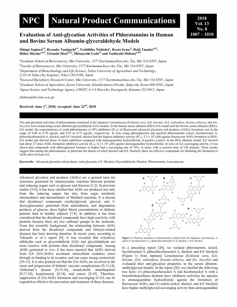
Figure 1: Chemical structures of phlorotannins isolated from the Japanese Lessoniaceae. a: eckol, b: fucofuroeckol A, c: phlorofucofuroeckol A, d: dieckol, e: 8,8´-bieckol.
In a preceding report [20], we isolated phlorotannins (eckol, fucofuroeckol A, phlorofucofuroeckol A, dieckol, and 8,8´-bieckol) (Figure 1) from Japanese Lessoniaceae (Ecklonia cava, Eck. kurome, Eck. stolonifera, Eisenia arborea, and Eis. bicyclis) and evaluated their anti-glycation properties in the serum albumin-methylglyoxal models. In the report [20], we clarified the following two facts: (1) phlorofucofuroeckol A and fucofuroeckol A with a benzobisbenzofuran skeleton have inhibitory activities far superior to aminoguanidine hydrochloride against the formation of fluorescent AGEs, and (2) eckols (eckol, dieckol, and 8,8´-bieckol) have higher methylglyoxal scavenging activity than aminoguanidine
OH
O
O
OH
OH
OH
O
OHOH
aOH
OH OH
O
O
OH
OH
O
OH
O
OH
bOHOH
OH
O
OH
OH
O
O
OH
O
O
OH OH
OH
c
OH
O
OH
O
O
OH
OH
O
OHOH
OH
O
OH
OH
O
O
OH
OH
d
OH
OH
O
O
OH
OH
OH
O
O
O
OH
OH
OH
OHOH
O
OH OHe
NPC Natural Product Communications 2018 Vol. 13 No. 8
1007 – 1010
1008 Natural Product Communications Vol. 13 (8) 2018 Sugiura et al.
hydrochloride. Phlorotannins are polyphenols that are specifically contained in brown algae and have a structure in which phloroglucinol (1,3,5-trihydroxybenzen) is polymerized [21]. In this study, to further evaluate the effectiveness of phlorotannins as a novel anti-glycation substance derived from natural plants, their inhibitory activity against the formation of fluorescent AGEs in the serum albumin-GA models and the GA scavenging activity of phlorotannins were examined.
In both human serum albumin (HSA)-GA and bovine serum albumin (BSA)-GA models, all the crude phlorotannins tested in this study inhibited the formation of fluorescent AGEs in a dose-dependent manner. Table 1 shows the concentration (IC50) of the crude phlorotannins at 50% inhibition of fluorescent AGEs formation. The IC50 values of crude phlorotannins were in the range of 0.48 to 0.70 mg/mL in the HSA-GA model and 0.52 to 0.75 mg/mL in the BSA-GA model, respectively (Table 1). In both models, crude phlorotannins prepared from Eis. bicyclis showed the highest inhibitory activity against fluorescent AGEs formation among the tested samples (Table 1). Aminoguanidine is a synthetic glycation inhibitor that inhibits the formation of AGEs and suppresses crosslinking and polymerization of proteins in vitro [22]. Aminoguanidine is not clinically applied because it has several adverse side effects on humans, but it is frequently used as a positive control in studies in the search for compounds having anti-glycation activity. Since the IC50 values of aminoguanidine hydrochloride obtained in this study were 1.10 mg/mL in HSA-GA model and 1.93 mg/mL in BSA-GA model, it was found that the crude phlorotannins of Eis. bicyclis has anti-glycation activity of about 2.3 times and 3.7 times with respect to aminoguanidine hydrochloride. Currently, Eck. kurome is cultivated as a supply source of phlorotannins in Kumamoto prefecture, Japan [23]. The crude phlorotannins were prepared from both naturally occurring and cultured versions of Eck. kurome, and their inhibitory activities on the formation of fluorescent AGEs were evaluated. As shown in Table 1, IC50 values of crude phlorotannins from cultured Eck. kurome in each model were almost the same as those of the natural plants of Eck. kurome. Therefore, as with the natural plants of Lessoniaceae, it was confirmed that crude phlorotannins of cultured Eck. kurome can be utilized as a natural product having an inhibitory effect against AGEs formation. Table 1: IC50 values of crude phlorotannins from Lessoniaceae against fluorescent AGEs formation.
Algae Specific area of origin HSA-GA (mg/mL) BSA-GA (mg/mL) Eck. cava Mie 0.70 0.75 Eck. kurome Fukuoka 0.58 0.55 Eck. kurome Kumamoto 0.61 0.59 Cultured Eck. kurome Kumamoto 0.52 0.58 Eck. stolonifera Yamaguchi 0.54 0.56 Eis. arborea Mie 0.51 0.61 Eis. bicyclis Fukuoka 0.48 0.52
All the data are expressed as the mean of three independent measurements. The IC50 values of aminoguanidine hydrochloride were 1.10 mg/mL in the HSA-GA model and 1.93 mg/mL in the BSA-GA model.
Table 2: IC50 values of phloroglucinol and isolated phlorotannins against fluorescent AGEs formation
Compounds HSA-GA (µM) BSA-GA (µM) Phloroglucinol 3.8 x 10³ 3.8 x 10³ Eckol 1.1 x 10³ 1.3 x 10³ Fucofuroeckol A 7.4 x 10² 1.4 x 10³ Phlorofucofuroeckol A 7.3 x 10² 1.4 x 10³ Dieckol 5.5 x 10² 8.7 x 10² 8,8´-Bieckol 5.7 x 10² 6.2 x 10² All the data are expressed as the mean of three independent measurements. The IC50 values of aminoguanidine hydrochloride were 1.0 x 10⁴ µM in the HSA-GA model and 1.7 x 10⁴ µM in the BSA-GA model.
In order to further analyze the inhibitory activity of phlorotannins against fluorescent AGEs formation in the albumin-GA models, tests were carried out using phloroglucinol and five kinds of
isolated compounds (eckol, fucofuroeckol A, phlorofucofuroeckol A, dieckol, and 8,8´-bieckol). Similar to the results in the albumin-methylglyoxal models obtained in the preceding report [20], phloroglucinol and the isolated eckols inhibited the formation of fluorescent AGEs in a concentration-dependent manner in both serum albumin-GA models (data not shown). As a result of calculating the IC50 value, dieckol and 8,8´-bieckol showed the effective activities in both models (Table 2). In the HSA-GA model, the IC50 value of dieckol was 5.5 x 10² µM (Table 2), which was found to have about 18 times activity compared with aminoguanidine hydrochloride. The IC50 value of 8,8´-bieckol obtained in the BSA-GA model was 6.2 x 10² µM (Table 2), and it had about 27 times inhibitory activity with respect to aminoguanidine hydrochloride. Even compounds with the lowest inhibition of AGEs also had activity about 9.1 times (eckol) in the HSA-GA model and about 12 times (fucofuroeckol A and phlorofucofuroeckol A) in the BSA-GA model as compared with aminoguanidine hydrochloride. Therefore, it was strongly suggested that the phlorotannins contained in Lessoniaceae, in particular dimers of eckol (dieckol and 8,8´-bieckol) have very excellent inhibitory activity on the formation of AGEs derived from GA.
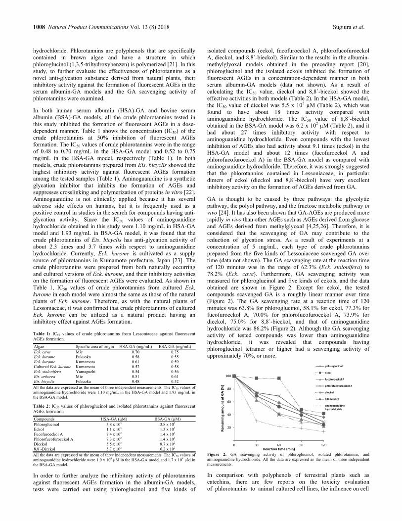
GA is thought to be caused by three pathways: the glycolytic pathway, the polyol pathway, and the fructose metabolic pathway in vivo [24]. It has also been shown that GA-AGEs are produced more rapidly in vivo than other AGEs such as AGEs derived from glucose and AGEs derived from methylglyoxal [4,25,26]. Therefore, it is considered that the scavenging of GA may contribute to the reduction of glycation stress. As a result of experiments at a concentration of 5 mg/mL, each type of crude phlorotannins prepared from the five kinds of Lessoniaceae scavenged GA over time (data not shown). The GA scavenging rate at the reaction time of 120 minutes was in the range of 62.3% (Eck. stolonifera) to 78.2% (Eck. cava). Furthermore, GA scavenging activity was measured for phloroglucinol and five kinds of eckols, and the data obtained are shown in Figure 2. Except for eckol, the tested compounds scavenged GA is a roughly linear manner over time (Figure 2). The GA scavenging rate at a reaction time of 120 minutes was 63.8% for phloroglucinol, 58.1% for eckol, 77.3% for fucofuroeckol A, 70.0% for phlorofucofuroeckol A, 73.9% for dieckol, 75.0% for 8,8´-bieckol, and that of aminoguanidine hydrochloride was 86.2% (Figure 2). Although the GA scavenging activity of tested compounds was lower than aminoguanidine hydrochloride, it was revealed that compounds having phloroglucinol tetramer or higher had a scavenging activity of approximately 70%, or more.
Figure 2: GA scavenging activity of phloroglucinol, isolated phlorotannins, and aminoguanidine hydrochloride. All the data are expressed as the mean of three independent measurements.
In comparison with polyphenols of terrestrial plants such as catechins, there are few reports on the toxicity evaluation of phlorotannins to animal cultured cell lines, the influence on cell
0
20
40
60
80
100
0 30 60 90 120
Remaining am
ount of GA (%)
Reaction time (min)
phloroglucinol
eckol
fucofuroeckol A
phlorofucofuroeckol A
dieckol
8,8'‐bieckol
aminoguanidinehydrochloride

Anti-glycation activities of phlorotannins in albumin-glyceraldehyde models Natural Product Communications Vol. 13 (8) 2018 1009
Figure 3: Cytotoxicity assay of phloroglucinol and isolated phlorotannins using MTT assay. Each value is the average of triplicate cultures.
proliferation, and bioavailability. Using phloroglucinol and the isolated compounds, their effect on the growth of HeLa (cancer cell line) and 3T3-L1 (normal cell line) cells was evaluated by 3-[4,5-dimethylthiazol-2-yl]-2,5 diphenyl tetrazolium bromide (MTT) assay (Figure 3). As a result of adding each compound to the medium at a concentration of 1 to 100 μg/mL, the survival rate of both cell lines was approximately 85%, or more (Figure 3). As a result of testing at concentrations of 50 and 100 μg/mL, each compound showed inhibitory activity in the range of about 10 to 20% against formation of fluorescent AGEs. Therefore, it was suggested that phloroglucinol and phlorotannins may not exhibit cytotoxicity at concentrations that exert anti-glycation properties. To our knowledge, there is no report on the evaluation of the anti-glycation properties of plant extracts or natural products in the serum albumin-GA models. The Lessoniaceae used in this study are the same algae used as a foodstuff in Japan and Korea. The results obtained in the preceding [20] and present studies strongly suggest that phlorotannins are superior anti-glycation substances derived from natural plants and that they may contribute to both alleviation of symptoms and prevention of onset of diseases caused by AGEs generated in vivo.
Experimental
Materials: For samples of brown algae (Ecklonia cava Kjellman, Eck. kurome Okamura, Eck. stolonifera Okamura, Eisenia arborea Areschoug, and Eis. bicyclis Kjellman), the algal plants used in the preceding report [20] were used. The cultured Eck. kurome was purchased from the Fisheries Cooperative Association of Amakusa in Kumamoto Prefecture, Japan, in 2015. The algae used for the extraction of phlorotannins were washed with filtered seawater, air-dried, and pulverized via pulverizing mill (ABS-W, Osaka Chemical). The algal powders were stored at -30°C until use. Aminoguanidine hydrochloride, HSA, and GA were purchased from Wako Pure Chemical Industries. BSA was obtained from Sigma-Aldrich. 1,3-Cyclohexanedione was purchased from Tokyo Chemical Industry. All other reagents used in this study were of analytical grade.
Extraction and purification of phlorotannins: Extraction of phlorotannins from algal powder was prepared according to the method described in the previous report [23]. Phlorotannins were purified by column chromatography and preparative HPLC using the same conditions described in the previous reports [27,28]. Each of the obtained phlorotannins (eckol, fucofuroeckol A, phlorofucofuroeckol A, dieckol, and 8,8´-bieckol) was confirmed to have a purity of 98% or more by three-dimensional HPLC (SPD-M10AV, Shimadzu) with an Inertsil ODS-3 column (4.6 mm i.d. x 250 mm, GL Science) [28]. The identification of the purified phlorotannins was carried out using LC/ESI/MS with the analysis condition reported in the preceding study [23]. The purified phlorotannins were stored at -30°C until used as samples. Serum albumin-GA assay: Sample, GA (400 mM), and albumin (HSA or BSA, 20 mg/mL) solutions were prepared separately by dissolving in a 100 mM phosphate buffer (pH 7.4). The measurement was carried out using a 96-well black plate (FLUOTRAC600, Greiner) and a microplate reader (Infinite 200, Tecan). The sample solution (40 µL), GA solution (10 µL), and albumin solution (50 µL) were added to the well in the plate. Measurement was carried out with fluorescence intensity at an excitation of 370 nm and an emission of 440 nm, and the obtained value was taken as a control value. After incubation at 37°C for 24 hours, the fluorescence intensities of each well were measured under the same measurement conditions. For blank wells, a 100 mM phosphate buffer was used instead of a sample solution. Aminoguanidine hydrochloride was used as a positive control. The inhibition rate (%) of fluorescent AGEs formation was calculated using the following formula: Inhibition rate (%) = {1 – [(fluorescence intensity of sample after incubation for 24 hours – fluorescence intensity of control of sample) / (fluorescence intensity of blank after incubation for 24 hours – fluorescence intensity of control of blank)]} x 100.
The IC50 value was calculated from the logarithmic function obtained by plotting the inhibitory rate of fluorescent AGEs formation against the sample concentration. GA-scavenging assay: The GA-scavenging activity of phlorotannins was measured using a modification of the derivatization method of GA by Usui et al [29]. Twenty-five mM GA was prepared using a 200 mM phosphate buffer (pH 7.4). The crude phlorotannins extracted from each brown algae were dissolved in the phosphate buffer to a concentration of 5 mg/mL. Purified phlorotannins, phloroglucinol, and aminoguanidine hydrochloride were dissolved in the phosphate buffer to a concentration of 25 mM each. 1,3-Cyclohexanedione (0.25 g) was dissolved in a mixture consisting of ammonium acetate (10 g), acetic acid (5 mL), and ultrapure water (50 mL), and it was used as a derivatization reagent. Each sample solution (50 µL) and a GA solution (50 µL) were mixed in well of a 96-well microplate (BioLite, Thermo Scientific) and incubated at 37°C for 30, 60, 90, and 120 minutes, respectively. After incubation, the derivatization reagent (100 µL) was added to the mixture and maintained at 60°C for 30 minutes. The amount of GA remaining in the reaction solution was measured at 370 nm using a microplate reader (Infinite 200, Tecan). For the blank test, the phosphate buffer was used instead of a GA solution. Aminoguanidine hydrochloride was used as a positive control. The scavenging rate of GA was calculated using the following formula: Scavenging rate of GA (%) = {1 – [concentration of GA remaining in the reaction solution (mM) / 25]} x 100.
Cytotoxicity assay: MTT (Dojindo) was used as an indicator of cell viability. Briefly, cell lines (HeLa or 3T3-L1) were cultured in 96-
0
20
40
60
80
100
120
0 1 10 20 50 100
Cell viab
ility(
%)
Concentration(μg/mL)
phloroglucinol
eckol
fucofuroeckol A
phlorofucofuroeckol A
dieckol
8,8´-bieckol
a HeLa cell
0
20
40
60
80
100
120
0 1 10 20 50 100
Cell viability(
%)
Concentration(μg/mL)
phloroglucinol
eckol
fucofuroeckol A
phlorofucofuroeckol A
dieckol
8,8´-bieckol
b 3T3‐L1 cell

1010 Natural Product Communications Vol. 13 (8) 2018 Sugiura et al.
well microplates (BioLite, Thermo Scientific) at a density of 5 x 10³ cells per well. After 24 hours cultivation in a CO2 incubator (CPE-2601, Hirasawa) with 5% CO2 at 37°C, the cell lines were washed with fresh medium (Dulbecco’s modified Eagle’s medium with glucose and pyruvate, containing 10% fetal bovine serum and 1% antibiotic-antimycotic, Gibco) and then treated with each sample solution (10 µL) for 24 hours in the incubator. Sample solutions were prepared by dissolving in Dulbecco’s phosphate buffered saline (DPBS) without calcium and magnesium (Gibco). For blank and control wells, DPBS was used instead of a sample solution. The cell lines were then rewashed with the medium, treated with 10 µL of MTT solution, and cultured for 4 hours in the incubator at 37°C. MTT solution was prepared by dissolving MTT (25 mg) in the
DPBS (5 mL). After removing the medium containing the MTT solution, 200 µL of the DPBS was added to each well and allowed to maintain for 1 minute at 37°C. Finally, in order to dissolve the formed formazan salt, dimethyl sulfoxide (200 μL) was added to each well from which DPBS had been removed. The absorbance of each well was measured at 535 nm using a microplate reader (Infinite 200, Tecan). The cell viability (%) was calculated using the following formula: Cell viability (%) = [(absorbance of sample well – absorbance of blank well) / (absorbance of control well – absorbance of blank well)] x 100.
Acknowledgments - This work was financially supported by the Japan Science and Technology Agency, CREST.
References
[1] McPherson JD, Shilton BH, Walton DJ. (1988) Role of fructose in glycation and cross-linking of proteins. Biochemistry, 27, 1901-1907. [2] Singh R, Barden A, Mori T, Beilin L. (2001) Advanced glycation end-products: a review. Diabetologia, 44, 129-146. [3] Thornalley PJ, Langborg A, Minhas HS. (1999) Formation of glyoxal, methylglyoxal and 3-deoxyglucosone in the glycation of proteins by glucose.
Biochemical Journal, 344, 109-116. [4] Takeuchi M, Makita Z, Bucala R, Suzuki T, Koike T, Kameda Y. (2000) Immunological evidence that non-carboxymethyllysine advanced
glycation end-products are produced from short chain sugars and dicarbonyl compounds in vivo. Molecular Medicine, 6, 114-125. [5] Takeuchi M, Makita Z. (2001) Alternative routes for the formation of immunochemically distinct advanced glycation end-products in vivo. Current
Molecular Medicine, 1, 305-315. [6] Sato T, Iwaki M, Shimogaito N, Wu X, Yamagishi S, Takeuchi M. (2006) TAGE (toxic AGEs) theory in diabetic complications. Current
Molecular Medicine, 6, 351-358. [7] McLellan AC, Thornalley PJ, Benn J, Sonksen PH. (1994) Glyoxalase system in clinical diabetes mellitus and correlation with diabetic
complications. Clinical Science, 87, 21-29. [8] Odani H, Shinzato T, Matsumoto Y, Usami J, Maeda K. (1999) Increase in three α,β-dicarbonyl compound levels in human uremic plasma: specific
in vivo determination of intermediates in advanced maillard reaction. Biochemical and Biophysical Research Communications, 256, 89-93. [9] Takeuchi M. (2012) Participation of toxic AGEs (TAGE) in a variety of diseases. Folia Pharmacologica Japonica, 139, 193-197. [10] Takeuchi M, Bucala R, Suzuki T, Ohkubo T, Yamazaki M, Koike T, Kameda Y, Makita Z. (2000) Neurotoxicity of advanced glycation end-
products for cultured cortical neurons. Journal of Neuropathology and Experimental Neurology, 59, 1094-1095. [11] Yonekura H, Yamamoto Y, Sakurai S, Petrova RG, Abedin MJ, Li H, Yasui K, Takeuchi M, Makita Z, Takasawa S, Okamoto H, Watanabe T,
Yamamoto H. (2003) Novel splice variants of the receptor for advanced glycation end-products expressed in human vascular endothelial cells and pericytes, and their putative roles in diabetes-induced vascular injury. Biochemical Journal, 370, 1097-1109.
[12] Iwamoto K, Kanno K, Hyogo H, Yamagishi S, Takeuchi M, Tazuma S, Chayama K. (2008) Advanced glycation end products enhance the proliferation and activation of hepatic stellate cells. Journal of Gastroenterology, 43, 298-304.
[13] Takeuchi M, Takino J, Yamagishi S. (2010) Involvement of TAGE-RAGE system in the pathogenesis of diabetic retinopathy. Journal of Opthalmology, 2010, 170393.
[14] Takeuchi M, Takino J, Yamagishi S. (2010) Involvement of the toxic AGEs (TAGE)-RAGE system in the pathogenesis of diabetic vascular complications: a novel therapeutic strategy. Current Drug Targets, 11, 1468-1482.
[15] Choei H, Sasaki N, Takeuchi M, Yoshida T, Ukai W, Yamagishi S, Kikuchi S, Saito T. (2004) Glyceraldehyde-derived advanced glycation end products in Alzheimer’s disease. Acta Neuropathologica, 108, 189-193.
[16] Koriyama Y, Furukawa A, Muramatsu M, Takino J, Takeuchi M. (2015) Glyceraldehyde caused Alzheimer’s disease-like alterations in diagnostic marker levels in SH-SY5Y human neuroblastoma cells. Scientific Reports, 5, 13313.
[17] Takino J, Kobayashi Y, Takeuchi, M. (2010) The formation of intracellular glyceraldehyde-derived advanced glycation end-products and cytotoxicity. Journal of Gastroenterology, 45, 646-655.
[18] Sakasai-Sakai A, Takata T, Takino J, Takeuchi M. (2017) Impact of intracellular glyceraldehyde-derived advanced glycation end-products on human hepatocyte cell death. Scientific Reports, 7, 14282.
[19] Takata T, Ueda T, Sakasai-Sakai A, Takeuchi M. (2017) Generation of glyceraldehyde-derived advanced glycation end-products in pancreatic cancer cells and the potential of tumor promotion. World Journal of Gastroenterology, 23, 4910-4919.
[20] Sugiura S, Minami Y, Taniguchi R, Tanaka R, Miyake H, Mori T, Ueda M, Shibata T. (2017) Evaluation of anti-glycation activities of phlorotannins in human and bovine serum albumin-methylglyoxal models. Natural Product Communications, 12, 1793-1796.
[21] Ragan MA, Glombitza KW. (1986) Phlorotannins, brown algal polyphenols. Progress in Phycological Research, 4, 1-241. [22] Brownlee M, Vlassara H, Kooney A, Ulrich P, Cerami A. (1986) Aminoguanidine prevents diabetes-induced arterial wall protein cross-linking.
Science, 232, 1629-1632. [23] Shibata T, Nagayama K, Sugiura S, Makino S, Ueda M, Tamaru Y. (2015) Analysis on composition and antioxidative properties of phlorotannins
isolated from Japanese Eisenia and Ecklonia species. American Journal of Plant Sciences, 6, 2510-2521. [24] Takeuchi M, Yamagishi S. (2004) Alternative routes for the formation of glyceraldehyde-derived AGEs (TAGE) in vivo. Medical Hypotheses, 63,
453-455. [25] Takeuchi M, Makita Z, Yanagisawa K, Kameda Y, Koike T. (1999) Detection of noncarboxymethyllysine and carboxymethyllysine advanced
glycation end products (AGE) in serum of diabetic patients. Molecular Medicine, 5, 393-405. [26] Takeuchi M, Yanase Y, Matsuura N, Yamagishi S, Kameda Y, Bucala R, Makita Z. (2001) Immunological detection of a novel advanced glycation
end-product. Molecular Medicine, 7, 783-791. [27] Shibata T, Ishimaru K, Kawaguchi S, Yoshikawa H, Hama Y. (2008) Antioxidant activities of phlorotannins isolated from Japanese Laminariaceae.
Journal of Applied Phycology, 20, 705-711. [28] Fujii Y, Tanaka R, Miyake H, Tamaru Y, Ueda M, Shibata T. (2013) Evaluation for antioxidative properties of phlorotannins isolated from the
brown alga Eisenia bicyclis, by the H-ORAC method. Food and Nutrition Sciences, 4, 8A, 78-82. [29] Usui T, Yoshino M, Watanabe H, Hayase F. (2007) Determination of glyceraldehyde formed in glucose degradation and glycation. Bioscience,
Biotechnology, and Biochemistry, 71, 2162-2168.
Natural Product Communications Vol. 13 (8) 2018 Published online (www.naturalproduct.us)
Anti-Melanogenic Effect of Chestnut Spike Extract through Downregulation of Tyrosinase-Related Proteins and Activation of ERK 1/2 Jung-Hee Byeon, Md Badrul Alam, Ki-Chan Kim, Sangsun Heo, Ji-young Lim, Yoon-Gyung Kwon, Peijun Zhao, Yeong-Ho Cha, Hee-Jeong Choi and Sang-Han Lee 1023
Analysis of the Volatile Components of Pouteria sapota (Sapote Mamey) Fruit by HS-SPME-GC-MS Candelario Rodríguez, Armando A. Durant-Archibold, Ana Santana, Enrique Murillo and Carlos M. Franco Abuín 1027
An Analysis of Volatile Components of the Liverworts Dumortiera hirsuta subsp. hirsuta and Dumortiera hirsuta subsp. nepalensis (Dumortieraceae) from Panama and Taxonomic Observations on the Species Armando A. Durant-Archibold, Noris Salazar Allen, Anette Garrido, Jose Gudiño Ledezma and Mahabir P. Gupta 1031
Terpenes and n-Alkanes in Needles of Pinus cembra Biljana Nikolić, Marina Todosijević, Mihajlo Ratknić, Iris Đorđević, Jovana Stanković, Mirjana Cvetković, PetarD. Marin and Vele Tešević 1035
Morphologic and Essential oil Profiles of Three Species from Asteraceae Melda Dolarslan and Tugba Gurkok 1039
Composition and Chemical Variability of Needle and Berry Oils from Corsican Juniperus communis var. communis Joséphine Ottavioli, Ange Bighelli, Joseph Casanova and Félix Tomi 1043
Antifungal and Insecticidal Properties of Juniperus thurifera Leaves Meryem El Jemli, Naima Khattabi, Khadija Lachqer, Driss Touati, Yousra El Jemli, Ilias Marmouzi, El Mahdi Wakrim, Yahia Cherrah and Katim Alaoui 1047
Antimicrobial Activity of two Mentha Species Essential Oil and its Dependence on Different Origin and Chemical Diversity Mária Pľuchtová, Teresa Gervasi, Qada Benameur, Vito Pellizzeri, Daniela Gruľová, Luca Campone, Vincent Sedlák and Nicola Cicero 1051
Seasonal Study of Methyleugenol Chemotype of Ocimum campechianum Essential Oil and Its Fungicidal and Antioxidant Activities Pablo Luis B. Figueiredo, Sebastião G. Silva, Lidiane D. Nascimento, Alessandra R. Ramos, William N. Setzer, Joyce Kelly R. da Silva and Eloisa Helena A. Andrade 1055
Evaluation of Antipneumonic Effect of Philippine Essential Oils Using Broth Microdilution Volatilization Method and Their Lung Fibroblasts Toxicity Marketa Houdkova, Ivo Doskocil, Klara Urbanova, Ea Kristine Clarisse B. Tulin, Johana Rondevaldova, Anabella B. Tulin, Tomas Kudera, Edgardo E. Tulin, Vaclav Zeleny and Ladislav Kokoska 1059
Accounts/Reviews The Roles of Natural Compounds in Epigenetics Yanhong Yang, Zuohua Chi, Ruiping Gao and Zili Lei 1067
Secondary Metabolites, Dietary Fiber and Conjugated Fatty Acids as Functional Food Ingredients Against Overweight and Obesity Kamila Kasprzak, Karolina Wojtunik-Kulesza, Tomasz Oniszczuk, Maciej Kuboń and Anna Oniszczuk 1073
The Sulfated Polysaccharides of Brown Algae and Products of Their Enzymatic Transformation as Potential Vaccine Adjuvants Tatyana A. Kuznetsova, Elena V. Persiyanova, Svetlana P. Ermakova, Maxim Yu. Khotimchenko and Natalya N. Besednova 1083

Natural Product Communications 2018
Volume 13, Number 8
Contents
Original Paper Page
Synthesis and Cytotoxic Evaluation of Artemisinin Derivatives Containing an Aminopropanol Group Le Nhat Thuy Giang, Doan Duy Tien, Dang Thi Tuyet Anh, Nguyen Tien Dung, Ngo Hanh Thuong, Luc Quang Tan, Nguyen Ha Thanh, Le Thi Tu Anh, Nguyen Van Tuyen and Phan Van Kiem 919
Biotransformation of Bicyclic Sesqui- and Diterpene 1,2-dials and Their Derivatives by the Fungus, Aspergillus niger Yoshinori Asakawa, Masako Sekita and Toshihiro Hashimoto 923
Synthesis of Ester-linked Taxol-oligosaccharide Conjugate and Its Drug Delivery System Using Bio-nanocapsules and Hybrid-bio-nanocapsules Hiroki Hamada, Shouta Okada, Noriyoshi Masuoka, Yuya Fujitaka, Kei Shimoda, Shouta Doi and Katsuhiko Mikuni 933
Monoaminergic Involvement in Decreased Locomotor Activity of Mice Treated with α and β-amyrin from Protium heptaphyllum Gislei F. Aragão, Manoel O. de Moraes Filho, Paulo N. Bandeira, Antônio P. Frota Junior, Yasmin Ingrid S. de Oliveira, Claudina F. Alves Balacó and Maria Elisabete A. de Moraes 935
Cytotoxic Evaluation of Compounds Isolated from the Aerial Parts of Hedyotis pilulifera and Methanol Extract of Inonotus obliquus Hoai Thi Nguyen, Duc Viet Ho, Phu Dinh Quynh Nguyen, Hung Quoc Vo, Thao Thi Do and Ain Raal 939
Production of the Anticancer Compound Withaferin A from Genetically Transformed Hairy Root Cultures of Withania Somnifera Zeynab Yousefian, Behnaz Hosseini, Hassan Rezadoost, Javier Palazón and Mohammad Hossein Mirjalili 943
New Oxygenated Steroid from the Marine-Derived Fungus Aspergillus flavus Meng-Yue Yang, Jian-Kun Yang, Jin-Kai Yang, Lian-Dong Hu, Hua-Jie Zhu and Fei Cao 949
Sulfated Glycosides from the Sea Cucumbers Block Ca2+ Flow in Murine Neuroblastoma Cells Evgeny A. Pislyagin, Ekaterina S. Menchinskaya, Dmitry L. Aminin, Sergey A. Avilov and Alexandra S. Silchenko 953
New Sesquiterpene Pyridine Alkaloids from Hippocratea excelsa Megumi Furukawa, Masakatsu Furukawa, Mitsuko Makino, Taketo Uchiyama, Yasuo Fujimoto and Keiichi Matsuzaki 957
Flavonoids from Milletia leucantha and Their Cytotoxicity Uraiwan Sriphana, Chavi Yenjai, Siriporn Tungnoi, Jongjai Srirapa and Auemporn Junsongduang 961
Inhibitory Effect of Pelargonidin on Secretory Group IIA Phospholipase A2 In-Chul Lee and Jong-Sup Bae 963
Skin Anti-aging Assays of Proanthocyanidin Rich Red Rice Extract, Oryzanol and Other Phenolic Compounds Supachai Yodkeeree, Pilaiporn Thippraphan, Wanisa Punfa, Jatupol Srisomboon and Pornngarm Limtrakul (Dejkriengkraikul) 967
Identification of Plant Origin of Propolis from Thailand Stingless Bees by Comparative Analysis Eriko Ishizu, Sari Honda, Boonyadist Vongsak and Shigenori Kumazawa 973
Leaves of Eugenia brasiliensis Used as a Folk Medicine Contain Cyclooxygenase Enzyme and Lipid Peroxidation Inhibitory Compounds Alessandra C. Dametto, Nivaldo Boralle, Chuan-Rui Zhang, Dulce H. S. Silva and Muraleedharan G. Nair 977
Difficulties to Determine the Absolute Configuration of Guaiaretic Acid Alfredo R. Ortega, Eleuterio Burgueño-Tapia and Pedro Joseph-Nathan 981
Comparison of Chemical Constituents in Magnoliae Officinalis Cortex Processed by “Sweating” and “Non Sweating” based on Ultra Fast Liquid Chromatography-Triple Quadrupole-Time of Flight Mass Spectrometry and Gas Chromatography-Triple Quadrupole Mass Spectrometry Combined with Multivariate Statistical Analysis Hui Zhao, Ying Yan, Cheng-cheng Wang, Li-si Zou, Xun-hong Liu, Shu-yu Chen and Jing-jing Shi 987
Potent α-Glucosidase Inhibitors from the Roots of Aruncus sylvester Zhang-Peng Li, Meng Que, Wen-Yuan Gao and Yan-Fang Su 993
Cytotoxic Compounds from the Seeds of Sophora alopecuroides Ping Song, Hao Chen, Yanzhang Wen, Yibing Lv, Shihao Deng and Xinzhou Yang 997
Antibacterial and Antibiofilm Effects of Zanthoxylum bungeanum Leaves against Staphylococcus aureus Shi-Yuan Chang, Kai Xiao, Jia-Qi Zhang, Kai Zhong, Elena Grosu, Zhen Gao, Yan-Ping Wu and Hong Gao 1001
Evaluation of Anti-glycation Activities of Phlorotannins in Human and Bovine Serum Albumin-glyceraldehyde Models Shingo Sugiura, Ryosuke Taniguchi, Yoshihiko Nishioka, Ryota Iwase, Reiji Tanaka, Hideo Miyake, Tetsushi Mori, Mitsuyoshi Ueda and Toshiyuki Shibata 1007
Stereoselective Total Synthesis of 1,4-Dideoxy-1,4-imino-L-ribitol by an Intramolecular Ring Opening of Epoxide with a Tethered Amide Dhudmal Chaya N, Dhanraj O Biradar, Maddipatla V. Satyanarayana and Basi V Subba Reddy 1011
Impact of Melittin on Microalgae Cell Wall: A Monolayer Study Magda Vargas-Perez, Gerardo Sierra-García, Hugo Luna Olvera, Abelardo Chavez-Montes and Azucena Gonzalez-Horta 1013
Phytochemical Profile and Anti-lipase Activity of Balkan Endemic Jurinea tzar-ferdinandii Antoaneta Trendafilova, Milka Todorova, Nikolina Kutova and Maya Guncheva 1017
The Chaenomeles sinensis Extract has the Potential to Exhibit Antioxidant Activity or Attenuate Liver Damage Young-Ji Choi, Young-Moo Choo, Seung-Il Jeong, Kang-Yeol Yu and Jiyoung Kim 1021
Continued inside backcover