volume 3, issue 5 july december -2017 - ecorfan
TRANSCRIPT

E
C
O
R
F
A
N
Journal-Republic of Guatemala
ECORFAN®
Volume 3, Issue 5 – July – December -2017
ISSN-On line: 2414-8849

ECORFAN-Republic of Guatemala
Indexing
- GOOGLE SCHOLAR
- RESEARCH GATE
- REBID
- Mendeley
- RENIECYT

ECORFAN-Republic of Guatemala
Directory
CEO
RAMOS-ESCAMILLA, María, PhD.
CAO
MARTÍNEZ-HERRERA, Erick Obed, MsC.
Director of the Journal
PERALTA-CASTRO, Enrique, MsC.
Institutional Relations
SERRUDO-GONZALES, Javier, BsC.
Editing Logistics
IGLESIAS-SUAREZ, Fernando, BsC.
Designer Edition
SORIANO-VELASCO, Jesus, BsC.
ECORFAN Journal-Republic of
Guatemala, Volume 3, Issue 5, July –
December 2017, is a journal edited semestral
by ECORFAN. Kilometer 16, American
Highway, House Terra Alta, House D7 Mixco
Zona 1, Republic of Guatemala. WEB:
www.ecorfan.org/republicofguatemala/,
[email protected]. Editor in Chief:
RAMOS-ESCAMILLA, María. ISSN On line:
2414-8849. Responsible for the latest update
of this number ECORFAN Computer Unit.
ESCAMILLA-BOUCHÁN, Imelda, LUNA-
SOTO, Vladimir, Kilometer 16, American
Highway, House Terra Alta, House D7 Mixco
Zona 1, Republic of Guatemala, last updated
December 31, 2017.
The opinions expressed by the authors do not
necessarily reflect the views of the editor of the
publication.
It is strictly forbidden to reproduce any part of
the contents and images of the publication
without permission of the Intellectual Property
Register, Republic of Guatemala.

Editorial Board
REYES-MONTES, María del Rocío, PhD.
Universidad Nacional Autónoma de México, Mexico
FRÍAS-DE-LEÓN, María Guadalupe, PhD.
Universidad Nacional Autónoma de México, Mexico
OCAÑA, Ely, MsC.
Universidad de San Carlos de Guatemala, Republic of Guatemala
DUARTE-ESCALANTE, Esperanza, PhD.
Universidad Nacional Autónoma de México, Mexico
SAMPIO. Paula, PhD.
University of Minho, Guimaraes, Portugal
PAIS, Celia, PhD.
University of Minho, Guimaraes, Portugal
SERRA, Lisandra, PhD.
Universidade Federal do Ceará-Brasil, Brazil
SAHAZA-CARDONA, Jorge, PhD.
Universidad de Antioquia, Colombia
CANTEROS, Cristina, PhD.
Instituto Nacional de Enfermedades Ifecciosas-ANLIS “Dr. Carlos G. Malbrán”, Argentina
SUAREZ, Roberto, PhD.
Instituto Nacional de Enfermedades Ifecciosas-ANLIS “Dr. Carlos G. Malbrán”, Argentina

Arbitration Committee
RIVERA-BECERRIL, Facundo, PhD.
Universidad Autónoma Metropolitana-México, Mexico
HERNÁNDEZ, Rigoberto, PhD.
Universidad Nacional Autónoma de México, Mexico
HÉRNANDEZ, Francisca, PhD.
Universidad Nacional Autónoma de México, Mexico
CASTAÑÓN, Rosio, PhD.
Universidad Nacional Autónoma de México, Mexico
ARENAS-GUZMÁN, Roberto, MsC.
Universidad Nacional Autónoma de México, Mexico
RÍOS-DE-GARCÍA, Vivian Matta, MsC.
Universidad de San Carlos de Guatemala, Republic of Guatemala
MORENO-COUTIÑO, Gabriela, MsC.
Universidad Nacional Autónoma de México, Mexico
TORRES-GUERRERO, Edoardo, MsC.
Universidad Nacional Autónoma de México, Mexico
ISA, Mariel, MsC.
Universidad Autónoma de Santo Domingo, Dominican Republic
BONIFAZ, Alejandro, MsC.
Universidad Nacional Autónoma de México, Mexico

Presentation
ECORFAN Journal-Republic of Guatemala is a research journal that publishes articles in the areas of:
Biological and Health Sciences, Medical Mycology, Dermatology, Immunology, Human
Ecology, Parasitology and Pediatric Infectious Diseases
In Pro-Research, Teaching and Training of human resources committed to Science. The content of the
articles and reviews that appear in each issue are those of the authors and does not necessarily the opinion
of the editor in chief.
In the first issue we present, In vitro induction of extracellular neutrophil traps (NET) with Trichophyton
rubrum, by PRIETO-CORREA, José Roberto, MAYORGA-RODRÍGUEZ, Jorge and ISLAS-
RODRÍGUEZ, Alfonso Enrique, with affiliation at Universidad de Guadalajara, as following article we
present, A Case of Inactivated Commercial Vaccine Functionality against Infectious Coryza Serotypes
A, B and C (Avibacterium Paragallinarum) in Birds of Combat, by CARRILLO-DÍAZ, Fernando,
MACÍAS-CORONEL, Humberto, SALGADO-MORENO, Socorro and MARTÍNEZ-GONZÁLEZ,
Sergio, with secondment in the Universidad Autónoma de Nayarit, as a next article we present, NBelyax
nanoparticle test as a disinfectant agent, applied in the hands of nursing staff. Hospital de la
Beneficencia Española and Hospital Nicolás San Juan, as case studies, by LEON-GUTIERREZ,
Gabriela, ALBARRAN, León, LEON-GUTIERREZ, Sergio and ARTEAGA-LOPEZ, Paola, as last
article we present, Response of stomatal conductance and xylem sap abscisic acid concentration of
Sorghum bicolor (L.) Moench cv. Tegemeo under re-watered and drought-stressed conditions, by NERI-
LUNA, Cecilia, VILLARREAL-RUIZ, Luis, HUERTA-MARTÍNEZ, Francisco Martín and ROBLES-
MURGUÍA, Celia, with affiliation in Universidad de Guadalajara, as last article we present Optimization
and characterization of the immobilization process of trypsin in calcium alginate beads, by TOVAR-
JIMÉNEZ, Xochitl, MURO-URISTA, Claudia Rosario and ARANA-CUENCA, Ainhoa.

Content
Article
Page
In vitro induction of extracellular neutrophil traps (NET) with Trichophyton
rubrum PRIETO-CORREA, José Roberto, MAYORGA-RODRÍGUEZ, Jorge and ISLAS-
RODRÍGUEZ, Alfonso Enrique
1-11
A Case of Inactivated Commercial Vaccine Functionality against Infectious
Coryza Serotypes A, B and C (Avibacterium Paragallinarum) in Birds of Combat
CARRILLO-DÍAZ, Fernando, MACÍAS-CORONEL, Humberto, SALGADO-
MORENO, Socorro and MARTÍNEZ-GONZÁLEZ, Sergio
12-15
NBelyax nanoparticle test as a disinfectant agent, applied in the hands of nursing
staff. Hospital de la Beneficencia Española and Hospital Nicolás San Juan, as case
studies
LEON-GUTIERREZ, Gabriela, ALBARRAN, León, LEON-GUTIERREZ, Sergio
and ARTEAGA-LOPEZ, Paola
16-20
Response of stomatal conductance and xylem sap abscisic acid concentration of
Sorghum bicolor (L.) Moench cv. Tegemeo under re-watered and drought-
stressed conditions
NERI-LUNA, Cecilia, VILLARREAL-RUIZ, Luis, HUERTA-MARTÍNEZ,
Francisco Martín and ROBLES-MURGUÍA, Celia
21-32
Optimization and characterization of the immobilization process of trypsin in
calcium alginate beads
TOVAR-JIMÉNEZ, Xochitl, MURO-URISTA, Claudia Rosario and ARANA-
CUENCA, Ainhoa
33-43
Instructions for Authors
Originality Format
Authorization Form

1
Article ECORFAN Journal December 2017 Vol.3 No.5 1-11
In vitro induction of extracellular neutrophil traps (NET) with Trichophyton
rubrum
PRIETO-CORREA, José Roberto1†, MAYORGA-RODRÍGUEZ, Jorge2 and ISLAS-RODRÍGUEZ,
Alfonso Enrique1*
1Universidad de Guadalajara. Departamento de Biología Celular y Molecular, Centro Universitario de Ciencias Biológicas
y Agropecuarias. Camino Ramón Padilla Sánchez 2100, Nextipac, 44600 Zapopan, Jal. México. 2Universidad de Guadalajara. Centro de Investigaciones en Inmunología y Dermatología del Centro Universitario de
Ciencias de la Salud.
Received June 12, 2017; Accepted November 18, 2017
Abstract
Tinea is a human dermatophytosis caused by Trichophyton rubrum, which infects keratinocytes,
generates severe lesions in immunosuppressed individuals, and is a public Health problem. The immune
system of mammals has kept his innate component based on the recognition of pathogen-associated
molecular patterns through its receptor recognition patterns on the surface of immune cells, including
macrophages and neutrophils, which besides its phagocytic function, are capable of destroying
extracellular pathogen forming traps made of DNA, histones, and antimicrobial peptides (NET). The aim
of this study was whether neutrophils isolated from peripheral blood of normal subjects by bringing them
into contact with sonicated conidia and conidia of T. rubrum, form NET. by means of In vitro culture of
T. rubrum, isolation and purification of conidia of T. rubrum, and obtaining a homogenate of T. rubrum
conidia by sonication for the induction of Neutrophil extracellular traps (NET). The results shows that
by putting in contact conidia and sonicated conidia of T. rubrum positively induced formation of NET.
It is concluded that both conidia as the sonicated conidia of T. rubrum may have a protective function as
a component of the innate immune response in individuals at risk of acquiring infection. Innate Immune Response, Neutrophils Extracellular Traps (NET), Trichophyton rubrum
Citation: PRIETO-CORREA, José Roberto, MAYORGA-RODRÍGUEZ, Jorge and ISLAS-RODRÍGUEZ, Alfonso Enrique. In vitro induction of extracellular neutrophil traps (NET) with Trichophyton rubrum. ECORFAN Journal-Republic of
Guatemala. 2017, 3-5: 1-11.
* Correspondence to Author (email: [email protected])
† Researcher contributing first author.
© ECORFAN Journal - Republic of Guatemala www.ecorfan.org/republicofguatemala

2
Article ECORFAN Journal December 2017 Vol.3 No.5 1-11
ISSN-On line: 2414-8849
ECORFAN® All rights reserved.
PRIETO-CORREA, José Roberto, MAYORGA-RODRÍGUEZ,
Jorge and ISLAS-RODRÍGUEZ, Alfonso Enrique. In vitro induction
of extracellular neutrophil traps (NET) with Trichophyton rubrum.
ECORFAN Journal-Republic of Guatemala. 2017
1. Introduction
Humans have infectious diseases caused by
fungal bacteria and pathogenic viruses that
develop adaptation mechanisms to evade the
Innate Immune Response (RII) and the Adaptive
Immune Response (RIA) of the host, survive in
it and generate these diseases that are becoming
more frequent among the population, since they
are resistant to treatment [2, 9, 22].
The most frequent fungal infections
among the population are dermatophytosis or
tinea infections caused by Trichophyton rubrum,
among others. In Mexico this is a problem that
occurs between 70 and 80% of all mycoses.
Because it has a very high frequency as an
infection in different parts of the body, it is
believed that Trichophyton rubrum has
developed survival modes to escape the host
defense mechanisms, as a result the disease is
expressed with bothersome and uncomfortable
symptoms for the patient [4 , 24, 31].
1.1 Justification
The NETs are related to many infectious
diseases, and possibly autoimmune as
mentioned, and have shown affinity for
pathogenic microorganisms destroying them.
Therefore, this mechanism is essential for the
innate immunity against T. rubrum, keeping the
host free from Ringworm.
1.2 Problem
Trichophyton rubrum is a pathogen that mainly
affects the keratinocytes, feeding on keratin,
which is a necessary nutrient for it to develop,
causing one of the most common fungal diseases
in humans; ringworm and athlete's foot among
others. This parasite shows to be rebellious to the
treatments, since it has developed mechanisms
of resistance and evasion [25, 19].
1.3 Hypotesis
The complete conidia and the sonicated extract
of conidia of the fungus Trichophyton rubrum,
induce the formation of extracellular networks of
neutrophils of human peripheral blood in in vitro
conditions.
1.4 Objectives
1.4.1 General objective
Experiment if the complete conidia and the
sonicated extract of conidia of the fungus
Trichophyton rubrum, are able to induce the
formation of extracellular DNA networks of
human peripheral blood neutrophils under in
vitro conditions.
1.4.2 Specific objectives
Isolation and Culture T. rubrum.
Isolation of conidia from T. rubrum.
Homogenate conidia of T. rubrum.
Isolation and culture of
polymorphonuclear neutrophils from
human peripheral blood.
Induction of extracellular neutrophil traps
(NET).
2. Theoretical framework
Classically, physical barriers such as skin,
mucous membranes, hair, body secretions, etc.,
constitute the first line of defense of the RII.
Antimicrobial peptides, phagocytosis and the
involvement of the complement system are
important mechanisms of this response [30].
The RII is also largely integrated by the
action of granulocytes and macrophages, its
main function is to phagocytose fungi and
destroy bacteria. They are very important cells
for immunity [15, 32].

3
Article ECORFAN Journal December 2017 Vol.3 No.5 1-11
ISSN-On line: 2414-8849
ECORFAN® All rights reserved.
PRIETO-CORREA, José Roberto, MAYORGA-RODRÍGUEZ,
Jorge and ISLAS-RODRÍGUEZ, Alfonso Enrique. In vitro induction
of extracellular neutrophil traps (NET) with Trichophyton rubrum.
ECORFAN Journal-Republic of Guatemala. 2017
Neutrophils are a type of granulocyte,
whose function is to participate in the initiation
of the inflammatory process and respond to the
stimulation of fungi, bacteria, and other parasites
to eliminate them, trying to make this
mechanism effective [3, 23, 26, 27, 39, 41].
Recent research indicates that neutrophils under
infection conditions can catapult extracellular
networks or fibers at the site of an infection in
response to pathogens, called traps.
Extracellular neutrophils, by its acronym
in English (NET), which are structures
composed mainly of DNA, histones, elastase and
antimicrobial peptides, which are present in the
nucleus and granules of the cytoplasm of
neutrophils [11, 12, 14, 34, 35, 37, 38, 43, 44,
47]. The DNA of the nucleus of the living cell
is violently expelled to the outside of the
membrane, and as a result the microorganisms
are trapped in the network, eliminating them and
initiating the process of healing and regeneration
of the tissue, all at the cost of the death of the
neutrophil. [5, 7, 8, 16, 17, 18, 20, 21, 33, 36, 42,
48].
There is still much to be deciphered
regarding the role of NETs in the development
or control of infectious diseases; however, the
excessive formation of NET or its persistence
would play a fundamental role in some
infectious and autoimmune diseases. It is not
clear if severe lung dysfunction or other diseases
in conjunction with viral infections could be due
to the formation of NETs as a consequence of the
inflammatory response. Finally, it should be
mentioned that the formation of NET is
preferably induced by pathogenic
microorganisms [28, 45, 46, 49].
3. Metodology
Isolation and Culture of T. rubrum.-
Trichophyton rubrum was directly isolated from
1 patient diagnosed with dermatophytosis (at the
Dermatological Institute of Jalisco, located at
North Federalism Street # 3102, Atemajac del
Valle, 45190 Guadalajara, Jalisco) according to
the technique of Dr. Jorge Mayorga of the
Mycology laboratory of the aforementioned
institute. It was cultivated in glass tube with
Agar Saboraud (J.T. Baker®) solid.
It was incubated at 37 ° C for 24 hrs (after
this time it was left to incubate at room
temperature between 7 and 15 days). The
nichrome handle was introduced with a self-
adhesive strip frame on the tube with
Trichophyton rubrum, it was scraped and a part
of the colonies was captured. The tape was
placed in an object holder and two drops of the
methylene blue dye (J.T. Baker®) were added,
the object cover was placed and the
identification of the microorganism was
observed in the compound microscope.
The strain of T. rubrum was inoculated
into the Petri dish containing the solidified
Sabouraud Agar. Petri dishes were labeled with
the name of the strain and incubated at 37 ° C for
24 hrs, the Petri dishes were left until their
maximum growth at room temperature [19].
Conidial isolation of T. rubrum.- For the
isolation of the conidia, 10mL of bidistilled
water was used (obtained from the laboratory of
Dr. Anne Santerre), sterilized by autoclave and
added to a culture box with the microorganism
in its maximum growth and scraped the agar
surface with a spatula. The liquid was recovered
and vortexed for 3 minutes (GENIES II®) three
times to detach the conidia. The suspension was
purified with sterile Wathman No.1 filters.

4
Article ECORFAN Journal December 2017 Vol.3 No.5 1-11
ISSN-On line: 2414-8849
ECORFAN® All rights reserved.
PRIETO-CORREA, José Roberto, MAYORGA-RODRÍGUEZ,
Jorge and ISLAS-RODRÍGUEZ, Alfonso Enrique. In vitro induction
of extracellular neutrophil traps (NET) with Trichophyton rubrum.
ECORFAN Journal-Republic of Guatemala. 2017
The conidia were quantified with a
Neubauer chamber (Loptik Labor®) obtaining
7X105 = 700,000 conidia of T. rubrum in 1 mL
of water. The conidial solution was divided into
two tubes of 15mL with a volume of 5mL per
tube, one tube was cooled to -20 ° C and the
second was placed in a sonicator (BRANSON®)
to break the conidia [19].
Homogenate of conidia of T. rubrum.-
Four sonication cycles of 30 minutes each were
used. The solution was filtered through a sterile
0.22 μ membrane (Millipore®). The breaking of
the conidia was confirmed by visualization in the
microscope. The solution with the conidial
homogenate was refrigerated at -20 ° C [19].
Isolation and culture of
polymorphonuclear neutrophils from human
peripheral blood. - 7mL (per donor) of human
peripheral blood was obtained from 6 clinically
healthy donors in heparin vacuator tubes (BD®),
a tube of 15mL was prepared with 7mL of
histopaque (Ficoll-SIGMA®) to perform the
first density gradient. The 7mL of blood was
slowly poured into the tube with the 7mL of
ficoll, taking care not to break the gradient,
centrifuged at 2500rpm for 20min, at room
temperature.
The different phases were discarded:
plasma, ficoll and mononuclear, the granulocyte
phase was taken and placed in a new tube. 10mL
of PBS pH 7.2 was added to the tube with the
granulocytes and mixed by inversion,
centrifuged at 1600rpm for 10min, (in a Jouan®
centrifuge). At the end of the centrifugation, the
supernatant was discarded and 4mL of PBS was
added, the cells were resuspended. This solution
was slowly passed to the tube containing 10mL
of the percoll gradients *. Centrifuged at
2,500rpm for 20min, at room temperature. The
polymorphonuclear ring was recovered and
added to a new tube, 10mL of PBS was added
and mixed by inversion, centrifuged at 1,600rpm
for 10min, at room temperature.
The supernatant was removed and 6mL of
RPMI medium (SIGMA®) was added, the cells
were resuspended. The cells were quantified in a
Neubauer chamber, concentration of 90μL of
trypan blue (SIGMA®) with 10μL of the sample
with the cells. [29, 1].
Formulation of the decreasing gradient
of Percoll (SIGMA®) .- The solution for the
percoll gradient was prepared: 36mL of percoll
were mixed with 4mL of 10% PBS, to have a
100% solution. In a new 15mL tube, 1.5mL of
Hank's solution was added with 8.5mL of percoll
leaving an 85% solution, 2mL of Hank's solution
was added with 8mL of percoll to another new
tube leaving the 80% solution , in the next tube,
2.5mL of Hank's solution (SIGMA®) was
placed with 7.5mL of percoll leaving the
solution at 75%, in the following tube 3mL of
Hank's solution was added with 7mL of percoll
to remain a 70% solution, and 3.5mL of Hank's
solution was added to the last tube with 6.5mL
of percoll to be 65%. In a new tube of 15mL
2mL of 85% percoll was added, slowly to the
same tube 2mL of 80% percoll, 2mL of 75%
percoll, 2mL of 70% percoll and finally 2mL of
65% percoll were added. (the solutions were
added as slowly to avoid breaking the gradient).
Induction of neutrophil extracellular
traps (NET). - 6 covers were placed round
objects 5 min in 70% ethanol, 5 min in 100%
ethanol and 5 min in poly-L-lysine (SIGMA®)
solution 1:10, subsequently They were placed on
the 6-well plate (one covers per well).
It was washed with 1mL of PBS pH 7.2 at
the periphery of the well to remove excess poly-
L-lysine. 3X10⁶ of neutrophils were added to
each well, 200μL of autologous serum, two wells
were left as negative control, no stimulus was
received (C-), two were established as positive
control (C +) with Forbol Miristato Acetate (for
its acronym in English PMA SIGMA ®) at a
concentration of 1μg / 1mL.

5
Article ECORFAN Journal December 2017 Vol.3 No.5 1-11
ISSN-On line: 2414-8849
ECORFAN® All rights reserved.
PRIETO-CORREA, José Roberto, MAYORGA-RODRÍGUEZ,
Jorge and ISLAS-RODRÍGUEZ, Alfonso Enrique. In vitro induction
of extracellular neutrophil traps (NET) with Trichophyton rubrum.
ECORFAN Journal-Republic of Guatemala. 2017
And the last two wells were the
experimental conditions and were stimulated
with 70,000 complete conidia of the fungus T.
rubrum in 100μL of bidistilled water each.
The plate was incubated for 3hrs at 37 ° C
with 5% CO2 (in a Thermo® incubator) After
the incubation time, each well was washed with
1mL of PBS to remove excess PMA and conidia.
500μL of 4% paraformaldehyde was added per
well, incubated for 20min at room temperature.
It was then washed with 1mL of PBS each well
to remove the excess. 100μL of DAPI in
concentration 1mg / 1mL was added to each well
and incubated for 1h at room temperature. Was
washed with 1mL of PBS each well to remove
excess DAPI (SIGMA®), incubated for 24h at 4
° C. The next day two drops of PBS pH 7.2 were
placed in the holder and covers were mounted,
samples were observed under the Karl Zeiss®
fluorescence microscope) and the images were
captured [10, 13].
4. Results
Isolation and Culture of T. rubrum.
As can be seen in Figure 1, it was possible to
maintain the viability and maximum growth of
the Trichophyton rubrum strain that was isolated
directly from a patient.
Figure 1 Maximum growth at room temperature of T. rubrum
inside the Petri dish containing the solidified Sabouraud Agar.
[19]
Induction of extracellular neutrophil traps
(NET)
After isolating, culturing and stimulating the
neutrophils of the seidonants with the different
controls and experimental conditions, it was
possible to observe, that the neutrophils of the
first duplicate, of each of the 6 experiments (one
from each donor), corresponding to the negative
control (C-) remain intact when no stimulus is
received, while the duplicates of each
experiment with cells that were stimulated with
PMA, corresponding to the positive control (C
+), showed the DNA released and defragmented.
It can be seen that the cell loses all its
structural properties, releasing their genetic and
cytoplasmic material to the extracellular space in
the form of a network (NET). The following
duplicates correspond to the stimulation of
complete conidia (3 first donors) and finally with
conidial homogenate (donors 4, 5 and 6), the
induction and release of the genetic material was
also observed, with the formation of NETs. (See
Figures 2-7).
Figure 2. Induction of NET in vitro with conidia of
Trichophyton rubrum, images obtained by fluorescence
microscopy (40X) (a). Neutrophils without stimulus (C-).
(b) Neutrophils stimulated with PMA (C +). (c)
Neutrophils stimulated with complete conidia of
Trichophyton rubrum.

6
Article ECORFAN Journal December 2017 Vol.3 No.5 1-11
ISSN-On line: 2414-8849
ECORFAN® All rights reserved.
PRIETO-CORREA, José Roberto, MAYORGA-RODRÍGUEZ,
Jorge and ISLAS-RODRÍGUEZ, Alfonso Enrique. In vitro induction
of extracellular neutrophil traps (NET) with Trichophyton rubrum.
ECORFAN Journal-Republic of Guatemala. 2017
Figure 3 Induction of NET in vitro with conidia of
Trichophyton rubrum, images obtained by fluorescence
microscopy (40X) (a). Neutrophils without stimulus (C-).
(b) Neutrophils stimulated with PMA (C +). (c)
Neutrophils stimulated with complete conidia of
Trichophyton rubrum
Figure 4 Induction of NET in vitro with conidia of
Trichophyton rubrum, images obtained by fluorescence
microscopy (40X) (a). Neutrophils without stimulus (C-).
(b) Neutrophils stimulated with PMA (C +). (c)
Neutrophils stimulated with complete conidia of
Trichophyton rubrum
Figure 5 In vitro induction of NET with sonic extract of
conidia of T. rubrum: images obtained by fluorescence
microscopy (40X). (to). Neutrophils without stimulus (C-
). (b) Neutrophils stimulated with PMA (C +). (c).
Neutrophils stimulated with complete conidia of
Trichophyton rubrum. (d) .Neutrophils stimulated with
sonic extract of conidia
Figura 6 Inducción in vitro de NET con extracto sonicado
de conidias de T. rubrum: imágenes obtenidas por
microscopía de fluorescencia (40X). (a). Neutrófilos sin
estímulo (C-) . (b). Neutrófilos estimulados con PMA
(C+). (c). Neutrófilos estimulados con conidias completas
de Trichophyton rubrum. (d).Neutrófilos estimulados con
extracto sonicado de conidias.

7
Article ECORFAN Journal December 2017 Vol.3 No.5 1-11
ISSN-On line: 2414-8849
ECORFAN® All rights reserved.
PRIETO-CORREA, José Roberto, MAYORGA-RODRÍGUEZ,
Jorge and ISLAS-RODRÍGUEZ, Alfonso Enrique. In vitro induction
of extracellular neutrophil traps (NET) with Trichophyton rubrum.
ECORFAN Journal-Republic of Guatemala. 2017
Figura 7 Inducción in vitro de NET con extracto sonicado
de conidias de T. rubrum: imágenes obtenidas por
microscopía de fluorescencia (40X). (a). Neutrófilos sin
estímulo (C-) . (b). Neutrófilos estimulados con PMA
(C+). (c). Neutrófilos estimulados con conidias completas
de Trichophyton rubrum. (d).Neutrófilos estimulados con
extracto sonicado de conidias.
Discussion
The nature of DNA as a polynucleotide that
contains deoxyribose and complementary
nitrogenous bases linked by phosphodiester
bonds and hydrogen bonds within its structure,
locate it as a hierarchical molecule, which
contains the genetic information of all biological
organisms. In this work, it was demonstrated that
not only is it a molecule that stores and protects
information, but it is also a utilitarian molecule
since it participates in the protection of the
organism against infectious agents, and can be
released into the extracellular space of
neutrophils and mast cells, in a of network in
conjunction with antimicrobial accessory
molecules [11, 50, 51].
Therefore the knowledge of this function,
has demonstrated this novel role of DNA in
granulocytes, and opens the possibility of new
hypotheses for the realization of research on this
mechanism different from apoptosis, called
"netosis", developed evolutionarily as protection
against infection, in different biological models
of invertebrates and vertebrates as an adaptation
mechanism.
Previous results of our work group
revealed that human keratinocytes in vitro,
respond to the antigenic stimulus of conidia and
the homogenate of the fungus T. rubrum,
proliferating and differentiating in response to
this stimulus, also by means of TLR-2 receptors,
TLR-4 and TLR-6 induce the production of
antimicrobial peptides such as beta-defensin 2
and interleukins 1β and 8 [19]. So it shows local
immunity.
Here it is shown that the neutrophils of
human peripheral blood also respond to the
stimulus of the complete conidia of T. rubrum,
forming the DNA networks, but importantly, in
this work it is demonstrated that the sonicate-
homogenate of these conidia also induces the
NET. Other works report that NET traps and
eliminates bacteria. Using Staphylococcus
aureus, it is reported that it reacts with
neutrophils and these suffer netosis,
subsequently releasing the NETs [17].
It has also been shown that DNA is the
main component of the NET since in an
experiment with Streptococcus pyogenes and
DNAse treatments it is not possible to trap the
bacteria, and therefore eliminate them, because
the networks are disintegrated by the DNAse
[12, 13]. The foregoing indicates the importance
of this mechanism. Furthermore, interestingly,
in other experiments with Staphylococcus
aureus, the results showed that pathogens are
trapped, but do not die by NETs, because they
promote the premature death of the immune cell,
as a mechanism of evasion [40].

8
Article ECORFAN Journal December 2017 Vol.3 No.5 1-11
ISSN-On line: 2414-8849
ECORFAN® All rights reserved.
PRIETO-CORREA, José Roberto, MAYORGA-RODRÍGUEZ,
Jorge and ISLAS-RODRÍGUEZ, Alfonso Enrique. In vitro induction
of extracellular neutrophil traps (NET) with Trichophyton rubrum.
ECORFAN Journal-Republic of Guatemala. 2017
It is demonstrated that Streptococcus
pneumoniae escapes NETs because they
produce DNADs and allow the diffusion of
pneumococci from the upper airways of the
lungs and from the lungs into the bloodstream
during pneumonia [6]. A similar study was
conducted with Mycobacterium tuberculosis,
and the result showed that mycobacteria
stimulate the neutrophils that release the NETs
but the networks fail to destroy the bacteria with
their accessory molecules [36]. So this important
mechanism can be evaded.
Several experiments have been carried out
to directly or indirectly induce the NETs with
different microorganisms, but it has never been
done with the conidia or with any structure of T.
rubrum, nor with the sonic extract of conidia, so
this would be the main contribution of this work.
It is likely that when the conidia are destroyed by
sonication, they release the molecular patterns of
the fungus, and in this way the ability of the
fungus to be able to parasitize is lost.
That complete conidia is needed among
other conditions in order to perpetuate its
species. The above, together, allows us to
hypothesize that the sonicate-homogenate of
conidia can have an effect similar to a vaccine,
since when homogenized by sonication, the
conidia breaks and does not have the danger of
parasitizing, but it does have the advantage of
start an immune response, what which can be
applied prophylactically in the case of the
prevention of infections in the skin and other
sites or of infectious diseases involving
neutrophils.
5. Conclusions
The conidia and their sonicate-homogenate of
the pathogenic microorganism Trichophyton
rubrum were placed in contact with neutrophils
isolated from human peripheral blood.
The above markedly induced in vitro the
neutrophils of human peripheral blood to the
production of NET. It is important to conclude
that the homogenate of T rubrum conidia that
does not cause infection could be an immunizing
and therefore protective prophylactic agent in
healthy subjects exposed to the causative agent
of tinea Trichophyton rubrum.
References
[1].Aga, E., D.M. Katschinski, G. Van
Zandbergen, H. Laufs, B. Hansen & K. Muller.
2002. Inhibition of the spontaneous apoptosis of
neutrophil granulocytes by the intracellular
parasite Leishmania major. Journal Immunol
169: 898–905.
[2].Almeida S.R. 2008. Immunology of
Dermatophytoses. Mycopathologia 166: 277-
283.
[3].Appelberg R. 2006. Neutrophils and
intracellular pathogens: beyond phagocytosis
and killing. Trends Microbiology 15: 87–92.
[4].Arenas R. 2002. Dermatofitosis en México.
Revista Iberoamericana de Micología 19: 63-67.
[5].Baker, VS, GE Imade, NB Molta, P Tawde,
SD Pam, MO Obadofin. 2008. Cytokine-
associated neutrophil extracellular traps and
antinuclear antibodies in Plasmodium
falciparum infected children under six years of
age. Malaria Journal 7: 41.
[6] Beiter, K. 2006. An endonuclease allows
Streptococcus pneumoniae to escape from
neutrophil extracellular traps. Curr. Biology 16:
401–407.
[7].Bianchi, M, A Hakkim, V Brinkmann, U
Siler, RA Seger, A Zychlinsky. 2009.
Restoration of NETs formation by gene therapy
in CGD controls aspergillosis. Blood 114: 2619–
2622.

9
Article ECORFAN Journal December 2017 Vol.3 No.5 1-11
ISSN-On line: 2414-8849
ECORFAN® All rights reserved.
PRIETO-CORREA, José Roberto, MAYORGA-RODRÍGUEZ,
Jorge and ISLAS-RODRÍGUEZ, Alfonso Enrique. In vitro induction
of extracellular neutrophil traps (NET) with Trichophyton rubrum.
ECORFAN Journal-Republic of Guatemala. 2017
[8].Binet F, S Chiasson, D Girard. 2009.
Interaction between arsenic trioxide (ATO) and
human neutrophils. Immunology Res 4: 25-61.
[9].Bonifaz A. Dermatofitosis. En: Méndez
editores (Ed.). Micología Médica Básica.
México, 2000: 35-95.
[10].Brinkmann V, Reichard U, Goosmann C,
Fauler B, Uhlemann Y, Weiss DS, Weinrauch Y,
Zychlinsky A. Neutrophil extracellular traps kill
bacteria. Science: 303. 2004; 1532–1535.
[11].Brinkmann V, Zychlinsky A. Beneficial
suicide: Why neutrophils die to make NETs. Nat
Rev Microbiol. 2007; 5: 577–582.
[12].Buchanan JT, Simpson AJ, Aziz RK, Liu
GY, Kristian SA, Kotb M. DNase expression
allows the pathogen group A Streptococcus to
escape killing in neutrophil extracellular traps.
Curr Biol. 2006; 16: 396–4.
[13].Camicia G, De Larrañaga G. Trampas
extracelulares de neutrófilos: un mecanismo de
defensa con 2 caras. Med Clin (Barc). 2013; 140
(2): 70-75.
[14].Clark SR, Ma AC, Tavener SA, McDonald
B, Goodarzi Z, Kelly MM. Platelet TLR4
activates neutrophil extracellular traps to
ensnare bacteria in septic blood. Nat Med. 2007;
13: 463–469.
[15].Dale DC, Boxer L, Liles WC. The
phagocytes: neutrophils and monocytes. Blood.
2008; 112: 935–45.
[16].Desloges N, Schubert C, Wolff MH,
Rahaus M. Varicella-zoster virus infection
induces the secretion of interleukin-8. Med
Microbiol Immunol. 2008; 197: 277–84.
[17].Fuchs TA, Abed U, Goosmann C, Hurwitz
R, Schulze I, Wahn V. Novel cell death program
leads to neutrophil extracellular traps. J Cell
Biol. 2007; 176: 231–241.
[18] Fuchs TA, Brill A, Duerschmied D,
Schatzberg D, Monestier M, Myers JrDD.
Extracellular DNA traps promote thrombosis.
Proc Natl Acad Sci. 2010; 107: 15880–5.
[19] García-Madrid LA, Huizar-López MR,
Flores-Romo L, Islas-Rodríguez AE.
Trichophyton rubrum manipulates the Innate
Immune functions of human keratinocytes. Cent
Eur J Biol. 2011; 6, (6): 902-910.
[20].Guimaráes AB, Nascimento MT, Froment
GS, Soares RP, Morgado FN, Conceicao F.
Leishmania amazonensis promastigotes induce
and are killed by neutrophil extracellular traps.
Proc Natl Acad Sci. 2009; 106: 6748–53.
[21] Hakkim A, Furnrohr BG, Amann K, Laube
1. B, Abed UA, Brinkmann V. Impairment of
neutrophil extracellular trap degradation is
associated with lupus nephritis. Proc Natl Acad
Sci. 2010; 107: 9813–8.
[22] Hawksworth D. The magnitude of fungal
diversity: the1.5 million species estimate
revisited. Mycol Res. 2001; 105: 1422–1432.
[23] Hoffbrand AV, Moss PAH, Pettit JE. 2006.
Essential haematology (5th ed.). Malden, MA:
Blackwell Publishing.
[24] Hohl TM, Rivera A, Pamer EG. Immunity
to fungi. Curr Op Immunol. 2006; 18: 1-8.
[25] Islas-Rodríguez A, García-Madrid L.
Keratinocytes from human skin respond as
typical immune cells alter the stimulation with
Trichophyton rubrum. Nat Prec. 2007; 930.1.

10
Article ECORFAN Journal December 2017 Vol.3 No.5 1-11
ISSN-On line: 2414-8849
ECORFAN® All rights reserved.
PRIETO-CORREA, José Roberto, MAYORGA-RODRÍGUEZ,
Jorge and ISLAS-RODRÍGUEZ, Alfonso Enrique. In vitro induction
of extracellular neutrophil traps (NET) with Trichophyton rubrum.
ECORFAN Journal-Republic of Guatemala. 2017
[26] Kopp EB, Medzhitov R. The Toll-receptor
family and control of innate immunity. Curr
Opin Immunol. 1999; 11: 13-18.
[27] Linehan SA., Martinez L, Gordon S.
Macrophage lectins in host defence. Microbes
Infect. 2000; 2: 279-88.
[28] Manzenreiter R, Kienberger F, Marcos V,
Schilcher K, Krautgartner WD, Obermayer A.
Ultrastructural characterization of cystic fibrosis
sputum using atomic force and scanning electron
microscopy. J Cyst Fibros. 2012; 11: 84–92.
[29] Mayorga J, Muñoz F, Barba J, Hurtado NA.
Dermatofitosis: Estudio epidemiológico en el
Instituto Dermatológico de Jalisco (1984-1992).
Dermatología Rev Mex. 1995; 39: 18-21.
[30] Medzhitov R. Inflammation: new
adventures of an old flame. Cell. 2010; 140:
771–776.
[31] Méndez LJ, López R, Hernández F.
Actualidades en Micología Médica. Micosis
superficiales: Dermatofitos. México, Facultad de
Medicina UNAM, 2004: 109-142.
[32] Nathan C. Neutrophils and immunity:
challenges and opportunities. Nat Rev Immunol.
2006; 6: 173–182.
[33] Nauseef WM. How human neutrophils kill
and degrade microbes: an integrated view.
Immunol Rev. 2007; 219: 88–102.
[34] Papayannopoulos V, Metzler KD, Hakkim
A, Zychlinsky A. Neutrophil enastase and
myeloperoxidase regulate the formation of
neutrophil extracellular traps. Cell. 2010;
191:677–691.
[35] Papayannopoulos V, Zychlinsky A. NETs:
a new strategy for using old weapons. Trends
Immunol. 2009; 30: 513–21.
[36] Ramos V, Mondragón R, Mondragón M,
González S, Muñiz S, Rojas O. Neutrophil
extracellular traps are induced by
Mycobacterium tuberculosis. Tuberculosis
Edinb. 2009; 89: 29-37.
[37] Romani L. Immunity to fungal infections.
Nat Rev Immunol. 2004; 43: 1-13.
[38] Segal A. How neutrophils kill microbes.
Annu Rev.Immunol. 2005; 23: 197–223.
[39] Stein J, Sonoda KH, Faunce D, Zhang J.
Regulation of adaptive immune responses by
innate cells expressing NK markers and antigen
transporting macrophages. J Leukoc Biol. 2000;
67: 488-94.
[40] Thammavongsa V, Missiakas VD,
Schneewind O. Staphylococcus aureus Degrades
Neutrophil Extracellular Traps to Promote
Immune Cell Death. Science. 2013; 342: 863-
866.
[41] Ulevitch RJ, Tobias PS. Receptor
dependent mechanism of cell stimulation by
bacterial endotoxin. Annu Rev Immunol. 1995;
13: 437-457.
[42] Urban CF, Reichard U, Brinkmann V,
Zychlinsky A. Neutrophil extracellular traps
capture and kill Candida albicans yeast and
hyphal forms. Cell Microbiol. 2006; 8: 668–676.
[43] Von Kockritz M, Goldmann O, Thulin P,
Heinemann K, Norrby A, Rohde M.
Phagocytosis-independent antimicrobial activity
of mast cells by means of extracellular trap
formation. Blood. 2008; 111: 3070–80.
[44] Wartha F, Beiter K, Albiger B, Fernebro J,
Zychlinsky A, Normark S. Capsule and D-
alanylatedlipoteichoic acids protect
Streptococcus pneumoniae against neutrophil
extracellular traps. Cell Microbiol. 2007; 9:
1162–1171.

11
Article ECORFAN Journal December 2017 Vol.3 No.5 1-11
ISSN-On line: 2414-8849
ECORFAN® All rights reserved.
PRIETO-CORREA, José Roberto, MAYORGA-RODRÍGUEZ,
Jorge and ISLAS-RODRÍGUEZ, Alfonso Enrique. In vitro induction
of extracellular neutrophil traps (NET) with Trichophyton rubrum.
ECORFAN Journal-Republic of Guatemala. 2017
[45] Woolhouse ME, Haydon T, Antia R.
Emerging pathogens: the epidemiology and
evolution of species jumps. Trends in Ecology
and evolution. 2005; 20: 5: 238-244.
[46] Xu J, Zhang X, Pelayo R, Monestier M,
Ammollo CT, Semeraro F. Extracellular
histones are major mediators of death in sepsis.
Nat Med. 2009; 15: 1318–21.
[47] Yousefi S, Gold JA, Andina N, Lee JJ,
Kelly AM, Kozlowski E. Catapult-like release of
mitochondrial DNA by eosinophils contributes
to antibacterial defense. Nat Med. 2008; 14:
949–953.
[48] Zhang Q, Itagaki K, Hauser CJ.
Mitochondrial DNA is released by shock and
activates neutrophils via p38 map kinase. Shock.
2010; 34: 55–59.
[49] Zhou J, Yang XQ, Fu Z, Zhao XD, Jiang
LP, Wang LJ. Increased patogénesis and
inflammation of airways from respiratory
syncytial virus infection in T cell deficient nude
mice. Med Microbiol Immunol. 2008; 197: 345–
51.
[50] Yu Y, Kwon K, Tsitrin T, Bekele S,
Sikorski P, Nelson KE, et al. Characterization of
Early-Phase Neutrophil Extracellular Traps in
Urinary Tract Infections. PLoS Pathog.
2017;13(1):e1006151.
[51] Campillo-Navarro M, Leyva-Paredes K,
Donis-Maturano L, González-Jiménez M,
Paredes-Vivas Y, Cerbulo-Vázquez A, et al.
Listeria monocytogenes induces Mast Cell
Extracellular Traps. Immunobiology. 2016.

12
Article ECORFAN Journal December 2017 Vol.3 No.5 12-15
A Case of Inactivated Commercial Vaccine Functionality against Infectious Coryza
Serotypes A, B and C (Avibacterium Paragallinarum) in Birds of Combat
CARRILLO-DÍAZ, Fernando†, MACÍAS-CORONEL, Humberto*, SALGADO-MORENO, Socorro
and MARTÍNEZ-GONZÁLEZ, Sergio Universidad Autónoma de Nayarit. Unidad Académica de Medicina Veterinaria y Zootecnia. Carretera Compostela-
Chapalilla Km 3.5. CP. 63700. Compostela, Nayarit, México
Received July 21, 2017; Accepted December 20, 2017
Abstract
Infectious coryza remains a serious problem for the poultry industry in many countries, despite the
widespread use of commercial vaccines. In order to reduce costs, caused by this disease, it was applied
the vaccine against infectious coryza of serotypes A, B and C to 200 birds of combat. The lines studied
were Leiper Hatch, Blueface, Yellow leg, Radio, Money and crosses of these in mixed flock. The vaccine
was applied at three weeks of age with a booster application two weeks (5 weeks old). The route of
administration was intramuscular in the breast, in doses of 0.5 ml/animal. The vaccine was stored at a
temperature between 4 and 8 °C, avoiding exposure to sunlight. Before applying the vaccine was stirred
until blended and applied to a temperature of 20 °C. Of the 200 animals that were administered the
vaccine, we obtained a 69% mortality attributable to infectious coryza, but 5% of mortality from other
causes. The survival rate was found 28.5% (27 females and 30 males), of which 5 females and 20 males
were one-eyed.
Infectious Coryza, Vaccine, Combat Birds
Citation: CARRILLO-DÍAZ, Fernando, MACÍAS-CORONEL, Humberto, SALGADO-MORENO, Socorro and
MARTÍNEZ-GONZÁLEZ, Sergio. A Case of Inactivated Commercial Vaccine Functionality against Infectious Coryza
Serotypes A, B and C (Avibacterium Paragallinarum) in Birds of Combat. ECORFAN Journal-Republic of Guatemala 2017,
3-5: 12-15.
* Correspondence to Author (email: [email protected])
† Researcher contributing first author.
© ECORFAN Journal - Republic of Guatemala www.ecorfan.org/republicofguatemala
13
Article ECORFAN Journal December 2017 Vol.3 No.5 12-15
ISSN-On line: 2414-8849
ECORFAN® All rights reserved.
CARRILLO-DÍAZ, Fernando, MACÍAS-CORONEL, Humberto,
SALGADO-MORENO, Socorro and MARTÍNEZ-GONZÁLEZ, Sergio. A
Case of Inactivated Commercial Vaccine Functionality against Infectious
Coryza Serotypes A, B and C (Avibacterium Paragallinarum) in Birds of
Combat. ECORFAN Journal-Republic of Guatemala 2017.
1. Introduction
Biosecurity is the set of management practices
designed to prevent the entry and transmission of
pathogens that may affect health on farms. It is a
fundamental part of any poultry business, as it
provides an increase in the productivity of the
flock and an increase in economic performance
(Ricaurte, 2006). Infectious coryza is an
infectious-contagious respiratory disease of
several species of birds, whose responsible
etiological agent is the bacterium Haemophilus
paragallinarum (Rajurkar et al., 2009).
The disease is characterized by producing
conjunctivitis, sinusitis, periorbital
inflammation and nasal discharge, being
common the complications with independent
gram-negative microorganisms, which facilitate
the magnitude of the injuries to the whole
respiratory system and other internal organs, in
this situation a more severe disease, called
complicated coryza, which can cause high
mortality (García, 2000).
The economic impact of the infection by
this bacterium lies in the losses that it causes to
poultry, due to growth retardation, weight loss,
increase in the number of birds eliminated and
predisposition to complicated chronic
respiratory disease (Vargas and Terzolo, 2004).
This disease is a frequent clinical problem,
especially in chickens that are intended for
posture, while in classic, uncomplicated coryza,
lesions are generally confined to the upper
respiratory tract. Sinusitis may be associated
with inflammation of chins, conjunctivitis or
keratitis (Colas et al., 2011).
It seems inappropriate to pay attention to
the old, tired and repeated problem of infectious
coryza. However, the fact remains that it remains
a serious problem for the poultry industries in
many countries, despite the widespread use of
commercial vaccines (Bragg, 2003).
Since the bacteria that cause this old
disease continue to appear and worrying,
although much more is known about it than
when it was first described in 1931. Its weakness
and extreme adaptation to the chicken, as well as
its inability to survive outside the host, indicated
a real way to control it (Bickford, 1979).
The protection and control of infectious
coryza, in chickens mainly, is through the use of
vaccines containing inactivated bacteria of the
disease (Dávila, 2010). The vaccines that the
different laboratories produce are subject to
strict quality controls and have made an effort to
offer the resource required by the different
epidemiological conditions, whether they are
alive or dead, alone or combined, of different
degrees of invasion, with variants or simple, of
standardized or autochthonous strains, cloned
and without cloning; lyophilized, frozen, etc.
This great variety makes it easier to choose the
most appropriate one (Flor, 2007).
2. Medical history
The present work was carried out in the farm
"Caminera" in Tepic Nayarit. The reason is that
year after year the problem of infectious coryza
has been presented, and although medicines such
as tylosin, tiamulin and sulfas have been used
among others, this causes excessive expenditure,
so, to reduce economic losses, the The cost of the
vaccine was the best option and it was expected
that this would protect the flock, therefore it was
decided to apply a commercial vaccine against
infectious coryza. At the time of application no
bird showed signs of the disease.
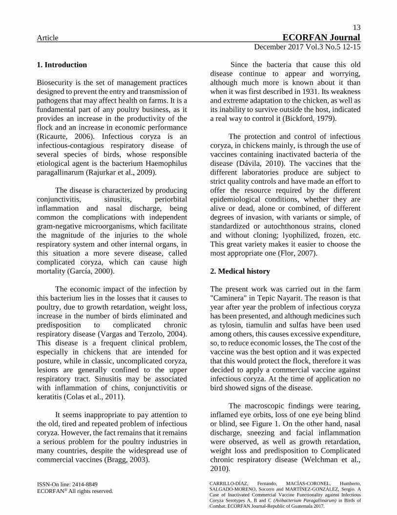
The macroscopic findings were tearing,
inflamed eye orbits, loss of one eye being blind
or blind, see Figure 1. On the other hand, nasal
discharge, sneezing and facial inflammation
were observed, as well as growth retardation,
weight loss and predisposition to Complicated
chronic respiratory disease (Welchman et al.,
2010).

14
Article ECORFAN Journal December 2017 Vol.3 No.5 12-15
ISSN-On line: 2414-8849
ECORFAN® All rights reserved.
CARRILLO-DÍAZ, Fernando, MACÍAS-CORONEL, Humberto,
SALGADO-MORENO, Socorro and MARTÍNEZ-GONZÁLEZ, Sergio. A
Case of Inactivated Commercial Vaccine Functionality against Infectious
Coryza Serotypes A, B and C (Avibacterium Paragallinarum) in Birds of
Combat. ECORFAN Journal-Republic of Guatemala 2017.
The findings at necropsy were similar to
those described by Welchman et al, (2010), such
as mucus in the trachea, swelling of the
infraorbital sinuses accompanied by congestion
of the mucous membranes of the nasal cavity and
paranasal sinuses and the formation of an excess
of mucus.
Figure 1 Fighting chicken with signs of infectious coryza
For the diagnosis, clinical signs and
necropsy findings presented by the birds were
taken into account together with the clinical
history of the disease, which quickly spread in
the geographical area where the infectious
coryza is the main manifestation. In addition,
microbial samples (exudates) were taken from
the nasal sinuses of sick birds, using cotton
swabs and subsequently inoculated, through
nasal route, to 5 healthy birds (Calnek, 2000).
The five birds were kept under observation and
isolated from the rest of the group, and the
coryza was produced 48 hours after inoculation.
As treatment, an avian bacterin in aqueous
suspension was used, which contains cultures in
artificial medium of avibacterium
paragallinarum serotypes A, B and C,
inactivated. This bacterin is used as an aid in the
prevention of infectious coryza of commercial
poultry. The route of administration was by
intramuscular injection in the breast, in birds
older than 3 weeks of age with doses of 0.5 ml
per animal. The vaccine was stored at a
temperature between 4 and 8 ° C.
It was applied at a temperature of 20 ° C,
avoiding exposure to sunlight at all times. It was
stirred well before being used until a
homogeneous mixture was obtained. Two weeks
after the first application, the reinforcement was
administered. The vaccine was applied to 200
fighter birds of the lines Leiper-hatch, Blueface,
Yellow leg, Radio, Gyros and crosses of these in
mixed flock.
3. Discussion and Comments
Of the 200 animals that received the vaccine, a
mortality of 69% attributable to the infectious
coryza was obtained, plus a 5% mortality from
other causes. The survival obtained was 28.5%
(27 females and 30 males), of which 5 females
and 20 males were one-eyed.
Rajukar et al (2009) mention that the
complications of infectious coryza can be
determined by stress factors. The natural
temperament of fighter birds leads to a state of
significant stress, which could determine the
lack of effectiveness of the vaccine.
A possible complication of the infectious
coryza with other bacterial or viral infections
should also be taken (Welchaman et al., 2010),
as in the complication with Mycoplasma
Gallisepticum (Abd El-Ghany, 2011), which
may have not allowed the effect favorable
vaccine. On the other hand it can be thought that
the presentation of the infectious coryza was
caused by a local strain, against which the
vaccine used had no effect. Blackall (1999)
comments that commercial vaccines are
produced in an international standard against
inactivated strains of Avibacterium
paragallinarum, but vaccines do not protect
against local variants of Avibacterium
paragallinarum, implying the need to produce
vaccines specific to the region ( -Ghany, 2011).

15
Article ECORFAN Journal December 2017 Vol.3 No.5 12-15
ISSN-On line: 2414-8849
ECORFAN® All rights reserved.
CARRILLO-DÍAZ, Fernando, MACÍAS-CORONEL, Humberto,
SALGADO-MORENO, Socorro and MARTÍNEZ-GONZÁLEZ, Sergio. A
Case of Inactivated Commercial Vaccine Functionality against Infectious
Coryza Serotypes A, B and C (Avibacterium Paragallinarum) in Birds of
Combat. ECORFAN Journal-Republic of Guatemala 2017.
4. Conclusions and recommendations
The vaccine did not meet the expectations
expected in the protection against avian
infectious coryza disease in fighting birds.
However, it is important to check the cold chain
to make sure that the vaccine management is
adequate. It is recommended to apply the
vaccine, experimentally, in a sample that is
representative of the total number of birds and
wait up to a week, and depending on the result,
make the application to the entire flock, in order
to reduce costs.
The birds of battle if they lose an eye are
waste animals for which the economic losses in
this operation were large.
5. References
ABD EL-GHANY, W.A. (2011). Evaluation of
autogenous Avibacterium paragallinarum
bacterins in Chickens. International Journal of
Paultry Science. 10 (1): 56-61.
BRAGG, R.R. (2004). Limitation of the spread
and impact of infectious coryza through the use
of a continuous disinfection programe.
Onderstepoort Journal of Veterinary Research.
71: 1-8.
BICKFORD A.A. (1979). Control del coriza en
dos zonas afectadas. California Poultry Letter. 7:
1-3.
CALNEK B.W. (2000). Enfermedades de las
aves. Editorial Manual Moderno. Segunda
edición, México.
COLAS C.M; Lamazares M.C; Pérez G.L; Sosa
T.I.M; Abeledo M.A; Merino L.A; Fuente D;
Gómez Á.E. (2011). Evaluación epidemiológica
de procesos respiratorios bacterianos en
reemplazos de ponedoras. Rev. Salud Anim. 33
(3): 178-183.
DÁVILA G.E. (2010). Vacunas e inmunidad
contra coriza infecciosa. Sistema de Revisiones
en Investigación Veterinaria de San Marcos
(SIRIVS). Universidad Mayor de San Marcos.
http://veterinaria.unmsm.edu.pe/files/Articulo_
davila_Final.pdf.
FLOR A.G. (2007). Vacunas y técnicas de
vacunación. VII Congreso Nacional de
Avicultura. http://www.amevea-
ecuador.org/datos/GALO%20FLOR.PDF.
GARCÍA F.J.A. (2000). Estudio microbiológico
de coriza infecciosa aviar en aves de postura
comercial provenientes de la región de los altos
de Jalisco, durante el periodo 1998-2001. Tesis
de Maestría. Universidad de Colima. Tecomán,
Colima.
RICAURTE G.S.L. (2006). Bioseguridad en
granjas avícolas.
http://www.engormix.com/MA-
avicultura/sanidad/articulos/bioseguridad-
granjas-avicolas-t868/165-p0.htm.
RAJURKAR G; Roy A; Mohan M.Y. (2009).
An overview on epidemiologic investigations of
Infectious coryza. Veterinary World. 2(10): 401-
403.
ROCA C E. (1996). La patología aviar en el
ultimo siglo. Universidad Autónoma de
Barcelona.
http://ddd.uab.cat/pub/selavi/selavi_a1996m5v3
8n5/selavi_a1996m5v38n5p29.pdf.
VARGAS E.S; Terzolo H.R. (2004).
Haemophilus paragallinarum: etiología de la
coriza infecciosa. Veterinaria México. 35 (3):
245-259.
WELCHMAN D.B; King S.A; Wragg P; et al.
(2010). Infectius coryza in chickens in Great
Britain. Veterinary Record. 912-913. .

16
Article ECORFAN Journal December 2017 Vol.3 No.5 16-20
NBelyax nanoparticle test as a disinfectant agent, applied in the hands of nursing
staff. Hospital de la Beneficencia Española and Hospital Nicolás San Juan, as case
studies
LEON-GUTIERREZ, Gabriela†, ALBARRAN, León, LEON-GUTIERREZ, Sergio and ARTEAGA-
LOPEZ, Paola*
Received July 25, 2017; Accepted December 16, 2017
Abstract
This study identified the presence of pathogenic microorganisms on inert surfaces of different areas
intended for direct medical care in two hospitals of the national public sector located in different places
of Mexico. In these hospitals the effectiveness of the nanoparticle NBelyax as a soap and hand soft cream
broad spectrum disinfectant agent was determined using the microbial process through the evaluation of
the percentage reduction of Colony Forming Units (UFC). Obtaining results of 100% effectiveness for
pathogenic microorganisms.
Nanoparticle, Nbelyax, Bacterial Challenge, Hospital, Hand, Handwashing, Nurses
Citation: LEON-GUTIERREZ, Gabriela, ALBARRAN, León, LEON-GUTIERREZ, Sergio and ARTEAGA-LOPEZ,
Paola. NBelyax nanoparticle test as a disinfectant agent, applied in the hands of nursing staff. Hospital de la Beneficencia
Española and Hospital Nicolás San Juan, as case studies. ECORFAN Journal-Republic of Guatemala. 2017, 3-5: 16-20.
*Correspondence to Author (email: [email protected]) †Researcher contributing first author.
© ECORFAN Journal - Republic of Guatemala www.ecorfan.org/republicofguatemala

17
Article ECORFAN Journal December 2017 Vol.3 No.5 16-20
ISSN-On line: 2414-8849
ECORFAN® All rights reserved.
LEON-GUTIERREZ, Gabriela, ALBARRAN, León, LEON-GUTIERREZ, Sergio and ARTEAGA-LOPEZ, Paola. NBelyax nanoparticle test as a
disinfectant agent, applied in the hands of nursing staff. Hospital de la
Beneficencia Española and Hospital Nicolás San Juan, as case studies.
ECORFAN Journal-Republic of Guatemala. 2017.
1. Introduction
Since the mid-nineteenth century, chemical
products applied to the skin have been used in
order to prevent infections. Semmelweis (1847)
introduces the practice of washing hands with
chlorinated compounds. Lister, years later;
expanded the use of phenolic solutions, both in
the hands and skin of patients and in the
instruments used. (1-6)
At present, even when the use of
antibiotics in clinical practice is very broad, the
use of antiseptics has not been eliminated as a
measure to prevent infections; On the contrary,
the formulations of old substances such as Iodine
have been perfected and other more elaborate
formulations have appeared. (7-9). However last
year, the Food and Drug Administration (FDA)
of the United States, released a statement on a
new regulation of the use of antiseptics. The rule,
published in the Federal Register of the United
States, "Safety and Effectiviness of Consumer
Antiseptics; Topical Antimicrobial Drug
Products for Over the Counter Human Use
"(https://www.gpo.gov/fdsys/pkg/FR 2016-09-
06 / pdf / 2016-21337.pdf) (10) establishes the
prohibition of the use of antiseptic products,
based on the recommendations of the
Supervisory Committee of Drugs Without
Prescription (NDAC, by its acronym in English).
The previous provision is based on the
results obtained when evaluating the
effectiveness of antibacterial products for
washing, compared with the effectiveness of
non-antibacterial products; the results showed
that there are no significant differences in the
effectiveness of both. However, when the effects
of the ingredients of these products were
evaluated it was found that they had a high risk
of producing reproductive alterations and
cancer. In addition to causing bacterial
resistance.
The studies carried out were in vitro to
evaluate the bacterial resistance and in vivo to
evaluate the toxicity as endocrine and
carcinogenic disruptors. The in vivo studies
were of the topical or dermal type and the
pharmacokinetics, dermal absorption, dermal
carcinogenesis, reproductive and developmental
toxicity, as well as the hormonal effects were
evaluated.
In total there were 19 ingredients that
presented these effects. The foregoing
demonstrates the need for the use of new
technologies to cover this field that remains
deserted, NBelyax is a good alternative since it
does not need the prohibited ingredients to be
effective against pathogens. As always, NBelyax
is at the forefront since it has been tested in
dermal toxicity studies and does not present
alterations, as well as its main asset in studies of
chronic and subchronic toxicity and it does not
present adverse effects when administered. At
the moment more sophisticated studies are being
developed to evaluate the reproductive and
carcinogenic effects of our product.
1.1 Justification
There is documentary evidence of the
importance of guaranteeing an inhospitable
environment free of germs, pathogens or with
maximum permissible levels (NMP) according
to current regulations, so it is currently
recommended to conduct environmental
sampling to identify the existence of pathogenic
microorganisms, before out-of-hospital
outbreaks or in case of trying to incorporate new
techniques or innovative products developed
with Nanotechnology.
The case before us is to determine the
antimicrobial effectiveness of the products: soap
and surgical antiseptic cream for its possible
incorporation in the process of asepsis,
sanitization and disinfection of the hands of the
nursing staff of both hospitals.

18
Article ECORFAN Journal December 2017 Vol.3 No.5 16-20
ISSN-On line: 2414-8849
ECORFAN® All rights reserved.
LEON-GUTIERREZ, Gabriela, ALBARRAN, León, LEON-GUTIERREZ, Sergio and ARTEAGA-LOPEZ, Paola. NBelyax nanoparticle test as a
disinfectant agent, applied in the hands of nursing staff. Hospital de la
Beneficencia Española and Hospital Nicolás San Juan, as case studies.
ECORFAN Journal-Republic of Guatemala. 2017.
1.2 Problem
Maintain safety and hygiene within the
framework of the effectiveness of proper
handwashing processes using nanotechnological
innovation products that reduce the risk of
intrahospital infections caused by the contact of
infected material and the hands of nurses.
1.3 Objectives
1.4.1 General objectives
Identify the presence and control of pathogenic
microorganisms in the hands of nurses at
random.
1.4.2 Specific objectives
Training and supervision of the proper
hand washing process.
Determine the quantitative value of germs
before and after the application of surgical
soap and antiseptic cream with the
NBelyax nanoparticle.
Determine the antimicrobial activity of the
products with the NBelyax nanoparticle in
hands, by applying the microbiology
method.
To determine the correct performance of
the safety and hygiene processes as well as
the effectiveness of products with the
NBelyax nanoparticle.
2. Materials and methods
The process of applying soap and cream to the
nursing staff was as follows:
Soap with NBelyax nanoparticle
1. Prior taking of the sample with isopos in
the hands of the nursing staff.
2. Hand washing with the soap, which
contains the NBelyax nanoparticle, with a
duration of approximately 20 seconds.
3. Rinsing process and taking the second
sample after washing.
Cream with NBelyax nanoparticle
1. Prior taking of the sample with isopos in
the hands of the nursing staff.
2. Application of the cream in hands
containing the NBelyax nanoparticle,
allowing it to be absorbed into the skin for
approximately 20 to 40 seconds.
3. Process of taking the second sample after
washing.
Evaluation by means of microbiological
techniques and obtaining results.
3. Results
Hospital of the Spanish Beneficence of
Torreón
Spanish Beneficence Torreón
Graph 1
Graph 1 shows the results of the effect of
the use of surgical grade soap and antiseptic
cream in hands. The results show the differences
between the before and after use of both products
on the final UFC count of mesophilic organisms,
coliforms and yeasts present in the hands of the
nursing staff of the Hospital de la Beneficencia
Española in Torreón. It was observed that a
decrease to zero of CFU is obtained after the
application of both products.

19
Article ECORFAN Journal December 2017 Vol.3 No.5 16-20
ISSN-On line: 2414-8849
ECORFAN® All rights reserved.
LEON-GUTIERREZ, Gabriela, ALBARRAN, León, LEON-GUTIERREZ, Sergio and ARTEAGA-LOPEZ, Paola. NBelyax nanoparticle test as a
disinfectant agent, applied in the hands of nursing staff. Hospital de la
Beneficencia Española and Hospital Nicolás San Juan, as case studies.
ECORFAN Journal-Republic of Guatemala. 2017.
Nicolás San Juan Hospital of the State of Mexico
Graph 2
Graph 2 shows the results of the effect of
the soap and surgical grade antiseptic cream in
hands on mesophilic organisms. The results
show the differences between the before and
after use of both products on the final UFC count
of mesophilic organisms, coliforms and yeasts
present in the hands of the nursing staff of the
Nicolás San Juan Hospital in the State of
Mexico. It was observed that a decrease to zero
of CFU is obtained after the application of both
products.
4. Discussion and Conclusions
The results obtained in this study, give evidence
of the bactericidal effect of the NBelyax
nanoparticle in its modality as soap and cream
development. The results of the sampling in two
hospitals, evidence the reduction of CFU of
mesophilic bacteria, coliforms and yeast present
in the hands of the nursing staff of both hospitals.
The results support the bactericidal effectiveness
of the NBelyax nanoparticle.
It is concluded that the study objectives
were fulfilled and the bactericidal effect of the
NBelyax nanoparticle is based on the results of
the microbiological study carried out. The study
invites to develop later ones to broaden the
microbiological spectrum of action.
New techno-scientific evidence was
obtained as novel disinfection tools that can
replace those products banned by the FDA. It is
important to highlight the usefulness of reducing
the presence of pathogenic organisms during the
hand-washing process of medical personnel in
general, particularly in hospital units that assist
in procedures aimed at the prevention, control
and epidemiological surveillance of Nosocomial
infections.
5. References
[1] Nosocomial Infection Surveillance. 1984.
MMWR CDC Surveill Summ 35 (No.1ss): 17ss,
1986. 20 -21.
[2] Wenzel RP, ed. Prevention and Control of
Nosocomial Infections. 2nd ed. Baltimore:
William & Wiekins; 1993. 15 -17.
[3] Edmond MB, Wenzel RP: Infection Control,
Mandells Infections Diseases, Principles and
Practice of Infection Diseases. 4th Edition.
Churchill Livignstone Inc. 1995, New York.
102.
[4]Malagón-Londoño/Hernández-Esquivel.
Infecciones Hospitalarias. Editorial Médica
Panamericana. 2ª Edición. Bogotá.1999: pg 78.
[5] International Society for Infectious Diseases,
Guía para el control de infecciones en el hospital,
Boston MA. USA, 2000.pg 45.
[6] Haley RW, Shaberg DR et al. Estimating the
extra charges and prolongation of hospitalization
due to intrahospitalization infection: a
comparison of methods. J. Infect. Dis. 1980;141-
248
[7] Crede W, Hierholzer WJ Jr. Linking hospital
epidemiology and quality assurance: seasoned
concepts in a new role. Infect Control 1988;9:42.

20
Article ECORFAN Journal December 2017 Vol.3 No.5 16-20
ISSN-On line: 2414-8849
ECORFAN® All rights reserved.
LEON-GUTIERREZ, Gabriela, ALBARRAN, León, LEON-GUTIERREZ, Sergio and ARTEAGA-LOPEZ, Paola. NBelyax nanoparticle test as a
disinfectant agent, applied in the hands of nursing staff. Hospital de la
Beneficencia Española and Hospital Nicolás San Juan, as case studies.
ECORFAN Journal-Republic of Guatemala. 2017.
[8] Gaynes RP, & Horan TC. Surveillance of
Intrahospitalaria Infections. In Mayhall CG.
Hospital Epidemiology and Infection Control.
Williams & Wilkins, Baltimore. 1996. Pp1017-
1031.
[9] Edmond MB & Wenzel RP. Infection
Control. In Mandell G, Bennett J, Dolin R.
Principles and Practice of Infectious Diseases.
Churchill Livingstone New York. 1995. pp2572-
2575.
[10] Safety and Effectiviness of Consumer
Antiseptics; Topical Antimicrobial Drug
Products for Over the Counter Human Use
(https://www.gpo.gov/fdsys/pkg/FR 2016-09-
06/pdf/2016-21337.pdf)

21
Article ECORFAN Journal December 2017 Vol.3 No.5 21-32
Response of stomatal conductance and xylem sap abscisic acid concentration of
Sorghum bicolor (L.) Moench cv. Tegemeo under re-watered and drought-stressed
conditions
NERI-LUNA, Cecilia1*†, VILLARREAL-RUIZ, Luis2, HUERTA-MARTÍNEZ, Francisco Martín1 and
ROBLES-MURGUÍA, Celia1
1Universidad de Guadalajara, Laboratorio de Ecofisiología Vegetal, Departamento de Ecología. CUCBA, México 2Laboratorio de Recursos Genéticos Microbianos & Biotecnología (LARGEMBIO), PREGEP-Genética, Colegio de
Postgraduados, Campus Montecillo, Edo. México, México
Received July 31, 2017; Accepted November 27, 2017
Abstract
The sorghum plant grows in a wide range of soils and environments, and its agronomic and economic
importance worldwide is incrementing yearly. This crop has several traits that make it a model for
research for the study of C4 species and stress tolerance. In this research, three methods for creating a
controlled environment to grow the sorghum plant using diverse substrates and nutrient solution
combinations were tested. After trials, the method that was composed of sand and nutrient solution by
infertile sandy soils was effective and sorghum plants were significantly bigger in height, and heavier in
fresh and dry weight compared to those that were cultivated by other two methodologies. Using this
artificial susbtrate, sorghum plants were grown-up and then were subjected to a drought conditions.
Following the drought-stressed period, the stomatal conductance was significantly reduced and xylem
sap abscisic acid concentration significantly increased by about 64% in plants developed in both
substrates. This experimental system can be used for future research contributing to sorghum study of
mechanisms in overcoming abiotic constrains such as drought and water relations and possible adaptation
to tropical and subtropical climates.
Sorghum, Drought, ABA Xylem Sap, Stomatal Conductance
Citation: NERI-LUNA, Cecilia, VILLARREAL-RUIZ, Luis, HUERTA-MARTÍNEZ, Francisco Martín and ROBLES-
MURGUÍA, Celia. Response of stomatal conductance and xylem sap abscisic acid concentration of Sorghum bicolor (L.)
Moench cv. Tegemeo under re-watered and drought-stressed conditions. ECORFAN Journal-Republic of Guatemala 2017,
3-5: 21-32.
* Correspondence to Author (email: [email protected])
† Researcher contributing first author.
© ECORFAN Journal - Republic of Guatemala www.ecorfan.org/republicofguatemala

22
Article ECORFAN Journal December 2017 Vol.3 No.5 21-32
ISSN-On line: 2414-8849
ECORFAN® All rights reserved.
NERI-LUNA, Cecilia, VILLARREAL-RUIZ, Luis, HUERTA-MARTÍNEZ,
Francisco Martín and ROBLES-MURGUÍA, Celia. Response of stomatal conductance and xylem sap abscisic acid concentration of Sorghum bicolor
(L.) Moench cv. Tegemeo under re-watered and drought-stressed conditions.
ECORFAN Journal-Republic of Guatemala 2017.
1. Introduction
Sorghum is the fifth most important cereal in the
world, and its relevance is increasing because of
its low demand for water, coupled with its
potential adaptation to global climate change
(Tsuboi, 2017). Furthermore, sorghum is
categorized as a competitive coarse grain in the
world markets and as an important food supply
to overcome the accelerated expansion of human
population (Thomason et al., 2017). In the same
line, sorghum planted area worldwide has been
increased by 66% over the past 50 years, and the
yield has improved by 2.4 fold (Qi et al., 2016).
This cereal is cultivated in at least 73
countries throughout Africa, America and Asia
in tropical and subtropical regions (Upadhyaya
et al., 2016). In the semiarid tropics of
undeveloped countries with low income human
populations, sorghum remains the primary
source for human nutrition.
Countries such as the United States,
Argentina, and Australia have been the major
sorghum producers for primarily commercial
use in livestock feed, bioenergy, and other non-
food and industrial uses (Qi, 2016, O’Brien,
2016). Recently, sorghum has been useful for
phytoremediation in poluted lands, whereby soil
contaminants (usually heavy metals) are taken
up by the plant and removed from the
environment (Tsoboi, 2017).
Sorghum plants possess inherent abilities
to survive and exceed under extreme
temperatures and drought conditions, and it has
also gained interest because contains most of the
traits in a model plant species such as large
embryos that are easy to rescue, plenty of seeds
production, moderate genome size and available
whole-genome sequences (Paterson et al., 2009;
Calviño and Messing, 2012; Rizal et al., 2014).
Sorghum also has a high photosynthetic
efficiency and use of water due to the effective
C4 carboxylation pathway (Nielsen and Vigil,
2017) and harbors genes for higher biomass and
other yield-related traits.
In spite of being one of the major crops in
the world, the use of sorghum in scientific
research lags far behind other cereals (Calviño
and Messing, 2012). Moreover, the outcomes of
sorghum research have not been used widely in
crop improvement compared with other cereals
like maize and rice (Izawa and Shimamoto,
1996; Rensink and Buell, 2004).
As a consequene, in order to promote
sorghum as a model species it is necessary to get
a better knowledge about sorghum
responsivness to water deficit and drought
starting with simple experimental model
designs, and then scaling up to bigger trials to
understand its adaptation under severe
environmental conditions (Calviño and Messing,
2012; Neri-Luna et al., 2016; Chen et al., 2017).
Despite the known resilience of sorghum
to drought stress, little is known about the
responses to water deficit and its drought
tolerance, specifically, the role of non-hydraulic
root-to-shoot signalling (i.e. changes in
concentration and flux rate of abscisic acid,
ABA) considered significant in regulating shoot
growth and water use when soil is drying,
without any demonstrable change in shoot water
or nutrient status (Hansen and Dörffling, 2003).
The xylem sap ABA concentration has a
distinctive role in stomatal regulation (which is
the main mechanism used by plants to control
gas exchange and transpiration) of hydric status
in water stress conditions (Dodd, 2003;
Pospíšilová, 2003).

23
Article ECORFAN Journal December 2017 Vol.3 No.5 21-32
ISSN-On line: 2414-8849
ECORFAN® All rights reserved.
NERI-LUNA, Cecilia, VILLARREAL-RUIZ, Luis, HUERTA-MARTÍNEZ,
Francisco Martín and ROBLES-MURGUÍA, Celia. Response of stomatal conductance and xylem sap abscisic acid concentration of Sorghum bicolor
(L.) Moench cv. Tegemeo under re-watered and drought-stressed conditions.
ECORFAN Journal-Republic of Guatemala 2017.
The aims of this research are: (i) to
compare three different methods for the
establishment and growth of sorghum plants
under controlled conditions in order to select the
most suitable; (ii) to provide the optimum
approach in order to evaluate the stomata
responses to changes in the concentration of
ABA in the xylem sap under a drought-stressed
and re-watered period.
2. Materials and Methods
2.1. Experimental set up
In this research three different artificial shorgum
culture methods suggested by Brundrett et al.
(1996), INVAM (1997), YMRG (1996) were
tested. Plastic pots (2 L, 15cm x 15cm x 12cm)
were thoroughly washed with water, and
disinfected with 1% (w/v) Virkon solution
(Antec International Limited) and rinsed with
destillated water before use (Vimard et al.,
1999).
For each method, the pots (n= 7) were
filled with the adequate substrate and the
containers were watered to field capacity
according to the appropriate nutrient solution
(Table 1).
Sorghum bicolor (L.) Moench cv.
Tegemeo seeds were washed with running tap
water, and surface sterilized with 10% NaOCl
for 20 min (Jarstfer and Sylvia, 1993) followed
by a final rinse with running distilled water. The
seeds were placed in a baker with autoclaved
distilled water for 16 h in darkness to imbibe
(Maiti, 1996).
Method Substrate 1Particle size
/disinfection
Nutrient Solution
(a) Brundrett et
al. (1996)
Silica sand (0.50-0.78 mm) autoclaved twice
for 1 h at 121C.
Snowball and
Robson
(g/L): (26) KH2PO4,
(43) K2SO4, (21) NH4NO3/2 weeks,
(43) CaCl22H2O, (6.4)
MgSO47H2O, (4.3) MnSO4H2O, (3)
ZnSO4 7H2O, (1.3)
CuSO45H2O, (0.2) H3BO3, (0.1)
CoSO47H2O, (0.05)
Na2MoO42H2O. Plants watered twice a
week with nutrient
solution
(b)
INVAM
(1997)
Silica sand:
loamy soil
(2:1 v/v).
Soil (1-0.5 mm)
steamed twice
for 1 h at 85C. Sand (0.50-0.78
mm) autoclaved
twice for 1 h at
121C.
Long Ashton (g/L):
(50.6) KNO3, (80.25)
Ca(NO3)24H2O, (52) NaH2PO42H2O, (46)
MgSO47H2O, (6.7)
FeNaEDTA
Plants watered twice a
week with 50% full strength of nutrient
solution
(c) YMRG
(2001)
Silica sand:
calcined attapulgite
clay soil conditioner
(50:50) +
bone meal
Sand:calcined
attapulgite clay soil conditioner
(0.50-1.00 mm) autoclaved twice
for 1 h at 121C.
Rorison (g/500 mL):
(62.01) MgSO47H2O, (119.02)
Ca(NO3)24H2O, (57.69)
K2HPO43H2O,
Trace elements: (6.250) FeEDTA,
(0.560) MnSO4H2O,
(0.716) H3BO3, (0.046)
(NH4)6Mo244H2O,
(0.110) ZnSO4 7H2O, (0.099) CuSO45H2O
Plants watered twice a week with half to one
fifth strencgth of
nutrient solution 1Gaur and Adholeya (2000).
Table 1 The three tested methods for shorgum growth
under greenhouse conditions controlled conditions

24
Article ECORFAN Journal December 2017 Vol.3 No.5 21-32
ISSN-On line: 2414-8849
ECORFAN® All rights reserved.
NERI-LUNA, Cecilia, VILLARREAL-RUIZ, Luis, HUERTA-MARTÍNEZ,
Francisco Martín and ROBLES-MURGUÍA, Celia. Response of stomatal conductance and xylem sap abscisic acid concentration of Sorghum bicolor
(L.) Moench cv. Tegemeo under re-watered and drought-stressed conditions.
ECORFAN Journal-Republic of Guatemala 2017.
Figure 1 Experimental set up with sorghum seedlings
growing with three differents methods under greenhouse
controlled conditions. (a) Brundrett et al. (1996). (b)
INVAM (1997). (c) YMRG (2001).
After imbibition, the sorghum seeds were
distributed on a damp layer of tissue paper and
placed in an envelope of aluminium foil and
stored in a dark cupboard for 48 h to allow
germination (Mace, 1999). After germination 5
sorghum seeds with a uniform radicle were
planted in each container and covered with the
substrate.
Finally, the planted pots were placed on a
greenhouse bench and randomly arranged every
week (Figure 1). After 2 weeks, the sorghum
seedlings were thinned to 1 plant per container
and received the nutrient solution and water
appropriate to the corresponding treatment.
Sorghum plants were cultivated in the
greenhouse under controlled conditions with a
photoperiod of 16h light/8h dark and a
Photosynthetic Active Radiation (PAR) of 300-
800 mol m-2 s-1. The temperature ranged from
16C to 26C with a relative humidity (rh)
between 45-70%.
A destructive harvest was performed at 9
weeks after planting. First, the height of each
plant was recorded, and the shoot components
(leaves, stem, and ligules) were separated and
the fresh weight (FW) was recorded.
The plant samples were placed in paper
bags, and dried at 60C for 48 h and allowed to
cool in a perspex single door desiccator chamber
with silica gel and subsequently the dry weight
(DW) was recorded using a Mettler AJ100
analytical balance. Statistical analyses were
carried out using SPSS v24 package.
All data was verified for normality using a
Kolmogorov-Smirnov Test and for homogeneity
of variances using the Levene Test. Plant
parameters at harvest were compared by One-
way ANOVA (Dytham, 2011). Based on these
results, the following experiment was designed
in order to test the sorghum response to stomatal
conductance and variations in xylem sap abscisic
acid concentration ([ABAxyl]).
2. Stomatal conductance measurements and
xylem sap collection
PVC pipe cylinders (10.5 cm diameter X 18cm
height) designed to fit inside a pressure chamber
were disinfected as mentioned before. Each
cylinder was sealed at the base with three layers
of plant fleece attached with waterproof tape.
The cylinders (n=8) were packed with sand (S)
substrate and watered with the nutrient solution
proposed by Snowball and Robson (1984) and
using the best sorghum growing system in
previous experiment (Brundrett, 1996). In order
to validate this artificial system, cylinders (n=8)
were packed with natural field soil (NS) for
comparative purposes. The natural soil is a sandy
clay loam with a pH 5.9 in H2O and 5.5 in CaCl2,
CEC 2.4 cmol kg-1, base saturation 88.4, texture
loamy sand (sand 73.9%, silt 20%, clay 6.1%),
organic matter 6.4%, Corg3.5% and Norg 449.6
mgN/100g.
Sorghum seeds were washed, surface
sterilized and germinated as explained before.
Three germinated seeds were planted in the
middle of each cylinder and the planted
cylinders were placed on a greenhouse bench
and randomly re-arranged every week.

25
Article ECORFAN Journal December 2017 Vol.3 No.5 21-32
ISSN-On line: 2414-8849
ECORFAN® All rights reserved.
NERI-LUNA, Cecilia, VILLARREAL-RUIZ, Luis, HUERTA-MARTÍNEZ,
Francisco Martín and ROBLES-MURGUÍA, Celia. Response of stomatal conductance and xylem sap abscisic acid concentration of Sorghum bicolor
(L.) Moench cv. Tegemeo under re-watered and drought-stressed conditions.
ECORFAN Journal-Republic of Guatemala 2017.
After emergence, the seedlings were
thinned to one per cylinder. Plants cultivated in
S were watered twice per week with 120 ml of
the nutrient solution (Snowball and Robson,
1984) and with deionised water at intervals
(twice a week) of nutrient additions. The plants
growing in NS were watered with tap water as
required. They were kept in the greenhouse
under the following environmental conditions:
TCmin 23C/TCmax 30C, 16-48% rh and a
mean PAR of 200 mol m-2 s-1 at plant level for
14 h d-1.
Drought period: Eight weeks after
sowing, the water was withheld from half of the
plants (n=4 in each substrate (S or NS). Firstly,
the planted cylinders were watered to field
capacity and allowed to drain overnight. In the
morning, the sorghum plants were re-watered
and allowed to drain until they stopped dripping,
then the drought-stressed period was induced.
The initial weight of each cylinder was
recorded using a digital balance (Ohaus, 5 kg
capacity). This step was repeated every 4 h
throughly, until the plants stopped losing weight,
and at this point they were moved to the lab for
xylem sap collection.
Throughout the drought-stressed period,
the stomatal conductance (gs) was recorded
several times using a Diffusion Porometer (AP4, ©Delta-T Devices, Cambridge, UK). Finally, the
control plants (n=4) for each susbtrate, were
watered as required.
Xylem sap was collected based on the
method described by Seel and Jeschke (1999).
The plants were sealed in the special lid of the
pressure vessel (Figure 2) with the two-
component silicon–based dental impression
material Blend-a-gum (Coltène PRESIDENT
fast microsystem regular body).
Figure 2 Schematic representation of the pressure
chamber. Inset (a) shows a section through the lid along
the line A-A with the V-shaped joint sealed with high
vacuum grease. Sealing of the stems within the lid also is
achieved by silicon-based dental impression material
which is, after sealing, compressed by the pressurizing
blocks (Prb)
A segment of 1 cm was cut out in the stem
base with the lower cut sloping downwards at
60 to the centre to retain exuding sap.
After sealing the plants into the pressure
vessel, the pneumatic pressure was raised
gradually until the first drops of xylem liquid
appeared; this balancing pressure sensu
Passioura and Munns (1984) was recorded.
Subsecuently, the xylem sap flow was induced
by increasing the pressure to 100 kPa. Xylem sap
samples (c. 500 L) were collected and stored in
the dark at -80C for further analysis.
The ABA concentration was analysed in
crude samples of xylem sap by the
radioimmunoassay technique described by
Quarrie et al. (1988).
26
Article ECORFAN Journal December 2017 Vol.3 No.5 21-32
ISSN-On line: 2414-8849
ECORFAN® All rights reserved.
NERI-LUNA, Cecilia, VILLARREAL-RUIZ, Luis, HUERTA-MARTÍNEZ,
Francisco Martín and ROBLES-MURGUÍA, Celia. Response of stomatal conductance and xylem sap abscisic acid concentration of Sorghum bicolor
(L.) Moench cv. Tegemeo under re-watered and drought-stressed conditions.
ECORFAN Journal-Republic of Guatemala 2017.
After thawing, 200 L 50% PBS were put
into 2 mL plastic centrifuge tubes. Subsequently
50 L of ABA standard or xylem sap sample was
added carefully to each tube. This solution was
mixed with 100 L of 3H-ABA in -globulin
solution (ca 9,000-10,000 cpm per vial) and 100
L MAC252 antibody solution. The solution
was mixed briefly by inverting tubes and then
spun down in a microcentrifuge (Centurion
8080, Norlab) for 30s.
The assay mixture was incubated at 4C in
darkness for 45 min to allow binding to take
place. Subsecuently, a saturated solution (500
L) of (NH4)2SO4 was added to each tube in
order to precipitate the ABA-antibody complex
and, after a brief shaking, the mixture was left in
the dark at room temperature for 30 min. Free
antibodies and those that bound to ABA in the
reaction mixture were precipitated and pelleted
by centrifugation for 4 min at 1400g, then the
supernatant was discarded.
The pellet was washed by suspending in 1
mL of 50% saturated (NH4)2SO4 solution in
order to remove the excess unbound
radioactivity (the total pellet was re-suspended
by shaking the rack of vials). The tubes were
centrifuged again for 4 min at 1400g and the
supernatant was discarded carefully and the
pellet resuspended in 100 L of deionized water.
The tubes were again spun for 30 s and
1.25 mL of scintillation cocktail (Ecoscint-H)
was added to all tubes and the content was mixed
thoroughly. Finally, the tubes were put inside the
glass scintillation vials and the radioactivity was
quantified in a liquid-scintillation counter
(Packard 4430, Berlshire, England), the tubes
where count up (on protocol 8), three times for 5
min on 3H setting.
Concentrations of ABA were calculated
from the radioactivity (cmp) present in the
pellets. A series of ABA standards was included
in each batch of assays in order to construct a
calibration curve. This was usually linearised by
subtraction of Bmin and plotting log-transformed
counts against the natural logarithm (ln) of the
unlabelled ABA present per vial, where:
Log (B/Bmax)= ln [B/Bmax / 1-(B/Bmax) ] (1)
B= Corrected cpm bound in the presence of an
ABA standard = (cpm-Bmin)
Bmax= The maximal bound radioactivity when
only 3H-ABA reacted with MAC252 (H2O
standards).
Bmin= The minimal bound radioactivity when a
large excess of non-labelled ABA was added to
the vial.
Sample ABA concentrations were
calculated from this formula and the final result
was expressed in pmol per mL. Statistical
analyses were carried out using SPSS v24
package. All data were verified for normality
using a Kolmogorov-Smirnov Test and for
homogeneity of variances using the Levene Test.
An independent sample t-Test was performed in
order to compare means between watered vs.
droghted plants treatments (Dytham, 2011).
3. Results
3.1. A comparison of methods using artificial
substrates
The effect of different non-natural substrates and
diverse nutrient solutions on sorghum´s
development was variable. For instance,
sorghum plants grown using Brundrett (1996)
method, were significantly bigger in height, and
heavier in fresh and the dry weight compared to
those that were cultivated by other methods
(Table 2). It is important to pointed out that the
sorghum germination process was not affected in
any of the three methods used (P=0.319).

27
Article ECORFAN Journal December 2017 Vol.3 No.5 21-32
ISSN-On line: 2414-8849
ECORFAN® All rights reserved.
NERI-LUNA, Cecilia, VILLARREAL-RUIZ, Luis, HUERTA-MARTÍNEZ,
Francisco Martín and ROBLES-MURGUÍA, Celia. Response of stomatal conductance and xylem sap abscisic acid concentration of Sorghum bicolor
(L.) Moench cv. Tegemeo under re-watered and drought-stressed conditions.
ECORFAN Journal-Republic of Guatemala 2017.
3.2. Response of stomatal conductance and
changes of [ABAxyl]
The main reduction in stomatal conductance (gs)
of sorghum leaves was at 53 h and 99 h after the
water was withheld for plants cultivated in S and
NS substrates respectively (Figure 3a). It
appears that when stomatal closure (within the
first 100 h after the water was suspended) the
weight loss from containers stopped (Figure 3b).
The combination of stomatal conductance
measurements and weight loss data clearly
indicated that plants were suffering water stress.
Daily weather conditions in the greenhouse
during the drought-stressed period were: mean
PAR of 1528 mol m-2 s-1 at plant height,
TCmin 23.5C/ TCmax 31C, 16-39% rh and
vapor pressure deficit between 1.7-3.4 kPa.
The mean balancing pressure (ρo) needed
to obtain xylem sap from the stems of sorghum
plants under re-watered conditions was ~1500
kPa in both S and NS substrates. The sorghum
plants subjected to a drought-stressed period
needed higher balancing pressure were before
xylem fluid appeared. For instance, ~52543
kPa in those plants grown-up in sand and
~3000 kPa in plants cultivated in NS were
needed.
Following the drought-stressed period, the
gs was significantly (P=0.006) reduced and
[ABAxyl] significantly increased (P=0.003) by
about 64% in plants developed in both substrates
(Figure 3c-d). During the first 8 h after
withholding water, the sand substrate allowed
more water loss (P<0.05) than the NS substrate,
and were not observed significant differences
until 76 h, when the plants developed in NS
substrate lost more water.
By the end of drought-stressed period, no
significant differences were found (P>0.05). It
appears that sand substrate permitted more rapid
gs reduction than NS, but the differences were
not significant (P>0.05).
4. Discussion
Sorghum can be used as a model system for
research because of its: (1) agronomic
importance, (a) C4 photosynthesis, (b) low input
levels of nutrients, (c) adaptation to stresses due
to its reservoir of genes for tolerance to high salt,
pH, drought and heat; (2) increase of its
economic significance in areas as a valuable
source of food, biofuel and phytoremediation
(Tsuboi et al., 2017); (3) representative of large
genomes like sugarcane and switchgrass
(Paterson et al., 2009); (4) ease to produce
different types of breeding materials
(Upadhyaya et al., 2016); (5) the big plant size
Plant
parameters
Brundrett
et al.
INVAM
UYRG
P
Germination
(%)
77.7 ±7.02 a 49.9±16.6 a 55.5±14.0 a NS
Height (cm-1)
32.19 ±3.52 b
30.2±3.55 b 18.57±2.13 a **
Leaves
FW (g) DW (g)
1.55±0.41 b 0.44±0.09 b
0.50±0.08 a 0.14±0.02 a
0.31±0.10 a 0.07±0.01 a
** **
Stem
FW (g) DW (g)
0.85±0.25 b 0.19±0.04 b
0.23±0.04 a 0.05±0.009 a
0.14±0.06 a 0.02±0.007 a
* ***
Total FW
DW
2.41±0.67 b
0.63±0.13 b
0.74±0.12 a
0.19±0.02 a
0.46±0.16 a
0.10±0.02 a
**
***
(§) Plants were harvested 9 weeks after plantation. Means ±1SE (n=7).
Significance values were calculated using One-Way ANOVA at 95%
CI and Student-Newman-Keuls Test. *P0.05; **P0.01; ***P0.001,
NS= Not significant. FW= Fresh Weight; DW= Dry Weight. Table 2 Parameters of sorghum plants (§) using three
different techniques

28
Article ECORFAN Journal December 2017 Vol.3 No.5 21-32
ISSN-On line: 2414-8849
ECORFAN® All rights reserved.
NERI-LUNA, Cecilia, VILLARREAL-RUIZ, Luis, HUERTA-MARTÍNEZ,
Francisco Martín and ROBLES-MURGUÍA, Celia. Response of stomatal conductance and xylem sap abscisic acid concentration of Sorghum bicolor
(L.) Moench cv. Tegemeo under re-watered and drought-stressed conditions.
ECORFAN Journal-Republic of Guatemala 2017.
Figure 3 Response of sorghum plants under re-watered
and drought-stressed conditions grown in S and NS
substrates. (A) gs in leaves. (B) Cumulative weight loss.
(C) Xylem sap ABA concentration. (D) gs in leaves before
harvest (gs in drought-stressed plants cultivated in sand
was zero). Data represent ±1SE of the mean (n=4).
Significance values were calculated using a t-Test.
*P0.05; **P0.01; ***P0.001.
With wide leaves, make it suitable for
physiological measurements, leaf anatomical
analysis, growth and development studies
(Roozeboom and Prasad, 2016).
However, despite all this, other crop plants
like maize and rice have still remained
considered the primarly models for the study of
cereals and C4 species (Izawa and Shimamoto,
1996; Strable and Scalon, 2009). Surprisingly,
the outcomes of Arabidopsis research have been
used widely in crop improvement (Rensink and
Buell, 2004).
Therefore, it is important to consider
sorghum, for biological research on stress
tolerance from harsh environmental conditions
due to its valuable agronomic traits (Rizal et al.,
2014). Given the multifunctionality responses by
plants to environmental factors, some
researchers have pointed out the need of studies
under controlled conditions in order to evaluate
the results more carefully (van der Heijden and
Kuyper, 2001; Lendzemo et al., 2005).
Therefore, in this research to get a better
knowledge about sorghum responsiveness to
water deficitency and drought tolerance, diverse
artificial procedures (instead of the complicate
use of natural soil) were tested. Our results
indicate that the substrate and nutrient solution
suggested by Brundrett et al. (1996) was
effective and allowed sorghum to grow well
under the greenhouse controlled environmental
conditions.
Furthermore, in order to validate this
experimental system, the sensitive parameters
such as stomatal conductance and [ABAxyl])
suggested in other drought experiments (Dodd,
2003; Hansen and Dörffling, 2003; Pospíšilová,
2003) were chosen to detect sorghum plants
response when subjected to a drought-stressed
period.

29
Article ECORFAN Journal December 2017 Vol.3 No.5 21-32
ISSN-On line: 2414-8849
ECORFAN® All rights reserved.
NERI-LUNA, Cecilia, VILLARREAL-RUIZ, Luis, HUERTA-MARTÍNEZ,
Francisco Martín and ROBLES-MURGUÍA, Celia. Response of stomatal conductance and xylem sap abscisic acid concentration of Sorghum bicolor
(L.) Moench cv. Tegemeo under re-watered and drought-stressed conditions.
ECORFAN Journal-Republic of Guatemala 2017.
The results shown that stomatal response
and changes in xylem sap concentration were
detectable and there were not different from
those obtained using field natural soil, thus,
confirming that using sand as substrate with the
proper nutrient solution is suitable for further
research.
Because, the germplasm of sorghum is
extraordinarily diverse (Upadhyaya et al., 2016),
could be a key species for the expanding
knowledge about the functions of beneficial
crops in agriculture, especially in hot and dry
regions of the world.
For instance, the method used for growing
sorghum under controlled conditions could be
useful for trials involving sorghum germoplasm
collections in order to identify new sources of
variations for stress resistance, phenology, seed
yield and quality, and for bioenergy uses
(Upadhyaya et al., 2016). In addition, this
system can be useful to know: (1) how sorghum
proceeds from one stage to another, (2) how it
accumulates dry matter along the way, and (3)
how it partitions the dry matter to tissue and
grain, essential for understanding how a
sorghum plant is likely to respond to
environmental factors (Roozeboom and Prasad,
2016).
On the other hand, a common problem in
trying to scale up results from greenhouse to
natural environment is the uncertainty about
whether the effects that take place in these
unrealistic conditions occur in field conditions
(Rillig, 2004). Furthermore, in nature, plant
biodiversity, fitness and adaptation rely on the
development of roots in combination with soil
microorganisms with complementary functions
in order to promote plant growth and health.
Therefore, it could be important to generate
proposals involving soil microorganism that
may influence water relations or lessen drought
straining in sorghum plants.
5. Conclusion and future research
The method proposed by Brundrett (1996) was
proven as a good alternative for sorghum
development under greenhouse controlled
conditions.
Data from sorghum plants grown-up in
this system related to stomatal conductance and
xylem sap ABA concentration subjected to a
drought-stressed period were successfully
measurable and were not different from those
obtained using field natural soil.
Therefore, this experimental system seems
to be reliable for future research in supporting
the idea of sorghum as a model cereal crop. Most
importantly, sorghum can be study in order to
understand the plant advanced mechanisms in
overcoming abiotic constrains such as water
relations and drought in its development of
strategies for crop stress tolerance and
adaptation to harsh environmental conditions
around the world.
6. References
Brundrett M, Bougher N, Dell B, Grove T and
Malajczuck N. 1996. Working with mycorrhizas
in forestry and agriculture. ACIAR Monograph
32: 374 p.
Calviño M and Messing J. 2012. Sweet sorghum
as a model system for bioenergy crops. Current
Opinion in Biotechnology 23: 323–329.
Chen J, Chopra R, Hayes C, Morris G, Marla S,
Burke J, Xin Z, and Burow G. 2017. Genome-
Wide Association Study of Developing Leaves’
Heat Tolerance during Vegetative Growth
Stages in a Sorghum Association Panel. Plant
Genome 10 (2): 1-15.
Dytham C. 2011. Choosing and using statistics:
a biologist’s guide. 3rd Edition. Blackwell
Publishing. United Kingdom. 292 p.

30
Article ECORFAN Journal December 2017 Vol.3 No.5 21-32
ISSN-On line: 2414-8849
ECORFAN® All rights reserved.
NERI-LUNA, Cecilia, VILLARREAL-RUIZ, Luis, HUERTA-MARTÍNEZ,
Francisco Martín and ROBLES-MURGUÍA, Celia. Response of stomatal conductance and xylem sap abscisic acid concentration of Sorghum bicolor
(L.) Moench cv. Tegemeo under re-watered and drought-stressed conditions.
ECORFAN Journal-Republic of Guatemala 2017.
Dodd IC. 2003. Hormonal interactions and
stomatal responses. Journal of Plant Growth
Regulation 22: 32-46.
Gaur A and Adholeya A. 2000. Effects of the
particle size of soil-less substrates upon AM
fungus inoculum production. Mycorrhiza 10: 43-
48.
Hansen H and Dörffling K. 2003. Root-derived
trans-zeatin riboside and abscisic acid in
drought-stressed and rewatered sunflower
plants: interaction in the control of leaf diffusive
resistance. Functional Plant Biology 30: 365-
375.
INVAM (International Culture Collection of
Arbuscular and Vesicular-Arbuscular
Mycorrhizal Fungi at West Virginia University,
USA). http://invam.caf.wvu.edu/.
Izawa T and Shimamoto K. 1996. Becoming a
model plant: the importance of rice to plant
science. Trends in Plant Science 1(3): 95-99.
Jarstfer AG and Sylvia DM. 1993. Inoculum
production and inoculation strategies for
vesicular-arbuscular mycorrhizal fungi. In:
Metting FB Jr (Ed.). Soil microbial ecology:
Applications in agricultural and environmental
management. Marcel Dekker, New York. pp.
349-377.
Jarstfer AG and Sylvia DM. 1993. Inoculum
production and inoculation strategies for
vesicular-arbuscular mycorrhizal fungi. In:
Metting FB Jr (Ed). Soil microbial ecology:
Applications in agricultural and environmental
management. Marcel Dekker, New York. pp.
349-377.
Lendzemo VW, Kuyper TW, Kropff MJ and van
Ast A. 2005. Field inoculation with arbuscular
mycorrhizal fungi reduces Striga hermonthica
performance on cereal crops and has the
potential to contribute to integrated Striga
management. Field Crops Research 91: 51-61.
Mace YJ. 1999. When the going gets tough: the
responses of Sorghum bicolor L. cv. Tegemeo
seedlings to soil penetration resistance. BSc.
Thesis. Department of Plant and Soil Science.
University of Aberdeen, UK. pp. 28-31.
Maiti R. 1996. Sorghum Science. Science
Publishers, Inc. USA. 352 p
Neri-Luna C, Villarreal-Ruiz L, Huerta-
Martínez FM and Robles-Murguía C. 2016.
Response to drought of sorghum (Sorghum
bicolor (L.) Moench cv. Tegemeo) grown with a
split-root system: a climate change perspective.
UTSOE-Journal of Multidisciplinary Science 3
(6): 54-72.
Nielsen DC and Vigil MF. 2017. Defining a
Dryland Grain Sorghum Production Function for
the Central Great Plains. Agronomy Journal 109:
1582–1590.
O’Brien D. 2016. Domestic and International
Sorghum Marketing. In: Ciampitti I. and Prasad
V. (Editors). Sorghum: State of the Art and
Future Perspectives. Agronomy Monograph 58.
doi:10.2134/agronmonogr58.2014.0079.
Passioura JB and Munns R. 1984. Hydraulic
resistance of plants. II. Effects of rooting
medium, and time of day, in barley and lupin.
Australian Journal of Plant Physiology 11: 341-
350.
Paterson AH, Bowers JE, Bruggmann R,
Dubchak I, Grimwood J, Gundlach H, Haberer
G, Hellsten U, Mitros T, Poliakov A. 2009. The
Sorghum bicolor genome and the diversification
of grasses. Nature, 457: 551-556.
31
Article ECORFAN Journal December 2017 Vol.3 No.5 21-32
ISSN-On line: 2414-8849
ECORFAN® All rights reserved.
NERI-LUNA, Cecilia, VILLARREAL-RUIZ, Luis, HUERTA-MARTÍNEZ,
Francisco Martín and ROBLES-MURGUÍA, Celia. Response of stomatal conductance and xylem sap abscisic acid concentration of Sorghum bicolor
(L.) Moench cv. Tegemeo under re-watered and drought-stressed conditions.
ECORFAN Journal-Republic of Guatemala 2017.
Pospíšilová J. 2003. Participation of
phytohormones in the stomatal regulation of gas
exchange during water stress. Biologia
Plantarum 46(4): 491-506.
Qi G, Li N, Sun XS and Wang D. 2016.
Overview of Sorghum Industrial Utilization. In:
Ciampitti I. and Prasad V. (Editors). Sorghum:
State of the Art and Future Perspectives.
Agronomy Monograph 58.
doi:10.2134/agronmonogr58.2014.0070.
Quarrie SA, Whitford PN, Appleford NEJ,
Wang TL, Cook SK, Henson IE and Loveys BR.
1988. A monoclonal antibody to (S)-abscisic
acid: its characterization and use in a
radioimmunoassay for measuring abscisic acid
in crude extracts of cereal and lupin leaves.
Planta 173: 330-339.
Rensink WA and Buell CR. 2004. Arabidopsis
to Rice. Applying Knowledge from a Weed to
Enhance Our Understanding of a Crop Species.
Plant Physiology 135(2): 622-629.
Rillig MC. 2004. Arbuscular mycorrhizae and
terrestrial ecosystem processes. Ecology Letters
7: 740-754.
Rizal G, Karki S, Alcasid M, Montecillo F,
Acebron K, Larazo N, Garcia R, Slamet-Loedin
IH, and Quick WP. 2014. Shortening the
Breeding Cycle of Sorghum, a Model Crop for
Research. Crop Science 54: 520–529.
Roozeboom KL and Prasad P.V.V. 2016.
Sorghum Growth and Development. In:
Ciampitti I. and Prasad V. (Editors). Sorghum:
State of the Art and Future Perspectives.
Agronomy Monograph 58.
doi:10.2134/agronmonogr58.2014.0062.
Seel WE and Jeschke WD. 1999. Simultaneous
collection of xylem sap from Rhinanthus minor
and the hosts Hordeum and Trifolium: hydraulic
properties, xylem sap composition and effects of
attachment. New Phytologist 143: 281-298.
Snowball K and Robson AD. 1984. Comparison
of the internal and external requirements of
wheat, oats and barley for copper. Australian
Journal of Agricultural Research 35: 359-365.
Strable, J and Scalon MJ. 2009. Maize (Zea
mays): a model organism for basic and applied
research in plant biology. Cold Spring Harb
Protoc. doi:10.1101/pdb.emo132.
Thomason W, Chim BK, Holshouser D, Behl H,
Balota M, Xia K, Frame W and Black T. 2017.
Comparison of Full-Season and Double-Crop
Soybean and Grain Sorghum Systems in Central
and Southeastern Virginia. Agronomy Journal
109: 1532–1539.
Tsuboi K, Shehzad T, Yoneda J, Uraguchi S, Ito
Y, Shinsei L, Morita S, Rai H, Nagasawa N,
Asari K, Suzuki H, Itoh R, Saito T, Suzuki K,
Takano I, Takahashi H, Sakurai K, Watanabe A,
Akagi H, Tokunaga T, Itoh M, Hattori H,
Fujiwara T, Okuno K, Tsutsumi N, and Satoh-
Nagasawa N. 2017. Genetic Analysis of
Cadmium Accumulation in Shoots of Sorghum
Landraces. Crop Science 57: 22–31.
Upadhyaya HD, Dwivedi SL, Wang Y-H and
Vetriventhan M. 2016. Sorghum Genetic
Resources. In: Ciampitti I. and Prasad V.
(Editors). Sorghum: State of the Art and Future
Perspectives. Agronomy Monograph 58.
doi:10.2134/agronmonogr58.2014.0056.5.
Van der Heijden EW and Kuyper TW. 2001.
Does origin of mycorrhizal fungus of mycorrizal
plant influence effectiveness of the mycorrhizal
symbiosis? Plant and Soil 230(2): 161-174.

32
Article ECORFAN Journal December 2017 Vol.3 No.5 21-32
ISSN-On line: 2414-8849
ECORFAN® All rights reserved.
NERI-LUNA, Cecilia, VILLARREAL-RUIZ, Luis, HUERTA-MARTÍNEZ,
Francisco Martín and ROBLES-MURGUÍA, Celia. Response of stomatal conductance and xylem sap abscisic acid concentration of Sorghum bicolor
(L.) Moench cv. Tegemeo under re-watered and drought-stressed conditions.
ECORFAN Journal-Republic of Guatemala 2017.
Vimard B, St-Arnaud M, Furlan V and Fortin
JA. 1999. Colonization potential of in vitro-
produced arbuscular mycorrhizal fungus spores
compared with a root-segment inoculum from
open pot culture. Mycorrhiza 8: 335-338.
YMRG (York Mycorrhiza Research Group).
2001. Techniques in arbuscular mycorrhiza
research. Laboratory Manual. Department of
Biology. The University of York. UK. 51 p.

33
Article ECORFAN Journal December 2017 Vol.3 No.5 33-43
Optimization and characterization of the immobilization process of trypsin in
calcium alginate beads
TOVAR-JIMÉNEZ, Xochitl*†, MURO-URISTA, Claudia Rosario and ARANA-CUENCA, Ainhoa
Received July 31, 2017; Accepted November 27, 2017
Abstract
The aim of this study was to optimize a procedure for the immobilization of trypsin on calcium alginate
(Ca-alginate) and to compare the structure of the Ca-alginate beads based on their stability, porosity and
enzymatic activity using bovine serum albumin (BSA) as a source of hydrolyzable protein. The optimal
reagent concentrations used in the immobilization process were determined using a central composite
routable design. The sodium alginate (C6H7O6Na) and calcium chloride (CaCl2) concentrations that
yielded the highest enzymatic activity and immobilization rate were 2.92% (w/v) and 1.28 M,
respectively. The beads generated under these conditions had a pore size of 10 μm. The stability of
immobilized trypsin under these optimized conditions was evaluated using residual enzyme activity
assays. There was a 30% decrease in enzymatic activity after 8 rounds of reuse, indicating that this
immobilization process could produce a stable biocatalyst. The optimum pH and temperature profile
were at pH 8.5 and 50 ºC, in addition, the thermal and storage stability of the trypsin was increased upon
immobilization. The kinetic parameters Km and Vmax were 5.46 mM/min and 6100 μmol/mL/min,
respectively.
Trypsin, Immobilization, Repeated Batch, Biocatalyst, Kinetic Parameters
Citation: TOVAR-JIMÉNEZ, Xochitl, MURO-URISTA, Claudia Rosario and ARANA-CUENCA, Ainhoa. Optimization
and characterization of the immobilization process of trypsin in calcium alginate beads. ECORFAN Journal-Republic of
Guatemala. 2017, 3-5: 33-43
* Correspondence to Author (email: [email protected])
† Researcher contributing first author.
© ECORFAN Journal - Republic of Guatemala www.ecorfan.org/republicofguatemala

34
Article ECORFAN Journal December 2017 Vol.3 No.5 33-43
TOVAR-JIMÉNEZ, Xochitl, MURO-URISTA, Claudia Rosario and
ARANA-CUENCA, Ainhoa. Optimization and characterization of the
immobilization process of trypsin in calcium alginate beads. ECORFAN
Journal-Republic of Guatemala. 2017.
ISSN-On line: 2414-8849
ECORFAN® All rights reserved.
Introduction
Alginate is a naturally occurring structural
component of marine brown algae and the
capsular polysaccharide of some soil bacteria. It
is a linear binary copolymer consisting of β-
(1,4)-linked D-mannuronic acid (M) and α-L-
guluronic acid (G) residues. The physical
properties of alginate depend on the sequence of
M and G residues as well as the average
molecular weight and molecule-weight
distribution of the polymer.
Alginate has some unique properties with
respect to non-toxicity, biocompatibility,
biodegradability, chelating ability, and
hydrophilicity and is currently being used as an
important biopolymer in the production of
microcapsules designed for drug delivery, tissue
engineering (Chico, Camacho, Pérez, Longo,
Sanromán, Pingarrón & Villalonga, 2009; Sun,
Wang, Zhong & Jiao, 2008; Wang, Wenk, Hu,
Castro, Meinel, Wang, Li, Merkle & Kaplan,
2007) and the immobilization of enzymes, such
as β-galactosidase, Saccharomyces cerevisiae
invertase or fungal phenol oxidase (Quiroga,
Llanes, Ochoa & Barberis, 2011; Won, Kim,
Kim, Park & Moon, 2005; Taqieddin & Amiji,
2004).
Ca-alginate beads are one of the most
widely used carriers for enzyme immobilization
because they offer several advantages over
enzymes in free solution including facilitation of
the recovery and reuse of enzymes. Enzyme
immobilization can be an alternative for the use
of enzymes in food applications, including the
use of trypsin, which can hydrolyse proteins into
bioactive peptides (Muro-Urista, Álvarez-
Fernández, Riera-Rodriguez, Arana-Cuenca,
Téllez-Jurado, 2011), or in analytical
applications, including biosensors, which are an
essential quality control method used in
medicine and food production.
In general, the trypsin immobilized can be
consumed directly, improving blood circulation
thereby increasing the supply of immune
substances in the affected tissue (Srivastava &
Singh, 2013).
Trypsin is the most widely used enzyme in
protein fragmentation and selectively cleaves at
lysine and arginine residues, typically resulting
in peptides that have a mass range suitable for
high-resolution/high-sensitivity mass mapping
using mass spectrometry. The concept of using
immobilized enzymes in protein analysis has
received much attention in recent years because
it offers several advantages over previous
protocols (Kakuta, Takashima, Nakahata,
Otsubo, Yamaguchi & Harada 2013; Ma, Zhang,
Liang, Zhang & Zhang, 2007) including larger
enzyme to substrate ratios, higher digestion
efficiencies, and the possibility of repeated use.
Moreover, the rate of enzyme denaturation
or inactivation is lower with immobilized
enzymes than with free enzymes, which
frequently lose their catalytic activity fairly
quickly, leading to a lack of reproducibility.
Immobilized trypsin can be used in batch-wise
experiments or packed into columns and used in
flow systems, such as immobilized-enzyme
reactors (IMERs) (Ma et al. 2007).
Currently, immobilized proteins/enzymes
are used routinely in the medical field to
diagnose and treat various diseases. For
example, immobilized antibodies, receptors, or
enzymes are used in biosensors and ELISAs to
detect various bioactive substances diagnostic of
different disease states. Encapsulated enzymes
are also used in bioreactors to remove waste
metabolites and to correct inborn metabolic
deficiencies (DiCosimo, McAuliffe, Poulose &
Bohlmann, 2013).

35
Article ECORFAN Journal December 2017 Vol.3 No.5 33-43
TOVAR-JIMÉNEZ, Xochitl, MURO-URISTA, Claudia Rosario and
ARANA-CUENCA, Ainhoa. Optimization and characterization of the
immobilization process of trypsin in calcium alginate beads. ECORFAN
Journal-Republic of Guatemala. 2017.
ISSN-On line: 2414-8849
ECORFAN® All rights reserved.
Although several supports have been
tested for immobilizing this enzyme including
polyelectrolyte microcapsules (PEMC), silica
gel-supported macroporous chitosan beads,
magnetic glycidyl methacrylate-based
nanoparticles and ether sulfone and vinylidene
fluoride membranes (Shen, Guo, Qi, Qiao, Wang
& Mao, 2013; Starke, Went, Prager & Schulze,
2013; Garbers, Mitlöhner, Georgieva &
Bäumler, 2007; Xi, Wu, Jia & Lin, 2005), Ca-
alginate beads are advantageous because they
increase enzyme stability compared to other
supports (Quiroga et al. 2011) and have not been
studied previously.
The concentrations of CaCl2 and
C6H7O6Na should be taken into account to avoid
the diffusion of nutrients and product through
the porous matrix. Ca-alginate beads have a wide
pore-size distribution due to the open lattice
structure of the matrix, which results in the
release of biomacromolecules from these
matrixes during storage. Alginate beads are also
used to control enzyme diffusion into the
surrounding solution (Ding, Shan, Xue, Zhu &
Cosnier, 2009).
The aim of this present study was to
optimize the concentrations of CaCl2 and
C6H7O6Na for trypsin immobilization and to
compare the structure of the Ca-alginate beads
based on their stability, porosity and enzymatic
activity using bovine serum albumin (BSA).
Methodology
Materials
Sodium alginate (C6H7O6Na), trypsin from
bovine pancreas, bovine serum albumin (BSA)
and CaCl2 were obtained from Sigma-Aldrich.
The viscosity of the 1 % (w/w) aqueous
C6H7O6Na solution at 20 ºC was measured with
a Rapid Visco-Analyzer (RVA, 3C, Newport
Scientific PTY Ltd., Sydney, Australia)
following the instruction manual, as 720 cps.
Experimental Design
A response surface method central composite
routable experimental design was applied to
determine the relationship between the
independent variables (C6H7O6Na and CaCl2
concentrations) and their responses as well as
their interactions in an effective model.
According to the model, it contains four full
factorial design points, four axial points, and five
central points. Two variables and two responses
were involved in the experimental design.
The dependent variables measured were
enzymatic activity and immobilization rate. The
independent variables are the sodium alginate
concentration and the CaCl2 concentration.
Preliminary trials were conducted before
applying the design to determine the conditions
under which the process yielded beads. The
process variables and their respective levels are
listed in Table 1. The data were analysed using
the program Statistic 7.0 for Windows.
Preparation of Beads
The immobilization procedure was performed
using the drip method reported by Ding et al.
(2009). Different concentrations (Table 1) of
C6H7O6Na and CaCl2 were evaluated. The beads
were prepared by dripping C6H7O6Na into a
solution (10 mL) containing trypsin (1.37 U/mg
protein). The resulting beads were stored in a
CaCl2 solution at 4 °C for 2 h to complete gel
formation. The solution was then decanted, and
the beads were collected and washed with
deionized water for subsequent use.
Determination of enzyme activity
The enzyme activity was determined in triplicate
using the method described by Kunitz (1965)
modificated, as substrate was used as a BSA.

36
Article ECORFAN Journal December 2017 Vol.3 No.5 33-43
TOVAR-JIMÉNEZ, Xochitl, MURO-URISTA, Claudia Rosario and
ARANA-CUENCA, Ainhoa. Optimization and characterization of the
immobilization process of trypsin in calcium alginate beads. ECORFAN
Journal-Republic of Guatemala. 2017.
ISSN-On line: 2414-8849
ECORFAN® All rights reserved.
The reaction mixture was made up of 100
μL of the free enzyme or two beads in a solution
of 100 μL Tris-HCl buffer and 200 μL of the
BSA dissolved in Tris-HCl 0.05 M, pH 8. These
were incubated at 37 ºC for 1 h, and then the
reaction was stopped by adding 500 μL of 10%
trichloroacetic acid (TCA) (J. T. Baker). This
was centrifuged at 13000 rpm for 5 min.
One unit of activity was defined as the
amount of enzyme required to release 1 μmol of
tyrosine per minute, under the assay conditions
described.
Determining the immobilization rate
The enzyme:substrate complexes immobilized
were filtered using quantitative cellulose filter
paper (Whatman) under a vacuum, and washed
three times with 50 mL of distilled water. The
filtrates and flow-through were collected to
determine the unabsorbed enzyme content using
the Lowry method as modified by Peterson
(1977) with BSA as a standard. The same
method was used to quantify the protein in the
enzymatic solution. The quantity of immobilized
enzyme was calculated based on the difference
between the proteins content in the enzymatic
solution and the content in both the filtrates and
flow-through combined. The immobilization
rate was calculated using equation (1):
𝐼𝑚𝑚𝑜𝑏𝑖𝑙𝑖𝑧𝑒𝑑 𝑒𝑛𝑧𝑦𝑚𝑒 (%) =Amount of immobilized protein
Amount of initial protein ∗ 100 (1)
Determination of the optimal pH and
temperature for the immobilized enzyme
The optimal pH of free and immobilized trypsin
was carried at pH range 4.0-9.0 at 37 ºC using
the proteolytic activity assays according to the
method of Kunitz (1965) modificated, as
substrate was used as a BSA. The optimal
temperatures of the free and immobilized
enzyme were determinate in the range 20-60 ºC
at their optimal pH value.
Stability studies
The thermal stability of free and immobilized
trypsin was determined by measuring the
residual enzymatic activity of two different
temperatures (55 y 65 ºC) in phosphate buffer
(0.1 M, pH 7.5) for 150 min. After every 30 min
time interval, a sample was removed and assayed
for enzymatic activity as describe Bayramoğlu,
Yilmaz & Arica, (2004). The results were given
as percentage activity. Activity of the free and
immobilized trypsin was expressed as a
percentage of this residual activity compared to
that of the initial activity.
Repeated use studies
The reusability of trypsin immobilized was
evaluated by measuring its enzymatic activity
before and after up to 20 repeated uses using the
Lowry method as modified by Peterson (1977).
The immobilized enzyme activity (U/mg
protein) was defined as amount of immobilized
enzyme required to catalyse the hydrolysis of 1
mole of BSA per minute. The reusability study
was performed in triplicate.
Determination of the kinetic parameters
The kinetic studies of the free and enzyme and
immobilized enzyme were conducted by
measuring the activity with BSA as the substrate
in accordance with the activity assay described
above; the substrate concentration varied from
1.25 to 2.50 mg/mL. The apparent Michaelis-
Menten constant (Km) and maximum velocity
(Vmax) of immobilized trypsin, were calculated
using the Lineweaver and Burk method and
compared with the free enzyme.

37
Article ECORFAN Journal December 2017 Vol.3 No.5 33-43
TOVAR-JIMÉNEZ, Xochitl, MURO-URISTA, Claudia Rosario and
ARANA-CUENCA, Ainhoa. Optimization and characterization of the
immobilization process of trypsin in calcium alginate beads. ECORFAN
Journal-Republic of Guatemala. 2017.
ISSN-On line: 2414-8849
ECORFAN® All rights reserved.
Scanning Electron Microscopy
The beads were mounted on brass stubs using
double-sided adhesive tape, and Micrographs
were taken with a scanning electron microscope
(JSM- 6610LV, Jeol Ltd, Tokyo, Japan) at the
required magnification (x1000) at room
temperature. An acceleration voltage of 20 kV
was used, and the image was detected with the
secondary electron.
Results
Optimization
A central composite design was used to identify
the optimal conditions for trypsin
immobilization including the C6H7O6Na and
CaCl2 concentrations. After defining the
experimental matrix, the experiments were
conducted, and the initial activity (Y) of the
immobilized enzyme was determined as shown
in Table 2.
The variables that displayed a significance
level lower than 0.05 (Prob>F) in these
experiments were most relevant to the model.
The results indicate that the CaCl2 concentration
(B), the square of the C6H7O6Na concentration
(A2), the C6H7O6Na concentration (A) and the
square of the CaCl2 concentration (B2) were
significant, as shown in equation (2):
𝐸𝐴 = 39.33 + 3.94 ∗ 𝐴 + 63.73 ∗ 𝐵 + 69.25 ∗ 𝐴2 + 3.86 ∗ 𝐵2 (2)
Where A and B are the coded values of the
concentrations of C6H7O6Na and CaCl2
concentrations, respectively, as shown in Table
2 and EA is enzyme activity.
The coefficient of determination (R2) of
the equation was 97 %, indicating that the
experimental data were adequately adjusted and
that these variables affect enzyme activity.
Therefore, 97 % of the variability in the
experimental data could be explained by the
empirical equation. Three additional
experiments were performed under optimal
conditions to validate the model, and the average
enzymatic activity of these tests was 121.4 ±
0.42 U/mg protein. Based on the model
[Equation (1)], the maximum enzyme activity
was 128.9 ± 1.7 U/mg protein when 3 %
C6H7O6Na and 1.36 M CaCl2 were used
(Treatment 4). To illustrate the effects of these
variables on enzymatic activity, the response
surface of these variables was constructed and is
presented in Graphic 1.
The region of maximum enzymatic
activity had higher alginate and CaCl2
concentrations, therefore, the different
concentrations of C6H7O6Na showed different
levels of substrate diffusion into the beads. This
is due to cross-linking, which is the main
problem associated with the use of immobilized
enzymes, especially when enzymes are acting on
macromolecular substrates (Bencina, Bencina,
Podgornik & Strancar, 2008). Because access of
the substrate to the enzyme active site is limited,
the accessible surface groups of the substrate
also limit enzymatic activity.
This steric hindrance may consequently
change the characteristic enzymatic cleavage
patterns of the products derived from
macromolecular substrates (Brena & Batista-
Viera, 2006) and the immobilization rate was
determined in order to examine these changes in
our system. The results of the tests were
compared with those reported by Ertan, Yagarb
& Balkan (2006) who described that the
optimum C6H7O6Na and CaCl2 concentrations
needed to achieve maximum enzymatic activity
for Aspergillus sclerotiorum α-amylase
immobilization were 3 % (w/v) y 0.3 M,
respectively.

38
Article ECORFAN Journal December 2017 Vol.3 No.5 33-43
TOVAR-JIMÉNEZ, Xochitl, MURO-URISTA, Claudia Rosario and
ARANA-CUENCA, Ainhoa. Optimization and characterization of the
immobilization process of trypsin in calcium alginate beads. ECORFAN
Journal-Republic of Guatemala. 2017.
ISSN-On line: 2414-8849
ECORFAN® All rights reserved.
Potumarthi, Subhakar, Pavani & Jetty
(2008) described a statistically optimized
calcium-alginate immobilization method for the
production of Bacillus licheniformis NCIM-
2042 alkaline protease in which they found that
the optimum C6H7O6Na and CaCl2
concentrations were 2.78 % (w/v) and 0.2 M,
respectively. Similarly, a thermostable Bacillus
subtilis α-amylase was immobilized by
entrapment in Ca-alginate gel capsules and
repeatedly used in batch processes of starch
hydrolysis. The authors of this study found that
the optimum C6H7O6Na and CaCl2
concentrations for immobilization were 2 %
(w/v) and 0.45 M, respectively (Konsoula &
Liakopoulou-Kyriakides, 2006).
Thus, the maximum enzymatic activities
for the different enzymes were achieved at
C6H7O6Na concentrations of approximately 3 %
(w/v). However, the CaCl2 concentrations in the
previous reports cited were lower (between 0.2
and 0.45 M) than those used in the present study
(1.36 M), which may be due to the size of the
trypsin substrate. Wang and Caruso (2005)
reported that it is important to control the pore
size of the beads and to treat the beads with an
agent that encapsulates the enzyme (for
example: polyvinyl diallylammonium, poly
(allylamine hydrochloride) or CaCl2 to form
cross-links that prevent enzyme leakage. The
inherent nature of the interactions in this method
of entrapment provides high levels of activity
and resistance to pH and temperature changes
(Volodkin, Larianova & Sukhorukov, 2004).
Graphic 2 shows the influence of the
C6H7O6Na and CaCl2 concentrations on the
immobilization rate (%) of trypsin, illustrating
that at higher C6H7O6Na and CaCl2
concentrations, the immobilization rate of
trypsin increases.
These data are likely attributable to more
complete gelation of the matrix due to the
increased concentration of guluronic acid
residues, which react with the calcium ions to
form a matrix structure that is firmer. Sankalia,
Mashru, Sankalia & Sutariya, (2006) reported
that the degree of crosslinking is higher at higher
C6H7O6Na concentrations given that increased
contact time favours the efficiency of
encapsulation. Likewise, when the C6H7O6Na
concentration is low, the immobilization
efficiency and substrate diffusion into the beads
decreases. These data are confirmed by the steric
restrictions mentioned above (Mahajan, Gupta &
Sharma, 2010).
With the use of response surface
methodology and overlapping surfaces, the
optimal level of enzyme immobilization was
calculated to find the optimal enzyme activity
and immobilization rate values that would
maximise enzymatic activity.
The conditions predicted by this model
(Equation 2) for trypsin immobilization were
then used to experimentally validate the model.
We initially selected the central point of the
optimal concentrations to ensure data
reproducibility. The concentrations of the
variables at the maximization point were: X1=
2.92 % C6H7O6Na (w/v) and X2= 1.28 M CaCl2
(Graphic 3). Under these conditions, we
achieved an enzymatic activity of 122 U/mg
protein and an immobilization rate of 95 %,
defined as the ratio of the enzymatic activity of
the immobilized enzyme and the total activity of
the soluble enzyme.
Effect of pH and temperature on activity
The effect of pH on the activity of free and
immobilized trypsin was carried out in the pH
range 4.0-9.0 and the results are presented in
Graphic 4.

39
Article ECORFAN Journal December 2017 Vol.3 No.5 33-43
TOVAR-JIMÉNEZ, Xochitl, MURO-URISTA, Claudia Rosario and
ARANA-CUENCA, Ainhoa. Optimization and characterization of the
immobilization process of trypsin in calcium alginate beads. ECORFAN
Journal-Republic of Guatemala. 2017.
ISSN-On line: 2414-8849
ECORFAN® All rights reserved.
Optimal conversion was obtained at pH 8
for the free enzyme, and optimum pH value for
the immobilized enzyme was shifted to more
alkaline region about pH 8.5. The immobilized
trypsin has also a different pH activity profile
and was broadened both in alkaline and acidic
region. This displacement toward to more
alkaline region could be possibly due to the
secondary interaction between the enzyme and
polymeric support (ionic interaction). Other
researchers have reported similar observations
upon immobilization of trypsin and other
enzymes (Bayramoğlu, 2004).
The temperature dependence of the
activities of the free and immobilized trypsin
was studies in the temperature range 20-60 ºC
(Graphic 5). The date revealed bell-shaped
curves with optimum activity at 37 ºC for the
free and at 50 ºC for the immobilized enzymes.
The increase in optimum temperature was cause
by the changing physical and chemical
properties of the enzyme.
Thermal stability of free and immobilized
trypsin
Thermal stability experiments were carried out
with the free and the immobilized trypsin, which
were incubated in the absence of substrate at two
different temperatures. The Graphic 6 shows the
heat inactivation curves at 55 and 65 ºC for the
free and immobilized enzyme, respectively. The
pattern of heat stability indicated that a smaller
rate of thermal inactivation was observed for the
immobilized enzyme.
At 55 ºC, the free enzyme retained 38% of
its initial activity after 120 min of heat treatment,
while the adsorbed enzyme showed significant
resistance to thermal inactivation, retaining
about 81 % of its initial activity after the same
period.
At 65 ºC, the free enzyme retained 10 % of
its initial activity after a 120 min of heat
treatment, on the other hand, the immobilized
trypsin was inactivated at much slower rate than
the native form and the immobilized form
showed significant resistance to thermal
inactivation because retain about 63 % of its
initial activity after the same time period.
These results suggest that the thermo
stability of adsorbed trypsin becomes
significantly at higher temperature. It should be
noted that the higher stability of immobilized
trypsin could partly be caused by the limitation
of autolysis and partly by the protein
conformational sytabilization. The extent of
autolysis in the inactivation of trypsin is difficult
to estimate in the native enzyme.
Moreover, in these experiment calcium
ions were added to buffer solutions to limit auto
destruction of enzyme. If the heat stability of
enzyme increased upon adsorption, the potential
application of this enzyme would be extended.
Increased thermal stability has been reported for
a number of adsorbed enzymes, and the polymer
network and multi-point attachment in ionic
adsorption method are supposed the tertiary
structure of trypsin (Bayramoğlu, 2004).
Repeated use studies
The reusability studies were performed using the
optimal conditions for trypsin immobilization,
and the results are shown in Graphic 7. The
relative activity for the rounds of reuse was
calculated relative to the first batch, which was
designated to have 100 % relative activity. There
were no significant differences in the enzymatic
activity of immobilized trypsin after 4 rounds of
reuse, and the activity decreased by 30 % after 8
rounds of reuse.

40
Article ECORFAN Journal December 2017 Vol.3 No.5 33-43
TOVAR-JIMÉNEZ, Xochitl, MURO-URISTA, Claudia Rosario and
ARANA-CUENCA, Ainhoa. Optimization and characterization of the
immobilization process of trypsin in calcium alginate beads. ECORFAN
Journal-Republic of Guatemala. 2017.
ISSN-On line: 2414-8849
ECORFAN® All rights reserved.
These results can be explained by the
mechanical damage caused by rupturing the
beads and thus releasing the enzyme from the
beads. Similarly, Won et al. (2005) reported a 72
% loss of activity after 3 rounds of reuse for a
lipase immobilized in alginate beads. These data
are also consistent with the reports by Konsoula
& Liakopoulou-Kyriakides (2006), who found
that α-amylase immobilized on Ca-alginate
beads retained 90 % of its initial efficiency after
20 batches of starch hydrolysis when
immobilized with 2 % (w/v) and 0.45 M of
C6H7O6Na and CaCl2, respectively.
The results in the present study indicate
that C6H7O6Na is a better immobilization
substrate for trypsin than silica gel-supported
macroporous chitosan beads when only retains
70 % of its activity after 4 rounds of reuse (Xi et
al. 2005), whereas it retained 70 % of its activity
after 8 rounds of reuse in our study.
Free and immobilized enzyme kinetic
parameters
Kinetic analyses of the free enzymes and
immobilized enzyme were conducted at optimal
pH (8.5) and temperature (50 ºC) using the
proteolytic activity assay with BSA as substrate.
The Michaelis-Menten constant was determined
using double-reciprocal plots showing 1/V vs
1/[S].
The kinetic parameters (Km y Vmax) for the
free enzyme the Km value was found to be 12.75
mM/min, and the Vmax was calculated to be 5621
mol/mL/min. Km value was found to be 5.46
mM/min for the immobilized enzyme and the
Vmax was calculated to be 6100 mol/mL/min.
The Km for the immobilized trypsin was
significantly greater compared with the free
enzyme, which might be due to the diffusional
resistance of the gel. Similar changes in the Km
were also reported for Wang, Chen, Wang &
Xing (2014).
Scanning electron microscopy studies
The scanning electron microscopy study was
performed to topographically characterize the
internal structure of the Ca-alginate beads
containing trypsin and evaluate the relationship
between the bead structure and trypsin activity.
As shown in Figure 1, the Ca-alginate beads
have irregular forms while maintaining a
constant lattice-form array on the surface of the
beads. In addition, when the concentrations of
C6H7O6Na and CaCl2 are increased, the surface
has fewer folds.
The reduction in the number of folds is
likely due to an increase in the viscosity and a
delay in the penetration of calcium into the
beads, resulting in a decline of cross-liking,
roughness and porosity on the surface of the
beads (Sankalia et al. 2006). Kakuta et al. (2013)
reported that the structure, porosity and Ca-
alginate size of the beads are influenced by the
pore size used to extrude the alginate solution
and its viscosity. These characteristics are very
important because the pore size of the beads
should be suitable enough for the substrate to
enter and hydrolysis to occur (Potumarthi et al.
2008; Wu, Wang, Li, Lin & Wei, 2010).
Wu et al. (2010) also reported that the
structure, porosity and Ca-alginate bead size are
influenced by the opening used to extrude the
C6H7O6Na solution and its viscosity, and in a
decrease in the enzymatic activity of
immobilized enzyme relative to soluble enzyme.
Figure 2 shows a photomicrograph of the cross-
section of the Ca-alginate beads containing
trypsin under optimum conditions (trypsin
immobilized activity of 120.87 U/mg protein
using C6H7O6Na and CaCl2 concentrations of
2.92 % (w/v) and 1.28 M, respectively) an x1000
magnification.

41
Article ECORFAN Journal December 2017 Vol.3 No.5 33-43
TOVAR-JIMÉNEZ, Xochitl, MURO-URISTA, Claudia Rosario and
ARANA-CUENCA, Ainhoa. Optimization and characterization of the
immobilization process of trypsin in calcium alginate beads. ECORFAN
Journal-Republic of Guatemala. 2017.
ISSN-On line: 2414-8849
ECORFAN® All rights reserved.
The beads had an irregular lattice structure
with an approximate pore size of 10 m, which
would allow the substrate to enter the beads and
hydrolysis to occur, which is shown by the fact
that there was measurable enzymatic activity.
Conclusions
The results of this study indicate that the optimal
conditions for trypsin immobilization were 2.92
% C6H7O6Na and 1.28 M CaCl2. These
conditions resulted in pore sizes of 10 m, which
provides an optimal immobilization rate and
maximum enzymatic activity. The enzyme
immobilized under these conditions retained 70
% of its initial activity after 8 rounds of reuse.
The optimum pH and temperature profile were
at pH 8.5 and 50 ºC, in addition, the thermal and
storage stability of the trypsin were increased
upon immobilization. The kinetic parameters Km
and Vmax were 5.46 mM/min and 6100
mol/mL/min, respectively.
References
Bayramoğlu, G. Yilmaz, M. Arica, M.Y. (2004).
Immobilization of a thermostable a-amilase onto
reactive membranes: Kinetics characterization
and application to continuous starch hydrolysis,
Food Chem, 84, 591-599.
Bencina, M., Bencina, K., Podgornik, A.,
Strancar, A. (2008). Monolithic bioreactors for
macromolecules, Method Mol. Biol, 421, 257-
274.
Brena, B.M., Batista-Viera, F. (2006).
Immobilization of enzymes and cells. In:
Methods in Biotechnology. Guisan JM (ed). 2nd
Humana Press Inc., Totowa, NJ, pp. 15-30.
Chico, B., Camacho, C., Pérez, M., Longo, M.,
Sanromán, M.A., Pingarrón, J.M., Villalonga, R.
(2009). Polyelectrostatic immobilization of gold
nanoparticles-modified peroxidase on alginate-
coated gold electrode for mediator less biosensor
construction, J. Electroanal Chem, 629, 126-
132.
DiCosimo, R., McAuliffe, J., Poulose, A.J.,
Bohlmann, G. (2013). Industrial use of
immobilized enzymes, Chem. Soc. Rev, 42,
6437-6474.
Ding, S.N., Shan, D., Xue, H.G., Zhu, D.B.,
Cosnier, S. (2009). Glucose oxidase
immobilized in alginate/layered double
hydroxides hybrid membrane and its biosensing
application, Anal. Sci, 25, 421-1425.
Ertan, F., Yagarb, H., Balkan, B. (2006).
Optimization of α‐amylase immobilization in
calcium alginate beads, Prep. Biochem. Biotech,
37, 195-204.
Garbers, E., Mitlöhner, R., Georgieva, R.,
Bäumler, H. (2007). Activity of immobilized
trypsin in the layer structure of polyelectrolyte
microcapsules (PEMC), Macromol. Biosci, 7,
1243-1249.
Kakuta, T., Takashima, Y., Nakahata, M.,
Otsubo, M., Yamaguchi, H., Harada, A. (2013).
Preorganized hydrogel: self-healing properties
of supramolecular hydrogels formed by
polymerization of host-guest-monomers that
contain cyclodextrins and hydrophobic guest
groups, Adv. Mater, 25, 2849-2853.
Konsoula, Z., Liakopoulou-Kyriakides, M.
(2006). Starch hydrolysis by the action of an
entrapped in alginate capsules α-amylase from
Bacillus subtilis, Process Biochem, 41, 343–349.
Kunit, N. (1965). Methods of enzymatic
analysis. 2nd ed. Velag, Chenie. Acad. Press
N.Y. London, pp. 807- 814.

42
Article ECORFAN Journal December 2017 Vol.3 No.5 33-43
TOVAR-JIMÉNEZ, Xochitl, MURO-URISTA, Claudia Rosario and
ARANA-CUENCA, Ainhoa. Optimization and characterization of the
immobilization process of trypsin in calcium alginate beads. ECORFAN
Journal-Republic of Guatemala. 2017.
ISSN-On line: 2414-8849
ECORFAN® All rights reserved.
Ma, J., Zhang, L., Liang, Z., Zhang, W., Zhang,
Y. (2007). Monolith‐based immobilized enzyme
reactors: Recent developments and applications
for proteome analysis, J. Sep. Sci, 30, 3050-
3059.
Mahajan, R., Gupta, V.K., Sharma, J. (2010).
Comparison and suitability of gel matrix for
entrapping higher content of enzymes for
commercial applications, Indian J. Pharm. Sci,
72, 223-228.
Muro-Urista, C.R. Álvarez-Fernández, F. Riera-
Rodríguez, Arana-Cuenca, A. Téllez-Jurado, A.
(2011). Review: Production and functionality of
active peptides from milk, Food Sci. Technol.
Int, 17, 293-317.
Peterson, G.L. (1977). A Simplification of the
protein assay method of Lowry et al. which is
more generally applicable, Anal. Biochem. 83,
346-356.
Potumarthi, R., Subhakar, C.H., Pavani, A.,
Jetty, A. (2008). Evaluation of various
parameters of calcium-alginate immobilization
method for enhanced alkaline protease
production by Bacillus licheniformis NCIM-
2042 using statistical methods, Bioresource
Technol, 99, 1776–1786.
Quiroga, E., Llanes, C.O., Ochoa, N.A.,
Barberis, S. (2011). Performance improvement
of araujiain, a cystein phytoprotease, by
immobilization within calcium alginate beads,
Process Biochem, 46, 1029-1034.
Sankalia, M.G., Mashru, R.C., Sankalia, J.M.,
Sutariya, V.B. (2006). Papain entrapment in
alginate beads for stability improvement and
site-specific delivery: Physicochemical
characterization and factorial optimization using
neural network modeling, AAPS Pharmscritech,
6, 209-222.
Shen, Y., Guo, W., Qi, L., Qiao, J., Wang, F.,
Mao, L. (2013). Immobilization of trypsin via
reactive polymer grafting from magnetic
nanoparticles for microwave-assisted digestion,
J. Mater. Chem. B, 1, 2260-2267.
Srivastava, P.K., Singh, S.A. (2013). Review:
Immobilization and applications of glucose-6-
phosphate dehydrogenase, Prep Biochem
Biotech, 43, 376-384.
Starke, S., Went, M., Prager, A., Schulze A.
(2013). A novel electron beam-based method for
the immobilization of trypsin on poly
(ethersulfone) and poly (vinylidene fluoride)
membranes, React. Funct. Polym, 73, 698-702.
Sun, W., Wang D.D., Zhong, J.H., Jiao, K.J.
(2008). Electrocatalytic activity of hemoglobin
in sodium alginate/SiO2 nanoparticle/ionic
liquid BMIMPF6 composite film, J. Solid State
Electr, 12, 655-661.
Taqieddin, E., Amiji. M., (2004). Enzyme
immobilization in novel alginate–chitosan core-
shell microcapsules, Biomaterials, 25, 1937-
1945.
Volodkin, D.V., Larianova, N.I., Sukhorukov,
G.B. (2004). Protein encapsulation via porous
CaCO3 microparticles templating,
Biomacromolecules, 5, 1962-1972.
Wang, W.Q., Wenk, E., Hu, X., Castro, G.R.,
Meinel, L., Wang, X., Li, C.M., Merkle, H.,
Kaplan, D.L. (2007). Silk coatings on PLGA and
alginate microspheres for protein delivery,
Biomaterials, 28, 4161-4169.
Wang, Y. Caruso, F. (2005). Mesoporous silica
spheres as supports for an enzyme
immobilization an encapsulation, Chem. Mater,
17, 953-961.

43
Article ECORFAN Journal December 2017 Vol.3 No.5 33-43
TOVAR-JIMÉNEZ, Xochitl, MURO-URISTA, Claudia Rosario and
ARANA-CUENCA, Ainhoa. Optimization and characterization of the
immobilization process of trypsin in calcium alginate beads. ECORFAN
Journal-Republic of Guatemala. 2017.
ISSN-On line: 2414-8849
ECORFAN® All rights reserved.
Wang, Y., Chen, H., Wang, J., Xing, L. (2014).
Preparation of active corn peptides from zein
through double enzymes immobilized with
calcium alginate–chitosan beads, Process
Biochem, 49, 1682-1690.
Won, K., Kim, S., Kim, K.J., Park, H.W., Moon,
S.J. (2005). Optimization of lipase entrapment in
Ca-alginate gel beads, Process Biochem, 40,
2149-2154.
Wu, J., Wang, J.L., Li, M.H., Lin, J.P, Wei, D.Z.
(2010). Optimization of immobilization for
selective oxidation of benzyl alcohol by
Gluconobacter oxydans using response surface
methodology, Bioresource Technol, 101, 8936-
8941.
Xi, F., Wu, J., Jia, Z., Lin, X. (2005). Preparation
and characterization of trypsin immobilized on
silica gel supported macroporous chitosan bead,
Process Biochem, 40, 2833-2840.

ECORFAN Journal-Guatemala
Instructions for authors
A. Submission of papers to the areas of analysis and modeling problems of the:
Biological and Health Sciences
Medical Mycology
Dermatology
Immunology
Human Ecology
Parasitology
Pediatric Infectious Diseases
B. The edition of the paper should meet the following characteristics:
-Written in English. It is mandatory to submit the title and abstract as well as keywords. Indicating the
institution of affiliation of each author, email and full postal address and identify the researcher and the
first author is responsible for communication to the editor
-Print text in Times New Roman #12 (shares-Bold) and italic (subtitles-Bold) # 12 (text) and #9 (in quotes
foot notes), justified in Word format. With margins 2 cm by 2 cm left-right and 2 cm by 2 cm Top-
Bottom. With 2-column format.
-Use Calibre Math typography (in equations), with subsequent numbering and alignment right: Example;
[𝑃𝑎
𝑀 +𝑃𝑖𝑀
[𝑃𝑃𝑃
𝑉]
1/2 ]
3/4
+ [𝑀𝑃𝑎
𝑎 + 𝑀𝑎𝑖
𝐴𝑐] + 𝜉2 (1)
-Start with an introduction that explains the issue and end with a concluding section.
- Items are reviewed by members of the Editorial Committee and two anonymous. The ruling is final in
all cases. After notification of the acceptance or rejection of a job, final acceptance will be subject to
compliance with changes in style, form and content that the publisher has informed the authors. The
authors are responsible for the content of the work and the correct use of the references cited in them.
The journal reserves the right to make editorial changes required to adapt the text to our editorial policy.
C. Items can be prepared by self or sponsored by educational institutions and business. The manuscript
assessment process will comprise no more than twenty days from the date of receipt.
D. The identification of authorship should appear in a first page only removable in order to ensure that
the selection process is anonymous.
E. Charts, graphs and figures support must meet the following:
-Should be self-explanatory (without resorting to text for understanding), not including abbreviations,

ECORFAN Journal-Guatemala
clearly indicating the title and reference source with reference down with left alignment number 9 with
bold typography.
-All materials will support gray scale and maximum size of 8 cm wide by 23 cm tall or less size, and
contain all content editable.
- Tables, graphs and figures should be simple and present relevant information. Prototype;
Graph 1 Hospital Activity
F. References are included at the end of the document, all its components will be separated by a comma
and must the following order:
- Articles: Beiter, T., Fragasso, A., Hartl, D., & Nieß, A. M. (2015). Neutrophil Extracellular Traps: A
Walk on the Wild Side of Exercise Immunology. Sports Medicine, 45(5), 625-640.
- Books: Iglesias García, M. (2015). Mesa 4. XIII Jornadas de Redes de Investigación en Docencia
Universitaria.
- WEB Resources: www.ecured.cu/Microbiología_Clínica, see: (October, 29-2015)
The list of references should correspond to the citations in the document.
G. The notes to footnotes, which should be used and only to provide essential information.
H. Upon acceptance of the article in its final version, the magazine tests sent to the author for review.
ECORFAN-Republic of Guatemala only accept the correction of types and errors or omissions from the
process of editing the journal fully reserving copyright and dissemination of content. Not acceptable
deletions, substitutions or additions which alter the formation of the article. The author will have a
maximum of 10 calendar days for the review. Otherwise, it is considered that the author (s) is (are) in
accordance with the changes made.
I.Append formats Originality and Authorization, identifying the article, author (s) and the signature, so
it is understood that this article is not running for simultaneous publication in other journals or publishing
organs.

ECORFAN Journal-Guatemala
Mixco, Republic of Guatemala ____, ____ 20_____
Originality Format
I understand and agree that the results are final dictamination so authors must sign before starting the
peer review process to claim originality of the next work.
___________________________________________________________________________________
Article
_____________________
Signature
_____________________
Name

ECORFAN Journal-Guatemala
Mixco, Republic of Guatemala ____, ____ 20_____
Authorization Form
I understand and accept that the results of evaluation are inappealable. If my article is accepted for
publication, I authorize ECORFAN to reproduce it in electronic data bases, reprints, anthologies or any
other media in order to reach a wider audience.
___________________________________________________________________________________
Article
_____________________
Signature
_____________________
Name

ECORFAN Journal-Guatemala
ECORFAN Journal-Republic of Guatemala
“In vitro induction of extracellular neutrophil traps (NET) with
Trichophyton rubrum” PRIETO-CORREA, José Roberto, MAYORGA-RODRÍGUEZ,
Jorge and ISLAS-RODRÍGUEZ, Alfonso Enrique Universidad de Guadalajara
“A Case of Inactivated Commercial Vaccine Functionality against
Infectious Coryza Serotypes A, B and C (Avibacterium
Paragallinarum) in Birds of Combat”
CARRILLO-DÍAZ, Fernando, MACÍAS-CORONEL, Humberto,
SALGADO-MORENO, Socorro and MARTÍNEZ-GONZÁLEZ,
Sergio
Universidad Autónoma de Nayarit
“NBelyax nanoparticle test as a disinfectant agent, applied in the hands
of nursing staff. Hospital de la Beneficencia Española and Hospital
Nicolás San Juan, as case studies”
LEON-GUTIERREZ, Gabriela, ALBARRAN, León, LEON-
GUTIERREZ, Sergio and ARTEAGA-LOPEZ, Paola
“Response of stomatal conductance and xylem sap abscisic acid
concentration of Sorghum bicolor (L.) Moench cv. Tegemeo under re-
watered and drought-stressed conditions”
NERI-LUNA, Cecilia, VILLARREAL-RUIZ, Luis, HUERTA-
MARTÍNEZ, Francisco Martín and ROBLES-MURGUÍA, Celia
Universidad de Guadalajara
“Optimization and characterization of the immobilization process of
trypsin in calcium alginate beads”
TOVAR-JIMÉNEZ, Xochitl, MURO-URISTA, Claudia Rosario
and ARANA-CUENCA, Ainhoa