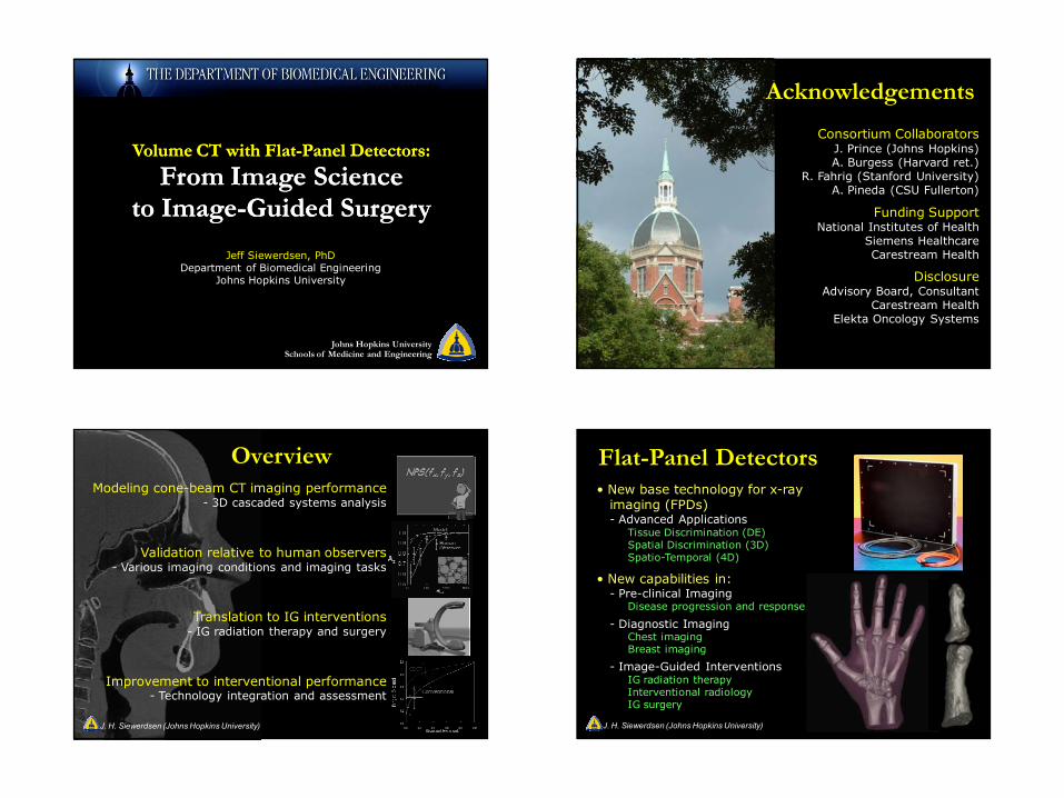
volume ct with flat-panel detectors: j. prince (johns...
TRANSCRIPT
8/30/2010
1
Volume CT with Flat-Panel Detectors:
From Image Scienceto Image-Guided Surgery
Volume CT with Flat-Panel Detectors:
From Image Scienceto Image-Guided Surgery
Jeff Siewerdsen, PhDDepartment of Biomedical Engineering
Johns Hopkins University
Johns Hopkins UniversitySchools of Medicine and Engineering
Acknowledgements
Consortium CollaboratorsJ. Prince (Johns Hopkins)A. Burgess (Harvard ret.)
R. Fahrig (Stanford University)A. Pineda (CSU Fullerton)
Funding SupportNational Institutes of Health
Siemens HealthcareCarestream Health
DisclosureAdvisory Board, Consultant
Carestream HealthElekta Oncology Systems
0 60 120 1800.5
0.6
0.7
0.8
0.9
1.0
B
θtot
Az
Human Observer
Model
0.0
0.2
0.4
0.6
0.8
1.0
0.0 0.2 0.4 0.6 0.8 1.0
CBCT
Conventional
Normal Excised
Targ
et
Exci
sed
OverviewModeling cone-beam CT imaging performance
- 3D cascaded systems analysis
Validation relative to human observers- Various imaging conditions and imaging tasks
Translation to IG interventions- IG radiation therapy and surgery
Improvement to interventional performance- Technology integration and assessment
NPS(fx,fy,fz)
J. H. Siewerdsen (Johns Hopkins University)
Flat-Panel Detectors• New base technology for x-ray
imaging (FPDs)- Advanced Applications
Tissue Discrimination (DE)Spatial Discrimination (3D)Spatio-Temporal (4D)
• New capabilities in:- Pre-clinical Imaging
Disease progression and response
- Diagnostic ImagingChest imagingBreast imaging
- Image-Guided InterventionsIG radiation therapyInterventional radiologyIG surgery
J. H. Siewerdsen (Johns Hopkins University)

8/30/2010
2
Cone-Beam CT
Projection dataMultiple projections (~100-500)
over ~180o-360o
Volume reconstructionSub-mm spatial resolution
+ soft tissue visibilityJ. H. Siewerdsen (Johns Hopkins University)
Tornai et al.
Platforms for CBCT (examples)
Jaffray et al.Sukovic et al.
Siewerdsen et al.
Feldkamp et al.
Boone et al.J. H. Siewerdsen (Johns Hopkins University)
Experimental Platform for CBCT
Repeat for ~360o
X-ray Tube50 – 150 kVp, Pulsed fluoro
φcone ~5o-15o
Flat-Panel Detectora-Si:H PD + CsI:Tl(1536 x 2048 pixels, 1-30 fps)
Optical Bench + Motion Control System8-axis translation / rotation, 180o+fan – 360o
J. H. Siewerdsen (Johns Hopkins University)
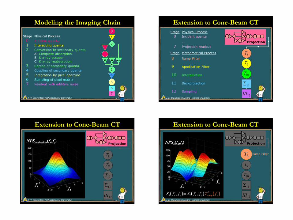
Stage Physical Process0 Incident quanta1 Interacting quanta2 Conversion to secondary quanta
A: Complete absorptionB: K x-ray escapeC: K x-ray reabsorption
3 Spread of secondary quanta4 Coupling of secondary quanta5 Integration by pixel aperture6 Sampling of pixel matrix7 Readout with additive noise
Stage Physical Process0 Incident quanta1 Interacting quanta2 Conversion to secondary quanta
A: Complete absorptionB: K x-ray escapeC: K x-ray reabsorption
3 Spread of secondary quanta4 Coupling of secondary quanta5 Integration by pixel aperture6 Sampling of pixel matrix7 Readout with additive noise
Stage Physical Process0 Incident quanta1 Interacting quanta2 Conversion to secondary quanta
A: Complete absorptionB: K x-ray escapeC: K x-ray reabsorption
3 Spread of secondary quanta4 Coupling of secondary quanta5 Integration by pixel aperture6 Sampling of pixel matrix7 Readout with additive noise
Stage Physical Process0 Incident quanta1 Interacting quanta2 Conversion to secondary quanta
A: Complete absorptionB: K x-ray escapeC: K x-ray reabsorption
3 Spread of secondary quanta4 Coupling of secondary quanta5 Integration by pixel aperture6 Sampling of pixel matrix7 Readout with additive noise
A CB
Modeling the Imaging Chain
J. H. Siewerdsen (Johns Hopkins University)
8/30/2010
3
Stage Physical Process0 Incident quanta1 Interacting quanta2 Conversion to secondary quanta
A: Complete absorptionB: K x-ray escapeC: K x-ray reabsorption
3 Spread of secondary quanta4 Coupling of secondary quanta5 Integration by pixel aperture6 Sampling of pixel matrix7 Readout with additive noise
Modeling the Imaging Chain
J. H. Siewerdsen (Johns Hopkins University)
Stage Physical Process0 Incident quanta
…
7 Projection readout
Stage Mathematical Process
8 Ramp Filter
9 Apodization Filter
10 Interpolation
11 Backprojection
12 Sampling
Projection
8T
12III
9T
10T
11Σ
Extension to Cone-Beam CT
J. H. Siewerdsen (Johns Hopkins University)
Extension to Cone-Beam CT
fx fz
Projection
8T
12III
9T
10T
11Σ
NPSprojection(fx,fz)
J. H. Siewerdsen (Johns Hopkins University)
Extension to Cone-Beam CT
fx
fz
NPS8(fx,fz)
12III
9T
10T
Projection
8T
11Σ
Ramp Filter
( )zx ffS ,7=( )zx ffS ,8 ( )xramp fT 2
J. H. Siewerdsen (Johns Hopkins University)

8/30/2010
4
Extension to Cone-Beam CT
fxfz
NPS9(fx,fz)
12III
10T
Projection
8T8T
9T
11Σ
ApodizationWindow
( )zx ffS ,8=( )zx ffS ,9 ( )xwin fT 2
J. H. Siewerdsen (Johns Hopkins University)
Extension to Cone-Beam CT
fxfz
NPS10(fx,fz)
12III
9T
Projection
8T8T
10T
11Σ
Interpolation
( )zx ffS ,9=( )zx ffS ,10 ( )zxinterp ffT ,2
J. H. Siewerdsen (Johns Hopkins University)
Extension to Cone-Beam CT
8T
Back-Projection
Projection
12III
NPS11(fx,fy,fz)
9T
10T
8T
11Σ
J. H. Siewerdsen (Johns Hopkins University)
Extension to Cone-Beam CT
fx fy
III12(fx ,fy ,fz)
fz 9T
10T
Projection
8T8T
11Σ
12III Sampling( ) ( )zz ffSffS ,, 1112 = ( )zff,III*** 12
J. H. Siewerdsen (Johns Hopkins University)

8/30/2010
5
NPS(fx, fy, fz) Axial NPSS(fx, fy)
Sagittal NPSS(fx, fz)
The 3-D Noise-Power Spectrum
Broken Symmetry
J. H. Siewerdsen (Johns Hopkins University)
NEQEffective number of quanta
used at each spatial frequency(Efficiency x Fluence)
DQEFraction of quanta
used at each each frequency.
Observations:3D DQE(0) ~ Projection DQE(0)
3D DQE(f) dependent on reconstruction parameters
The 3-D NEQ and DQE
J. H. Siewerdsen (Johns Hopkins University)
Model Observer Detectability
Generalized NEQDetector TypeReconstruction FilterVoxel SizeBackground Noise
Task FunctionTask TypeObject sizeObject contrast
Model ObserversPrewhitening (PW)PW + Eye Filter + Internal Noise (PWEI)Non-Prewhitening (NPW)NPW + Eye Filter (NPW)NPWE + Internal Noise (NPWEI)J. H. Siewerdsen (Johns Hopkins University)
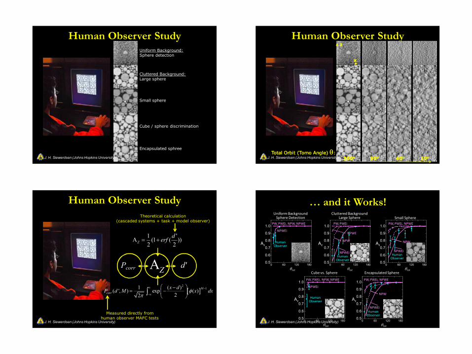
9AFC Tests
• Darkened reading room
• Diagnostic-quality display
• Fixed win / level [90%min, 110%max]
• 6 observers (physicists)
• Training set distinct from test set
• 5 repeats (distinct stimuli)
• Randomized reading order
• ~100 minutes for each observer
Human Observer Study
J. H. Siewerdsen (Johns Hopkins University)
8/30/2010
6
Human Observer StudyUniform Background:Sphere detection
Cluttered Background:Large sphere
Small sphere
Cube / sphere discrimination
Encapsulated sphree
J. H. Siewerdsen (Johns Hopkins University)
Human Observer Study
1010oo4040oo9090oo360360ooTotal Orbit (Total Orbit (TomoTomo Angle) Angle) θθ::
xx
zz
J. H. Siewerdsen (Johns Hopkins University)
1 'A (1 ( ))2 2Z
derf= +
[ ]2
11 ( )( ', ) exp ( )22
Mcorr
x dP d M x dxφπ
∞ −
−∞
−= −
∫
Theoretical calculation(cascaded systems + task + model observer)
AZ
Measured directly from human observer MAFC tests
Pcorr d'
Human Observer Study
J. H. Siewerdsen (Johns Hopkins University) 0 60 120 1800.5
0.6
0.7
0.8
0.9
1.0
B
0 60 120 1800.5
0.6
0.7
0.8
0.9
1.0
B
0 60 120 1800.5
0.6
0.7
0.8
0.9
1.0
B
0 60 120 1800.5
0.6
0.7
0.8
0.9
1.0
B
0 60 120 1800.5
0.6
0.7
0.8
0.9
1.0
B
θtot
Az
NPWEi
PW, PWEi, NPW, NPWE
θtot
AzHuman
Observer
NPWEi
PW, PWEi, NPW, NPWE
θtot
Az
PW, PWEi
Human Observer
θtot
Az
PW, PWEi, NPWE
Human Observer
θtot
Az
PW, PWEi, NPWE
Human Observer
Uniform BackgroundSphere Detection
NPWE
… and it Works!
Human Observer
NPWEi
NPW
NPWEi
NPW
NPWEi
NPW
Cluttered BackgroundLarge Sphere Small Sphere
Cube vs. Sphere Encapsulated Sphere
J. H. Siewerdsen (Johns Hopkins University)
8/30/2010
7
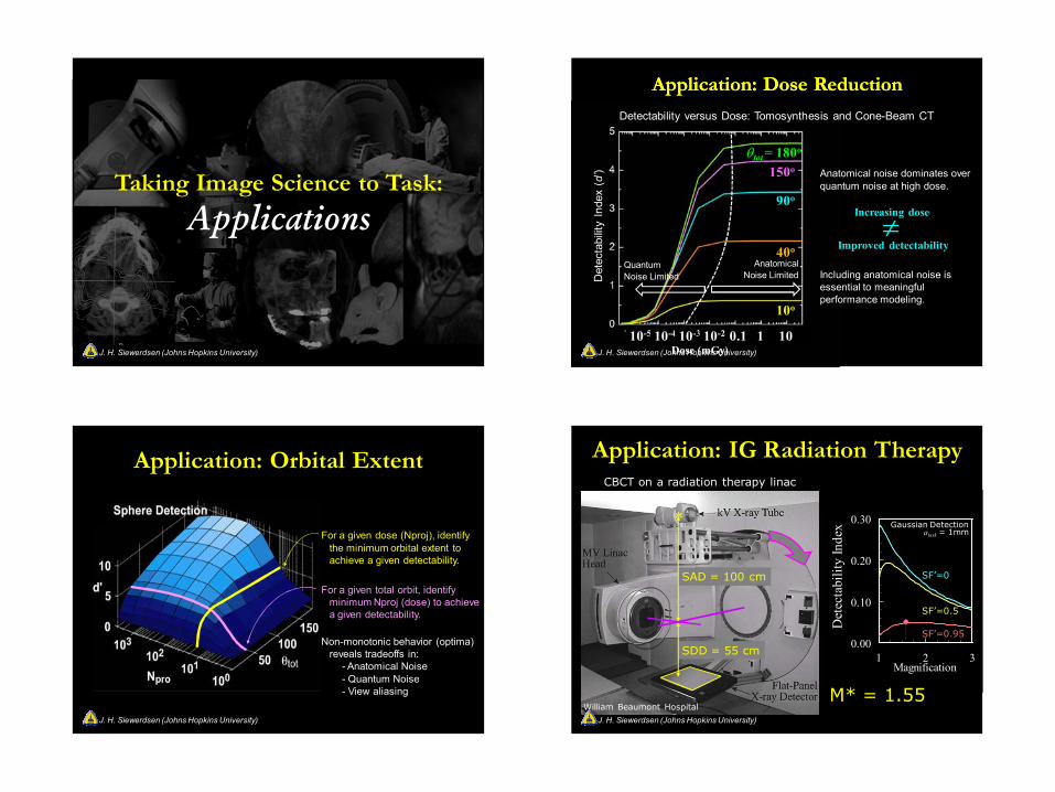
Taking Image Science to Task:
Applications
J. H. Siewerdsen (Johns Hopkins University) Dose (mGy)
Det
ecta
bilit
y In
dex
(d’)
Detectability versus Dose: Tomosynthesis and Cone-Beam CT
10-5 10-4 10-3 10-2 0.1 1 10
40o
90o
150oθtot = 180o
10o
Det
ecta
bilit
y In
dex
(d’) Anatomical noise dominates over
quantum noise at high dose.
Including anatomical noise is essential to meaningful performance modeling.
Increasing dose
Improved detectability≠
Application: Dose ReductionApplication: Dose Reduction
Quantum Noise Limited
Anatomical Noise Limited
J. H. Siewerdsen (Johns Hopkins University)
Application: Orbital Extent
For a given dose (Nproj), identify the minimum orbital extent to achieve a given detectability.
For a given total orbit, identify minimum Nproj (dose) to achieve a given detectability.
Non-monotonic behavior (optima) reveals tradeoffs in:
- Anatomical Noise- Quantum Noise- View aliasing
J. H. Siewerdsen (Johns Hopkins University)
0.00
0.10
0.20
0.30
1 2 3Magnification
SF’=0
SF’=0.5
SF’=0.95
Det
ecta
bilit
y In
dex Gaussian Detection
atask = 1mm
William Beaumont Hospital
SDD = 55 cm
SAD = 100 cm
M* = 1.55
*
CBCT on a radiation therapy linac
Application: IG Radiation Therapy
J. H. Siewerdsen (Johns Hopkins University)

8/30/2010
8
Application: IG Radiation TherapyHead-and-Neck Lung
Liver Metastases
Sarcoma
Prostate
kV CBCT ImagingMV Treatment Beam
J. H. Siewerdsen (Johns Hopkins University)
Multiple projection images acquired over ~180o
2D Image acquisition- Nominal: 60 s- High-speed: 10-20 s
3D Image reconstruction- Nominal: 60 s- High-speed : 10 s
Radiation dose- ~1/10th that of Dx CT
Application: Mobile C-Arm for IGI
J. H. Siewerdsen (Johns Hopkins University)
Mobile Isocentric C-ArmCone-Beam CT-Capable C-Arm
Pre-clinical platform for multi-mode Fluoro / CBCT guidance
Image Acquisition3D Reconstruction
Control System
J. H. Siewerdsen (Johns Hopkins University)
Orthopedics
Spine
Head and Neck Ear
LungBrachytherapy
Cone-Beam CT-Guided Interventions
J. H. Siewerdsen (Johns Hopkins University)
8/30/2010
9
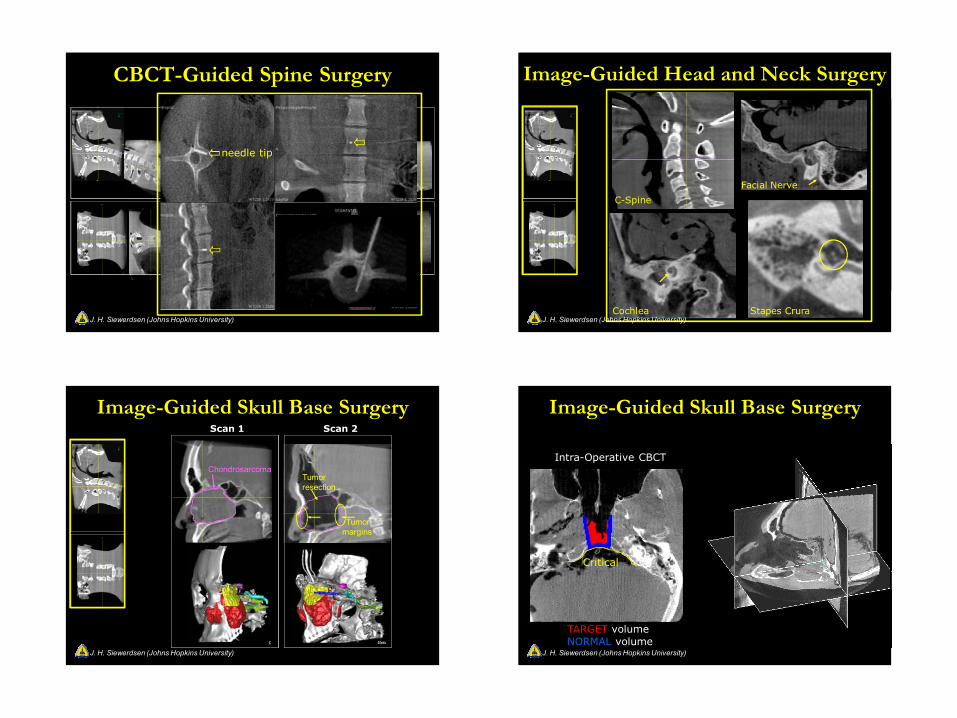
CBCT-Guided Spine Surgery
needle tip
J. H. Siewerdsen (Johns Hopkins University)
Facial Nerve
C-Spine
Cochlea Stapes Crura
Image-Guided Head and Neck Surgery
J. H. Siewerdsen (Johns Hopkins University)
ChondrosarcomaTumorresection
Tumormargins
Scan 1 Scan 2
Image-Guided Skull Base Surgery
J. H. Siewerdsen (Johns Hopkins University)
Intra-Operative CBCT
TARGET volumeNORMAL volume
Critical
Image-Guided Skull Base Surgery
J. H. Siewerdsen (Johns Hopkins University)
8/30/2010
10
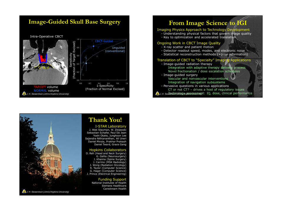
Intra-Operative CBCT
TARGET volumeNORMAL volume
Post-Operative CBCT
TARGET RemainingNORMAL Remaining
Critical Critical
0.0
0.2
0.4
0.6
0.8
1.0
0.0 0.2 0.4 0.6 0.8 1.0
CBCT-Guided
Unguided(conventional)
1-Specificity(Fraction of Normal Excised)
Sen
sitivi
ty(F
ract
ion o
f Ta
rget
Exc
ised
)
Image-Guided Skull Base Surgery
J. H. Siewerdsen (Johns Hopkins University)
Imaging Physics Approach to Technology Development- Understanding physical factors that govern image quality- Key to optimization and accelerated translation
Ongoing Work in CBCT Image Quality- X-ray scatter and patient motion- Detector readout speed, modes, and electronic noise- Statistical reconstruction methods (+prior information)
Translation of CBCT to “Specialty” Imaging Applications- Image-guided radiation therapy
Integration with adaptive therapy delivery processNovel fractionation / dose escalation schedules
- Image-guided surgeryVascular and nonvascular interventionsIntegration of navigation subsystems
- Pervasive questions in various applicationsCT or not CT? – drives a host of regulatory issuesTechnology assessment: IQ, dose, clinical performance
From Image Science to IGI
J. H. Siewerdsen (Johns Hopkins University)
Thank You!I-STAR Laboratory
J. Web Stayman, W. ZbijewskiSebastian Schafer, Paul De Jean
Yoshi Otake, Junghoon LeeSajendra Nithiananthan, Ali Uneri
Daniel Mirota, Prakhar PrakashDaniel Tward, Grace Gang
Hopkins CollaboratorsD. Reh (Head and Neck Surgery)
G. Gallia (Neurosurgery)J. Khanna (Spine Surgery)J. Carrino (MSK Radiology)
J. Wong (Radiation Oncology)R. Taylor (Computer Science)G. Hager (Computer Science)
J. Prince (Electrical Engineering)
Funding SupportNational Institutes of Health
Siemens HealthcareCarestream Health
J. H. Siewerdsen (Johns Hopkins University)