water-equivalent plastic scintillation detectors for … plastic scintillation detectors for...
TRANSCRIPT

Water-equivalent plastic scintillation detectors for high-energy beam dosimetry: I. Physical
characteristics and theoretical considerations
This article has been downloaded from IOPscience. Please scroll down to see the full text article.
1992 Phys. Med. Biol. 37 1883
(http://iopscience.iop.org/0031-9155/37/10/006)
Download details:
IP Address: 143.111.80.27
The article was downloaded on 16/11/2011 at 16:50
Please note that terms and conditions apply.
View the table of contents for this issue, or go to the journal homepage for more
Home Search Collections Journals About Contact us My IOPscience

~
Phys. Med. B i d , 1992, Vol. 37, NO 10, 1883-1900. Printed in the UK
Water-equivalent plastic scintillation detectors for high- energy beam dosimetry: I. Physical characteristics and theoretical considerations
A S Beddart, T R Mackie and F H Attix Depanment of Medical Physics, University of Wisconsin Medical School, Madison, W l 537%. USA
Received 27 FebNary 1992
AbstracL A minimally penurbing plastic rintillalion detector has been developed for the dosimetry ot high-energy teams in radiotherapy. The detector syslem consists of two identical parallel sets of radialion-resistanl optical fibre bundles, each wnnecled lo independent photomultiplier l u k s ( P m ) . One fibre bundle is conneaed lo a miniature waler-equivalent plastic szintillalor and KI rinlillation as well as Cerenkov light generated in the fibres is delecled at its Pm. ?he other 'backgmund' bundle is not wnnected Io the scintillator and y, only Cerenkov light is detected bj its PMI. The tackground signal is sublracled to yield only the signal hom the scinlillator. The walerquivalence of plaslic scinlillation dewclon is studied lor photon and electmn beams in the radiotherapy range. Application of Burlin ravity theory show thal the energy dependence of such dela'tars is expecled to be tetter than the wmmonly used systems (ionizalion chambers, LiF thermoluminescent dosimeters, film and Si diodes). I1 is also shown that they are not affected bj temperature variations and ah ib i t much less radiation damage than either photon or electron diode detectors.
1. Intmduction
One of the few methods of dose measurement not commonly encountered in the field of high-energy radiotherapy beams is scintillation dosimetry using low atomic number phosphor organic scintillators. While a scintillation dosimeter may never supplant the cavity ionization chamber, it nevertheless has some very attractive features that deserve more attention than previously has been shown. Small plastic scintillators can provide the ideal means of dose measurement in tissue-equivalent phantoms. The close matching of these plastic scintillators to water avoids the usual conversion of dose measurements from one medium to another, which strongly depends on the accuracy of the correction factors involved.
The purpose of this investigation is to characterize water-equivalent plastic scin- tillators which can be used for the measurement of radiation doses in high-energy photon and electron beams.
t Present a d d r w : Ontario Cancer InstituteRrincw Margaret Hospital, Clinical Physics Deparlment, 500 Sherboume Street, Bmnto. Ontaria M4X 1K9. Canada.
0031-91S5/92/101883+18$M.S0 @ 1992 IOP Publishing U d 1883

1884
2. Theory of scintillation dosimetry
Any transparent material that luminesces in a suitable wavelength range when ion- izing radiation passes through it can serve as a detector. Scintillators that are of particular interest are those that exhibit high transparency to the emitted light, with correspondingly high ionizing radiation sensitivity and fast response. In addition, the chosen scintillation material should convert the kinetic energy of charged particles into detectable light with a large enough scintillation efficiency. This conversion should be linear such that the light yield is proportional to deposited energy over a wide energy range. Finally, the density and atomic composition should resemble water as closely as possible. A relatively small fractipn of the kinetic energy transferred to a scintillator is converted into fluorescent light energy. The rest of the energy is dissipated non-radiatively, mainly in the form of lattice vibrations or heat. The scintillation efficiency (fraction of the particle energy converted to fluorescent light energy) differs for each type of scintillator (liquid or solid) and also depends on the type of charged particle producing the ionization.
For dosimetry purposes, the measurement of the fluorescent light generated by a scintillator would be a direct measure of dose only if the scintillation efficiency is independent of energy of the charged particles. Thus, the light yield should have a linear dependence upon the energy deposited by the charged particles intereacting within the scintillator. For organic scintillators such as anthracene, stilbene, and many other commerciaiiy avaiiabie iiquio ana piastic scinriiiaters, &r FCS~JRSC ti' e ! e c w ~ s is linear for particle energies above about 125 keV (Brannen and Olde 1962, Smith ef a1 1968 and Craun and Smith 1970). The rcsponsc to heavier particles is linear only above much higher energies (Goading and Pugh 1960, Smith er al 1%8 and McParland 1986). Smith ef a1 (1968) systematically studied several commonly used organic scintillators Over relatively wide ranges of particle energy and found that their response to electrons is quite linear with particle energy above approximately 100 key Furthermore, linear extrapolations of the electron response from higher energies generally pass within a few keV of the origin (Horrocks 1970). This fall-off of the scintillation efficiency at low energies (< 100 keV) has vely little effect in the present context when one considers the photon and electron spectra commonly encountered in the megavoltage energy beams used in radiation therapy.
A S Beddar et a1
3. Description uf the winl i l la t io~~ detector
The plastic scintillator chosen for this study is 'BC-400' (Bicron Corporation Premium Plastic and Liquid Scintillators, Newbury, OH, USA) which is similar to NE-102 manufactured by Nuclear Enterprises (Monmouth Junction, NJ, USA). The relevant characteristics of this plastic are its high scintillation efficiency and its desirable spec- tral response when compared to other organic plastic scintillators. The principal characteristics of this scintillator are listed in table 1.
The detector system is composed of a small polystyrene probe which encloses the scintillator, a parallel-paired fibre light guide and two identical P M n . The spectral response of the PMT (Hamamatsu R1635, Hamamatsu Corp., Bridgewater, NJ, USA) is well matched to the emission spectrum of the plastic scintillator and the transmis- sion spectrum of the optical fibres. A detailed diagram of the plastic scintillation detector system is shown in figure 1. The polystyrene probe, which is cylindrically

Pluslic scintillulion detectors for dosimetfy: I 1885
lhbk I. Physical characteristics of the 'BC-4W' Plasric Scinlillators (manufactured by Bicron Corporation).
l ight oulpul (% anthracene): 65
Decay mnstant main mmponent (ns): 2.4
Wavelength of maximum emision (nm): 423
No. of H Atoms/ No. of C atoms: 1.104
Densily (g 1.032
Refractive indu: 1.58
Melting or soflening point (OC!): I5
shaped (5.0 mm outer diameter and 7.0 mm in length), contains a miniature plas- tic scintillator (1.0 mm diameter and 4.0 mm in length) imbedded in its centre. A longitudinal cross section of the proximal fibre light guide end and the polystyrene probe, respectively, are shown in inserts (a ) and (b) of figure 1. The parallel-paired fibre light guide consists of two optical fibre light guides running parallel to each other through a 3 mm outer diameter black polyethylene jacket to provide a light shield and protection from the outside environment. One, which will be referred to as the 'signal' fibre light guide, is optically coupled to the plastic scintillator to collect and transmit the light output generated to one of the PMIS. The second one, which is light-shielded at the proximal tip and coupled to the second PMT at the distal tip, allows subtraction of the background signal due to the radiation-induced light generated in the optical fibres (Beddar et U / 1989) and will be referred to as the 'background' fibre light guide. The polyethylene jacket, which contains both fibre light guides, is bifurcated at its end to allow each to be coupled to their respective PMT. Each fibre light guide contains a bundle of seven single multi-mode step-index fibres packed in a hexagonal fashion. The physical and optical characteristics of the optical fibres are presented in table 2.
The polystyrene probe enclosing the scintillator is optically coupled to the parallel- paired light guide with an optically clear expoxy adhesive ma-Bond F114, 'Ita-Con, Inc., Melford, MA, USA). The same epoxy is used to tightly secure the bundled single fibres in their polystyrene adaptors. The bifurcated tail of the paired fibre light guide is coupled to both PMTS using two small Lucite guides. These guides are polished at both ends and machined in a conical shape in such a way as to diffuse the exiting light from the fibre light guide to the PMT faces. The clear epoxy adhesive is used at the fibre light guide joints, while silicone grease is used to couple the distal ends of the bifurcated light guide to the PMD.
The DC output signal of a PMT, under normal operation, is a very precise measure of the total light intensity output that is transmitted through the fibre light guide. The 'signal' fibre light guide transmits the total light intensity produced by the plastic scin- tillator plus the radiation-induced light intensity generated by the irradiated portion

1886 A S Beddar el a1
Photomultiplier 'Signal' Fiber Light Guide
Tubes 1 \ PMT Bases 7
Metalic Case ' J 7 \ \ T s c i n t i l l a t o r
'Background Fiber Light Guide
'Background, / Fiber Guide
\LPolysfyrene Adaptor
Figure 1. Diagram of the plastic suinliiialion drircior qsteni aDd IongiiiidifiaI c i i r s sections of (a) the proximal fibre light guide end and (b) the plyslyrene probe that contains the scintillator.
Table 2 Fibre pmpenies (Model No: FHP 200/220/240, Manufactured k 4 PolymiCN 'Ethnologies).
Core diameter (pm): 2 W i 5
Clad diameter "(pm): 220i5
BuKer diameter b(pm): 240f5
Care refractive index (@ 588 nm):
Numerical apenure (NA):
1.458
0.22f02 @ 8w nm
Atlenuation: < 10 db km-' @ 8W nm < 25 db km-' @ 5CU nm < 100 db km-' @ 365 nm
Packing fraction: 69% mre area per fibre
Opelaling environment: -100 10 +360 T. &lW% relative humidily
Including fibre core. Including fibre core and cladding.
of the fibre light guide (Beddar el al 1992a). The 'background' fibre light guide Only transmits the light induced by radiation in the fibre light guide. Assuming that the radiation-induced light signals in both fibre light guides are identical and both P M B have identical gain, the light signal produced by the plastic scintillator alone can be

Plasfic scintillafion defectors for dosimefry: I 1887
obtained by subtraction of the background PMT output from the signal PMT output. The high voltage supply to both PMB is delivered through a potentiometer, allowing the balancing of the output signals of the two PM?S when they receive equal light out- puts. This can be accomplished by shielding the scintillation probe against radiation (in a lead brick) while a portion of the parallel-paired fibre light guide is exposed to either an x-ray or an electron beam, balancing the output of both PMlb due to the induced Cerenkov light through the potentiometer adjustment. This will also account for differences in transmission efficiencies through optical interfaces as well. The high voltage to both P M B is delivered through a 1 megohm potentiometer, and the PMT output currents are passed to ground through two accurately matched load resistors whose resistance is equal to 1 megohm within 1%, thus generating ‘signal’ and ‘background’ voltage outputs. The scintillator signal would be proportional to the voltage difference between the two outputs. This method will probably be the way in which the detection will be used in future practice. However, the method chosen for this investigation is the charge-integration method which allows greater reproducibility through time averaging. Tho different electrometers (Keithley, Model 602), calibrated against each other, are used to measure both the scintillator (signal output) and dummy fibre (background output) PMT currents individually hy charge integration. Thus, in this method, the light intensity signal produced by the scintillator is obtained by measuring both outputs using the integration method and subtracting the background output from the signal output.
4. Water-equivalence
Water-equivalence for a dosimeter is achieved by matching the sensitive material of the detector to the medium in which the absorbed dose is to be measured (i.e. water) in order to minimize the perturbation of the panicle Auence. If the detector sensitive volume is surrounded by a ’wall’ material, then ideally the atomic composition of the wall should be matched to both the medium and the detector material. In the case of the plastic scintillation detector, the detector (polyvinyltoluene), the wall (polystyrene) and the medium (water) are closely matched. Actually, this is the best combination of materials for matching purposes among all the known dosimeters with the exception of Fricke dosimeters. (However, Fricke dosimeters are not sensitive to absorbed doses commonly encountered in radiotherapy, and do not give real time dose measurements.) Some of the relevant characteristics of this plastic scintillator compared to those of polystyrene and water are listed in table 3.
The concept of effective atomic number, Z,,, (Mayneord 1937, Spiers 1946) is often used to evaluate water-equivalency. However, since for radiotherapy beams the predominant mode of interaction in low-Z materials is Compton scattering, this criterion should not be used. Instead, a direct comparison of the mean mass energy- absorption coefficients and the mean mass collision stopping powers of the material to those of water would be more relevant to describe water-equivalency for x-rays. In the case of electron beams, the mass collision stopping powers and the mass angular scattering powers should be comparable to those of water. A logarithmic plot of the mass energy-absorption coefficients (Hubbell 1982) for the plastic scintillator, water and polystyrene for monoenergetic photons ranging from 10 keV up to 20 MeV is shown in figure 2(a). For the plastic scintillator, the mass energy-absorption coeffi- cients were computed using the Bragg rule for compounds and mixtures. lb appreci- ate their close matching, figure 2(a) is re-plotted using the same data but on a linear

1888 A S Beddar et a1
6 water 1 Polystyrene I
Photon Energy (MeV)
Figure 2. Maw energy-absorption mefificients for the plastic xintillator mmpared to those of water and plystyrene as a function ot photon energy. (a ) logarithm4ogarithm plot, (b) linear-logarilhm plat.
Table 3. Physical characteristia of the plastic Yinlillalor mmpared U) plystyrene and water (ICRU 1984a).
~
Paramctcr Polystyrene Scintillator Wdter
Density (g "3): 1.MO 1.032 1.002
Ralio of number of e- in lhe compound lo lhe molecular weight 0.5377 0.5414 0.5551 V/4: Electron density: ( ioz3 e-g-')
Cammsilion
3.238 3.272 3.343
(2: Iraction by weighl (C)): k7.74 1:8.47 111.19 k92.26 691.53 8:88.81
* Manufactured by Bicron Corporation, lype: BC-400, vinyltoluene based.

Plastic scintillation detectors for dosimetry: I 1889
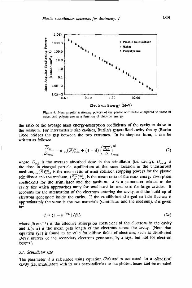
vertical scale, for photon energies ranging kom 100 keV up to 20 MeV This is shown in figure 2(b). The mass energy-absorption coefficients for the scintillator are some- what lower than water but above 100 keV closer to water than those of polystyrene. Mass collision stopping powers (ICRU 1984) of the plastic scintillator material were compared to those of polystyrene and water for monoenergetic electrons ranging from 10 keV to 50 MeV and are shown in figure 3. The collision stopping powers using logarithm-logarithm scales are shown in figure 3(a), while the Same coefficients using linear-logarithm scales are shown in figure 3(b). Once again, we notice that the mass collision stopping powers of the plastic scintillator and polystyrene are similar, and somewhat less than those of water. The mass angular scattering powers [ @ / p l ] of the plastic scintillator material were computed, using ICRU Report 35 (ICRU 1985), by means of the following equation:
where [ p / ( p l ) l S c , represents the mass angular scattering power of the scintillator m?terial, W, is the weight percentage of element i and [ @ / p l ] , is the mass angu- lar scattering power corresponding to element i. Mass angular scattering powers of the scintillator material were compared to those of polystyrene and water for mono- energetic electrons 10 keV to 50 MeV and are shown in figure 4. The mass angular scattering powers of the scintillator material are found to be almost identical to those of polystyrene, and lower than those of water throughout the whole range of electron energies (10 keV up to 50 MeV).
Attix (1986) discussed the advantages of media matching of a dosimeter to the medium of interest (i.e. water), thoroughly, and pointed out that the most obvious parameter to match is the atomic composition. However, the density state (gaseous versus condensed) also influences the collision-stopping-power ratio of the wall to the sensitive volume by the polarization effect. This effect, which is also known as density effect, has some important consequences in radiation dosimetry measurements in electron (or even photon) beams above approximately 2 MeV When measuring absorbed dose in the gas of an ionization chamber which is surrounded by a condensed state medium, the polarization effect will influence the energy deposition. Electrons interacting in condensed medium, result in dipole distortion of the atoms near the track of a passing fast electron. This dipole distortion weakens the coulomb force field experienced by the more distant atoms, thus decreasing the energy loss cross section. Because of this, the mass collision stopping power is decreased in condensed media (Sternheimer 1956, Sternheimer and Peierls 1971). The percentage reduction of collision stopping power of, for instance, carbon (Z = 6) versus gases of the same atomic number Z due to this effect is approximately 3% for 1 MeV, 12% for 10 MeV and 20% at 50 MeV (Attix 1986 and Berger and Seltzer 1983). In our case, the detector is composed of solid media (plastic scintillator surrounded by polystyrene) inserted in another solid medium (i.e. water or a phantom material). The plastic scintillator, the polystyrene wall and water (or phantom material) are very similar in atomic composition, thus, nearly the Same polarization effect would be experienced everywhere. Therefore, the energy deposition will neither be reduced nor overestimated by this effect, unlike the case of an ionization chamber, where the detecting volume and the surrounding medium are of different density states (e.g. air versus condensed-medium wall).

1890 A S Beddar el al
0 Plastic Scintillator water
E 10.0 A Polystyrene
6 1.0
“ 4 I 0.01 0.10 1.00 10.00
3.0 a
f j 1.0 J ’ J
0’ 5 10 15 20 25 30 35 40 45 50
Electron Energy (MeV)
Figure 3. Mass collision stopping pavers for the plastic Scintillator compared to those of water and polystyxne as a fundion of electron energy. (a) logarithm-logarithm plot, ( b ) linear-logarithm plot.
In addition to media matching, the size of the detector should be small enough to minimize particle fluence perturbations but large enough to be subjected to a large number of interactions to yield a response that can be measured with precision. These requirements are met for this prototype scintillation detector as shown elsewhere (Beddar er a1 1992b).
5. Interpretation in terms of Burlin cavity theory as a function of scintillator size, energy and material dependence
In order to determine the absorbed dose in the plastic scintillator (the detecting volume material), it is necessary to h o w if the detecting volume behaves as a small cavity (Bragg-Gray cavity), a large cavity, or lies somewhere between these extremes. For a small cavity, the ratio of the average absorbed dose in the cavity, Dsci, to the absorbed dose in the surrounding medium Dmed in the absence Of the cavity (plastic scintillator) is equal to the ratio of the average mass collision stopping powers of the cavity to that of the medium. In the case of a large cavity, assuming that charged particles are completely generated by photon interactions occurring in the cavity and their energy is completely deposited inside the cavity, the ratio of the doses is equal to
Plastic scintillation detectors for dosimetry: I
1.OE4
1000.0 I I.
9, 2s 100.0 3 N 10.0
a
.- I k "
M$
$ e 0.1
m ~ 3 1.0 6 s - 3 1.OE-2 s
1.OE-3
1891
0 Plastic ScinUllator 0 rater 1 Polystyrene
88 6 s
8 8
8 8 8 8
88 8 8
8 8
8 8 8
8 8 -
Figure 4. Mass angular scattering powers of the plastic scintillalor mmpared 10 those Of waler and plyslyrene as a function of eleclmn energy.
the ratio of the average mass energy-absorption coefficients of the cavity to those in the medium. For intermediate size cavities, Burlin's generalized cavity theory (Burlin 1966) bridges the gap between the two extremes. In its simplest form, it can be written as follows:
where Dsci is the average absorbed dose in the scintillator (i.e. cavity), D,,, is the dose in charged particle equilibrium at the same location in the undisturbed medium, ,,,(S):kd is the mean ratio of m a s collision stopping powers for the plastic scintillator and the medium, (FY;h,, ir the mean ratio of the mass energy absorption coefficients for the scintillator and the medium. d is a parameter related to the cavity size which approaches unity for small cavities and zero for large cavities. It accounts for the attenuation of the electrons entering the cavity, and the build up of electrons generated inside the cavity. If the equilibrium charged particle fluence is approximately the same in the hvo materials (scintillator and the medium), d is given by:
d = (1 - e - P L ) / p L (a) where P(cm-') is the effective absorption coelficient of the electrons in the cavity and L(cm) is the mean path length of the electrons across the cavity. (Note that equation (2a) is found to be wlid for diffuse fields of electrons, such as distributed p r a y sources or the secondary electrons generated by x-rays, but not for electron beams.)
5.1. Scintillator size 7he parameter d is calculated using equation (2a) and is evaluated for a cylindrical cavity (i.e. scintillator) With its axis perpendicular to the photon beam and surrounded

1892
by polystyrene. p is evaluated by using Burlin’s suggested formula (exp(-pt,,,) = 0.01) using a anstant equal to 0.04 instead of 0.01. This value is found by Janssens el a1 (1974) to improve agreement with experimental results. Thus, t,,, which is defined as the maximum depth of electron penetration, is taken as the depth where the secondary Ruence is attenuated to 4% of its original value instead of 1% of its original value as originally suggested by Burlin. L is defined as the mean chord length across the volume which is taken as 4V/S where V is the volume of the cavity and S is the surface area (Ogunleye 1982). The details of this calculation are presented elsewhere (Beddar 1990). A plot of d versus monoenergetic photon energy is shown in figure 5 for two scintillator sizes (1.0 mm and 3.0 mm in diameter, both 4.0 mm in length). A ‘d’ value of 0.5 is obtained at approximately 1.25 MeV and 20 MeV for the 1.0 mm diameter and 3.0 mm diameter scintillator, respectively. It is clear that for these scintillator sues in the energy range indicated above, the scintillator behaves as an intermediate cavity, justifying the application of Burlin theory. If such a detector is used in the orthovoltage x-ray range (< 250 kev) then the detecting volume can be considered as a large cavity. Small cavity behaviour is reached only if the mean beam energy is well above 20 MeV.
A S Beddar ef a1
1,000
Scintillator size . 1.0 mm **am b, 4.0“ LIngtb I 3.0 mm dlam. by 1.0 mm length
3 0.100
8 0.010 a
o.ooi 5.000E-4
0.1 1.0 1o.o
Photon Energy ( M e V )
Figure 5. Burlin cavity theory parameter ‘d’ as a function of photon energy for two cylindrical plastic scintillator sizes. lhis parameter represenls lhe franion of lhe dose deposited in the cavity due lo electrons generated outside the cavity.
5.2. Energy dependence wilh respect to water assuming ‘thick’ and k r o ’ polystyrene wall
In section 2, we discussed that, in order to ensure energy independence of the scintillation detector response, it is required that the scintillation efficiency of the plastic scintillator should be independent of the energy of charged particles. This requirement is fulfilled only if the fluorescent light yield has a linear dependence on the initial energy of the charged particle interacting within the scintillator. Previously cited literature showed that this was the case for the chosen plastic scintillator, and that its response is proportional to energy for electrons with particle energies above 100 k e y
Since the plastic scintillator is embedded in a polystyrene probe, the detecting volume is surrounded by polystyrene in every direction except at the coupling interface with the fibres, (which could also be designed to include a polystyrene layer). A

Plastic scintillation detectors for dosimetry: I 18%
relatively thin wall of polystyrene (which closely matches the scintillator) is used since:
(i) The Smaller the overall size of the probe, and the closer the wall matches the sensitive volume with respect to mass energy-absorption coefficient and mass collision stopping power, the less it perturbs the radiation field at the measurement location;
(ii) the probe must be usable for electron beams as well as x-rays; and (iii) it is not feasible to provide equilibrium-thickness walls for all radiotherapy
x-ray energies anyway, since the range of secondary electrons of energy T MeV approximates T / 2 cm in polystyrene, making the probe huge for T equal to tens of MeV.
The energy dependence of the response of the plastic scintillator detector may be determined by considering two extreme thicknesses of the pbstyrene wall: (i) thick enough to provide transient charged particle equilibrium (TCPE), and (ii) zero thickness.
For the first situation, Burlin theory (equation (1)) was applied and evaluated for scintillators of 1.0 mm and 3.0 mm diameter by 4.0 mm in length to determine the ratio of the mean dose in the scintillator to the equilibrium dose in polystyrene for monoenergetic photons. The average starting electron energy (average energy transferred to the electron) To was determined using the Klein-Nishina interaction cross sections for monoenergetic photons (To = hu(a,, /a), where atP is the energy. transfer cross section and a is the total cross section). The mean electron energy T, for which the collision stopping power was evaluated to use in equation (l), was taken as T’/2 (Attix 1986). The dose to the polystyrene wall was related to the equilibrium dose in the water medium, through a ratio of mass cnergy-absorption coefficients as shown in equation (3):
~,,l, = Dwate, (5””” , (3) water
Thus, the ratio of the mean dose in the scintillator to the equilibrium dose in water, at the probe location is given by
(4) POLY - D.,i/D,,t,, = (~~,,i/D,,,’)(~,,,,/D,,,,,) = (o,,i/Dp,,y)(~~”/P)water.
For higher-energy beams (hu >, 1 MeV), where CPE fails but TCPE takes its place, the dose in a medium is given by:
where V is the photon energy h e n c e and /3 is the ratio of absorbed dose by the collision part of Kerma (KJ.
In such cases relating D,,, to D,,,,, when using: med
med
water
requires the evaluation of 0 = D/KC for the medium and watert. However, the value of /3 is generally not much greater than unity for x-ray energies up to a few
t Note lhat this p is not lhe Same quantily as in equation Z(4). However, the literature for both of these quantities. No mnlusion should mull, however.
is conventionally used in

1894
tens of MeV and it is not strongly dependent on atomic number (Attk 1986). In the case of the plastic scintillator and water psCi and bwater are nearly the same (within less than 0.2%), thus their ratio is equal to 1 and equation (6) is still valid (within less than 0.2%).
A S Beddar ef a1
; .s y 0.90
: a 0.85
7 1.00 n
-, 0.95 7 2
II)
1.10,
-
.
.
0.80 1 0.1 1.0 10.0
1,lO ,
0.80 J - 0.1 1.0 10.0
Photon Energy (MeV)
Figure 6. Ralio of absorbed dose in the plaslic scinlillator lo that of waler lor scintillator sizes of (a) 1.0 mm in diameler by 4.0 mm in lenglh and (b) 3.0 mm in diameter by 4.0 mm in length for thick polystyrene wall and a wall-less mvily.
The ratio of the mean absorbed dose in the plastic scintillator to that in water is shown in figure 6. This ratio is almost identical for both diameters, and stays nearly constant from 2MJ keV up to 3 MeV. It then decreases with increasing photon energies (an approximate 9% decrease from the plateau region is reached at 20 MeV).
For the second situation (zero wall thickness), the energy dependence of the scin- tillation detector with respect to water was determined directly using the Burlin equa- tion (1) for monoenergetic photon beams in which the surrounding medium ("ed') was water. The same general procedure was followed as in the previous section, but omitting the step (equations (3)-(6)) involving the mass energy-absorption coefficient ratios of polystyrenehvater. The ratio of the absorbed dose in the scintillator to that of water for this case is also shown in figure 6 for both scintillator sizes. This dose ratio stays practically constant throughout the whole range of monoenergetic pho-

Plastic scintillation dereciors for dosimetry: I 1895
ton energies considered (200 keV-20 MeV). Notice, again, that this ratio is basically identical for both scintillator diameters.
Since the actual polystyrene wall thickness lies between zero and an equilibrium thickness, these two limiting values of the dose ratios will bracket the true value of the mean dose in the scintillator with respect to water. Thus, the true energy-dependence curve relating the scintillation detector reading to the water dose will lie between the upper and lower curves of figure 6 Furthermore, this true energy-dependence curve will most likely be closer, if not almost identical, to the upper curve (zero wall thickness) since the thickness of polystyrene wall surrounding the scintillator is equal to 2.0 mm (see figure 1). a value that is much closer to zero wall thickness than to an equilibrium-thickness wall especially at high radiotherapy x-ray energies where the difference between the two curves is more pronounced.
5.3. Comparison to other dosimeter media
The plastic scintillator material is compared to other common detecting media used for radiotherapy dosimetry, (air for ionization chambers, LiF for thermoluminescent dosimeters, and silicon for diodes) to determine which dosimeter detecting medium will have the least energy dependence with respect to water and/or the closest dose agreement with water. The Burlin equation was used for monoenergetic photon beams and was evaluated for the same sensitive detecting volume of 1.0 mm in diameter and 4.0 mm in length for the scintillator, air, and LiF media. The ratio of the absorbed dose in the detecting medium to that of water is shown in figure 7. Values of this ratio are also presented in table 4. In the case of the diode, the Burlin equation is evaluated using a value of unity for parameter d. Because the detecting layer in the silicon junction is very thin, the detecting volume should be taken as a Bragg-Gray cavity, Figure 7 shows clearly that of the four different materials, the scintillator exhibits the least energy dependency with respect to water and remains closest to unity. By inspection of table 4, one may expect that the dose ratio, D,,i/D,,,,,, to be closely approximated by a constant equal to 0.980 for any x-ray spectrum (within i0.5%) in the radiotherapy range. The steep rise of the airiwater curve above 2 MeV is caused by the difference in polarization effect between gaseous air and unit density water (Stemheimer and Peierls 1971).
-
6. Temperature dependence
The temperature dependence of the scintillation detector was evaluated by immersing the detecting probe in a water-filled beaker and varying its temperature from O°C to 50 OC. A Pyrex beaker large enough to provide full scatter was filled with ice to ml the temperature of the plastic scintillator to OOC. The beaker, which contained a magnetic stirrer in its bottom, was placed on a temperature heater/controller with a stirring capability. The scintillator probe was left in ice for 45 min to allow the scintillator to reach temperature equilibrium. From O°C to m0C, the water temperature was allowed to reach the ambient temperature without any external disturbances to allow the detector to stay in thermal equilibrium with the surrounding water bath. From 20°C to 30°C, hot water was gradually added to the water bath. The temperature was carefully monitored in this range to keep it stabilized at the meaSUrement p in t s (22, 25, and 30'12) for at least 15 minutes before proceeding with the measurements. From 40'C to 5OoC, the heater was used to gradually bring the water bath temperature up
--

18% A S Beddar er a1
- 1.05
4
5 1.00 2 0.95 s E
i 0.90 ;; 0.85 :
a-. SolnUllaLor .-a WFTUI'a .-. *Ir .-. SI Dlods . . . . . . . . . . . . . . . . . . . . . . . . . . . . . . . . . .
-9 / -2,-
-.--- /-
/
- .-.-.-.- -.-._. -
'hbk 4 length.
rnocon energy @ieVj Air U! n c Si a!ode Sc:~~tiiiatar
Dmed/Dwater for a detecting volume of 1.0 mm diameter and 4.0 mm in
-. - ..
0.20 0.892 0.834 0.7h9 OM7 0.30 0.893 a833 0.775 0.972 0.40 0.50 0.64 0.80 1.00 1.50 200 3.00 4.00 5.w 6.00 8.00
10.0 15.0 20.0
0.894 0.895 0.894 0.895 0.891 0.891 0.892 0.899 0.908 0.914 0.921 0.934 0.945 0.967 0.986
0.832 0.832 0.830 0.824 0.820 0.819 0.815 0.813 0.811 0.811 0.810 0.809 0.808 0.806 0.804
0.784 0.792 0.995 0.998 0.801 0.804 0.808 0.814 0.824 0.826 0.83 0.839 0.844 0.852 0.860
0.975 0.978 0.976 0.979 0.984 0.987 0.987 0.984 0.982 0.978 0.976 0.978 0.975 0.975 0.975
and to keep it stabilized at the measurement pints . A Varian Clinac 4, providing a 4 MV photon beam, was used in this experiment. The output signal of the detector is insensitive to temperature changes from 18'C to 3O'C (plateau region) as shown in figure 8. A slight increase is noticed as the temperature is increased corresponding to a 0.2% increase at 50°C. From 18OC to O'C, the output signal decreases by 0.7% from plateau region (at 5OC the output signal decreases by 0.2%). It is almost impossible to discern these variations from normal output signal reproducibility. Thus, one may conclude that temperature variations in the range of 5OC to 50°C do not significantly affect the response of such a detector system, and within f 5 O of normal room temperature (22'C), the response is constant.

Plastic scintillation detectors for dosimetry: I 1897
Temperature ("C)
Figure 8. 'the plastic scintillator response WRUS temperature.
7. Radiation damage properties
The plastic scintillation detector was exposed to a fairly large accumulated dose to evaluate radiation damage properties. A cesium irradiator was used to deliver a total dose of lo4 Gy to the plastic scintillator at a dose rate of 7.31 Gy min-'. The output signal of the scintillation detector (corrected for stem effect background) was monitored during the entire irradiation.
1.00
0.95
a
e: 0.85
5 0.80 $
c 0.90 a
a > 3
0.75 =--. Electron Diode, 15 MeV Ele~trons
0.70" ' ' " ' ~ " " ' ' ' ' 0 1 2 3 4 5 6 7 8 9 10
Dose (kGy) Figure 9. ' h e relative response of the plastic scintillation detector as a function of total accumulated dose when subjected to Cesium-I37 gamma rays and mmpared lo a photon diode detector exposed U) gamma rays and an electron diode detector exposed to a 15 MeV electron beam.
The relative response of the scintillation detector as a function of accumulated dose is shown in figure 9. Its response is compared to that of a photon detector diode (Victoreen, Model 30-490) hnd an electron detector diode (Victoreen, Model 30-495). The photon diode and the electron diode were exposed to soCO gamma rays and to a 15 MeV electron beam, respectively. The photon diode response exhibited a
1898
9% decrease, while the electron diode exhibited a 25% decrease in output for a total accumulated dose of lo4 Gy. The plastic scintillation detector output exhibited only a 28% decrease for the same total dose. Therefore, the plastic scintillator is much less susceptible to radiation damage than either of these diodes.
A S Beddar ef a1
8. Discussion
Ai was shown above, the scintillator material was well matched to water with respect to all the criteria for water-equivalence. It ii closer to being water-equivalent than polystyrene which makes it superior to all other known detectors used in the dosimetry of radiotherapy photon and electron beams.
The close matching to water avoids the usual conversion of dose from one medium to another (i.e. water) which strongly depends on the accuracy of factors such as mass collision stopping powers and mass energy-absorption coefficients that are needed for cavity theory considerations. If these scintillators, for instance, are used in plastic phantoms, no corrections need be applied. Furthermore, they do not require detec- tor specific factors such as the ion-collection efficiency (Aion) and the replacement correction factor (Prep[). Theory suggests that it may be possible to calibrate these detectors in terms of absorbed dose in water and use them in any x-ray or elec-
stability of light detection systems such as PMIS may preclude this possibility, however. Application of Burlin cavity thcory demonstrates that the energy dependence of
the detector improves as the polystyrene wall thickness is reduced. This work has led to the use of a 1 mm wall of water-equivalent plastic (Constantinou et a1 1982) for the mmmercial prototype detector (Radiation Measurements, Inc., Madison, WI, USA).
The proposed detector can be physically made in any size to lend itself to in vivo measurements at any site in the human body. It can be directly placed in the oesophagus, bronchus, sinus, stomach, etc. Furthermore, it offers the advantage of real-time dose measurements of treatment delivery which is not possible with thermoluminescent dosimeters. This capability is invaluable for external beam as well as brachytherapy, and particularly for intra-operative therapy where the build up of body fluid may cause a significant discrepancy between the dose delivered and the prescribed dosc to thc tumour.
tron radiation iieia to measure dose wirhoui ihr ncrd ei a y ""LILT! - . CC',!TLLIC'LLI..
9. Conclusion
A prototype water-equivalent scintillation detector which has characteristics that make it an excellent choice for use in radiotherapy dosimetry has been described. The detector has proved to be rugged, resistant to radiation damage, has no need for waterproofing and is not affected by changes in temperature or pressure. Polariza- tion effects are minimal which makes it ideal for use in the dosimetry of high-energy electrons. It is more water-equivalent than polystyrene and exhibits the best en- ergy independence when compared to other dosimeters that are commonly used in radiotherapy measurements.

Plasfic scintillation defectors for dosimety: I 1899
Acknowledgment
The authors gratefully acknowledge the Wtsconsin Innovarium Limited and Radiation Measurements, Inc. for their support.
R6sumi
Elecreun B scintillations en plaslique 6qquivalenl eau pour la dasimClrie des faisceaux de haule bergie: 1. CaractCristiques physiques el wnsidCralions th6oriques
Ips auleum on1 mis au p i n 1 un dilecleur scinlillatians en plaslique, A penurbation minimale, p u r la dosimelrie des faisceaux de haute bnergie en radiothCrapie. Le Vteme de d6lecteurs esl w m p 6 de deux jeux idenliques, parall&la, de paquels de fibres optiquea radiodsistantes. mnnectCs chacun B des tubes pholomulliplicateurs indCpendanls (Pm). Un paquet de fibres est connect6 A un scinlillaleur miniature en plastique Cquivalent eau e l ainsi, une scintillation ou de la lumitre &&e dans la fibres par effet Cerenkov, a t d6teclie par le pholomultiplicaleur asaci6. U u l r e paquel, mrrespndanl au 'bNil de fond', des1 pas connect6 au scinlillaleur, e l ainsi, le dklecteur associC dClecle Seulement la lumitre produite par I'effel Cerenkov: f f i n d'obtenir uniquement le signal pmvenant du scinlillaleur le bruit de fond est soustrail. Equivalence A lleau des dilecleun A xinlillations en plaslique 8 1 CludiCe p u r des bisceaux de photons et d'6lectrons u l i l i sh en radiolhCrapie. bpplication de la UlCorie de la cavil6 de Burlin manlre que la dCpendance vis B vis de I'Cnergie de lek dCleneurs doit &re moindre que d e des systemes utilids gCnCralemen1 (chambres dionisation, dosimklres lhemoluminescents de fluoNre de lilhium, film el diodes en silicium). II a1 ausi montr6 que la dClecleurs ne sont pas affectis par des variations de lemphlure el son1 beaucoup moins expods aux dommages cauds par les radiations que les autres d6leeleurs i diode p u r les Cleclmns ou les photons
Zusammenfassung
Wdsser-lquivalenle Plasliliszinlillaliansdelektoren fiir die Dosimelrie hochenergelischer Slrahlen: Eil I. Physikalirhe Grundlagen und lheorelische Uberlegungen.
Ein neuaniger P l a s l i l i s z i n t i l l a l i o n ~ ~ l ~ k l ~ ~ wurde enlwickell fiir die Dosimetrie hochenergelischer Smhlen in der Slrahlentherapie. Das Delektorsystem ksteht aus m e i identirh parallelen Reihen von slrahlen- xsislenten aplischen Fikrglasbhdeln, wbe i jede Glasfiber unabhlngig mil einem Photomultiplier (PM) verbunden i t . Eine Glasfiber is1 verbunden mil einem sehr kleinen Wdser-lquivalentem Plastikszintil- lalor. Sa kann sowohl das Szinlillationslicht wie auch die in der Glasfiber erzeugle Cerenkov-Slrahlung durch den PM nachgewiesen werden. Das andere 'Unlergrundbiindel' kt nicht mil dem Szintillalor verbun- den, deshalb wird hier nur Cerenkov-Strahlung durch die PM nachgewiesen. D a s UntergNndsignal wird dam abgezogen und man erhilt nur nOch das Signal vom Szintillalor. D ie Wasserlquivalenz der Plastik- szinlillationsdelektoren wurde fiir Pholonen- und Eleklronenstrahlen im Bereich slrahlenlherapeutischer Energien untenucht. D ie Anwendung der Hohlraumlheorie von Burlin zeigr, da0 die Energieabhlngigkeit solcher Detektoren ksser is1 ak die der iibliche-eise verwendelen Sysleme (lonisationskammer, UF- ~Ihermoluminesz~nzdasimeter, Filmdosimeter und Si-Diode"). Fs wird ebenfalls gezeigl. da0 die Plas- tiks~inlillalionsdetektoren nichl keinRu0t werden durch Tkmperalunchwankungen und auch weniger empfindlich gegeniiber Slrahlenschlden sind ah Photonen- oder Eleklronendiodendetekloren.
Referrnces
Attix F H 1986 Innohclion P RodioIo~co/ Physics md Rodlorion Physics (New York Wlley) Beddar A S 1990 Wter-equivalent plastic scintillalion deleclor for high-energy photon and electron beams
Beddar A S, Allix F H and Mackie T R 1989 On the nalure of the lighl induced by radialion in
Beddar A S, Mackie T R and Allix F H 1992a Cerenkov lighl generated in optical fibres and other light
PhLJ i'hesir Univeniq of Wisconsin (Madison, WI: Medical Phpics Publishing Corporation)
transparent media used in radiotherapy Med. Phys. 16 683
pipes irradiated by eleclron teams Phys. Mcd BiOL 37 925-35

1900
Beddar A S, Mackie T R and Attix F H 1992b Mter-equivalent plastic scintillation detectors for high-
Berger M J and Seltzer S M 1983 Stopping powers and ranges of electrons and p i l rons NBSlR 82-2550-A
Brannen B and Olde G L 1962 The response of organic scintillaton 10 electron energy d e p i t e d in them
Burlin T E 1966 A General Theoly of Cavity Ionization BE 1 M i o L 39 727-34 Constanlinou C, Attix F H and Paliwal B 1982 A solid water phantom material far radiotherapy %ray
Craun R Land Smith D L 1970 Analysis of response data for several organic scintillators NucL h r m
Goading T J and Pugh H G 1960 me response of plastic scintillators to high-energy particles N u d
Horrock D L 1970 Ilu Cwrrnr Srom of Lip id Scinrilkuian Cowuing ed E D Bransomc Jr (New York
Hubbell J H 1982 Photon mass attenuation and energy-atsorption coefficients f" 1 keV to U) MeV
ICRU 1984 Stopping powers for elecIrons and positmns ICRU Repon 37 - 1985 Radiation dosimetry: electron beams wilh energies between 1 and 50 MeV ICRU Repon 35
(Bethesda, M D ICRU) Janssens A Eggermont G, Jambs R and Ihielens G 1974 Spectrum perturbation and energy deposilion
models for slopping power ratio calculations in general cavity theoly Phys. Mcd BioL 19 619-30 Mayneord W V 1937 n e significance of the roentgen Acra Inr Union againrr Cmcn 1 27142 McParland B J 1986 Ihe response of NE-102 plastic 9'intillator to stopped 3He nuclei NucL Inrrmm
A S Beddar el a1
energy team dosimetry: 11. Properties and Measurements Phys Med Bh1 37 submitted
(Washinglon. D C NBS) '
Rad Rrr 16 1-6
and -pray team calibration Med Phys. 9 4 3 U l
Merho& 80 23944
Inrmrm. Methcds 7 189-92
Grune and Stratton) ch 3, pp 25-43
Int 1 AppL Radiar Isorope 53 1269-90
Mefhodr A 251 412-13
RodioL 55 588-90
elecrrons, protons and deuterons NucL Immm Mea& 64 15746
n p n i ~ c n T r w ! n ~ o c n ~ or P ! P c ! ~ ~ p h tngh !hPnq E.? 1.
Smith D L Polk R G and Miller T G 19158 Measurement of the response of organic 9'inlillators 10
Spiers F W 1946 Effective Atomic Number and Energy-Absorplion in lissues BI: J . Rodid 19 5 2 4 3 Slernheimer R M 1956 Density effecl for lhe ionimtion 10s in various materials Phys. Rev. 103 511-15 Slemheimer R M and Peierls R F 1971 General exprersion for the density effect for the ionization loss
!hp R~?!W~CE of n~r!in':
of charged panicles Phys. Rn! B 3 3681-92