watershed cerebral infarction in a patient with acute ... · olan 70 yaşındaki kadın hastada...
TRANSCRIPT
| Journal of Clinical and Analytical Medicine1
Böbrek Yetmezliği ile Watershed İnfarkt / Watershed Infarction with Renal Failure
Watershed Cerebral Infarction in a Patient with Acute Renal Failure
Akut Böbrek Yetmezliği olan bir Hastada Watershed Serebral İnfarkt
DOI: 10.4328/JCAM.3771 Received: 20.07.2015 Accepted: 12.08.2015 Printed: 01.10.2015 J Clin Anal Med 2015;6(suppl 5): 671-3Corresponding Author: Ruya Ozelsancak, Nefroloji BD, Başkent Üniversitesi Tıp Fakültesi, Adana Uygulama ve Araştırma Hastanesi, Yüreğir, Adana, Türkiye.T.: +90 3223444444 F.: +90 3223444445 GSM: +905325864019 E-Mail: [email protected] / [email protected]
Özet
Akut böbrek yetmezliği duygu durum değişikliği, konsantrasyon bozukluğu, tremor,
stupor, koma, asteriksis,dizartri gibi nörolojik bulgulara neden olabilir. Bu bulgular
aynı zamanda serebral infarktın da bir belirtisidir. Burada, kontrast ve anjiotensin
dönüştürücü enzim inhibitörü kullanımına sekonder gelişen akut böbrek yetmezliği
olan 70 yaşındaki kadın hastada görülen watershed serebral infarkt vakasını sun-
duk. Hasta dizartri nedeniyle magnetik rezonans görüntüleme ile değerlendirildi.
Görüntülemede her iki serebral hemisferin frontal ve parietal derin beyaz cevhe-
rinde, iç sınır bölgesini etkileyen watershed serebral infarkt ile uyumlu milimet-
rik akut iskemik lezyonlar saptandı. Yatışının beşinci gününde böbrek fonksiyon-
ları normale döndü (BUN 32 mg/dl, kreatinin 1.36 mg/dl) ve hasta taburcu edildi.
Dizartri yirmi gün devam etti.
Anahtar Kelimeler
Akut Böbrek Yetmezliği; Dizartri; Watershed İnfarkt
Abstract
Acute renal failure can cause neurologic manifestations such as mood swings,
impaired concentration, tremor, stupor, coma, asterixis, dysarthria. Those findings
can also be a sign of cerebral infarct. Here, we report a case of watershed cere-
bral infarction in a 70-year-old female patient with acute renal failure secondary
to contrast administration and use of angiotensin converting enzyme inhibitor.
Patient was evaluated with magnetic resonance imaging because of dysarthria.
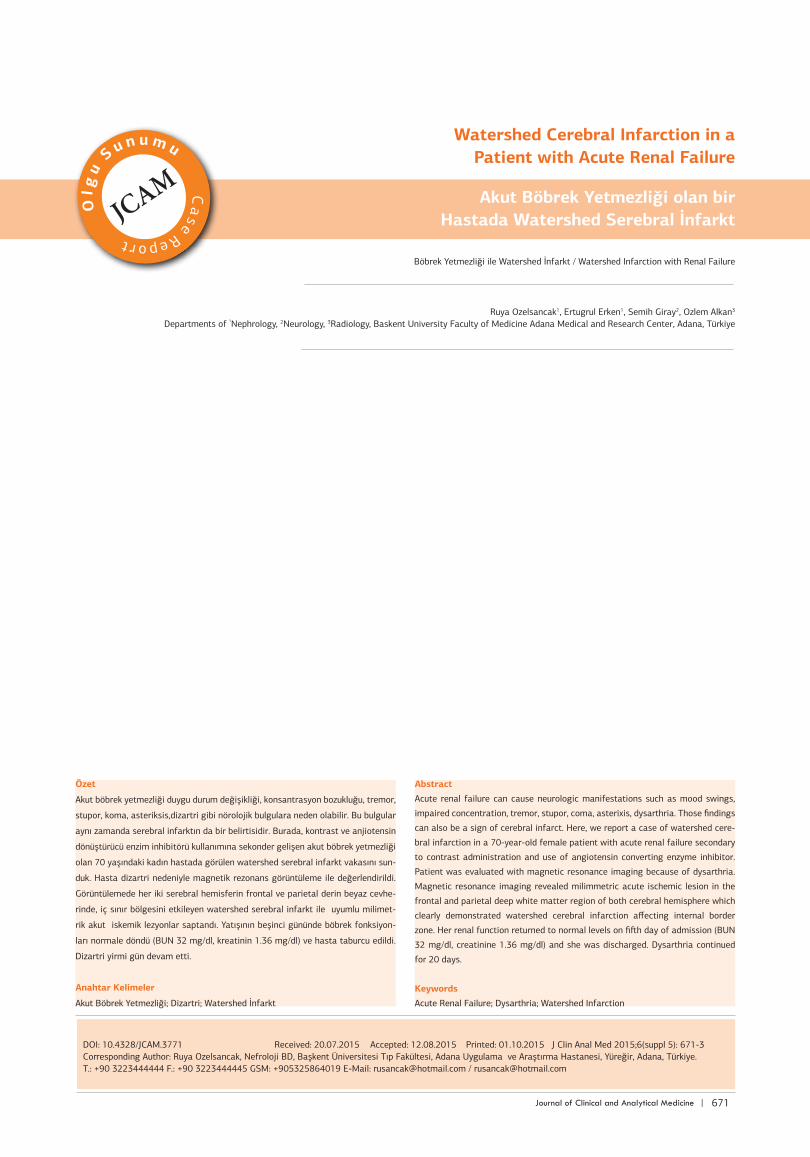
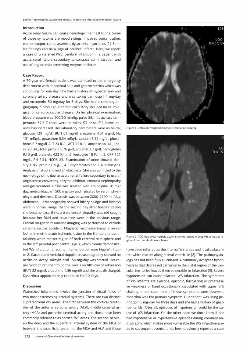
Magnetic resonance imaging revealed milimmetric acute ischemic lesion in the
frontal and parietal deep white matter region of both cerebral hemisphere which
clearly demonstrated watershed cerebral infarction affecting internal border
zone. Her renal function returned to normal levels on fifth day of admission (BUN
32 mg/dl, creatinine 1.36 mg/dl) and she was discharged. Dysarthria continued
for 20 days.
Keywords
Acute Renal Failure; Dysarthria; Watershed Infarction
Ruya Ozelsancak1, Ertugrul Erken1, Semih Giray2, Ozlem Alkan3
Departments of ¹Nephrology, 2Neurology, 3Radiology, Baskent University Faculty of Medicine Adana Medical and Research Center, Adana, Türkiye
Journal of Clinical and Analytical Medicine | 671
| Journal of Clinical and Analytical Medicine
Böbrek Yetmezliği ile Watershed İnfarkt / Watershed Infarction with Renal Failure
2
IntroductionAcute renal failure can cause neurologic manifestations. Some of those symptoms are mood swings, impaired concentration, tremor, stupor, coma, asterixis, dysarthria, myoclonus [1]. Simi-lar findings can be a sign of cerebral infarct. Here, we report a case of watershed (WS) cerebral infarction in a patient with acute renal failure secondary to contrast administration and use of angiotensin converting enzyme inhibitor.
Case Report A 70-year-old female patient was admitted to the emergency department with abdominal pain and gastroenteritis which was continuing for one day. She had a history of hypertension and coronary artery disease and was taking perindopril 4 mg/day and metoprolol 50 mg/day for 3 days. She had a coronary an-giography 3 days ago. Her medical history included no neurolo-gical or cerebrovascular disease. On her physical examination, blood pressure was 100/60 mmHg, pulse 88/min, axillary tem-perature 37.3 C there were no ralles, S3 or soufflé, bowel so-unds has increased. Her laboratory parameters were as below; glucose 139 mg/dl, BUN 61 mg/dl, creatinine 4.51 mg/dl, Na 131 mEq/L, potassium 5.34 mEq/L, calcium 8.35 mg/dl, phosp-horus 6.7 mg/dl, ALT 24 IU/L, AST 33 IU/L, amylase 44 U/L, lipa-se 20 U/L, total protein 5.76 g/dl, albumin 3.7 g/dl, hemoglobin 9.13 g/dl, platelets 423 K/mm3, leukocyte 18 K/mm3, CRP 121 mg/L, PH 7.34, HCO3 25. Examination of urine showed den-sity 1012, protein 0.9 g/L, 4-6 erythrocytes and 2-4 leukocytes. Analysis of stool showed amebic cysts. She was admitted to the nephrology clinic due to acute renal failure secondary to use of angiotensin converting enzyme inhibitor, contrast nephropathy and gastroenteritis. She was treated with amlodipine 10 mg/day, metronidazole 1500 mg/day and hydrated by serum physi-ologic and dextrose. Diuresis was between 2000-2500 ml /day. Abdominal ultrasonography showed biliary sludge and kidneys were in normal range. On the second day after hospitalization she became dysarthric, uremic encephalopathy was not sought because her BUN and creatinine were in the previous range. Cranial magnetic resonance imaging was performed to exclude cerebrovascular accident. Magnetic resonance imaging revea-led milimmetric acute ischemic lesion in the frontal and parie-tal deep white matter region of both cerebral hemisphere and in the left parietal post central gyrus, which clearly demonstra-ted WS infarction affecting internal border zone Figure1, Figu-re 2. Carotid and vertebral doppler ultrasonography showed no occlusion. Acetyl salicylic acid 150 mg/day was started. Her re-nal function returned to normal levels on fifth day of admission (BUN 32 mg/dl, creatinine 1.36 mg/dl) and she was discharged. Dysarthria approximatelly continued for 20 days.
DiscussionWatershed infarctions involve the junction of distal fields of two nonanastomosing arterial systems. There are two distinct supratentorial WS areas. The first between the cortical territo-ries of the anterior cerebral artery (ACA), middle cerebral ar-tery (MCA) and posterior cerebral artery and those have been commonly referred to as cortical WS areas. The second, betwe-en the deep and the superficial arterial system of the MCA or between the superficial system of the MCA and ACA and those
have been referred as the internal WS areas and it take place in the white matter along lateral ventricule [2]. The pathophysio-logy has not been fully elucidated. A commonly accepted hypot-hesis is that decreased perfusion in the distal region of the vas-cular territories leaves them vulnerable to infarction [3]. Severe hypotension can cause bilateral WS infarction. The symptoms of WS infarcts are syncope, episodic, fluctuating or progressi-ve weakness of hand occasionally associated with upper limb shaking. In our case none of those symptoms were observed, dysarthia was the primary symptom. Our patient was using pe-rindopril 5 mg/day for three days and she had a history of gast-roenteritis. After all, episodes of hypotension could be the ca-use of WS infarction. On the other hand we don’t know if she had hypotensive or hypertensive episodes during coronary an-giography, which makes more vulnerable the WS infarction are-as to subsequent events. It has been previously reported a case
Figure 1. Diffusion-weighted magnetic resonance imaging.
Figure 2. ADC map show multiple acute ischemic lesions in deep white matter re-gion of both cerebral hemispheres
| Journal of Clinical and Analytical Medicine672
Böbrek Yetmezliği ile Watershed İnfarkt / Watershed Infarction with Renal Failure

| Journal of Clinical and Analytical Medicine
Böbrek Yetmezliği ile Watershed İnfarkt / Watershed Infarction with Renal Failure
3
of WS infarction in a hemodialysis patient who had recurrent episodes of intradialytic hypotension [4]. Renal failure can also cause neurologic manifestations such as mood swings, impa-ired concentration, tremor, stupor, coma, asterixis, dysarthria, myoclonus. Advanced uremic encephalopathy is usually charac-terized by a reduced level of consciousness, anorexia, asteri-xis, myoclonus, and upper motor neuron signs that result in dis-turbances of gait and speech. Twenty percent of patients with acute kidney injury in an intensive care unit setting develop ne-urologic impairment [1]. The treatment of those patients is re-nal replacement therapy. But in a patient with acute renal failu-re and stable renal function, as in our case, a careful search for other causes should be initiated before it is considered a clinical feature of uremia requiring renal replacement therapy. In conclusion, in case of stable renal function, every patient with renal failure and symptoms of uremic encephalopathy shoud be evaluated for cerebral disorders. We could save the patients from unnecessary procedures and complications
Competing interestsThe authors declare that they have no competing interests.
References1. Seifter JL. Neurologic complications of chronic kidney disease. In: Feehaly J, Flo-ege J, Johnson RJ, editors. Comprehensive Clinical Nephrology. 3rd ed. Philadelp-hia: Mosby Elsevier; 2007.p.887-912. Momjian-Mayor I, Baron JC. The pathophysiology of watershed infarction in internal carotid artery disease: review of cerebral perfusion studies. Stroke 2005;36(3):567-773. Mangla R, Kolar B, Almast J, Ekholm SE. Border zone infarcts: pathophysiologic and imaging characteristics. Radiographics 2011;31(5):1201-14.4. Davenport A, Buscombe JR. Watershed cerebral infarction in a hemodialysis pa-tient. Kidney Int 2010;77(12):1140
How to cite this article:Ozelsancak R, Erken E, Giray S, Alkan O. Watershed Cerebral Infarction in a Patient with Acute Renal Failure. J Clin Anal Med 2015;6(suppl 5): 671-3.
Journal of Clinical and Analytical Medicine | 673
Böbrek Yetmezliği ile Watershed İnfarkt / Watershed Infarction with Renal Failure