we have reviewed this material in accordance with u.s. copyright law and have tried to maximize your...
TRANSCRIPT

We have reviewed this material in accordance with U.S. Copyright Law and have tried to maximize your ability to use, share, and adapt it. The citation key on the following slide provides information about how you may share and adapt this material.
Copyright holders of content included in this material should contact [email protected] with any questions, corrections, or clarification regarding the use of content.
For more information about how to cite these materials visit http://open.umich.edu/education/about/terms-of-use.
Any medical information in this material is intended to inform and educate and is not a tool for self-diagnosis or a replacement for medical evaluation, advice, diagnosis or treatment by a healthcare professional. Please speak to your physician if you have questions about your medical condition.
Viewer discretion is advised: Some medical content is graphic and may not be suitable for all viewers.
Author(s): University of Michigan Medical School, Department of Cell and Developmental Biology
License: Unless otherwise noted, this material is made available under the terms of the Creative Commons Attribution–Non-commercial–Share Alike 3.0 License: http://creativecommons.org/licenses/by-nc-sa/3.0/

Citation Keyfor more information see: http://open.umich.edu/wiki/CitationPolicy
Use + Share + Adapt
Make Your Own Assessment
Creative Commons – Attribution License
Creative Commons – Attribution Share Alike License
Creative Commons – Attribution Noncommercial License
Creative Commons – Attribution Noncommercial Share Alike License
GNU – Free Documentation License
Creative Commons – Zero Waiver
Public Domain – Ineligible: Works that are ineligible for copyright protection in the U.S. (17 USC § 102(b)) *laws in your jurisdiction may differ
Public Domain – Expired: Works that are no longer protected due to an expired copyright term.
Public Domain – Government: Works that are produced by the U.S. Government. (17 USC § 105)
Public Domain – Self Dedicated: Works that a copyright holder has dedicated to the public domain.
Fair Use: Use of works that is determined to be Fair consistent with the U.S. Copyright Act. (17 USC § 107) *laws in your jurisdiction may differ
Our determination DOES NOT mean that all uses of this 3rd-party content are Fair Uses and we DO NOT guarantee that your use of the content is Fair.
To use this content you should do your own independent analysis to determine whether or not your use will be Fair.
{ Content the copyright holder, author, or law permits you to use, share and adapt. }
{ Content Open.Michigan believes can be used, shared, and adapted because it is ineligible for copyright. }
{ Content Open.Michigan has used under a Fair Use determination. }

M1 - GI Sequence
Oral Cavity and
Salivary glands
Winter, 2009
Cell and Developmental biology
Wheater 14.1

Layers of the Digestive TractDigestive Tube (GI tract) Mucosa (mucous membrane)
epitheliumlamina propriamusculris mucosa
SubmucosaMuscularis Externa
inner-circularouter-longitudinal(3RD layer in stomach)
Serosa or adventitiaGlands
- Glands within the GI Tract - Glands outside - Salivary glands, Liver, Pancreas
Wheater 14.1

Oral Mucosa
1. Lining Mucosa: lip, cheek, floor of mouth, soft palate, ventral surface of tongue
Epithelium - non-keratinizedSubmucosa contains salivary glands
2. Masticatory Mucosa: gingiva, hard palateEpithelium - keratinized or parakeratinizedSubmucosa - absent
3. Specialized Mucosa: dorsal surface of tongue 1. Filiform Papillae – keratinized epithelium
2. Fungi form Papillae - non-keratinized epithelium 3. (Foliate Papillae) - rudimentary in human 4. Circumvallate Papillae – non-keratinized epithelium with
associated taste buds and von Ebner’s salivary glands

Slide 115
Oral cavity
Vestibule Oral cavity proper
Michigan Medical School Histology Slide Collection

Slide 114 Lip
Oral mucosa:
St. sq. non-keratinized epithelium
Labial salivary glands in submucosa
Skin:
Hair follicles sebaceous glands sweat glands
Vermillion border (zone)
Dilated venules and veins lacks salivary glands
Orb
icu
lari
s o
ris
mu
scle
Michigan Medical School Histology Slide Collection

absence of salivary glands dilated vessels
Michigan Medical School Histology Slide Collection

Muco-gingival Junction
Source Undetermined
Source Undetermined

Orofacial Histology and Embryology, Moss-Salentijn, L., et al., F.A. Davis Co.

Tooth Structure
(95%)
(mineral content)
(65%)
(45-50%)
Orofacial Histology and Embryology, Moss-Salentijn, L., et al., F.A. Davis Co.
Cell and Tissue Biology, L. Weiss 6th Ed. Pp. 597

Diagram of a tooth (incisor) in its alveolar
socket
Sam Fentress, Wikimedia CommonsSource Undetermined

Teeth in Alveolar Bone (Sockets)
Source Undetermined

Periodontal Ligaments (fibers)
Orofacial Histology and Embryology, Moss-Salentijn, L., et al., F.A. Davis Co
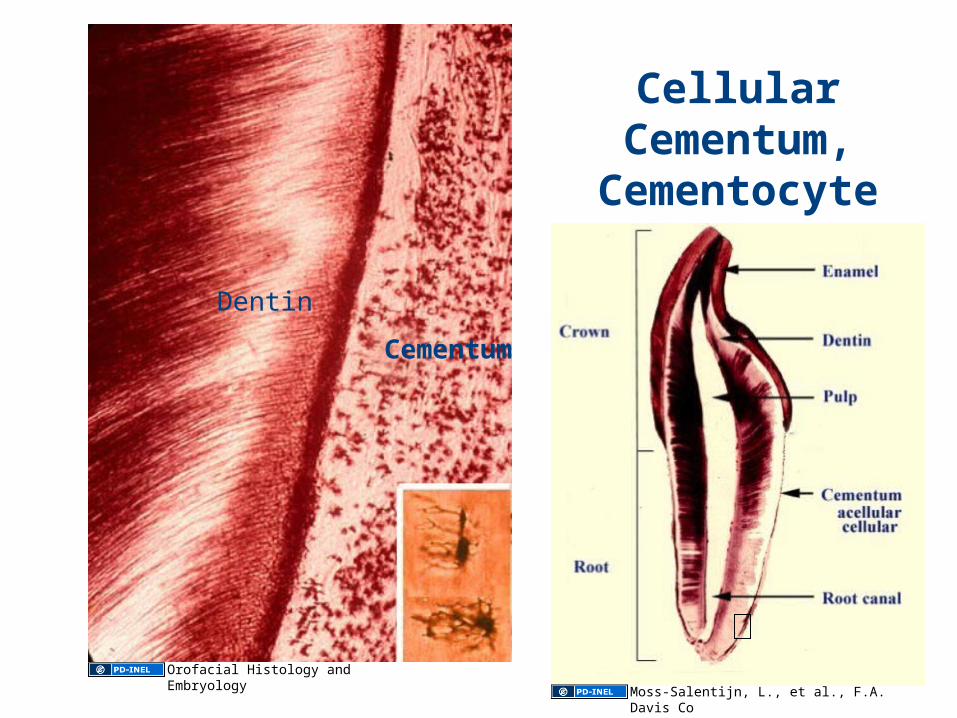
Cellular Cementum,
Cementocytes, and Dentin
Dentin
Cementum
Orofacial Histology and Embryology
Moss-Salentijn, L., et al., F.A. Davis Co

Deciduous and Permanent Teeth
Source Undetermined

Forming Tooth
Dentin
Enamel
Michigan Medical School Histology Slide Collection

Source Undetermined

Orofacial Histology and Embryology, Moss-Salentijn, L., et al., F.A. Davis Co
Cormack D., p.485

Erosion of Enamel and Cavity Formation
Source Undetermined

Orofacial Histology and Embryology, Moss-Salentijn, L., et al., F.A. Davis Co

Weiss/Greep, Histology, 4th ed. P.637 Drosenbach, Wikipedia
The Epithelial Attachment

Indiana University

X-section of the Tongue
Intrinsic and Extrinsic MusclesSource Undetermined

Filiform and Fungiform Papillae
Non-keratinized epithelium with secondary papillae and scattered taste buds.
Keratinized epithelium, no taste buds
Source Undetermined
Source Undetermined

Abnormal Keratinization of Filiform Papillae
Geographic tongue
Hairy tongue Over keratinized
Under keratinized
Source Undetermined
Source Undetermined
Source Undetermined
Source Undetermined

Circumvallate papillae and Taste Buds
US Federal Government, Wikipedia
NEUROtiker, Wikimedia Commons

A Visual Approach to Histology, Wismar and Ackerman

Taste Buds
Source Undetermined
NEUROtiker, Wikimedia Commons
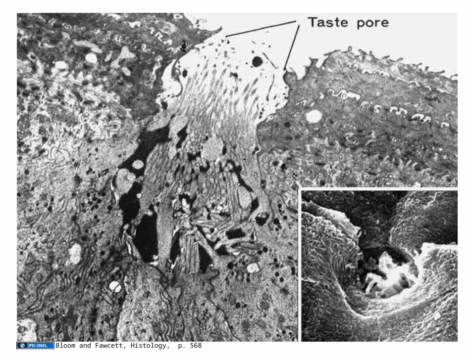
Bloom and Fawcett, Histology, p. 568

Areas of Taste Perception
Wela49, Wikimedia Commons

Major Salivary Glands
1. Parotid
2. Submandibular
3. Sublingual
US Federal Government, Wikimedia Commons

Saliva
Secretion About 1,000 ml/day
Submandibular Glands 65%Parotid Glands 23%Sublingual Glands 4%Minor Salivary Glands 8%
Flow Rate 0.3 ml/min (Unstimulated)
Stimulation Autonomic Nervous System
Composition Varies with flow rate

Composition of Saliva
Water
Ions: Bicarbonate, potassium, sodium, chloride, etc
Glycoproteins: Mucus
Proteins: Enzymes – Amylase (parotid gland), nucleases, etc.
Cells: Desquamated Epithelial cellsLeukocytes
pH: ~ 7.0

Glandular Lobules and Lobes
Many Lobules form a Lobe
Acini, Intralobular duct, Interlobular duct
Kierszenbaum p. 53
Text/Atlas of Histology, Philadelphia, WB Saunders, 1968

Structural and functional Unit of Salivary Gland
Source Undetermined

Myoepithelial Cell
Kim, SK


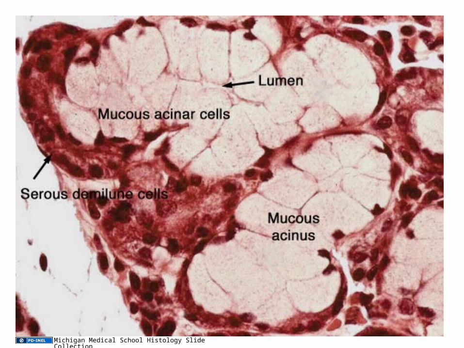
Mixed, Sero-mucous Gland
Mucous acini
Serous acini
Michigan Medical School Histology Slide Collection

Submandibular and Sublingual Gland
Michigan Medical School Histology Slide CollectionMichigan Medical School Histology Slide Collection
Michigan Medical School Histology Slide Collection
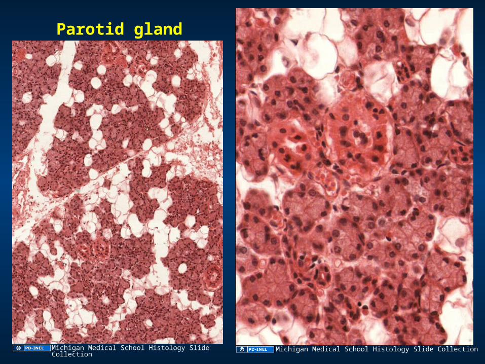
Parotid gland
Michigan Medical School Histology Slide Collection Michigan Medical School Histology Slide Collection
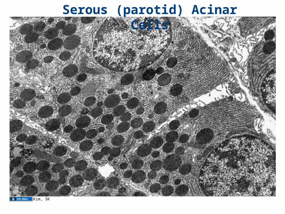
Serous (parotid) Acinar Cells
Kim, SK

Innervations of the Acinar Cells
Salivary Gland secretionis regulated by the autonomic nervous SystemNE: Nerve endings of
postganglionic fibers
NE
NE
Hand, A.R., J. Cell Biol. 47:541, 1970

Exocytosis
Kim, S.K. Bloom and Fawcett p. 695 Kim, S.K.
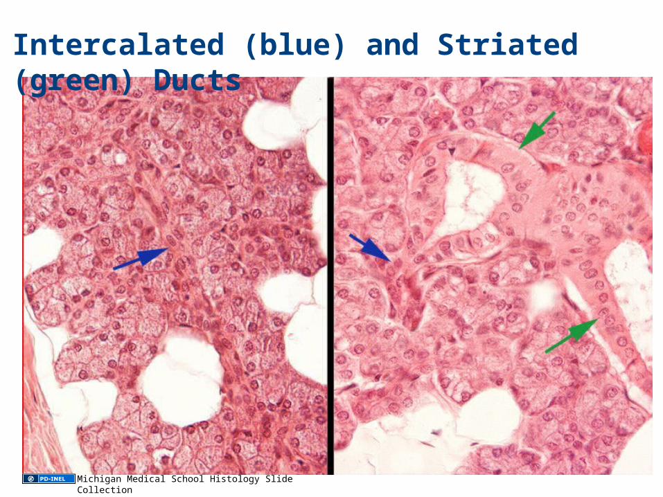
Intercalated (blue) and Striated (green) Ducts
Michigan Medical School Histology Slide Collection

Salivary Gland Ducts
Source Undetermined

EM of Striated Duct Cells
Source Undetermined

Role of Striated Ducts in Saliva Production
Image of ion flow through striated ducts removed
Regents of the University of Michigan

Junqueira/Carneiro 3rd ed. P. 340

Intra and Inter Lobular Ducts
Source Undetermined

Intra (left) and Inter (right) Lobular Ducts
Michigan Medical School Histology Slide Collection

Slide 3: Wheater 14.1
Slide 4: Wheater 14.1Slide 6: Michigan Medical School Histology Slide Collection
Slide 7: Michigan Medical School Histology Slide CollectionSlide 8: Michigan Medical School Histology Slide CollectionSlide 9: Sources UndeterminedSlide 10: Orofacial Histology and Embryology, Moss-Salentijn, L., et al., F.A. Davis Co.
Slide 11: Cell and Tissue Biology, L. Weiss 6th Ed. Pp. 597; Orofacial Histology and Embryology, Moss-Salentijn, L., et al., F.A. Davis Co. Slide 12: Sam Fentress, Wikimedia Commons, http://commons.wikimedia.org/wiki/File:ToothSection.jpg, CC: BY-SA http://creativecommons.org/licenses/by-sa/2.0; Source Undetermined Slide 13: Source UndeterminedSlide 14: Orofacial Histology and Embryology, Moss-Salentijn, L., et al., F.A. Davis Co Slide 15: Orofacial Histology and Embryology; Moss-Salentijn, L., et al., F.A. Davis CoSlide 16: Sources UndeterminedSlide 17: Michigan Medical School Histology Slide CollectionSlide 18: Source UndeterminedSlide 19: Orofacial Histology and Embryology, Moss-Salentijn, L., et al., F.A. Davis Co; Cormack D., p.485 Slide 20: Source UndeterminedSlide 21: Orofacial Histology and Embryology; Moss-Salentijn, L., et al., F.A. Davis CoSlide 22: Drosenbach, Wikipedia, http://en.wikipedia.org/wiki/File:The_Periodontium.jpg; Weiss/Greep, Histology, 4th ed. P.637Slide 23: Indiana University, http://anatomy.iupui.edu/courses/histo_D502/D502f04/lecture.f04/upperdigf04/uppergif04.html Slide 24: Source UndeterminedSlide 25: Sources UndeterminedSlide 26: Sources UndeterminedSlide 27: US Federal Government, Wikipedia, http://en.wikipedia.org/wiki/File:Illu04_tongue.jpg; NEUROtiker, Wikimedia Commons, http://commons.wikimedia.org/wiki/File:Taste_bud.svg, CC: BY-SA 3.0, http://creativecommons.org/licenses/by-sa/3.0/ Slide 28: A Visual Approach to Histology, Wismar and Ackerman Slide 29: Source Undetermined; NEUROtiker, Wikimedia Commons, http://commons.wikimedia.org/wiki/File:Taste_bud.svg, CC: BY-SA 3.0, http://creativecommons.org/licenses/by-sa/3.0/ Slide 30: Bloom and Fawcett, Histology, p. 568 Slide 31: Wela49, Wikimedia Commons, http://commons.wikimedia.org/wiki/File:Tongue_flavor.jpg, CC: BY-SA 30 http://creativecommons.org/licenses/by-sa/3.0/ Slide 32: US Federal Government, Wikimedia Commons, http://en.wikipedia.org/wiki/File:Illu_quiz_hn_02.jpg
Slide 35: Text/Atlas of Histology, Philadelphia, WB Saunders, 1968; Kierszenbaum p. 53
Slide 36: Source UndeterminedSlide 37: Sun-Kee Kim
Additional Source Informationfor more information see: http://open.umich.edu/wiki/CitationPolicy

Slide 38: Gray’s Anatomy Plate 1025, Wikimedia Commons, http://commons.wikimedia.org/wiki/File:Gray1025.png Slide 39: Michigan Medical School Histology Slide CollectionSlide 40: Michigan Medical School Histology Slide CollectionSlide 41: Michigan Medical School Histology Slide CollectionSlide 42: Michigan Medical School Histology Slide CollectionSlide 43: Sun-Kee KimSlide 44: Hand, A.R., J. Cell Biol. 47:541, 1970 Slide 45: Sun-Kee Kim; Bloom and Fawcett p. 695 Slide 46: Michigan Medical School Histology Slide CollectionSlide 47: Source UndeterminedSlide 48: Source UndeterminedSlide 49: Regents of the University of MichiganSlide 50: Junqueira/Carneiro 3rd ed. P. 340 Slide 51: Source UndeterminedSlide 52: Michigan Medical School Histology Slide Collection