· web viewemerging technologies for next-generation point-of-care testing sandeep kumar vashist...
TRANSCRIPT

Emerging technologies for next-generation point-of-care testing
Sandeep Kumar Vashist a*, Peter B. Luppa a, Leslie Y. Yeo b, Aydogan Ozcan c,d,e and John H.T.
Luong f*
a Institute of Clinical Chemistry and Pathobiochemistry, Klinikum rechts der Isar der Technischen
Universität München, Ismaninger Str. 22, D-81675 Munich, Germany.
b Micro/Nanophysics Research Laboratory, RMIT University, Melbourne, VIC 3001, Australia.
c Electrical Engineering Department, University of California, Los Angeles, CA 90095, USA.
d Bioengineering Department, University of California, Los Angeles, CA 90095, USA.
e California NanoSystems Institute (CNSI), University of California, Los Angeles, CA 90095, USA.
f Innovative Chromatography Group, Irish Separation Science Cluster (ISSC), Department of Chemistry
and Analytical, Biological Chemistry Research Facility (ABCRF), University College Cork, Cork,
Ireland.
*Corresponding authors. (SKV) E-mail: [email protected]; Tel.: +49 89 41404071; Fax:
+49 89 41404875. (JHTL) E-mail: [email protected]

Supplementary Table S1. Cell phone-based point-of-care testing (CP-POCT).
Types Analyte(s) detected Characteristics Ref.Colorimetric detection
Peanut Detects 1-25 ppm of peanut within 20 min [1]
25-hydroxyvitamin D AuNP-based immunoassay (IA), linear detection range (LDR): 15-110 nM
[2]
Herpesvirus nucleic acid
DNA sequences [1 mM] from Kaposi’s sarcoma associated herpes virus (KSHV) using a nanoparticle (NP) assay in microfluidic chips
[3]
CRP CRP [0.035-0.182 µg mL-1] with limit of detection (LOD) of 0.026 µg mL-1
[4]
CRP Rapid sandwich IA for CRP [0.3-81 ng mL-1] with LOD of 0.4 ng mL-1 using a smartphone-based colorimetric reader
[5]
Cortisol Detects cortisol in 10 min with LDR of 0.01-10 ng mL-1 and LOD of 0.01 ng mL-1
[6]
Cholesterol Detects the total cholesterol level in blood [140-400 mg dL-1] within 1 min
[7]
Proteases A quantum dot (QD) based multiplex assay for the activity of trypsin, chymotrypsin and enterokinase [pM-nM]
[8]
Cocaine AuNP and aptamer-based assay for cocaine with LDR of 0.2-0.8 mg mL-1
[9]
HE4 Microchip ELISA for HE4 in urine with LDR of 19.5-1250 ng mL-1 and LOD of 19.5 ng mL-1
[10]
Hg2+ Plasmonic AuNP and aptamer-based assay for Hg2+ with LOD of ~3.5 ppb
[11]
Albumin Detects albumin in the LDR of 0-100 mg dL-1 with LOD of 6 mg dL-1
[12]
Glucose Detects glucose in the LDR of 0-300 mg dL-1 with LOD of 40 mg dL-1
[12]
Triplex detection of infectious diseases
Detects HIV antibody, treponemal specific antibody for syphilis, and non-treponemal antibody for active syphilis infection in 15 min
[13]
Mumps, measles and herpes simplex virus IgGs
CP-based hand-held micro-plate reader based ELISAs for mumps, measles and herpes simplex virus IgGs
[14]
Multi-analyte sensing arrays
An image processing algorithm for ascorbic acid, glucose, protein, ketones, urobilinogen, bilirubin and RBC using paper-based urine strips
[15]
Heavy metals (Cu2+, Ni2+, Cd2+ and Cr6+)
Detects Cu2+, Ni2+, Cd2+ and Cr6+ with LODs of 0.29, 0.33, 0.19 and 0.35 ppm, respectively
[16]
2,2ʹ,4,4ʹ-tetrabromodiphenyl ether (BDE-47)
Detects BDE-47 in the LDR of 10-3-104 ng mL-1 using CP-interfaced lab-on-a-chip device based ELISA
[17]
Fluorescent detection
M. tuberculosis M. tuberculosis bacilli in Auramine O-stained sputum smears. [18]
Salmonella typhimurium DNA
A fluorescent microscope and paper microfluidics-based assay for detecting S. typhimurium down to 104 CFU mL-1
[19]
DNA amplification Detects the DNA fragment of the Hepatitis B virus plasmid by convective polymerase chain reaction (cPCR)
[20]
Recombinant bovine somatotropin (rbST) Abs
Detects rbST Abs in milk extracts by an IA using magnetic polystyrene microspheres and QD-labelled detection Ab
[21]
Salmonella and E.coli O157
Lateral flow IA (LFIA) strips for Salmonella and E. coli O157 with LOD of 105 CFU mL-1
[22]

β-D-galactosidase A paper microfluidics-based IA for β-D-galactosidase, with LDR of 0.7-12 nM and LOD of 0.7 nM
[23]
Single NPs & viruses Fluorescent imaging of isolated 100 nM fluorescent NPs and human cytomegaloviruses
[24]
microRNA Fluorescent molecular beacon assay to detect microRNA with LOD of 10 pM.
[25]
Red blood cells (RBCs)White blood cells (WBCs)Haemoglobin (Hb)
Imaging cytometry platform for Hb concentration and the density of RBCs and WBCs
[26]
E. coli Detects E.coli O157 by QD-based sandwich IA in glass capillaries with LOD of ~5-10 CFU mL-1
[27]
WBCsGiardia lamblia cysts
Imaging labelled WBCs in whole blood and water-borne pathogenic Giardia lamblia cysts [resolution of 10 µm and FOV of 81 mm2]
[28]
Trypanosoma cruzi T. cruzi genomic DNA [0.1 to100 fg µL-1] with LOD of 9.6 ag [29]Collagenase and Trypsin
Detects collagenase and trypsin with LODs of 3.75 µg mL-1
and 930 pg, respectively, using a simple CP and tablet-based fluorescence detection system
[30]
Luminescent detection
Bile acid An lateral flow assay (LFA) minicartridge format for total bile acid in serum and oral fluid with LDR and LOD of 0.5-100 µM and 0.5 µM
[31]
Cholesterol LFA minicartridge format for total cholesterol in serum with LDR and LOD of 20-386 mg dL-1 and 20 mg dL-1
[31]
Salivary cortisol LFIA for salivary cortisol [0.3-60 ng mL-1] with LOD of 0.3 ng mL-1
[32]
Spectrophotometric detection
microRNA CP fluorimeter system to detect specific microRNA sequence with LOD of 10 pM using molecular beacon FRET assay
[33]
Additives in beverages and food
CP-based spectroscopic analysis using CP’s screen for illumination and front camera for imaging.
[34]
SPR detection β2 microglobulin (β2M)
Angle-resolved SPR detection system for β2M with LOD of 0.1 µg mL-1
[35]
Glycerol Fiber optic SPR sensor to detect glycerol concentrations of 0-20%
[36]
IgG Fiber optic SPR sensor to detect various concentrations of IgG binding to protein A-bound sensing element
[37]
Electrochemical detection
Plasmodium falciparum histidine-rich protein 2 (PfHRP2) biomarker for malaria,
Detects PfHRP2 in 15 min with LOD of 16 ng mL-1 using a compact embedded circuit, disposable microfluidic chip and capillary flow
[38]
blood glucose and trace heavy metals (Pb2+, Cd2+, Zn2+) in water, Na+ in urine, and PfHRP2
Universal low-cost CP-based electrochemical detector for the detection of blood glucose and heavy metals in water, Na+ in urine, and PfHRP2 biomarker
[39]
2,4,6-trinitrotoluene (TNT)
A CP-based electrochemical biosensor, using screen-printed electrodes modified with TNT-specific peptides, to detect TNT with LOD of 1 µM
[40]
iHealth Wireless Smart Gluco-Monitoring System
Detects pathophysiological blood glucose concentrations using a miniaturized glucometer interfaced with SP via a smart application
[41]
iHealth Align Detects pathophysiological blood glucose concentrations using the smallest Food and Drug Administration (FDA) approved blood glucose meter
[42]

Escherichia coli CP-based electrochemical biosensor pre-concentrates E.coli and detects it with a LDR and LOD of 10-100 cells mL -1 and 10 cells mL-1, respectively
[43]
Nitrate Audio jack-based electrochemical sensor that detects nitrate concentration in water with LOD of 0.2 ppm and LDR of 1.4-70 ppm within 1 min
[44]
Cytometry Optofluidic fluorescent imaging-based flow cytometer
Compact, light-weight and cost-effective CP-based flow cytometer with fluorescent resolution of ~2 µm and capability to analyze samples >0.1 mL
[45]
RBCs, WBCs and Hb Determines the density of RBCs, WBCs and Hb in blood from a 10 µL sample
[26]
Other detection techniques
Microscopy and spectroscopy
Stained and unstained blood smears, and transmission and fluorescence spectroscopy of diffuse tissue
[46]
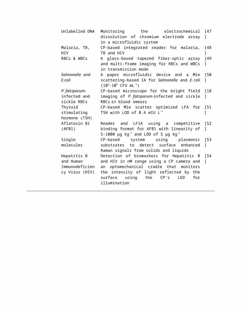
Unlabelled DNA Monitoring the electrochemical dissolution of chromium electrode array in a microfluidic system
[47]
Malaria, TB, HIV CP-based integrated reader for malaria, TB and HIV [48]RBCs & WBCs A glass-based tapered fiber-optic array and multi-frame
imaging for RBCs and WBCs in transmission mode[49]
Salmonella and E.coli A paper microfluidic device and a Mie scattering-based IA for Salmonella and E. coli (102-105 CFU mL-1)
[50]
P. falciparum-infected and sickle RBCs
CP-based microscope for the bright field imaging of P. falciparum-infected and sickle RBCs in blood smears
[18]
Thyroid stimulating hormone (TSH)
CP-based Mie scatter optimized LFA for TSH with LOD of 0.4 mIU L-1
[51]
Aflatoxin B1 (AFB1) Reader and LFIA using a competitive binding format for AFB1 with linearity of 5-1000 µg kg-1 and LOD of 5 µg kg-1
[52]
Single molecules CP-based system using plasmonic substrates to detect surface enhanced Raman signals from solids and liquids
[53]
Hepatitis B and Human Immunodeficiency Virus (HIV)
Detection of biomarkers for Hepatitis B and HIV in nM range using a CP camera and an optomechanical cradle that monitors the intensity of light reflected by the surface using the CP’s LED for illumination
[54]

Supplementary Figure S1. (A) Schematic of a CP-based device for the colorimetric detection of
Hg2+ [11]. Reprinted with permission from the American Chemical Society. (B) vitaAID
accessory on an iPhone for the colorimetric detection of vitamin D [2]. Reprinted with
permission from the Royal Society of Chemistry. (C) CP attachment and minicartridge for
biochemiluminescent detection of cholesterol [31]. Reprinted with permission from the
American Chemical Society. (D) A CP-based colorimetric reader using screen-based bottom
illumination provided by gadgets [5]. Reprinted with permission from the Elsevier B.V. (E) CP
attachment for wide-field fluorescent and dark field imaging along with its schematic [28].
Reprinted with permission from the Royal Society of Chemistry. (F) Schematic of a CP-based
fluorescence microscope and its ray-tracing diagram [24]. Reprinted with permission from the
American Chemical Society (G) A CP-based contact microscopy platform and a schematic of the
CP attachment [49]. Reprinted with permission from the Royal Society of Chemistry.
References
1. Coskun, A.F., et al. (2013) A personalized food allergen testing platform on a cellphone.
Lab. Chip. 13, 636-640.
2. Lee, S., et al. (2014) A smartphone platform for the quantification of vitamin D levels. Lab.
Chip. 14, 1437-1442.
3. Mancuso, M., et al. (2014) Detection of Kaposi's sarcoma associated herpesvirus nucleic
acids using a smartphone accessory. Lab. Chip. 14, 3809-3816.
4. McGeough, C.M. and O'Driscoll, S. (2013) Camera phone-based quantitative analysis of C-
reactive protein ELISA. IEEE Trans. Biomed. Circuits Syst. 7, 655-659.

5. Vashist, S.K., et al. (2015) A smartphone-based colorimetric reader for bioanalytical
applications using the screen-based bottom illumination provided by gadgets. Biosens.
Bioelectron. 67, 248-255.
6. Choi, S., et al. (2014) Real-time measurement of human salivary cortisol for the assessment
of psychological stress using a smartphone. Sensing Bio-Sensing Research 2, 8-11.
7. Oncescu, V., et al. (2014) Cholesterol testing on a smartphone. Lab. Chip. 14, 759-763.
8. Petryayeva, E. and Algar, W.R. (2014) Multiplexed homogeneous assays of proteolytic
activity using a smartphone and quantum dots. Anal. Chem. 86, 3195-3202.
9. Smith, J.E., et al. (2014) Colorimetric detection with aptamer-gold nanoparticle conjugates
coupled to an android-based color analysis application for use in the field. Talanta 121, 247-
255.
10. Wang, S., et al. (2011) Integration of cell phone imaging with microchip ELISA to detect
ovarian cancer HE4 biomarker in urine at the point-of-care. Lab. Chip. 11, 3411-3418.
11. Wei, Q., et al. (2014) Detection and spatial mapping of mercury contamination in water
samples using a smart-phone. ACS Nano 8, 1121-1129.
12. Yetisen, A.K., et al. (2014) A smartphone algorithm with inter-phone repeatability for the
analysis of colorimetric tests. Sens. Actuators B Chem. 196, 156-160.
13. Laksanasopin, T., et al. (2015) A smartphone dongle for diagnosis of infectious diseases at
the point of care. Sci. Trans. Med. 7, 273re271-273re271.
14. Berg, B., et al. (2015) Cellphone-Based Hand-Held Micro-Plate Reader for Point-of-Care
Testing of Enzyme-Linked Immunosorbent Assays. ACS Nano 9, 7857-7866.
15. Hong, J.I. and Chang, B.Y. (2014) Development of the smartphone-based colorimetry for
multi-analyte sensing arrays. Lab. Chip. 14, 1725-1732.

16. Wang, H., et al. (2014) Paper-based three-dimensional microfluidic device for monitoring of
heavy metals with a camera cell phone. Anal. Bioanal. Chem. 406, 2799-2807.
17. Chen, A., et al. (2014) Smartphone-interfaced lab-on-a-chip devices for field-deployable
enzyme-linked immunosorbent assay. Biomicrofluidics 8, 064101.
18. Breslauer, D.N., et al. (2009) Mobile phone based clinical microscopy for global health
applications. PLoS One 4, e6320.
19. Fronczek, C.F., et al. (2014) Paper microfluidic extraction and direct smartphone-based
identification of pathogenic nucleic acids from field and clinical samples. RSC Adv. 4,
11103.
20. Lee, D., et al. (2011) DNA detection using commercial mobile phones. Biosens.
Bioelectron. 26, 4349-4354.
21. Ludwig, S.K., et al. (2014) Cellphone-based detection platform for rbST biomarker analysis
in milk extracts using a microsphere fluorescence immunoassay. Anal. Bioanal. Chem. 406,
6857-6866.
22. Rajendran, V.K., et al. (2014) Smartphone based bacterial detection using biofunctionalized
fluorescent nanoparticles. Microchim. Acta 181, 1815-1821.
23. Thom, N.K., et al. (2014) Quantitative Fluorescence Assays Using a Self-Powered Paper-
Based Microfluidic Device and a Camera-Equipped Cellular Phone. RSC Adv. 4, 1334-
1340.
24. Wei, Q., et al. (2013) Fluorescent imaging of single nanoparticles and viruses on a smart
phone. ACS Nano 7, 9147-9155.
25. Chun, H.J., et al. (2014) Paper-based glucose biosensing system utilizing a smartphone as a
signal reader. Biochip J. 8, 218-226.

26. Zhu, H., et al. (2013) Cost-effective and rapid blood analysis on a cell-phone. Lab. Chip. 13,
1282-1288.
27. Zhu, H., et al. (2012) Quantum dot enabled detection of Escherichia coli using a cell-phone.
Analyst 137, 2541-2544.
28. Zhu, H., et al. (2011) Cost-effective and compact wide-field fluorescent imaging on a cell-
phone. Lab. Chip. 11, 315-322.
29. Walker, F.M., et al. (2014) Transformation of personal computers and mobile phones into
genetic diagnostic systems. Anal. Chem. 86, 9236-9241.
30. Wargocki, P., et al. (2015) Medically relevant assays with a simple smartphone and tablet
based fluorescence detection system. Sensors 15, 11653-11664.
31. Roda, A., et al. (2014) Integrating biochemiluminescence detection on smartphones: mobile
chemistry platform for point-of-need analysis. Anal. Chem. 86, 7299-7304.
32. Zangheri, M., et al. (2015) A simple and compact smartphone accessory for quantitative
chemiluminescence-based lateral flow immunoassay for salivary cortisol detection. Biosens.
Bioelectron. 64, 63-68.
33. Yu, H., et al. (2014) Smartphone fluorescence spectroscopy. Anal. Chem. 86, 8805-8813.
34. Iqbal, Z. and Eriksson, M. (2013) Classification and quantitative optical analysis of liquid
and solid samples using a mobile phone as illumination source and detector. Sens. Actuators
B-Chem. 185, 354-362.
35. Preechaburana, P., et al. (2012) Surface plasmon resonance chemical sensing on cell phones.
Angew. Chem. Int. Ed. Engl. 51, 11585-11588.
36. Bremer, K. and Roth, B. (2015) Fibre optic surface plasmon resonance sensor system
designed for smartphones. Optics express 23, 17179-17184.

37. Liu, Y., et al. (2015) Surface Plasmon Resonance Biosensor Based on Smart Phone
Platforms. Sci. Rep. 5, 12864.
38. Lillehoj, P.B., et al. (2013) Rapid electrochemical detection on a mobile phone. Lab Chip
13, 2950-2955.
39. Nemiroski, A., et al. (2014) Universal mobile electrochemical detector designed for use in
resource-limited applications. Proc. Natl. Acad. Sci. USA 111, 11984-11989.
40. Zhang, D.M., et al. (2015) Smartphone-based portable biosensing system using impedance
measurement with printed electrodes for 2,4,6-trinitrotoluene (TNT) detection. Biosens.
Bioelectron. 70, 81-88.
41. http://www.ihealthlabs.com/glucometer/wireless-smart-gluco-monitoring-system/.
42. http://www.ihealthlabs.com/.
43. Jiang, J., et al. (2014) Smartphone based portable bacteria pre-concentrating microfluidic
sensor and impedance sensing system. Sens. Actuators B-Chem 193, 653-659.
44. Wang, X., et al. (2015) Audio Jack Based Miniaturized Mobile Phone Electrochemical
Sensing Platform. Sens. Actuators B-Chem. 209, 677-685.
45. Zhu, H., et al. (2011) Optofluidic fluorescent imaging cytometry on a cell phone. Anal.
Chem. 83, 6641-6647.
46. Smith, Z.J., et al. (2011) Cell-phone-based platform for biomedical device development and
education applications. PLoS One 6, e17150.
47. Huang, Y.W. and Ugaz, V.M. (2013) Smartphone-based detection of unlabeled DNA via
electrochemical dissolution. Analyst 138, 2522-2526.
48. Mudanyali, O., et al. (2012) Integrated rapid-diagnostic-test reader platform on a cellphone.
Lab Chip 12, 2678-2686.

49. Navruz, I., et al. (2013) Smart-phone based computational microscopy using multi-frame
contact imaging on a fiber-optic array. Lab Chip 13, 4015-4023.
50. Park, T.S., et al. (2013) Smartphone quantifies Salmonella from paper microfluidics. Lab
Chip 13, 4832-4840.
51. You, D.J., et al. (2013) Cell-phone-based measurement of TSH using Mie scatter optimized
lateral flow assays. Biosens. Bioelectron. 40, 180-185.
52. Lee, S., et al. (2013) Performance improvement of the one-dot lateral flow immunoassay for
aflatoxin B1 by using a smartphone-based reading system. Sensors 13, 5109-5116.
53. Ayas, S., et al. (2014) Counting Molecules with a Mobile Phone Camera Using Plasmonic
Enhancement. ACS Photonics 1, 17-26.
54. Giavazzi, F., et al. (2014) A fast and simple label-free immunoassay based on a smartphone.
Biosens. Bioelectron. 58, 395-402.