jensenbio.weebly.com€¦ · web viewunit 6, part 2 notes – meiosis ap biology what is meiosis...
TRANSCRIPT

Name: ______________________________________ Date: _________________________ Period: ________
Unit 6, Part 2 Notes – MeiosisAP Biology
1. What is meiosis and how is it different from mitosis? A. Our last notes packet focused on mitosis, a type of cell division that makes cells with the same number of
chromosomes as the parent cell. The chart below (from your Part 1 Notes) summarizes some key information about mitosis.
Purpose of Mitosis in Unicellular Eukaryotes
Asexual reproduction
Purpose of Mitosis in Multicellular Eukaryotes
-Growth -Tissue repair (replacement of dead or damaged cells)-Asexual reproduction (only in some organisms)
Starts With One diploid (2n) parent cell. In humans, this cell contains 46 replicated chromosomes. (These chromosomes look like X’s.)
In humans, this parent cell is a normal body cell (aka somatic cell) like a blood cell, bone cell, muscle cell, skin cell, etc.
Ends With Two diploid daughter cells. In humans, these daughter cells each contain 46 un-replicated chromosomes. (These chromosomes look like single lines or rods.)
In humans, these daughter cells are somatic cells like blood cells, bone cells, muscle cells, skin cells, etc.
Practice Question: Let’s review some vocabulary from the Part 1 notes. Record the definitions of the following terms below.
Asexual reproduction
Sexual reproduction
Diploid cell (2)
Haploid cell (n)
Un-replicated chromosomes
Replicated chromosomes
B. Today, we will be focusing on a different type of cell division—meiosis—which is used by many multicellular eukaryotes. Meiosis is used to create sex cells (i.e., eggs and sperm) with half the number of chromosomes as a normal body cell. The chart on the next page summarizes some key information about meiosis.
Purpose of Meiosis in Multicellular Eukaryotes
Create sex cells (aka gametes) for sexual reproduction.
Starts With One diploid (2n) parent cell. In humans, this cell contains 46 replicated chromosomes. (These chromosomes look like X’s.)
In humans, this parent cell is a cell within the testes in men or the ovaries in women. Ends With Four haploid (n) daughter cells. In humans, these daughter cells each contain 23 un-
replicated chromosomes. (These chromosomes look like single lines or rods.)

C. The following image summarizes the differences between mitosis and meiosis.
D. In sexual reproduction in humans, a sperm cell (male sex cell) combines with an egg cell (female sex cell) in a process called fertilization. Together, they make a fertilized egg, which is also called a zygote. This is the first cell in the developing embryo.
E. Again, for the baby’s first cell to have the correct number of chromosomes (46), the sperm and egg cell must have half the number of chromosomes of a normal human body cell. As such, the sperm and egg cell each have 23 chromosomes.
F. Also remember that your chromosomes are arranged in 23 homologous pairs. Chromosomes within a pair have the same types of genes in the same places. For example, each chromosome within a pair may have a gene for eye color, though one chromosome may contain a blue eye gene, and the other chromosome may contain a brown eye gene. One chromosome in the homologous pair came from your mother and one chromosome in the pair came from your father
The image to the right shows two un-replicated homologous chromosomes with genes for eye color and hair color in the same locations.
G. Cells in the ovaries (in women) and testes (in men) that are destined to divide using meiosis to become eggs and sperm go through the following cell cycle stages.
Interphase (G1, S, G2) – Remember, during interphase the cell grows (G1), copies its DNA (S stage), and prepares for cell division by making extra proteins and organelles (G2).
Meiosis I – The parent cell (diploid, 46 replicated chromosomes) divides to create two daughter cells (haploid, 23 replicated chromosomes).
Interkinesis – In some species, the daughter cells of meiosis I enter a brief period of rest before meiosis II. Some sources refer to this as a second interphase, or they call it “interkinesis.” It is less accurate to call it a second interphase because the DNA is not copied again (so some of the events that happen in the interphase before Meiosis I do not occur in this “interkinesis” between meiosis I and meiosis II).
Meiosis II – The two daughter cells of meiosis I (haploid, 23 replicated chromosomes) divide to create a total of four daughter cells (haploid, 23 un-replicated chromosomes).
The image to the right shows the changes in chromosome number and ploidy (diploid vs. haploid) during meiosis to create human sperm cells.

2. What happens during Meiosis I?
A. Meiosis I occurs in the following stages—prophase I, metaphase I, anaphase I, and telophase I. This is followed by cytokinesis to divide the cytoplasm and organelles equally between the daughter cells.
B. During Prophase I, the chromosomes arrange themselves in homologous pairs. This pairing of homologous chromosomes is called synapsis. Together, the two homologous chromosomes in a pair are called a tetrad. “Tetra” means “four,” and this refers to the number of chromatids present. There are two chromatids per chromosome, so four chromatids total.
C. Once the homologous chromosomes are paired together, they exchange genes (sections of DNA) during a process called crossing over. The locations on the chromosomes where DNA is exchanged are called chiasmata (singular: chiasma).
D. Crossing over occurs in each male testis cell or female ovary cell that goes through meiosis. Different sections of DNA are exchanged each time crossing over occurs. This ensures that no two eggs created by the same woman are genetically identical to each other. Similarly, no two sperm created by the same man are genetically identical to each other.
E. The stages of meiosis I are summarized in the chart below.
Stage Description PictureStarting cell One diploid parent cell containing 46 replicated
chromosomes in humansProphase I The chromatin coils up into chromosomes
Synapsis and crossing over take place (see B-D above for more information)
The nuclear envelope (aka nuclear membrane) and nucleolus break down
The centrosomes move to opposite poles of the cell and mitotic spindle fibers begin to form from the centrosomes
Metaphase I Spindle fibers attach to chromosomes Homologous chromosomes line up in pairs at the
center of the cell (metaphase plate)o Note: this is different from metaphase of
mitosis! They are lining up with a “matching partner” rather than in a single file!
o Independent Assortment (discussed later on)
*Note: The kinetochore is the part of the centromere of a chromosome that attaches to the spindle fiber.
Anaphase I Spindle fibers pull the homologous chromosomes away from each other to opposite ends of the dividing cell (chromatids on a single chromosome DO NOT separate yet, the chromosomes are still replicated!)
Segregation = separation of homologous partners to different daughter cells
*Note: we just divided the number of chromosomes going into the daughter cells in half

Telophase I The homologous chromosomes reach opposite ends of the dividing cell. In some species, the chromosomes uncoil back into chromatin during telophase I. In other species, the chromosomes stay coiled in preparation for meiosis II.
In some species, nuclear membranes and nucleoli reform in both daughter cells.
In some species, the mitotic spindle breaks down.Cytokinesis Cytokinesis occurs at the same time as Telophase I.
Cytokinesis divides the cytoplasm and separates the organelles into the two daughter cells. Look back to your Part 1 Notes to review the difference between cytokinesis in animal cells vs. plant cells.
Ending Cells Two haploid daughter cells with 23 replicated chromosomes in humans.
3. What happens during meiosis II?
A. Remember, in some species, the daughter cells of meiosis I enter a brief period of rest before meiosis II. Some sources refer to this as a second interphase, or they call it “interkinesis.” It is less accurate to call it a second interphase because the DNA is not copied again (so some of the events that happen in the interphase before Meiosis I do not occur in this “interkinesis” between meiosis I and meiosis II).
B. The daughter cells of meiosis I are the starting cells in meiosis II. C. Meiosis II occurs in the following stages—prophase II, metaphase II, anaphase II, and telophase II.
This is followed by cytokinesis to divide the cytoplasm and organelles equally between the daughter cells. D. Meiosis II proceeds identical to mitosis except the starting cells are haploid rather than diploid.E. The stages of meiosis II are summarized in the chart below.
Stage Description PictureStarting Cells Two haploid daughter cells with 23
replicated chromosomes in humans (the daughter cells of Meiosis I)

Prophase II If the chromosomes unwound (uncoiled) into chromatin during telophase I, they wind back up again
If the nuclear envelope and nucleolus reformed during telophase I, they break down again
If the mitotic spindle broke down during telophase I, it is reformed by the centrosomes, which have moved to opposite ends of the cell
Metaphase II Spindle fibers attach to chromosomes and push them to the center of the dividing cell (metaphase plate)
The chromosomes line up one-by-one (single file) along the metaphase plate because they no longer have a homologue. This is different from meiosis I where the chromosomes line up in pairs.
Anaphase II Spindle fibers split the two sister chromatids from each chromosome and pull them to opposite ends of the dividing cell. Once the chromatids have split, they are called daughter chromosomes (un-replicated).
Telophase II Chromosomes uncoil into chromatin Nuclear membranes and nucleoli reform
in the daughter cells The mitotic spindle breaks down.
Cytokinesis Cytokinesis occurs at the same time as telophase II. Cytokinesis divides the cytoplasm and separates the organelles into the daughter cells. Look back to your Part 1 Notes to review the difference between cytokinesis in animal cells vs. plant cells.
Ending Cells Four haploid daughter cells with 23 un-replicated chromosomes in humans.
4. How does meiosis increase genetic variation in the sex cells?
A. Remember, crossing over during prophase I ensures that each egg cell created by one woman contains a different combination of her genes. (Same goes for each sperm cell created by one man.) In other words, crossing over increases genetic variation (aka genetic diversity) in the egg cells and sperm cells.
B. Another process that increases genetic variation in gametes is called independent assortment. Independent assortment means that each pair of homologous chromosomes arranges itself separately from the other pairs of homologous chromosomes along the metaphase plate during metaphase I.
C. Remember, each homologous chromosome pair contains one maternal chromosome and one paternal chromosome. The homologous chromosome pairs do not necessarily all arrange themselves with their maternal chromosomes on the same side of the metaphase plate and their paternal chromosomes on the other side of the metaphase plate.
D. For example, let’s say there are three homologous chromosome pairs. The pairs COULD all arrange themselves with their maternal chromosomes on the left and their paternal chromosomes on the right.

E. However, because each pair assorts (arranges) itself independently (separately) from the other pairs, there are a variety of alternate arrangements. For example, in the image to the right, the first homologous chromosome pair arranges itself with its maternal chromosome on the left and its paternal chromosome on the right. The second chromosome pair arranges itself with its paternal chromosome on the left and its maternal chromosome on the right. The third chromosome pair arranges itself with its maternal chromosome on the left and its paternal chromosome on the right.
F. Due to independent assortment, there are 2n possible arrangements of maternal and paternal chromosomes in the daughter cells. “n” stands for the number of homologous chromosome pairs. For the example given above, that would be 23 = 8 possible combinations.
G. For human cells, there are 23 pairs of homologous chromosomes. Therefore, there will be 223 = 8,388,608 possible combinations of maternal and paternal chromosomes in the egg cells created by a woman or the sperm cells created by a man.
H. But how does this result in different genes in the daughter cells? Remember, homologous chromosomes have the same types of genes in the same places. However, they don’t have to have the same gene forms. For example, both chromosomes may have an eye color gene, though one chromosome may have a blue eye gene, and the other chromosome may have a brown eye gene. Similarly, both chromosomes may have a hair texture gene, though one chromosome may have a straight hair gene, and the other chromosome may have a curly hair gene.
I. In the series of images below, there are two homologous chromosome pairs, indicated by the letters A/a and B/b. Capital A and capital B represent the maternal chromosomes in each pair. Lowercase “a” and lowercase “b” represent the paternal chromosomes in each pair. Within a single chromosome, the two chromatids are indicated by numbers (1 and 2).
J. Notice that for chromosome pair A/a, the maternal chromosome (A) carries the blue eye gene, and the paternal chromosome (a) carries the brown eye gene.
K. For chromosome pair B/b, the maternal chromosome (B) carries the straight hair gene, and the paternal chromosome (b) carries the curly hair gene.
L. In the image shown to the right, we can see that during metaphase I of meiosis, both chromosome pairs can line up with their maternal chromosomes (A and B) on the left and their paternal chromosomes (a and b) on the right.
Metaphase I

Metaphase II: The images below show the daughter cells of meiosis I going through metaphase II
Daughter Cells of Meiosis II
Trait combination in the two daughter cells pictured above: blue eye gene and straight hair gene

Trait combination in the two daughter cells pictured above: brown eye gene and curly hair gene
*Notice that the arrangement of chromosomes during metaphase I of meiosis results in two possible chromosome combinations in the gametes—A/B and a/b. This gives the gametes (eggs or sperm) two possible gene combinations—blue eye gene/straight hair gene and brown eye gene/curly hair gene.
Alternately, the homologous chromosome pairs can line up differently from one another during metaphase I of meiosis. In the image shown to the right, we can see that the first chromosome pair lines up with its maternal chromosome (A) on the left and its paternal chromosome (a) on the right. The second chromosome pair lines up with its paternal chromosome (b) on the left and its maternal chromosome (B) on the right. This would result in a gamete with A/b and a/B combinations rather than the A/B and a/b combinations we saw in the first example.
Overall: Therefore, if there are two pairs of homologous chromosomes there will be four possible combinations of maternal and paternal chromosomes in the daughter cells (A/B, a/b, A/b, and a/B). In the example given, this results in the following four possible gene combinations in the daughter cells—blue eye gene/straight hair gene, brown eye gene/curly hair gene, blue eye gene/curly hair gene, and brown eye gene/straight hair gene. This fits with the 2n calculation because 22 =4.
M. Because both crossing over and independent assortment result in different combinations of maternal and paternal genes in the gametes (eggs or sperm), they are often described as methods of genetic recombination. In other words, the genes are being “recombined” or “mixed” in different ways to create genetically unique eggs or sperm.
5. How are eggs and sperm different from one another?
A. Remember, an egg cell and a sperm cell each contribute 23 chromosomes to create a fertilized egg (aka zygote) with 46 chromosomes.
B. Though they each contribute the same amount of DNA, an egg cell is much larger than a sperm cell. The egg cell is responsible for contributing nearly all of the cytoplasm and organelles to the zygote. (The sperm cell really just contributes DNA).
C. To create such large egg cells, cytokinesis must occur unevenly during meiosis in human ovary cells. (Meiosis in human ovary cells is called oogenesis.) Because cytokinesis occurs unevenly during meiosis I and meiosis II, the result is three tiny daughter cells called polar bodies and one large egg cell. All four daughter cells have the same amount of DNA, but the polar bodies cannot survive with so little cytoplasm and organelles. They die and disintegrate. Therefore, meiosis in an ovary cell can only produce one functional egg cell.


Note: Ovum (plural: ova) is another name for an egg cell, so “oogenesis” is the creation of egg cells.
D. In contrast, cytokinesis occurs evenly during meiosis in human testis cells—which is called spermatogenesis. This results in four functional sperm cells that are the same size (see image below).
6. Do some people have more (or less) chromosomes than 46?
A. Yes, some people have different amounts of chromosomes in their cells (typically 45 or 47).
B. This abnormal chromosome number is called aneuploidy. Aneuploidy results from an error during meiosis called nondisjunction.
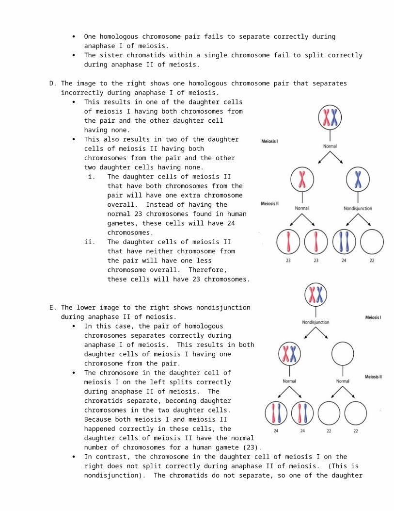
C. Nondisjunction occurs when one of two events takes place… One homologous chromosome pair fails to separate correctly during anaphase I of meiosis. The sister chromatids within a single chromosome fail to split correctly during anaphase II of
meiosis.
D. The image to the right shows one homologous chromosome pair that separates incorrectly during anaphase I of meiosis.
This results in one of the daughter cells of meiosis I having both chromosomes from the pair and the other daughter cell having none.

This also results in two of the daughter cells of meiosis II having both chromosomes from the pair and the other two daughter cells having none.
i. The daughter cells of meiosis II that have both chromosomes from the pair will have one extra chromosome overall. Instead of having the normal 23 chromosomes found in human gametes, these cells will have 24 chromosomes.
ii. The daughter cells of meiosis II that have neither chromosome from the pair will have one less chromosome overall. Therefore, these cells will have 23 chromosomes.
E. The lower image to the right shows nondisjunction during anaphase II of meiosis. In this case, the pair of homologous chromosomes separates correctly during anaphase I of
meiosis. This results in both daughter cells of meiosis I having one chromosome from the pair. The chromosome in the daughter cell of meiosis I on the left splits correctly during anaphase II of
meiosis. The chromatids separate, becoming daughter chromosomes in the two daughter cells. Because both meiosis I and meiosis II happened correctly in these cells, the daughter cells of meiosis II have the normal number of chromosomes for a human gamete (23).
In contrast, the chromosome in the daughter cell of meiosis I on the right does not split correctly during anaphase II of meiosis. (This is nondisjunction). The chromatids do not separate, so one of the daughter cells of meiosis II receives both chromatids (daughter chromosomes), and the other daughter cell receives none.
i. The daughter cell that receives both chromatids (daughter chromosomes) has one extra chromosome overall, for a total of 24.
ii. The daughter cell that receives neither chromatid (daughter chromosome) has one less chromosome overall, for a total of 22.
F. Suppose an egg cell contains 24 chromosomes due to nondisjunction. If it is fertilized by a normal sperm cell containing 23 chromosomes, the zygote (fertilized egg) will have 47 chromosomes. The zygote then divides many times by mitosis to create a multicellular developing baby with 47 chromosomes in each of his or her cells.
G. Suppose an egg cell contains 22 chromosomes due to nondisjunction. If it is fertilized by a normal sperm cell containing 23 chromosomes, the zygote (fertilized egg) will have 45 chromosomes. The zygote then divides many times by mitosis to create a multicellular developing baby with 45 chromosomes in each of his or her cells.
H. What are some real disorders caused by nondisjunction in humans? Down syndrome occurs when a baby receives three
chromosomes instead of two chromosomes for the 21st
homologous chromosome pair. (Two of the chromosomes came from one parent due to nondisjunction in that parent’s cells. The other chromosome came from the other parent due to normal meiosis in that parent’s cells.) For this reason, Down syndrome is often known as trisomy 21. (Remember, “tri” means “three.”) Therefore, a person with Down syndrome will have a total of 47 chromosomes in his or her cells.
Patau syndrome occurs when a baby receives three chromosomes instead of two chromosomes for the 13th
homologous chromosome pair. For this reason, it is often called trisomy 13.
Klinefelter syndrome occurs when a baby receives three sex chromosomes instead of two for the 23rd homologous chromosome pair. The baby has two X chromosomes and one Y chromosome (XXY). The baby is a boy because he has a Y chromosome.
Turner syndrome occurs when a baby receives one sex chromosome instead of two for the 23rd homologous chromosome pair. This is called monosomy, because “mono” means “one.” The baby has one X chromosome and is a girl because she does not have a Y chromosome. We often write “X0” for the sex chromosomes of a person who has Turner syndrome, and the “0” means there is no second sex chromosome. A person with Turner syndrome has 45 chromosomes in each of her cells.
Note: We will learn about the symptoms of these disorders in class if we have time.
Notes Questions
1. Complete the chart below that compares mitosis and meiosis.
Mitosis MeiosisHow many times does the parent cell divide?How many daughter cells are produced?Are the daughter cells haploid or diploid?Are the daughter cells body cells (aka somatic cells) or sex cells (aka gametes)?Are the daughter cells identical to each other and the parent cell? (Yes or No)Is this process used for asexual reproduction and growth OR sexual reproduction?
2. Why do scientists often call the brief rest that some cells go through between meiosis I and meiosis II interkinesis rather than interphase II?

3. Define the following terms…
A. Synapsis
B. Tetrad
C. Crossing over
D. Chiasmata
4. When in meiosis does crossing over occur?
5. For each description given below, identify the stage of Meiosis I that corresponds to this description. Your options are prophase I, metaphase I, anaphase I, and telophase I / cytokinesis. The stage names may be used more than once.
Description Stage NameA Pairs of homologous chromosomes separate and travel to
opposite ends of the dividing cellB Pairs of homologous chromosomes come together and
exchange segments of DNAC The nuclear membrane and nucleoli reform around the
chromosomes in the two daughter cellsD The fibers of the mitotic spindle push the pairs of homologous
chromosomes to the center of the dividing cell. E The cell membrane pinches in to divide the cytoplasm
(including organelles) of the two daughter cells. F The mitotic spindle is built.
6. For each image given below, identify the stage of Meiosis I that corresponds to this image. Your options are prophase I, metaphase I, anaphase I, and telophase I / cytokinesis.
Image Stage NameA
B

C
D
7. For each description given below, identify the stage of Meiosis II that corresponds to this description. Your options are prophase II, metaphase II, anaphase II, and telophase II / cytokinesis. The stage names may be used more than once.
Description Stage NameA The nuclear membrane and nucleolus break down.
B The fibers of the mitotic spindle push the chromosomes to the center of the dividing cell, where they line up single file.
C The mitotic spindle breaks down.
D Pairs of chromatids separate and the daughter chromosomes move to opposite ends of the dividing cell.
E Daughter chromosomes uncoil into chromatin.
8. For each image given below, identify the stage of Meiosis II that corresponds to this image. Your options are prophase II, metaphase II, anaphase II, and telophase II / cytokinesis.
Image Stage NameA

B
C
D
9. Does the image below show Meiosis I or Meiosis II? How do you know? (Provide three pieces of evidence to support your claim.)
10. For each of the statements listed below, write a I or II to indicate whether this statement applies to Meiosis I or Meiosis II.
Statement I or II?A The end result of this process is four daughter cells.
B The end result of this process is two daughter cells.
C This process is similar to mitosis.
D This process converts a parent diploid cell to haploid daughter cells.
E This process converts a parent haploid cell to haploid daughter cells.
F Chromosomes line up single file along the metaphase plate.
G Chromosomes line up in homologous pairs along the metaphase plate.
H Synapsis (pairing of homologous chromosomes) and crossing over take place.
I Spindle fibers separate chromatids at the centromere.

J Spindle fibers separate pairs of homologous chromosomes.
11. How does crossing over result in genetic variation (aka genetic diversity) in the gametes created during meiosis?
12. Describe independent assortment (pictured below) using the following terms—homologous chromosome pairs, maternal chromosomes, paternal chromosomes.
13. How does independent assortment result in genetic variation in the gametes created during meiosis?
14. Why are crossing over and independent assortment often described as processes of “genetic recombination”?
15. Explain how and why cytokinesis occurs differently during oogenesis and spermatogenesis.
16. Identify the two times during meiosis where nondisjunction can occur. (Make sure you define nondisjunction.)
17. Explain how nondisjunction can cause Down syndrome. Also, explain why Down syndrome is often called trisomy 21.

18. Explain how nondisjunction can cause Turner syndrome. Also, explain why Turner syndrome is often described as monosomy.
19. The two images below compare mitosis and meiosis.
A. Based on these images, how are meiosis I and meiosis II different from one another?
B. Based on these images, which process is most similar to mitosis—meiosis I or meiosis II?