qibawiki.rsna.org€¦ · web view · 2017-04-19notation in this template. template element....
TRANSCRIPT

document.docx
QIBA Profile:<Title of the Profile> (<Acronym>)
Stage: A. Initial Draft
Notation in this TemplateTemplate Element Appears as InstructionsBoilerplate text Plain black text Don't change.
Should appear in all profiles.Example text Plain grey text Provides an example of content and wording
appropriate to that location.Rewrite it to your needs and change the text color back to Automatic (which will make it black).
Placeholder <text in angle brackets> Replace text and <> with your text.Use Find/Replace for ones that appear frequently.
Guidance Comment with "GUIDANCE" at the top.
Delete it when you've followed it and don't need it anymore.
5
10

document.docx
Table of ContentsChange Log:................................................................................................................................................. 4Open Issues:.................................................................................................................................................5Closed Issues:...............................................................................................................................................51. Executive Summary..................................................................................................................................62. Clinical Context and Claims......................................................................................................................73. Profile Activities....................................................................................................................................... 9
3.1. Pre-delivery.....................................................................................................................................103.1.1 Discussion..................................................................................................................................103.1.2 Specification..............................................................................................................................10
3.2. Installation...................................................................................................................................... 103.2.1 Discussion..................................................................................................................................103.2.2 Specification..............................................................................................................................10
3.3. Periodic QA..................................................................................................................................... 103.3.1 Discussion..................................................................................................................................103.3.2 Specification..............................................................................................................................11
3.4. Subject Selection.............................................................................................................................113.4.1 Discussion..................................................................................................................................113.4.2 Specification..............................................................................................................................11
3.5. Subject Handling............................................................................................................................. 113.4.1 Discussion..................................................................................................................................113.4.2 Specification..............................................................................................................................11
3.6. Image Data Acquisition................................................................................................................... 123.6.1 Discussion..................................................................................................................................123.6.2 Specification..............................................................................................................................12
3.7. Image Data Reconstruction.............................................................................................................123.7.1 Discussion..................................................................................................................................123.7.2 Specification..............................................................................................................................12
3.8. Image QA.........................................................................................................................................123.8.1 Discussion..................................................................................................................................123.8.2 Specification..............................................................................................................................13
3.9. Image Distribution...........................................................................................................................133.9.1 Discussion..................................................................................................................................133.9.2 Specification..............................................................................................................................13
3.10. Image Analysis...............................................................................................................................133.10.1 Discussion................................................................................................................................143.10.2 Specification............................................................................................................................14
3.11. Image Interpretation.....................................................................................................................143.11.1 Discussion................................................................................................................................143.11.2 Specification............................................................................................................................14
4. Assessment Procedures......................................................................................................................... 154.1. Assessment Procedure: Voxel Noise...............................................................................................154.2. Assessment Procedure: <Parameter Y>..........................................................................................154.3. Assessment Procedure: PET Calibration Factor...............................................................................16
References................................................................................................................................................. 17
15
20
25
30
35
40
45
50
55

document.docx
Appendices................................................................................................................................................ 18Appendix A: Acknowledgements and Attributions.................................................................................18Appendix B: Background Information....................................................................................................18Appendix C: Conventions and Definitions..............................................................................................18Appendix D: Model-specific Instructions and Parameters.....................................................................19
60
65

document.docx
Change Log:This table is a best-effort of the authors to summarize significant changes to the Profile.
Date Sections Affected Summary of Change2015.10.10 All Major cleanup based on comments resolved in the Process Cmte.
Also had to remove a few hundred extraneous paragraph styles.2015.10.21 All Approved by Process Cmte2015.11.04 2 (Claims)
3 (Requirements)
Incorporating the more refined form of the claim language and referenced a separate claim template.Added Voxel Noise requirement to show example of the linkage between the requirement and the assessment procedure.
2015.12.16 Minor changes to remove reference to "qualitative" measurements, fix reference to guidance and clean some formatting.
2016.01.06 1, 3.8.1 Rewording to avoid the term "accuracy".70

document.docx
Open Issues:The following issues are provided here to capture associated discussion, to focus the attention of reviewers on topics needing feedback, and to track them so they are ultimately resolved. In particular, comments on these issues are highly encouraged during the Public Comment stage.
Q. A.
Q. A.
Closed Issues:The following issues have been considered closed by the biomarker committee. They are provided here to forestall discussion of issues that have already been raised and resolved, and to provide a record of the rationale behind the resolution.
Q. Is this template open to further revisions?A. Yes.
This is an iterative process by nature.Submit issues and new suggestions/ideas to the QIBA Process Cmte.Q. A.
75
80

document.docx
1. Executive SummaryThe goal of a QIBA Profile is to help achieve a useful level of performance for a given biomarker.
Profile development is an evolutionary, phased process; this Profile is in the <Consensus> stage. The performance claims represent expert consensus and will be empirically demonstrated at a subsequent stage. Users of this Profile are encouraged to refer to the following site to understand the document’s context: http://qibawiki.rsna.org/index.php/QIBA_Profile_Stages.
The Claim (Section 2) describes the biomarker performance.The Activities (Section 3) contribute to generating the biomarker. Requirements are placed on the Actors that participate in those activities as necessary to achieve the Claim. Assessment Procedures (Section 4) for evaluating specific requirements are defined as needed.Conformance (Section 5) regroups Section 3 requirements by Actor to conveniently check Conformance.
This QIBA Profile (<Title of the Profile>) addresses tumor volume change which is often used as a biomarker of disease progression or response to treatment. It places requirements on Acquisition Devices, Technologists, Radiologists, Reconstruction Software and Image Analysis Tools involved in Subject Handling, Image Data Acquisition, Image Data Reconstruction, Image QA and Image Analysis.
The requirements are focused on achieving known (ideally negligible) bias and avoiding unnecessary variability of the tumor volume measurements.
The clinical performance target is to achieve a 95% confidence interval for the tumor volume change with precision of-25% to +30%.
This document is intended to help clinicians basing decisions on this biomarker, imaging staff generating this biomarker, vendor staff developing related products, purchasers of such products and investigators designing trials with imaging endpoints.
Note that this document only states requirements to achieve the claim, not “requirements on standard of care.” Conformance to this Profile is secondary to properly caring for the patient.
QIBA Profiles addressing other imaging biomarkers using CT, MRI, PET and Ultrasound can be found at qibawiki.rsna.org.
85
90
95
100
105
110

document.docx
2. Clinical Context and ClaimsClinical Context
Quantifying the volumes of tumors and measuring tumor longitudinal changes within subjects; i.e. evaluating growth or regression with image processing of CT scans acquired at different time points.
Conformance to this Profile by all relevant staff and equipment supports the following claim(s):
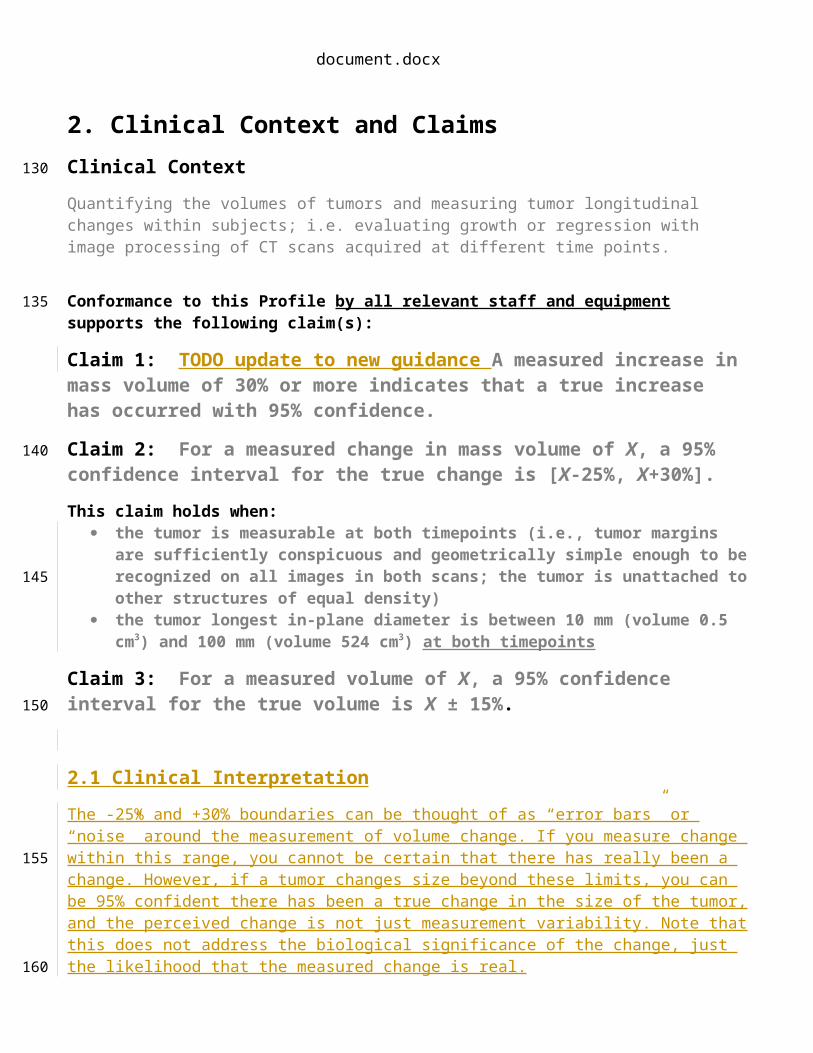
Claim 1: TODO update to new guidance A measured increase in mass volume of 30% or more indicates that a true increase has occurred with 95% confidence.
Claim 2: For a measured change in mass volume of X, a 95% confidence interval for the true change is [X-25%, X+30%].
This claim holds when: the tumor is measurable at both timepoints (i.e., tumor margins are sufficiently conspicuous
and geometrically simple enough to be recognized on all images in both scans; the tumor is unattached to other structures of equal density)
the tumor longest in-plane diameter is between 10 mm (volume 0.5 cm3) and 100 mm (volume 524 cm3) at both timepoints
Claim 3: For a measured volume of X, a 95% confidence interval for the true volume is X ± 15%.
2.1 Clinical Interpretation
The -25% and +30% boundaries can be thought of as “error bars” or “noise” around the measurement of volume change. If you measure change within this range, you cannot be certain that there has really been a change. However, if a tumor changes size beyond these limits, you can be 95% confident there has been a true change in the size of the tumor, and the perceived change is not just measurement variability. Note that this does not address the biological significance of the change, just the likelihood that the measured change is real.
Clinical interpretation with respect to the magnitude of true change: The magnitude of the true change is defined by the measured change and the error bars (+-83%). If you measure the volume to be 200mm3 at baseline and 380mm3 at follow-up, then the measured change is a 90% increase in volume (i.e., 100x(380-200)/200). The 95% confidence interval for the true change is a 7% to 173% increase in volume. The asymmetric range in Claim 1 (-25% to +30%) is due to the way change is conventionally expressed (as a percentage of the first measurement rather than, say, a percentage of the smaller measurement) and how measurements are performed.
Clinical interpretation with respect to progression or response:TBA
115
120
125
130
135
140
145
document.docx
2.2 Discussion
These claims are based on estimates of the within-nodule coefficient of variation (wCV) for nodules in this size range. In the claim statement the CI is expressed as Y ± 1.96 × Y × wCV. The claim assumes that the wCV is constant for nodules in the specified size range and that there is negligible bias in the measurements (i.e. bias < 5%). For estimating the critical % change, the % Repeatability Coefficient (%RC) is used: 2.77 × wCV × 100.
The -25% and +30% boundaries can be thought of as “error bars” or “noise” around the measurement of volume change. If you measure change within this range, you cannot be certain that there has really been a change. However, if a tumor changes size beyond these limits, you can be 95% confident there has been a true change in the size of the tumor, and the perceived change is not just measurement variability. Note that this does not address the biological significance of the change, just the likelihood that the measured change is real.
Clinical interpretation with respect to the magnitude of true change: The magnitude of the true change is defined by the measured change and the error bars (+-83%). If you measure the volume to be 200mm3 at baseline and 380mm3 at follow-up, then the measured change is a 90% increase in volume (i.e., 100x(380-200)/200). The 95% confidence interval for the true change is a 7% to 173% increase in volume. The asymmetric range in Claim 1 (-25% to +30%) is due to the way change is conventionally expressed (as a percentage of the first measurement rather than, say, a percentage of the smaller measurement) and how measurements are performed.
Clinical interpretation with respect to progression or response:TBA
The lower bound on the tumor longest in-plane diameter is set to limit the variability introduced when approaching the resolution of the dataset, e.g. partial volume. The upper bound is set to limit the variability introduced by more complex tumor morphology and organ involvement, and also to keep performance assessment procedures manageable.
While Claim 1 has been informed by an extensive review of the literature and expert consensus that has not yet been fully substantiated by studies that strictly conform to the specifications given here. The expectation is that during field test, data on the actual field performance will be collected and any appropriate changes made to the claim or the details of the Profile. At that point, this caveat may be removed or re-stated.
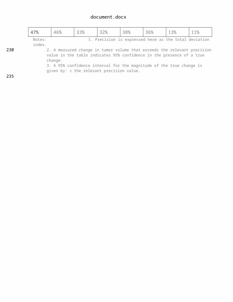
The performance values in Claim 1 reflect the likely impact of variations permitted by this Profile. The Profile permits different compliant actors (acquisition device, radiologist, image analysis tool, etc.) at the two timepoints (i.e. it is not required that the same scanner or image analysis tool be used for both exams of a patient). If one or more of the actors are the same, the implementation is still compliant with this Profile and it is expected that the measurement performance will be improved. To give a sense of the possible improvement, the following table presents expected precision for alternate scenarios, however except for the leftmost, these precision values are not Claims of this Profile.
Table 1: Expected Precision for Alternate Scenarios (Informative)Different Same
150
155
160
165
170
175
180
185
document.docx
Acquisition Device Acquisition DeviceDifferent
RadiologistSame
RadiologistDifferent
RadiologistSame
RadiologistDifferent Analysis
Tool
Same Analysis
Tool
Different Analysis
Tool
Same Analysis
Tool
Different Analysis
Tool
Same Analysis
Tool
Different Analysis
Tool
Same Analysis
Tool47% 46% 33% 32% 38% 36% 13% 11%
Notes: 1. Precision is expressed here as the total deviation index.2. A measured change in tumor volume that exceeds the relevant precision value in the table indicates 95% confidence in the presence of a true change. 3. A 95% confidence interval for the magnitude of the true change is given by: ± the relevant precision value.
190
195

document.docx
3. Profile ActivitiesThe Profile is documented in terms of “Actors” performing “Activities”. Equipment, software, staff or sites may claim conformance to this Profile as one or more of the “Actors” in the following table.
Conformant Actors shall support the listed Activities by conforming to all requirements in the referenced Section.
Table 1: Actors and Required Activities
Actor Activity Section
Acquisition Device Pre-delivery 3.1.
Subject Handling 3.5.
Image Data Acquisition 3.6.
Technologist Subject Handling 3.5.
Image Data Acquisition 3.6.
Image Data Reconstruction 3.7.
Radiologist Subject Handling 3.5.
Image QA 3.8.
Image Analysis 3.10.
Reconstruction Software Image Data Reconstruction 3.7.
Image Analysis Tool Image Analysis 3.10.
The requirements in this Profile do not codify a Standard of Care; they only provide guidance intended to achieve the stated Claim. Failing to conform to a “shall” in this Profile is a protocol deviation. Although deviations invalidate the Profile Claim, such deviations may be reasonable and unavoidable and the radiologist or supervising physician is expected to do so when required by the best interest of the patient or research subject. How study sponsors and others decide to handle deviations for their own purposes is entirely up to them.
The sequencing of the Activities specified in this Profile are shown in Figure 1:
<activity sequence diagram>Figure 1: <Title of the Profile> - Activity Sequence
200
205
210
215

document.docx
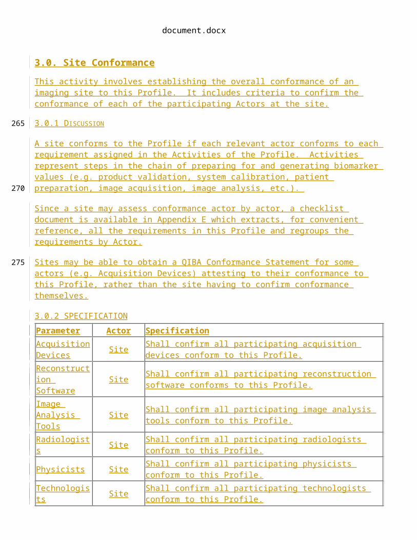
3.0. Site Conformance
This activity involves establishing the overall conformance of an imaging site to this Profile. It includes criteria to confirm the conformance of each of the participating Actors at the site.
3.0.1 D ISCUSSION
A site conforms to the Profile if each relevant actor conforms to each requirement assigned in the Activities of the Profile. Activities represent steps in the chain of preparing for and generating biomarker values (e.g. product validation, system calibration, patient preparation, image acquisition, image analysis, etc.).
Since a site may assess conformance actor by actor, a checklist document is available in Appendix E which extracts, for convenient reference, all the requirements in this Profile and regroups the requirements by Actor.
Sites may be able to obtain a QIBA Conformance Statement for some actors (e.g. Acquisition Devices) attesting to their conformance to this Profile, rather than the site having to confirm conformance themselves.
3.0.2 SPECIFICATION
Parameter Actor SpecificationAcquisition Devices Site Shall confirm all participating acquisition devices conform to this
Profile.Reconstruction Software Site Shall confirm all participating reconstruction software conforms to this
Profile.Image Analysis Tools Site Shall confirm all participating image analysis tools conform to this
Profile.Radiologists Site Shall confirm all participating radiologists conform to this Profile.Physicists Site Shall confirm all participating physicists conform to this Profile.Technologists Site Shall confirm all participating technologists conform to this Profile.
3.1. Staff Qualification
This activity involves evaluating the human Actors (Radiologist, Physicist, and Technologist) prior to their participation in the Profile. It includes training, qualification or performance assessments that are necessary to reliably meet the Profile Claim.
3. 1 .1 DISCUSSIONThese requirements, as with any QIBA Profile requirements, are focused on achieving the Profile Claim. Evaluating the medical or professional qualifications of participating actors is beyond the scope of this profile.
220
225
230
235
document.docx
3. 1 .2 SPECIFICATION
Parameter Actor Specification
Tumor VolumeChange Repeatability
Radiologist
Shall, if operator interaction is required by the Image Analysis Tool to perform measurements, be validated to achieve tumor volume change repeatability with:
an overall repeatability coefficient of less than or equal to 16%. a small subgroup repeatability coefficient of less than 21% a large subgroup repeatability coefficient of less than 21%
See section 4.4. Assessment Procedure: Tumor Volume Change Repeatability.
3.2. Product Validation
This activity involves evaluating the product Actors (Acquisition Device, Reconstruction Software, and Image Analysis Tool) prior to their use in the Profile (e.g. at the factory). It includes validations and performance assessments that are necessary to reliably meet the Profile Claim.
3. 2 .1 DISCUSSION
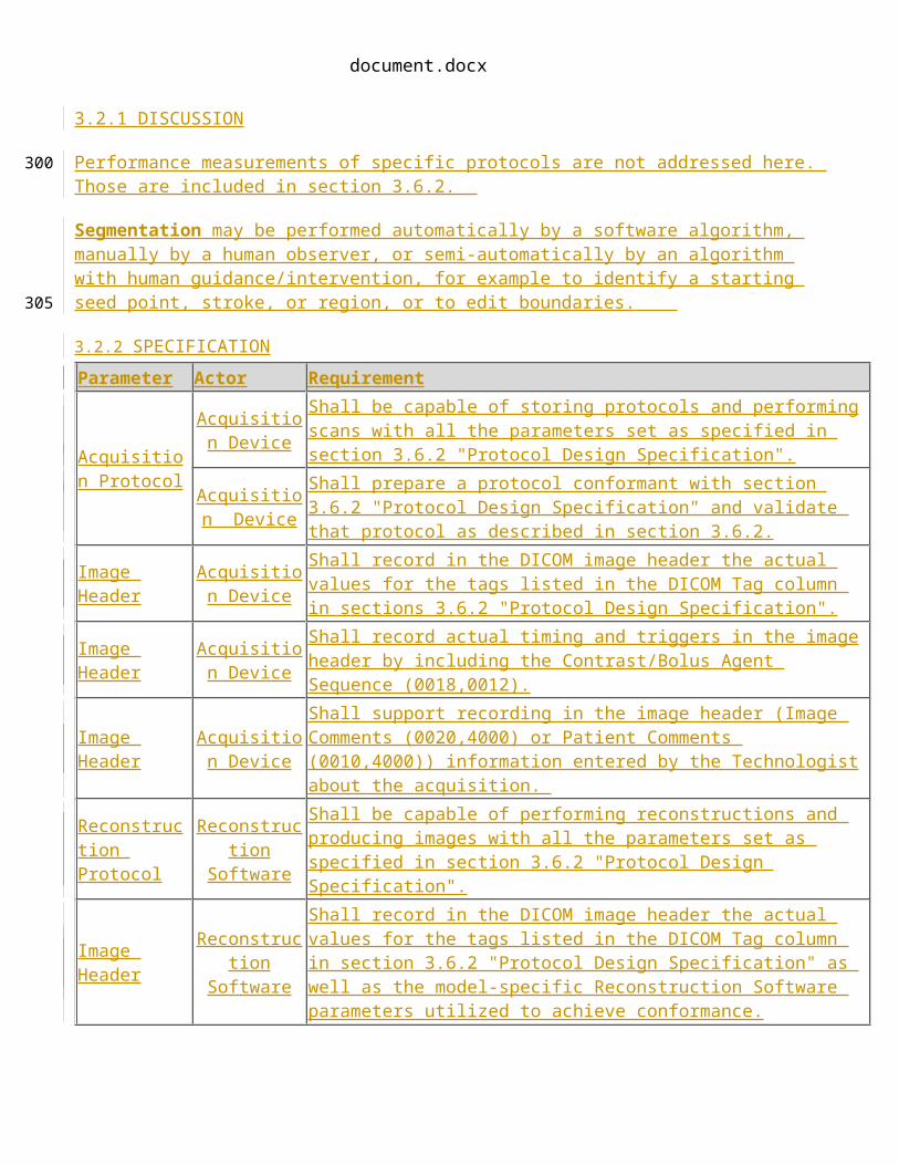
Performance measurements of specific protocols are not addressed here. Those are included in section 3.6.2.
Segmentation may be performed automatically by a software algorithm, manually by a human observer, or semi-automatically by an algorithm with human guidance/intervention, for example to identify a starting seed point, stroke, or region, or to edit boundaries.
3. 2 .2 SPECIFICATION
Parameter Actor Requirement
Acquisition Protocol
Acquisition Device
Shall be capable of storing protocols and performing scans with all the parameters set as specified in section 3.6.2 "Protocol Design Specification".
Acquisition Device
Shall prepare a protocol conformant with section 3.6.2 "Protocol Design Specification" and validate that protocol as described in section 3.6.2.
Image Header Acquisition Device
Shall record in the DICOM image header the actual values for the tags listed in the DICOM Tag column in sections 3.6.2 "Protocol Design Specification".
Image Header Acquisition Device
Shall record actual timing and triggers in the image header by including the Contrast/Bolus Agent Sequence (0018,0012).
Image Header Acquisition Device
Shall support recording in the image header (Image Comments (0020,4000) or Patient Comments (0010,4000)) information entered by the Technologist about the acquisition.
Reconstruction Protocol
Reconstruction Software
Shall be capable of performing reconstructions and producing images with all the parameters set as specified in section 3.6.2 "Protocol Design Specification".
240
245
250

document.docx
Parameter Actor Requirement
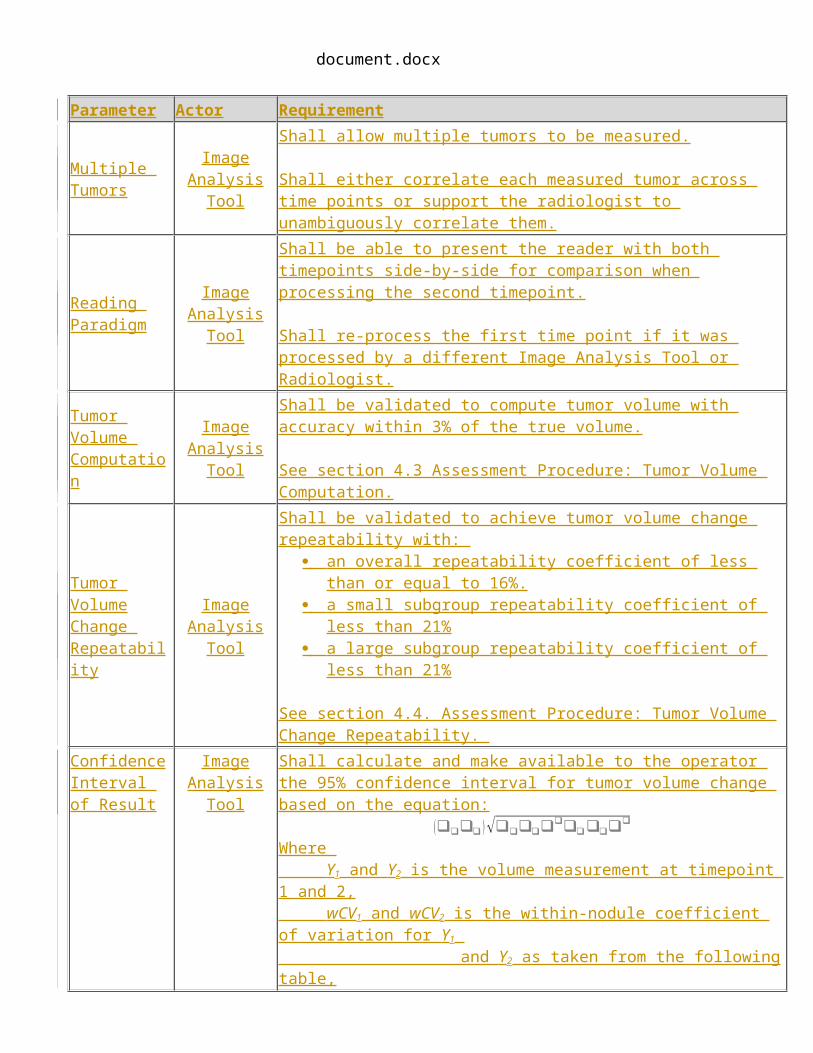
Image Header Reconstruction Software
Shall record in the DICOM image header the actual values for the tags listed in the DICOM Tag column in section 3.6.2 "Protocol Design Specification" as well as the model-specific Reconstruction Software parameters utilized to achieve conformance.
Multiple Tumors
Image Analysis Tool
Shall allow multiple tumors to be measured.
Shall either correlate each measured tumor across time points or support the radiologist to unambiguously correlate them.
Reading Paradigm
Image Analysis Tool
Shall be able to present the reader with both timepoints side-by-side for comparison when processing the second timepoint.
Shall re-process the first time point if it was processed by a different Image Analysis Tool or Radiologist.
Tumor Volume Computation
Image Analysis Tool
Shall be validated to compute tumor volume with accuracy within 3% of the true volume.
See section 4.3 Assessment Procedure: Tumor Volume Computation.
Tumor VolumeChange Repeatability
Image Analysis Tool
Shall be validated to achieve tumor volume change repeatability with: an overall repeatability coefficient of less than or equal to 16%. a small subgroup repeatability coefficient of less than 21% a large subgroup repeatability coefficient of less than 21%
See section 4.4. Assessment Procedure: Tumor Volume Change Repeatability.
Confidence Interval of Result
Image Analysis Tool
Shall calculate and make available to the operator the 95% confidence interval for tumor volume change based on the equation:
(❑❑❑❑ )√❑❑❑❑❑❑❑❑❑❑❑
❑
Where Y1 and Y2 is the volume measurement at timepoint 1 and 2, wCV1 and wCV2 is the within-nodule coefficient of variation for Y1 and Y2 as taken from the following table, D1 and D2 is the longest in-plane diameter of the volume at timepoint 1 and 2:
D1, D2 10-34mm 35-49mm 50-100mmwCV1,wCV2
0.141 0.103 0.085
Result Recording
Image Analysis Tool
Shall record percentage volume change relative to baseline for each tumor.
Shall record the confidence interval of result for each change measurement.
Shall record the image analysis tool version.

document.docx
3.13. Pre-delivery
This activity describes calibrations, phantom imaging, performance assessments or validations prior to delivery of equipment to a site (e.g. performed at the factory) that are necessary to reliably meet the Profile Claim.
3. 1 3 .1 DISCUSSION
3. 1 3 .2 SPECIFICATION
Parameter Actor Requirement
3.24. Installation
This activity describes calibrations, phantom imaging, performance assessments or validations following installation of equipment at the site that are necessary to reliably meet the Profile Claim.
3. 2 4 .1 DISCUSSION
3. 2 4 .2 SPECIFICATION
Parameter Actor Requirement
3.35. Periodic QA
This activity describes calibrations, phantom imaging, performance assessments or validations performed periodically at the site, but not directly associated with a specific subject, that are necessary to reliably meet the Profile Claim.
3. 3 5 .1 DISCUSSION
3. 3 5 .2 SPECIFICATION
Parameter Actor RequirementPET Calibration Physicist Shall assess the current PET Calibration Factor at least quarterly.
255
260
265
270
275
document.docx
Parameter Actor Requirement
Factor
See 4.3 Assessment Procedure: PET Calibration Factor.Shall record the date/time of the calibration for auditing.
Acquisition Device
Shall be capable of performing the PET Calibration Factor assessment.Shall record the most recent PET Calibration Factor for use in subsequent activities.
Qualification Physicist Shall be a Qualified Medical Physicist (QMP) as defined by AAPM.
Time sync Physicist Shall confirm on a weekly basis that all device clocks are synchronized to within +- 1 minute.
3.6. Protocol Design
This activity involves designing acquisition and reconstruction protocols for use in the Profile. It includes constraints on protocol acquisition and reconstruction parameters that are necessary to reliably meet the Profile Claim.
3. 6 .1 DISCUSSION The Profile considers Protocol Design to take place at the imaging site, however, sites may choose to make use of protocols developed elsewhere.
The approach of the specifications here is to focus as much as possible on the characteristics of the resulting dataset, rather than one particular technique for achieving those characteristics. This is intended to allow as much flexibility as possible for product innovation and reasonable adjustments for patient size (such as increasing acquisition mAs and reconstruction DFOV for larger patients), while reaching the performance targets. Again, the technique parameter sets in the Conformance Statements for Acquisition Devices and Reconstruction Software may be helpful for those looking for more guidance.
The purpose of the minimum scan duration requirement is to permit acquisition of an anatomic region in a single breath-hold, thereby preventing respiratory motion artifacts or anatomic gaps between breath-holds. This requirement is applicable to scanning of the chest and upper abdomen, the regions subject to these artifacts, and is not required for imaging of the head, neck, pelvis, spine, or extremities.
Pitch is chosen so as to allow completion of the scan in a single breath hold.
3. 6 .2 SPECIFICATION
Parameter Actor Requirement DICOM Tag
3.47. Subject Selection
This activity describes criteria and procedures related to the selection of appropriate imaging subjects
280
285
290
295
300

document.docx
that are necessary to reliably meet the Profile Claim.
3. 4 7 .1 DISCUSSION
3. 4 7 .2 SPECIFICATION
Parameter Actor Requirement
3.58. Subject Handling
This activity describes details of handling imaging subjects that are necessary to reliably meet the Profile Claim.
3. 4 8 .1 DISCUSSION
3. 4 8 .2 SPECIFICATION
Parameter Actor Requirement
3.69. Image Data Acquisition
This activity describes details of the data acquisition process that are necessary to reliably meet the Profile Claim. It may also include calibrations, performance assessments or validations during acquisition (such as laying the subject on a calibrator or placing a pocket phantom next to the subject) that are necessary to reliably meet the Profile Claim.
3. 6 9 .1 DISCUSSION
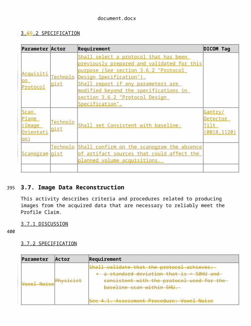
3. 6 9 .2 SPECIFICATION
Parameter Actor Requirement DICOM TagAcquisition Protocol
Technologist Shall select a protocol that has been previously prepared and validated for this purpose (See section 3.6.2 "Protocol Design Specification").
305
310
315
320
document.docx
Parameter Actor Requirement DICOM TagShall report if any parameters are modified beyond the specifications in section 3.6.2 "Protocol Design Specification".
Scan Plane (Image Orientation)
Technologist Shall set Consistent with baseline.Gantry/Detector Tilt (0018,1120)
Scanogram Technologist Shall confirm on the scanogram the absence of artifact sources that could affect the planned volume acquisitions.
3.7. Image Data Reconstruction
This activity describes criteria and procedures related to producing images from the acquired data that are necessary to reliably meet the Profile Claim.
3.7.1 DISCUSSION
3.7.2 SPECIFICATION
Parameter Actor Requirement
Voxel Noise Physicist
Shall validate that the protocol achieves: a standard deviation that is < 50HU and consistent with the
protocol used for the baseline scan within 5HU.
See 4.1. Assessment Procedure: Voxel Noise
Reconstruction Protocol Technologist
Shall select a protocol that has been previously prepared and validated for this purpose (See section 3.6.2 "Protocol Design Specification").Shall report if any parameters are modified beyond those specifications.
3.8. Image QA
This activity describes criteria and evaluations of the images that are necessary to reliably meet the Profile Claim.
3.8.1 DISCUSSION Tumor Size can affect the bias and precision of measurements. Both theoretical considerations and the groundwork projects done by QIBA indicate that for tumors that are small, errors in measurement represent a greater percentage of the measured size. For tumors that are smaller than the limits defined in this profile, please see the profile produced by the QIBA Small Nodule group for more information on imaging recommendations and performance claims. For tumors that are extremely large, the limitations on measurement are based less on imaging physics and more on anatomy. Such tumors are likely to cross anatomical boundaries and abut structures that make consistent segmentation difficult.
325
330
335
340
document.docx
Tumor Margin Conspicuity refers to the clarity with which the boundary of the tumor can be discerned from the surroundings. Conspicuity can directly impact the ability to segment the tumor to properly determine its volume. Conspicuity problems can derive from poor contrast enhancement, from the inherent texture, homogeneity or structure of the tumor, or from attachment of the tumor to other structures.
3.8.2 SPECIFICATION
Parameter Actor RequirementPatient Motion Artifacts Radiologist Shall confirm the images containing the tumor are free from artifact due
to patient motion.Dense Object Artifacts Radiologist Shall confirm the images containing the tumor are free from artifact due
to dense objects, materials or anatomic positioning.
Tumor Size Radiologist
Shall confirm (now or during measurement) that tumor longest in-plane diameter is between 10 mm and 100 mm. (For a spherical tumor this would roughly correspond to a volume between 0.5 cm3 and 524 cm3.)
Tumor Margin Conspicuity Radiologist
Shall confirm the tumor margins are sufficiently conspicuous and unattached to other structures of equal density to distinguish the volume of the tumor.
3.9. Image Distribution
This activity describes criteria and procedures related to distributing images that are necessary to reliably meet the Profile Claim.
3.9.1 DISCUSSION
3.9.2 SPECIFICATION
Parameter Actor Requirement
3.10. Image Analysis
This activity describes criteria and procedures related to producing quantitative measurements from the images that are necessary to reliably meet the Profile Claim.
345
350
355
360

document.docx
3.10.1 DISCUSSION
3.10.2 SPECIFICATION
Parameter Actor Requirement
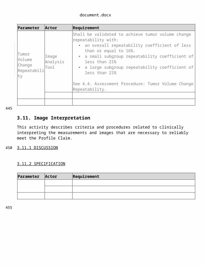
Tumor VolumeChange Repeatability
Image Analysis Tool
Shall be validated to achieve tumor volume change repeatability with: an overall repeatability coefficient of less than or equal to 16%. a small subgroup repeatability coefficient of less than 21% a large subgroup repeatability coefficient of less than 21%
See 4.4. Assessment Procedure: Tumor Volume Change Repeatability.
3.11. Image Interpretation
This activity describes criteria and procedures related to clinically interpreting the measurements and images that are necessary to reliably meet the Profile Claim.
3.11.1 DISCUSSION
3.11.2 SPECIFICATION
Parameter Actor Requirement
365
370
375
380

document.docx
4. Assessment ProceduresTo conform to this Profile, participating staff and equipment (“Actors”) shall support each activity assigned to them in Table 1.
To support an activity, the actor shall conform to the requirements (indicated by “shall language”) listed in the specifications table of the activity subsection in Section 3.
Although mMost of the requirements described in Section 3 can be assessed for conformance by direct observation, however some of the performance-oriented requirements are assessed using a procedure. When a specific cannot, in which case the requirement will reference an assessment procedure is required or to provide clarity, those procedures are defined in a subsections here in Section 4 and the subsection is referenced from the corresponding requirement in Section 3.
Formal claims of conformance by the organization responsible for an Actor shall be in the form of a published QIBA Conformance Statement. Vendors publishing a QIBA Conformance Statement shall provide a set of “Model-specific Parameters” (as shown in Appendix D) describing how their product was configured to achieve conformance. Vendors shall also provide access or describe the characteristics of the test set used for conformance testing.
4.1. Assessment Procedure: Voxel Noise
This procedure can be used by a vendor or an imaging site to assess the voxel noise of reconstructed images. Voxel noise is assessed in terms of the standard deviation of pixel values when imaging a material with uniform density.
The assessor shall first warm up the scanner’s x-ray tube and perform calibration scans (often called air-calibration scans) according to scanner manufacturer recommendations. The assessor shall then scan a phantom of uniform density, such as the ACR CT Accreditation Program (CTAP) Phantom’s module 3, which is a 20 cm diameter cylinder of water equivalent material. The phantom shall be placed at the isocenter of the scanner. The acquisition protocol and reconstruction parameters shall conform to this Profile (See Section 3.6.2 and 3.7.2). The same protocol and parameters shall be used when performing the assessments in 4.1 and 4.2.
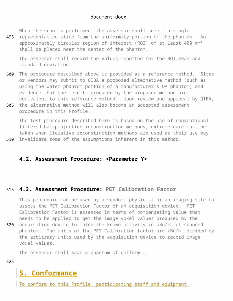
When the scan is performed, the assessor shall select a single representative slice from the uniformity portion of the phantom. An approximately circular region of interest (ROI) of at least 400 mm2 shall be placed near the center of the phantom.
The assessor shall record the values reported for the ROI mean and standard deviation.
The procedure described above is provided as a reference method. Sites or vendors may submit to QIBA a proposed alternative method (such as using the water phantom portion of a manufacturer’s QA phantom) and evidence that the results produced by the proposed method are equivalent to this reference method. Upon review and approval by QIBA, the alternative method will also become an accepted assessment procedure in this Profile.
The test procedure described here is based on the use of conventional filtered backprojection reconstruction methods; extreme care must be taken when iterative reconstruction methods are used as their use may invalidate some of the assumptions inherent in this method.
385
390
395
400
405
410
415

document.docx
4.2. Assessment Procedure: <Parameter Y>
4.3. Assessment Procedure: PET Calibration Factor
This procedure can be used by a vendor, physicist or an imaging site to assess the PET Calibration Factor of an acquisition device. PET Calibration Factor is assessed in terms of compensating value that needs to be applied to get the image voxel values produced by the acquisition device to match the known activity in kBq/mL of scanned phantom. The units of the PET Calibration factor are kBq/mL divided by the arbitrary units used by the acquisition device to record image voxel values.
The assessor shall scan a phantom of uniform …
5. ConformanceTo conform to this Profile, participating staff and equipment (“Actors”) shall support each activity assigned to them in Table 1 in Section 3.
To support an activity, the actor shall conform to the requirements (indicated by “shall language”) listed in the Specifications table of the activity. Each activity has a dedicated subsection in Section 3. For convenience, the Specification table requirements have been duplicated and regrouped by actor in the form of a checklist in Appendix E.
Some requirements reference a specific assessment procedure in section 4 which shall be used to assess conformance to that requirement.
If a QIBA Conformance Statement is already available for an actor (e.g. your analysis software), you may choose to provide a copy of that statement rather than confirming each of the requirements in that Actors checklist yourself.
Formal claims of conformance by the organization responsible for an Actor shall be in the form of a published QIBA Conformance Statement.
Vendors publishing a QIBA Conformance Statement shall provide a set of “Model-specific Parameters” (as shown in Appendix D) describing how their product was configured to achieve conformance. Vendors shall also provide access or describe the characteristics of the test set used for conformance testing.
420
425
430
435
440
445

document.docx
References450
document.docx
AppendicesAppendix A: Acknowledgements and Attributions
Appendix B: Background Information
Appendix C: Conventions and Definitions
455
460

document.docx
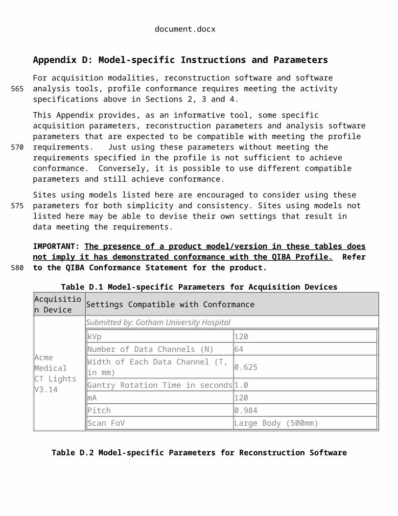
Appendix D: Model-specific Instructions and Parameters
For acquisition modalities, reconstruction software and software analysis tools, profile conformance requires meeting the activity specifications above in Sections 2, 3 and 4.
This Appendix provides, as an informative tool, some specific acquisition parameters, reconstruction parameters and analysis software parameters that are expected to be compatible with meeting the profile requirements. Just using these parameters without meeting the requirements specified in the profile is not sufficient to achieve conformance. Conversely, it is possible to use different compatible parameters and still achieve conformance.
Sites using models listed here are encouraged to consider using these parameters for both simplicity and consistency. Sites using models not listed here may be able to devise their own settings that result in data meeting the requirements.
IMPORTANT: The presence of a product model/version in these tables does not imply it has demonstrated conformance with the QIBA Profile. Refer to the QIBA Conformance Statement for the product.
Table D.1 Model-specific Parameters for Acquisition DevicesAcquisition Device Settings Compatible with Conformance
Acme MedicalCT LightsV3.14
Submitted by: Gotham University Hospital
kVp 120Number of Data Channels (N) 64Width of Each Data Channel (T, in mm) 0.625Gantry Rotation Time in seconds 1.0mA 120Pitch 0.984Scan FoV Large Body (500mm)
Table D.2 Model-specific Parameters for Reconstruction SoftwareReconstruction Software Settings Compatible with Conformance
Acme MedicalCT WSV3.14
Reconstructed Slice Width, mm 1.25Reconstruction Interval 1.0mmDisplay FOV, mm 350Recon kernel STD
465
470
475
480

document.docx
Appendix E: Conformance Checklists TODO Check styles below.
QIBA Checklist:CT Tumor Volume Change for Advanced Disease (CTV-AD)
INSTRUCTIONS
This Checklist is organized by "Actor" for convenience. If a QIBA Conformance Statement is already available for an actor (e.g. your analysis software), you may choose to provide a copy of that statement rather than confirming each of the requirements in that Actors checklist yourself.
Within an Actor Checklist the requirements are grouped by the corresponding Activity in the QIBA Profile document. If you are unsure about the meaning or intent of a requirement, additional details may be available in the Discussion section of the corresponding Activity in the Profile.
Conforms (Y/N) indicates whether you have performed the requirement and confirmed conformance.
Site Opinion allows you to indicate how the requirement relates to your current, preferred practice. If a requirement is not feasible or not worth it to achieve the Profile Claim, please explain to help us understand why.
Since several of the requirements mandate the use of specific assessment procedures, those are also included at the end to minimize the need of referring to the Profile document.
Feedback on all aspects of the Profile and associated processes is welcomed.
Site checklist Page 2Acquisition Device checklist Page 3Image Analysis Tool checklist Page 4Radiologist checklist Page 6Physicist checklist Page 9Technologist checklist Page 10
485
490
495
500
505
document.docx
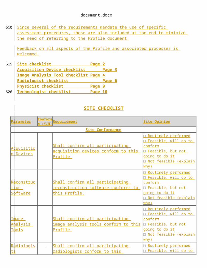
S ITE CHECKLIST
Parameter Conforms (Y/N) Requirement Site Opinion
Site Conformance
Acquisition Devices
Shall confirm all participating acquisition devices conform to this Profile.
□ Routinely performed□ Feasible, will do to conform□ Feasible, but not going to do it□ Not feasible (explain why)
Reconstruction Software
Shall confirm all participating reconstruction software conforms to this Profile.
□ Routinely performed□ Feasible, will do to conform□ Feasible, but not going to do it□ Not feasible (explain why)
Image Analysis Tools
Shall confirm all participating image analysis tools conform to this Profile.
□ Routinely performed□ Feasible, will do to conform□ Feasible, but not going to do it□ Not feasible (explain why)
Radiologists Shall confirm all participating radiologists conform to this Profile.
□ Routinely performed□ Feasible, will do to conform□ Feasible, but not going to do it□ Not feasible (explain why)
Physicists Shall confirm all participating physicists conform to this Profile.
□ Routinely performed□ Feasible, will do to conform□ Feasible, but not going to do it□ Not feasible (explain why)
Technologists Shall confirm all participating technologists conform to this Profile.
□ Routinely performed□ Feasible, will do to conform□ Feasible, but not going to do it□ Not feasible (explain why)
510

document.docx
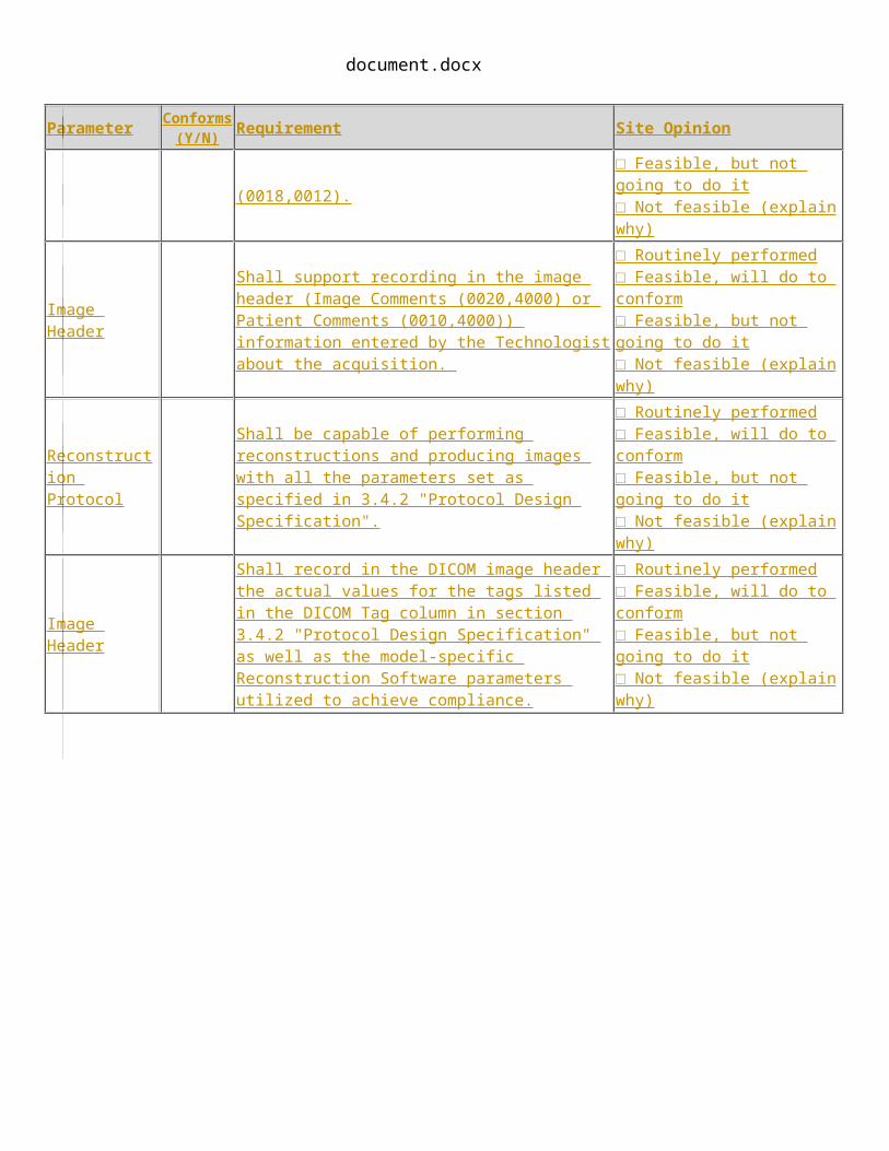
ACQUISITION DEVICE AND R ECONSTRUCTION S OFTWARE C HECKLIST
Parameter Conforms (Y/N) Requirement Site Opinion
Product Validation (section 3.2)
Acquisition Protocol
Shall be capable of storing protocols and performing scans with all the parameters set as specified in section 3.4.2 "Protocol Design Specification".
□ Routinely performed□ Feasible, will do to conform□ Feasible, but not going to do it□ Not feasible (explain why)
Shall prepare a protocol conformant with section 3.4.2 "Protocol Design Specification" and validate that protocol as described in section 3.4.2.
□ Routinely performed□ Feasible, will do to conform□ Feasible, but not going to do it□ Not feasible (explain why)
Shall validate that the protocol achieves an f50 value that is between 0.3 mm-1 and 0.75 mm-1 .
See section 4.1. Assessment Procedure: In-plane Spatial Resolution
□ Routinely performed□ Feasible, will do to conform□ Feasible, but not going to do it□ Not feasible (explain why)
Shall validate that the protocol achieves: a standard deviation that is < 60HU.
See 4.2. Assessment Procedure: Voxel Noise
□ Routinely performed□ Feasible, will do to conform□ Feasible, but not going to do it□ Not feasible (explain why)
Image HeaderShall record in the DICOM image header the actual values for the tags listed in the DICOM Tag column in sections 3.4.2 "Protocol Design Specification".
□ Routinely performed□ Feasible, will do to conform□ Feasible, but not going to do it□ Not feasible (explain why)
Image HeaderShall record actual timing and triggers in the image header by including the Contrast/Bolus Agent Sequence (0018,0012).
□ Routinely performed□ Feasible, will do to conform□ Feasible, but not going to do it□ Not feasible (explain why)
Image Header
Shall support recording in the image header (Image Comments (0020,4000) or Patient Comments (0010,4000)) information entered by the Technologist about the acquisition.
□ Routinely performed□ Feasible, will do to conform□ Feasible, but not going to do it□ Not feasible (explain why)
Reconstruction Protocol
Shall be capable of performing reconstructions and producing images with all the parameters set as specified in 3.4.2 "Protocol Design Specification".
□ Routinely performed□ Feasible, will do to conform□ Feasible, but not going to do it□ Not feasible (explain why)
Image Header
Shall record in the DICOM image header the actual values for the tags listed in the DICOM Tag column in section 3.4.2 "Protocol Design Specification" as well as the model-specific Reconstruction Software parameters utilized to achieve compliance.
□ Routinely performed□ Feasible, will do to conform□ Feasible, but not going to do it□ Not feasible (explain why)
515
document.docx
IMAGE ANALYSIS TOOL C HECKLIST
Parameter Conforms (Y/N) Requirement Site Opinion
Product Validation (section 3.2)
Multiple Tumors Shall allow multiple tumors to be measured.
□ Routinely performed□ Feasible, will do to conform□ Feasible, but not going to do it□ Not feasible (explain why)
Multiple Tumors
Shall either correlate each measured tumor across time points or support the radiologist to unambiguously correlate them.
□ Routinely performed□ Feasible, will do to conform□ Feasible, but not going to do it□ Not feasible (explain why)
Reading Paradigm
Shall be able to present the reader with both timepoints side-by-side for comparison when processing the second timepoint.
□ Routinely performed□ Feasible, will do to conform□ Feasible, but not going to do it□ Not feasible (explain why)
Reading Paradigm Shall re-process the first time point if it was processed by a
different Image Analysis Tool or Radiologist.
□ Routinely performed□ Feasible, will do to conform□ Feasible, but not going to do it□ Not feasible (explain why)
Tumor Volume Computation
Shall be validated to compute tumor volume with accuracy within 3 % of the true volume.
□ Routinely performed□ Feasible, will do to conform□ Feasible, but not going to do it□ Not feasible (explain why)
Tumor Volume Computation
See section 4.3 Assessment Procedure: Tumor Volume Computation.
□ Routinely performed□ Feasible, will do to conform□ Feasible, but not going to do it□ Not feasible (explain why)
Tumor VolumeChange Repeatability
Shall be validated to achieve tumor volume change repeatability with:
an overall repeatability coefficient of less than or equal to 16%.
a small subgroup repeatability coefficient of less than 21%
a large subgroup repeatability coefficient of less than 21%
See section 4.4. Assessment Procedure: Tumor Volume Change Repeatability.
□ Routinely performed□ Feasible, will do to conform□ Feasible, but not going to do it□ Not feasible (explain why)
Tumor Volume Bias& Linearity
Shall be validated to achieve: an overall tumor volume %bias of less than the
Allowable Overall %Bias a tumor volume %bias for each shape subgroup
(spherical, ovoid, lobulated) of less than the Allowable Shape Subgroup %Bias
slope (β̂1¿ between 0.98 and 1.02
□ Routinely performed□ Feasible, will do to conform□ Feasible, but not going to do it□ Not feasible (explain why)
document.docx
Parameter Conforms (Y/N) Requirement Site Opinion
The Allowable Overall %Bias and the Allowable Shape Subgroup %Bias are taken from Table 3.1.2-2 based on the overall repeatability coefficient achieved by the Image Analysis Tool using the assessment procedure in section 4.4.
See section 4.5 Assessment Procedure: Tumor Volume Bias and Linearity.
Confidence Interval of Result
Shall calculate and make available to the operator the 95% confidence interval for tumor volume change based on the equation:
(❑❑❑❑ )√❑❑❑❑❑❑❑❑❑❑❑
❑
Where Y1 and Y2 is the volume measured at timepoint 1 and 2, wCV1 and wCV2 is the within-nodule coefficient of variation for Y1 and Y2 as taken from the following table, D1 and D2 is the longest in-plane diameter of the volume at timepoint 1 and 2:
D1, D2 10-34mm 35-49mm 50-100mmwCV1,wCV2
0.141 0.103 0.085
Result Recording
Shall record percentage volume change relative to baseline for each tumor.
□ Routinely performed□ Feasible, will do to conform□ Feasible, but not going to do it□ Not feasible (explain why)
Result Recording
Shall record the confidence interval of result for each change measurement.
□ Routinely performed□ Feasible, will do to conform□ Feasible, but not going to do it□ Not feasible (explain why)
Result Recording Shall record the image analysis tool version.
□ Routinely performed□ Feasible, will do to conform□ Feasible, but not going to do it□ Not feasible (explain why)
520

document.docx
Table 3.1.2-2: Allowable Tumor Volume %Bias based on Repeatability Coefficient
OverallRepeatability Coefficient R̂C
p
AllowableOverall %Bias
(RMSE Target: 7.1%)
AllowableShape Subgroup %Bias
(RMSE Target: 7.8%)5% <6.7% <7.4%6% <6.5% <7.3%7% <6.3% <7.1%8% <6.1% <6.8%9% <5.8% <6.6%
10% <5.5% <6.3%11% <5.1% <5.9%12% <4.6% <5.6%13% <4.1% <5.1%14% <3.4% <4.6%15% <2.6% <4.0%16% <1.1% <3.2%17% n/a (failed repeatability) n/a (failed repeatability)
525
document.docx
RADIOLOGIST C HECKLIST
Note: The Radiologist is responsible for the protocol parameters, although they may choose to use a protocol provided by the vendor of the acquisition device. The Radiologist is also responsible for ensuring that the protocol has been validated, although the Physicist actor is responsible for performing the validation.
Parameter Conforms (Y/N) Specification Site Opinion
Staff Qualification (section 3.1)
Tumor VolumeChange Repeatability
Shall, if operator interaction is required by the Image Analysis Tool to perform measurements, be validated to achieve tumor volume change repeatability with:
an overall repeatability coefficient of less than or equal to 16%.
a small subgroup repeatability coefficient of less than 21%
a large subgroup repeatability coefficient of less than 21%
See 4.4. Assessment Procedure: Tumor Volume Change Repeatability.
□ Routinely performed□ Feasible, will do to conform□ Feasible, but not going to do it□ Not feasible (explain why)
Protocol Design (section 3.6.2)
Acquisition Protocol
Shall prepare a protocol to meet the specifications in section 3.4-protocol design.
□ Routinely performed□ Feasible, will do to conform□ Feasible, but not going to do it□ Not feasible (explain why)
Acquisition Protocol
Shall ensure technologists have been trained on the requirements of this profile.
□ Routinely performed□ Feasible, will do to conform□ Feasible, but not going to do it□ Not feasible (explain why)
Total Collimation Width
Shall set to Greater than or equal to 16mm.
Total Collimation Width(0018,9307)
□ Routinely performed□ Feasible, will do to conform□ Feasible, but not going to do it□ Not feasible (explain why)
IEC Pitch Shall set to Less than 1.5.
Spiral Pitch Factor(0018,9311)
□ Routinely performed□ Feasible, will do to conform□ Feasible, but not going to do it□ Not feasible (explain why)
Nominal Tomographic Section Thickness (T)
Shall set to Less than or equal to 1.5mm.
Single Collimation Width(0018,9306)
□ Routinely performed□ Feasible, will do to conform□ Feasible, but not going to do it□ Not feasible (explain why)
Scan Duration for Thorax
Shall achieve a table speed of at least 4cm per second, if table motion is necessary to cover the required anatomy.
Table Speed(0018,9309)
□ Routinely performed□ Feasible, will do to conform□ Feasible, but not going to do it□ Not feasible (explain why)
Reconstruction Shall prepare a protocol to meet the □ Routinely performed
530
document.docx
Parameter Conforms (Y/N) Specification Site Opinion
Protocol specifications in this table. □ Feasible, will do to conform□ Feasible, but not going to do it□ Not feasible (explain why)
Reconstruction Protocol
Shall ensure technologists have been trained on the requirements of this profile.
□ Routinely performed□ Feasible, will do to conform□ Feasible, but not going to do it□ Not feasible (explain why)
Reconstructed Image Thickness
Shall set to between 1.0mm and 2.5mm (inclusive).
Slice Thickness (0018,0050)
□ Routinely performed□ Feasible, will do to conform□ Feasible, but not going to do it□ Not feasible (explain why)
Reconstructed Image Interval
Shall set to less than or equal to the Reconstructed Image Thickness (i.e. no gap, may have overlap).
Spacing Between Slices (0018,0088)
□ Routinely performed□ Feasible, will do to conform□ Feasible, but not going to do it□ Not feasible (explain why)
Subject Handling (section 3.8)
Contrast Protocol
Shall prescribe a contrast protocol that achieves enhancement consistent with baseline.
□ Routinely performed□ Feasible, will do to conform□ Feasible, but not going to do it□ Not feasible (explain why)
Use of intravenous contrast
Shall determine whether the selected contrast protocol, if any, will achieve sufficient tumor conspicuity.
□ Routinely performed□ Feasible, will do to conform□ Feasible, but not going to do it□ Not feasible (explain why)
Use of oral contrast
Shall determine whether the selected contrast protocol, if any, will achieve sufficient tumor conspicuity.
□ Routinely performed□ Feasible, will do to conform□ Feasible, but not going to do it□ Not feasible (explain why)
Image QA (section 3.8)
Patient Motion Artifacts
Shall confirm the images containing the tumor are free from artifact due to patient motion.
□ Routinely performed□ Feasible, will do to conform□ Feasible, but not going to do it□ Not feasible (explain why)
Dense Object Artifacts
Shall confirm the images containing the tumor are free from artifact due to dense objects, materials or anatomic positioning.
□ Routinely performed□ Feasible, will do to conform□ Feasible, but not going to do it□ Not feasible (explain why)
Clinical Conditions
Shall confirm that there are no clinical conditions affecting the measurability of the tumor.
□ Routinely performed□ Feasible, will do to conform□ Feasible, but not going to do it□ Not feasible (explain why)
Tumor Size
Shall confirm (now or during measurement) that tumor longest in-plane diameter is between 10 mm and 100 mm. (For a spherical tumor this would roughly correspond to a volume between 0.5 cm3 and 524 cm3 .)
□ Routinely performed□ Feasible, will do to conform□ Feasible, but not going to do it□ Not feasible (explain why)

document.docx
Parameter Conforms (Y/N) Specification Site Opinion
Tumor Margin Conspicuity
Shall confirm the tumor margins are sufficiently conspicuous and unattached to other structures of equal density to distinguish the volume of the tumor.
□ Routinely performed□ Feasible, will do to conform□ Feasible, but not going to do it□ Not feasible (explain why)
Contrast Enhancement
Shall confirm that the phase of enhancement and degree of enhancement of appropriate reference structures (vascular or tissue) are consistent with baseline.
□ Routinely performed□ Feasible, will do to conform□ Feasible, but not going to do it□ Not feasible (explain why)
Tumor Measurability
Shall disqualify any tumor they feel might reasonably degrade the consistency and accuracy of the measurement.
Conversely, if artifacts or attachments are present but the radiologist is confident and prepared to edit the contour to eliminate the impact, then the tumor need not be judged non-conformant to the Profile.
□ Routinely performed□ Feasible, will do to conform□ Feasible, but not going to do it□ Not feasible (explain why)
Consistency with Baseline
Shall confirm that the tumor is similar in both timepoints in terms of all the above parameters.
□ Routinely performed□ Feasible, will do to conform□ Feasible, but not going to do it□ Not feasible (explain why)
Image Analysis (section 3.9)
Reading Paradigm
Shall re-process the first time point if it was processed by a different Image Analysis Tool or Radiologist.
□ Routinely performed□ Feasible, will do to conform□ Feasible, but not going to do it□ Not feasible (explain why)
ResultVerification
Shall review & approve margin contours produced by the tool.
□ Routinely performed□ Feasible, will do to conform□ Feasible, but not going to do it□ Not feasible (explain why)

document.docx
PHYSICIST CHECKLIST
Note: The role of the Physicist actor may be played by an in-house medical physicist, a physics consultant or other staff (such as vendor service or specialists) qualified to perform the validations described.
Parameter Conforms (Y/N) Requirement Site Opinion
Periodic QA (section 3.5)
QCShall perform relevant quality control procedures as recommended by the manufacturer.Shall record the date/time of QC procedures for auditing.
□ Routinely performed□ Feasible, will do to conform□ Feasible, but not going to do it□ Not feasible (explain why)
Protocol Design (section 3.6.2)
In-plane Spatial Resolution
Shall validate that the protocol achieves an f50 value that is between 0.3 mm-1 and 0.75 mm-1 .
See section 4.1. Assessment Procedure: In-plane Spatial Resolution
□ Routinely performed□ Feasible, will do to conform□ Feasible, but not going to do it□ Not feasible (explain why)
Voxel Noise
Shall validate that the protocol achieves: a standard deviation that is < 60HU.
See section 4.2. Assessment Procedure: Voxel Noise
□ Routinely performed□ Feasible, will do to conform□ Feasible, but not going to do it□ Not feasible (explain why)
535
document.docx
TECHNOLOGIST CHECKLIST
Parameter Conforms (Y/N) Specification Site Opinion
Subject Handling (section 3.8)
Use of intravenous contrast
Shall use the prescribed intravenous contrast parameters.
□ Routinely performed□ Feasible, will do to conform□ Feasible, but not going to do it□ Not feasible (explain why)
Use of intravenous contrast
Shall document the total volume of contrast administered, the concentration, the injection rate, and whether a saline flush was used.
□ Routinely performed□ Feasible, will do to conform□ Feasible, but not going to do it□ Not feasible (explain why)
Use of oral contrast Shall use the prescribed oral contrast parameters.
□ Routinely performed□ Feasible, will do to conform□ Feasible, but not going to do it□ Not feasible (explain why)
Use of oral contrast Shall document the total volume of contrast administered and
the type of contrast.
□ Routinely performed□ Feasible, will do to conform□ Feasible, but not going to do it□ Not feasible (explain why)
Subject Positioning
Shall position the subject consistent with baseline. If baseline positioning is unknown, position the subject Supine if possible, with devices such as positioning wedges placed as described in section 3.5.1.
□ Routinely performed□ Feasible, will do to conform□ Feasible, but not going to do it□ Not feasible (explain why)
Artifact Sources
Shall remove or position potential sources of artifacts (specifically including breast shields, metal-containing clothing, EKG leads and other metal equipment) such that they will not degrade the reconstructed CT volumes.
□ Routinely performed□ Feasible, will do to conform□ Feasible, but not going to do it□ Not feasible (explain why)
Table Height & Centering
Shall adjust the table height for the mid-axillary plane to pass through the isocenter.
□ Routinely performed□ Feasible, will do to conform□ Feasible, but not going to do it□ Not feasible (explain why)
Table Height & Centering
Shall position the patient such that the “sagittal laser line” lies along the sternum (e.g. from the suprasternal notch to the xiphoid process).
□ Routinely performed□ Feasible, will do to conform□ Feasible, but not going to do it□ Not feasible (explain why)
Breath hold
Shall instruct the subject in proper breath-hold and start image acquisition shortly after full inspiration, taking into account the lag time between full inspiration and diaphragmatic relaxation.
□ Routinely performed□ Feasible, will do to conform□ Feasible, but not going to do it□ Not feasible (explain why)
Breath hold Shall ensure that for each tumor the breath hold state is consistent with baseline.
□ Routinely performed□ Feasible, will do to conform□ Feasible, but not going to do it□ Not feasible (explain why)
Image Header Shall record factors that adversely influence subject positioning or limit their ability to cooperate (e.g., breath
□ Routinely performed□ Feasible, will do to conform

document.docx
Parameter Conforms (Y/N) Specification Site Opinion
hold, remaining motionless, agitation in subjects with decreased levels of consciousness, subjects with chronic pain syndromes, etc.).
□ Feasible, but not going to do it□ Not feasible (explain why)
Contrast-based Acquisition Timing
Shall ensure that the time-interval between the administration of intravenous contrast (or the detection of bolus arrival) and the start of the image acquisition is consistent with baseline (i.e. obtained in the same phase; arterial, venous, or delayed).
□ Routinely performed□ Feasible, will do to conform□ Feasible, but not going to do it□ Not feasible (explain why)
Contrast-based Acquisition Timing
Shall ensure that the time-interval between the administration of oral contrast and the start of the image acquisition is consistent with baseline. (Note that the tolerances for oral timing are larger than for intravenous).
□ Routinely performed□ Feasible, will do to conform□ Feasible, but not going to do it□ Not feasible (explain why)
Image Data Acquisition (section 3.6)
Acquisition Protocol
Shall select a protocol that has been previously prepared and validated for this purpose (See section 3.4.2 "Protocol Design Specification").
□ Routinely performed□ Feasible, will do to conform□ Feasible, but not going to do it□ Not feasible (explain why)
Acquisition Protocol
Shall report if any parameters are modified beyond the specifications in section 3.4.2 "Protocol Design Specification".
□ Routinely performed□ Feasible, will do to conform□ Feasible, but not going to do it□ Not feasible (explain why)
Scan Plane (Image Orientation)
Shall set Consistent with baseline.
Gantry/Detector Tilt (0018,1120)
□ Routinely performed□ Feasible, will do to conform□ Feasible, but not going to do it□ Not feasible (explain why)
Tube Potential (kVp)
Shall set Consistent with baseline (i.e. the same kVp setting if available, otherwise as similar as possible).
KVP (0018,0060)
□ Routinely performed□ Feasible, will do to conform□ Feasible, but not going to do it□ Not feasible (explain why)
ScanogramShall confirm on the scanogram the absence of artifact sources that could affect the planned volume acquisitions.
□ Routinely performed□ Feasible, will do to conform□ Feasible, but not going to do it□ Not feasible (explain why)
Scan Duration for Thorax
Shall achieve a table speed of at least 4cm per second, if table motion is necessary to cover the required anatomy.
Table Speed(0018,9309)
□ Routinely performed□ Feasible, will do to conform□ Feasible, but not going to do it□ Not feasible (explain why)
Anatomic Coverage
Shall ensure the tumors to be measured and additional required anatomic regions are fully covered.
Anatomic Region Sequence(0008,2218)
□ Routinely performed□ Feasible, will do to conform□ Feasible, but not going to do it□ Not feasible (explain why)
Anatomic Coverage
Shall, if multiple breath-holds are required, obtain image sets with sufficient overlap to avoid gaps within the required anatomic
Anatomic Region Sequence
□ Routinely performed□ Feasible, will do to conform□ Feasible, but not going to do it

document.docx
Parameter Conforms (Y/N) Specification Site Opinion
region(s), and shall ensure that each tumor lies wholly within a single breath-hold.
(0008,2218) □ Not feasible (explain why)
Image Header
Shall enter on the console any factors that adversely influenced subject positioning or limited their ability to cooperate (e.g., breath hold, remaining motionless, agitation in subjects with decreased levels of consciousness, subjects with chronic pain syndromes, etc.).
Image Comments (0020,4000) or Patient Comments (0010,4000
□ Routinely performed□ Feasible, will do to conform□ Feasible, but not going to do it□ Not feasible (explain why)
Acquisition Field of View (FOV)
Shall set Consistent with baseline.
Data Collection Diameter (0018,0090)
□ Routinely performed□ Feasible, will do to conform□ Feasible, but not going to do it□ Not feasible (explain why)
Image Data Reconstruction (section 3.7)
Reconstruction Protocol
Shall select a protocol that has been previously prepared and validated for this purpose (See section 3.4.2 "Protocol Design Specification").Shall report if any parameters are modified beyond those specifications.
□ Routinely performed□ Feasible, will do to conform□ Feasible, but not going to do it□ Not feasible (explain why)
In-plane Spatial Resolution
Shall either select the same protocol as used for
the baseline scan, or select a protocol with a recorded f50
value within 0.2 mm-1 of the f50 value recorded for the baseline scan protocol.
See section 3.4.2 for further details.
□ Routinely performed□ Feasible, will do to conform□ Feasible, but not going to do it□ Not feasible (explain why)
Voxel Noise
Shall either select the same protocol as used for
the baseline scan, or select a protocol with a recorded
standard deviation within 5HU of the standard deviation recorded for the baseline scan protocol.
See section 3.4.2 for further details.
□ Routinely performed□ Feasible, will do to conform□ Feasible, but not going to do it□ Not feasible (explain why)
Reconstructed Image Thickness
Shall set to between 1.0mm and 2.5mm (inclusive) and consistent (i.e. within 0.5mm) with baseline.
□ Routinely performed□ Feasible, will do to conform□ Feasible, but not going to do it□ Not feasible (explain why)
Reconstructed Image Interval
Shall set to less than or equal to the Reconstructed Image Thickness (i.e. no gap, may have overlap) and consistent with baseline.
□ Routinely performed□ Feasible, will do to conform□ Feasible, but not going to do it□ Not feasible (explain why)
document.docx
Parameter Conforms (Y/N) Specification Site Opinion
Reconstruction Characteristics
Shall set the reconstruction kernel and parameters consistent with baseline (i.e. the same kernel and parameters if available, otherwise the kernel most closely matching the kernel response of the baseline).
Convolution Kernel Group (0018,9316), Convolution Kernel (0018,1210)
□ Routinely performed□ Feasible, will do to conform□ Feasible, but not going to do it□ Not feasible (explain why)
ReconstructionField of View
Shall ensure the Field of View spans at least the full extent of the thoracic and abdominal cavity, but not substantially greater than that, and is consistent with baseline.
Reconstruction Field of View (0018,9317)
□ Routinely performed□ Feasible, will do to conform□ Feasible, but not going to do it□ Not feasible (explain why)
540