ucfsarc.files.wordpress.com€¦ · web view · 2018-02-02powerpoint 4a – interactions cells...
TRANSCRIPT
Powerpoint 4A – Interactions Cells and Environment
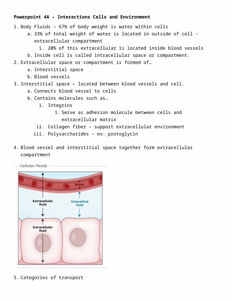
1. Body Fluids – 67% of body weight is water within cellsa. 33% of total weight of water is located in outside of cell - extracellular compartment
i. 20% of this extracellular is located inside blood vesselsb. Inside cell is called intracellular space or compartment.
2. Extracellular space or compartment is formed of…a. Interstitial spaceb. Blood vessels
3. Interstitial space – located between blood vessels and cell. a. Connects blood vessel to cellsb. Contains molecules such as…
i. Integrins1. Serve as adhesion molecule between cells and extracellular matrix
ii. Collagen fiber – support extracellular environmentiii. Polysaccharides – ex: protoglycin
4. Blood vessel and interstitial space together form extracellular compartment
5. Categories of transporta. Carrier Mediated transport
i. Could be passive (facilitated diffusion) or active (primary/secondary active)ii. Characteristics of carrier mediated transport
1. Stereospecificitya. Only certain molecules allowed to pass
b. Ex: D-glucose(natural) is transport but not L-glucose(not as common or natural)
2. Saturationa. Transport rate increases as concentration of the solute increases, until the
carriers are saturated. 3. Competition
a. Structurally related solutes compete for transport sites on carrier molecules.i. Galactose in competitive inhibitor of glucose transport in small
intestineb. Passive – no need of ATP
i. Simple diffusion – means moving a molecule from high concentration to low concentration area (high low). There are some factors which influence this type of diffusion. No equations needed for exam. These are…
1. Not carrier mediated2. Diffusion and osmosis3. Factors affecting rate of diffusion
a. Size of solute – smaller means more diffusionb. Concentration of solute – more concentrated means fasterc. Permeability of membraned. Structure of solute or substance – when substance is lipid(cholesterol) it can
pass through membrane easily - lipophilic, protein cannot pass through membrane.
ii. Facilitated diffusion1. Carrier-mediated so exhibits stereospecificity, saturation, and competition2. From high to low concentrations3. Does not require energy4. More rapid than diffusion5. Good for transport of larger molecules that cannot diffuse naturally across
membrane6. When first molecule helps second molecule transport to target cell. Means second
molecule can’t enter cell without first molecule’s help. 1st facilitates 2nd. 7. Example:
a. Insulin and glucose in blood vessel. (1st) Insulin binds to receptor on cell membrane. (2nd) Insulin activates molecules in cytoplasm called GLUT4. These molecules normally exist inside cytoplasm. (3rd) – GLUT4 inserts into cell membrane then they act as a channel for glucose entrance. (4th) Glucose can enter into target cell through GLUT4 channels. Then the target cell uptakes glucose. (5th) Glucose is broken down for formation for ATP which is required for active transport. In this way, insulin facilitates glucose entrance.

i. Type 2 diabetes: hyperglycemia is sign or symptom for type II diabetes since there is not enough insulin to help glucose get into cell.
Cell is hungryc. Active – need ATP.
i. Primary active transport1. Against concentration gradient. Transport from low to high concentrations (low
high)2. Requires energy3. Is carrier-mediated and thus exhibits stereospecificity, saturation, and competition4. 3 Examples
a. 1st example: Na-K Pump (at Neuromuscular)i. Mechanism: Neuron(top) and muscle fiber(bottom) in neuromuscular
junction. Neuron is called presynaptic and muscle fiber is called postsynaptic. Synaptic cleft is in between neuron and muscle.

Step 1: Axon receives stimulus from outside. Step 2: Calcium channels open and calcium enters into neuron. Step 3: When calcium concentration increases in presynaptic, Ach vesicle forms. Step 4: Secretory vesicle gets pushed down at high calcium concentrations. Step 5: Causes exocytosis which releases Ach into synaptic cleft. Step 6: Ach binds to receptors on postsynaptic membrane. Step 7: This binding activates sodium potassium channels and this pump requires ATP. Because of this Na-K pump, this opens calcium
channels and calcium enters into cell. Calcium causes stimulus SRS – sarcoplasmic reticulum
b. 2nd example: Calcium pump – more about this next note setc. 3rd example – more about this next note set
6. Neuromuscular junctiona. Innervation of muscle fiber or motor neuron
i. Cell body of neuron produces Ach.b. Between pre and post we have synaptic cleft
7. 1st example of primary active transport: Na-K pump (neuromuscular junction)i. (1st) Any action potential or stimulus to axon of presynaptic.

(2nd) leads to opening of calcium channels and calcium flows into presynaptic compartment.(3rd) When calcium concentration increases in presynaptic compartment, (4th) this leads to exocytosis of vesicle which is full of Ach – achetylcholine. (5th) Ach in synaptic cleft binds to its specific receptor on postsynaptic compartment membrane. (6th) Because of binding of Ach to its receptor, it leads to opening of sodium potassium channels which requires ATP for sodium potassium pump. This is first example for primary active transport. (7th) A few calcium ions enter into postsynaptic compartment which is not sufficient for muscle contraction. (8th) This calcium stimulates SR – sarcoplasmic reticulum which is full of calcium. After stimulation of SR, it releases its calcium into the cytoplasm of postsynaptic compartment. (9th) When calcium concentration increases in cytoplasm, which is important ion for muscle contraction, it activates microfilaments (such as actin and myosin) which leads to muscle contraction.
ii. After each contraction, we have a relaxation phase. Relaxion of skeletal muscle which occurs after each contraction is by reaccumulating of calcium by SR from cytoplasm. It means less calcium in cytoplasm leads to relaxation phase in muscle.
8. 2nd example of primary active transport (calcium pump)a. Calcium ATPase Pump from SR into cytoplasm against gradient and is 2nd example for primary
active transport.
9. 3rd example of primary active transport is hydrogen ion potassium pump (or proton pump)i. Process
1. [Parietal cells in stomach releases HCl into stomach.] (1): Parietal cell from stomach secretes hydrogen ion (HCl) into lumen of stomach. (2): It requires ATP to do this. (3): When hydrogen ion gets into lumen, then the pump absorbs potassium into the cell. This is antiport pump because hydrogen comes out and K comes in. This is called hydrogen ion potassium pump or proton pump.
a. Pathology Conditioni. When hydrogen ion is high in lumen of stomach, it increases risk of
gastritis (itis means inflammation).

1. Omeprazole blocks this pump, proton pump, since it is an antacid and can decrease or inhibit hydrogen ion secretion into lumen of stomach and prevents irritation of stomach.
10. Secondary active transport –a. It is active transport which requires ATP and has two substances transported.b. The first ion transport consumes ATP for transport and can help other ions or substances transport
after itself by either symport or antiport. c. Example
i. Inside kidney we have millions of nephrons. Nephrons are surrounded by capillary or blood vessel. Nephron produces urine and urine excretes or releases into urinary bladder or urethra. After filtration of many ions or substances into nephron, then capillary that surrounds the nephron absorbs all these ions. The 67% of sodium is absorbed by capillary from nephron. This sodium requires ATP which is active transport. Sodium (Na+) helps other ions transport out of nephrons into capillary.

1. Symport (same direction): potassium, glucose, amino acids, phosphate, chloride, and HCO3
+ (bicarbonate). It symports many substances. 2. Antiport (opposite directions): Sodium antiports H+ into urine.
ii. Pathological Condition1. Healthy person’s urine cannot have glucose in it. This only happens in pathology
condition such as…a. Endocrine hormone disorder like type 1 or type 2 diabetesb. Nephropathy which is destruction of nephrons in kidney which leads to
excretion of glucose into urine.
11. Osmosisa. We have two chambers separated by semipermeable membranes. Chamber A on left and chamber
B on right. Chamber A contains fluid with concentrated solute. Part B contains fluid with low concentration of solutes. Semipermeable membranes only allow water to pass through since they are only permeable to water.
b. Chamber A with more solutes is said to have higher osmotic pressure. Fluid flows from less concentrated to more concentrated or from part B to part A.
c. Concentrated part is called hyperosmotic and part B with less solute is called hypo-osmotic. After flow of fluid from part B to part A, part A is said to have been diluted (water was added). After dilution, osmolarity is same level on both sides and they are both called iso-osmotic.
i. Example1. Pretend part A is blood vessel and part B is cell or interstitial space. Blood vessels
contain large proteins such as albumin. Albumin cannot pass through semipermeable wall. Having albumin in blood vessel increases blood’s osmolarity so that the blood vessels absorb extra fluid from cell to prevent edema which is water accumulation in cell.
a. Exam question example: 25-year-old female had a genetic disorder and anemia (which is reduction of red blood cells). The blood tests show that patient has decreased red blood cells, decreased white blood cells, decreased blood proteins. What type of risk might she have after having all these signs and symptoms.
i. Option A: respiratory disorder ii. Option B: edema
iii. Option C: kidney problemiv. Option D: vision problem
1. Answer is both edema because of reduction of blood proteins and respiratory problem because of red blood cells which means reduction of oxygen molecules.
12. Cystic fibrosis – example for osmosis. a. Definition - genetic disorder that occurs when there is any mutation to a protein structure in
cytoplasm called CFTR (cystic fibrosis transmembrane regulator).i. Normal function of CFTR
1. Control excretion of chloride ion from cell into lumen of organs.

ii. (THIS IS ACTUALLY NOT HOW CYSTIC FIBROSIS WORKS)In pathology condition (cystic fibrosis), which occurs when there is mutation to this protein (CFTR), then it cannot control excretion of chloride which leads to…
1. Overexcretion of chloride ion into different organs’ lumen from epithelial cells. After excretion of chloride, excretion of sodium chloride also. Lumen side is now really concentrated which increases osmotic pressure. This increases the lumen’s absorption of water. This attracts white blood cells and causes local inflammation because white blood cells are the sign of inflammation. In time, this leads to formation of mucus which closes the lumen.
iii. (REAL MECHANISM FOR CYSTIC FIBROSIS)1. Since CFTR is damaged, Cl^- followed by Na^+ cannot be excreted from epithelial
cells into lumen. Since lumen is not concentrated enough, water does not flow into lumen and flush out mucus. This causes mucus buildup in lumen. Bacteria come and live in mucus causes really bad infections.
b. Signs and symptomsi. Obstruction of lumen by mucus formation can occur in organs such as…
1. Respiratory system – lung. Mucus formation can lead to respiratory infection and inflammation. In severe condition, this can cause newborn death.
2. Pancreas - which leads to pancreatitis which is inflammation in pancreas. Inflammation of pancreas leads to endocrine hormone disorder like type 1 and type 2 diabetes. Also, causes pancreatic disorders which leads to malabsorption of proteins, carbohydrates, fat in small intestines.
3. Intestine - leads to colitis – which is inflammation of large intestine. 4. Small intestine - leads to diarrhea, malabsorption, malnutrition. Vitamin A is
important for red blood cells and deficiency leads to anemia. Iron deficiency also leads to anemia.
5. Spermatid cord - is connected to testicle and contains vas deferens which carries mature sperm into urethra. If formation of mucus occurs in spermatid cord, means patient has erectile dysfunction, obstruction of vas deferens muscle, and infertility.
ii. Treatment1. There is no treatment for cystic fibrosis. You can control mucus by taking cortisol
which is anti-inflammatory. We can control signs and symptoms but cannot treat it directly.
13. Edema – accumulation of fluid inside cell and interstitial space. a. Cause 1
i. When we have accumulation of fluid, due to deficiency of albumin and other proteins in blood vessel. Albumin is major protein in blood plasma and is important for osmolality.
b. Cause 2 i. Destruction of capillary or blood vessels by infection, congenital disease, malformation.
c. Cause 3 i. Could be due to kidney problem, heart problem, hypertension. For example, after filtration
of ions, kidney cannot produce urine. Nephropathy.d. Cause 4
i. Any obstruction to lymphatic system.

1. Lymph nodes accumulate fluids and release into vein system. We have two main veins in neck. Thoracic duct that opens into left vein. We have lymphatic duct on right side. Viral or bacterial infection or congenital problem of lymphatic system leads to failure of accumulation of fluid by lymphatic symptom which causes severe edema.
14. Regulating Blood osmolalitya. Step 1: When a person has severe dehydration, this leads to change in blood osmolarity and blood
becomes concentrated. After sweating, body loses fluid and blood becomes concentrated. 2: Any change to blood osmolarity leads to stimulation of osmoreceptors which is located on the wall of large blood vessels such as aorta which is largest blood vessel in body. 3: By stimulation of osmoreceptors, this leads to stimulation of two cranial nerves. Cranial nerve 10 and 9. These nerves take information about blood osmolarity to the central nervous system, specifically to hypothalamus.4: Hypothalamus contains two nuclei which are supra-aortic and paraventricular. These two nuclei are located in hypothalamus. 5: By stimulation of these nuclei, these release one hormone called ADH – antidiuretic hormone. 6: ADH is carried by blood to the nephron. This hormone binds to V2 receptor in nephron tubule. 7. After binding of ADH to V2 receptor, this activates the intracellular molecules in the capillary which absorbs fluid. This leads to (1) absorption of water (fluid) from urine by capillary which (2) decreases osmotic pressure in blood and (3) prevents urine excretion and prevents further dehydration.
b. Normal osmolality in plasma is about 280-303 milli-osmoles per kilogram