welcome to biology. increasing reliability a drug company wants to test the effectiveness of a new...
TRANSCRIPT

Welcome to Biology

Increasing reliability
A drug company wants to test the effectiveness of a new drug against asthma. How can they increase the reliability of their results?

Increasing reliability
A drug company wants to test the effectiveness of a new drug against asthma. How can they increase the reliability of their results?
Use a large test group of asthmatics (1000 +)
Have a mixed group (ages, gender & fitness levels)
Use the same number & mix in the control group
Give the control group a placebo
Run the trial over a long period
Swap the groups over and repeat the trial
Repeat the trial with another group
Get someone else to replicate the trial
Measure accurately and often changes in incidence of asthma & any side effects, etc

Milk bubbles experimentFor Part A, identify the:• Hypothesis• Independent variable• Dependent variable• Control• Variables you controlled • How you increased reliability
For Part B, you’ll need to identify these before you design & carry out your experiment

Milk bubbles experimentFor Part A, identify the:• Hypothesis – skim milk will produce smaller bubbles than full cream milk
• Independent variable – the type of milk
• Dependent variable – size of bubbles
• Control – full cream milk
• Variables you controlled – temperature, volume of milk, strength of blowing, etc
• How you increased reliability – repeat experiment, replicate experiment, etc
For Part B, you’ll need to identify these before you design & carry out your experiment

Reviewing graphs

Reviewing graphs
Title
Dependent variable 3 Grid
goes on the
Vertical axis (Y) 2 Plotted points or line or curve
Use an even scale 1
and include units
0
1 2 3 4 5
Independent variable goes on the Horizontal axis (X)
Use an even scale and include units

Interpreting graphsBubbles produced by yeast
0
10
20
30
40
50
60
70
80
0 5 10 20 30 40 50 60 70
Temperature (Celcius)
Nu
mb
er o
f b
ub
ble
s/m
in
Bubbles/min

Parts of the microscope

Parts of the microscope
Ocular lens
Objective lensArm
Stage
Iris wheel diaphragm
Light
Coarse focus
Fine focus

Using the microscope
• Changing the magnification
• Changing the focus
• Adjusting the light or contrast

Using the microscope
• Changing the magnification
• Changing the focus
• Adjusting the light or contrast

Calculating magnification
• x10 ocular lens and x4 objective lens =
• x10 ocular lens and x10 objective lens =
• x10 ocular lens and x40 objective lens =

Calculating magnification
• x10 ocular lens and x4 objective lens = x40
• x10 ocular lens and x10 objective lens = x100
• x10 ocular lens and x40 objective lens = x400

Working distance
This is the distance between the objective lens and your slide.
The higher the magnification of the lens, the larger the lens
The higher the magnification of the lens, the smaller the working distance

What you see under the microscope
Everything is reversed as well as magnified

What you see under the microscope 2
• If the object appears to be at the top of the slide it is really
You need to move the slide
• If the object appears to be at the left of the slide it is really
You need to move the slide

What you see under the microscope 2
• If the object appears to be at the top of the slide it is really at the bottom
You need to move the slide towards you
• If the object appears to be at the left of the slide it is really on the right
You need to move the slide to the right

Diameter of field of view
• Distance across centre of field
• Measured with
• Measured in

Diameter of field of view
• Distance across centre of field
• Measured with a minigrid
• Measured in micrometres (m)

Millimetres and micrometres
• 1 mm = m
• 2.4 mm = m
• 340 m = mm
• 4400 m = mm

Millimetres and micrometres
• 1 mm = 1 000 m
• 2.4 mm = 2 400 m
• 340 m = 3 400 mm
• 4400 m = 4 400 mm

Using a minigridEach square is 1 mm by 1 mm in size
The centre grid is further subdivided into 0.1 mm grid squares

Magnification and field of view 1
x40 x100 x400

Magnification and field of view 2
• As magnification increases, field of view
• As magnification decreases, field of view

Magnification and field of view 2
• As magnification increases, field of view decreases by the same factor
• As magnification decreases, field of view increases by the same factor

Magnification and field of view 3
If field of view at x100 is 1600 m, then
• Field of view at x400 =
• Field of view at x40 =
If field of view at x400 = 200 m, then
• Field of view at x100 =
• Field of view at x40 =

Magnification and field of view 3
If field of view at x100 is 1600 m, then
• Field of view at x400 = 400 m
• Field of view at x40 = 4 000 m
If field of view at x400 = 200 m, then
• Field of view at x100 = 800 m
• Field of view at x40 = 2 000 m

Size of objects under the microscope 1
If given a scale – 0 1 2 mm

Size of objects under the microscope 1
If given a scale – 0 1 2 mm
Measure object with rulerMeasure scale with rulerUse scale to convert ruler measurement
of object to real one
Size = 0.6mm = 600 µm

Size of objects under the microscope 2
If given a field of view –
Magnification x100, field of view 2000 m
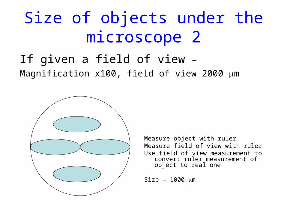
Size of objects under the microscope 2
If given a field of view –
Magnification x100, field of view 2000 m
Measure object with rulerMeasure field of view with rulerUse field of view measurement to convert
ruler measurement of object to real one
Size = 1000 m

Size of objects under the microscope 3
If given a magnification –
Magnification x 200

Size of objects under the microscope 3
If given a magnification –
Magnification x 200
Measure object with rulerDivide size by magnification factor to get
real size
Size = 4cm ÷ 200 = 0.02 cm = 0.2 mm = 200 m