welcome to chiropractic - joy gayler · welcome to chiropractic ... thoracic spine aka dorsal...
TRANSCRIPT
2
Welcome to Chiropractic
Chiropractic: focuses on the body's inborn capacity to maintain and restore its own health. This inborn wisdom, or innate intelligence, helps the body constantly adapt to changes in its internal and external environment to help the body survive and thrive in changing conditions. The nervous system (through the brain and spinal cord) coordinates and controls the function of all of the body's cells, organs and systems. Vertebral Subluxations: are misalignments in the vertebrae of the spinal column, that interfere with the transmission of information from the brain, over the spinal cord to the spinal nerves and, ultimately, to all systems of the body. These misalignments disrupt proper nerve function and lead to decreased performance and health. The Chiropractic Objective (LACVS): Chiropractors Locate, Analyze and gently Correct Vertebral Subluxations by using a broad range of adjusting techniques. By restoring proper nerve flow, the chiropractor helps people enjoy more of their full potential for health and optimal performance. Chiropractic Philosophy: Chiropractic involves the study of philosophy, science and art. Philosophy considers the fundamental nature of existence, of human beings and of their interaction with the environment. The sciences deal with human biology - especially the relationship between the spinal segments and the nerve system, and the effect this relationship has on the body's innate striving to express and maintain its own health. Chiropractic's art is the expression of its philosophy and is concerned with locating and correcting vertebral subluxations. Ultimately, chiropractic is concerned with improving the body's expression of life and health What is Straight Chiropractic: “Straight” chiropractic uses chiropractic methods of examination, analysis and adjusting to accomplish the objective of correcting vertebral subluxation. "Straight" simply means a total commitment to the teaching, research and practice of chiropractic focused on correcting vertebral subluxation.
3
Introduction to Chiropractic Palpation Chiropractic Palpation is the art of analyzing the spine through the sense of touch, in order to assess the position and motion of spinal structures. The various forms of palpation include:
• Static: palpation of the static bony structures of the spine and pelvis • Motion: palpation of the moving structures (joints) of the spine and pelvis • Muscle: palpation of the muscles of the spine and pelvis
In this class we will be focusing on static and motion palpation Palpation is an art and skill that takes practice and repetition to master. The harder you work at it the better you will become. YOU WILL NOT master the art palpation unless you practice EVERY DAY OUTSIDE CLASS. Needed every class period:
• Be in your gown every day ready to palpate • Model spine • Notes from the bookstore • Skin marking pencil
On a personal note: • We will be palpating on skin and at times, palpating structures in the pelvic
and sacral area and as such we must maintain a professional attitude. • Personal hygiene. It seems silly to have to address this issue however poor
personal hygiene can really make this class stink! • Lastly, finger nails that are trimmed make for a more pleasurable palpation
experience for your patient.
4
Anatomical Terminology
Anatomical Positional Terms • Superior / Sup / S - up / away from gravity • Inferior / Inf / I - down / toward gravity • Anterior / Ant / A - forward • Posterior / Post / P - backward • Left Lt (L) / Right Rt (R) • Lateral / Lat - away from or further from midline - either (L) or (R) • Medial / Med - toward or closer to the midline - either (L) or (R) • Median / Mn - on the midline
Morphological Terms (relate one body part to another) • Cranial - close to the head / Caudal - close to the feet • Proximal closer to the body / Distal further from the body • Dorsal toward the back / Ventral toward the belly • Palmar palm of the hand / Plantar sole of the foot
Introduction to Spinal Anatomy
Spine: consists of 24 movable vertebrae Spinal Column (spine + sacrum + coccyx) is divided into 4 regions:
Cervical spine: refers to the neck • Top 7 vertebrae named from top to bottom (C1 - C7) • Smallest vertebrae • C1 AKA atlas / C2 AKA axis or epistropheus
Thoracic Spine aka Dorsal Spine: refers to the thorax • Next 12 vertebrae named from top to bottom (T1 - T12) • All have ribs attached
Lumbar Spine • Next 5 vertebrae • Refers to the small of the back • Largest of the true vertebrae • Named from top to bottom (L1 - L5)
Sacro-coccygeal Spine – consists of the sacrum and coccyx • Sacrum: consists of 5 fused segments (S1 – S5 ) • Coccyx: 3 – 5 fused segments, usually 4
5
The Vertebral Column
Neural Canal / Spinal Canal / Vertebral Canal Tube-like passage within the spinal column through which passes the spinal cord. Boundaries include:
• Superior - foramen magnum of occiput • Inferior - sacral hiatus (inferior sacrum) • Anterior - posterior vertebral bodies / IVDs • Lateral - pedicles / IVFs • Posterior - laminae
Spinal Cord • Begins at the Foramen Magnum in the Occiput and extends to about L1 • Cauda-Equina (horses tail) extends from l through sacrum and coccyx
Spinal Nerves: 31 pairs of spinal nerves • Cervical nerves: named C1 - C8 spinal nerves which exit the neural
canal above vertebra of the same number (note C8 spinal nerve exits below the C7 vertebra)
• Thoracic nerves: named T1 - T12 spinal nerves which exit neural canal below vertebra of the same number
• Lumbar nerves: named L1 - L5 spinal nerves which exit neural canal below vertebra of the same number
• Sacral nerves: named S1 - S5 spinal nerves which exit neural canal below segment of the same number (S5 exits through the sacral hiatus)
• Coccygeal nerves exit through the sacral hiatus aka pudendal nerves Intervertebral Foramina (IVFs): exit pathways for spinal nerves
• 31 pairs of IVFs Intervertebral Discs (IVDs) from C2/C3 through L5/S1
• 23 pad-like ligaments attaching vertebral body to vertebral body which separate vertebrae, allow for spinal flexibility, limit spinal ROM, shock absorbers, shape spinal curves, helps form the IVFs and neural canal
• Consists of 2 distinct parts: Annulus fibrosus: outer portion of the IVD
• Thick, tough strong fibrous layers layers arranged in a crossed pattern (appears like the growth rings of a tree)
Nucleus pulposus: inner portion of the IVD • Semi-gelatinous center of the IVD • Is a pivot point for the vertebral body
6
Anatomy of a Vertebra
Body of the vertebra • The anterior mass of bone attached by the IVD’s • Cylindrical w/ concave sidewalls
Vertebral arch: surrounds the spinal cord • Entire vertebra posterior to the body including pedicles, lamina,
spinous, transverses, zygapophyses Pedicles
• Two short, thick, cylinders of bone projecting posterior from the vertebral body and attaching vertebral arch to vertebral body
• Top and bottom notches form the IVF Lamina: forms the base for the spinous process
• Project in an arc posterior and medial from right and left pedicles • Fuse in the midline to complete the arch (line of inter-laminar fusion)
Lamina-Pedicle Junction (LPJ or Lam Ped Junction) • Area where pedicle meets lamina
Spinous Process (SP or Spinous) • Most posterior process, projects posterior from the lamina • Forms lever arm for adjustments and a surface for muscle attachment
Transverse Processes (TP) • Two lateral processes of bone projecting from lamina-pedicle junction • Forms lever arm for adjustments and a surface for muscle attachment
Articular Processes: project from lam ped junction (LPJ) Superior articular processes R/L (AKA pre-zygapophyses)
• Project upward from each LPJ • Articulate w/ inferior articular processes above
Inferior articular processes R/L (AKA post-zygapophyses) • Project downward from each LPJ • Articulate w/ superior articular processes below
Pars-Interarticularis (area between the articulations) • Bony bridge between the pre and post-zygapophyses • Distinct feature only in lumbars
Neural Foramen • Hole through all vertebrae through which the spinal cord passes; • Borders are the; body anterior / pedicles lateral / lamina posterior • Vertebra stacked up forms the neural canal
7
The Upper Cervical Region Occiput: not a vertebra
• Articulation point between skull and spine • Attachment for many spinal muscles / ligaments • Large, cupped (externally convex, internally concave), trapezoidal bone • Prominent hole in center = foramen magnum • Forms posterior base of the skull and includes:
External Occipital Protuberance (EOP): palpable bony tubercle Superior Nuchal Line: palpable bony ridge
Occipital Condyles: non-palpable Two large convex masses located at the base of the occiput
Articulates with the lateral masses of the atlas No rotation (would result in disarticulation) Allows for flexion and extension along the long axis Lateral flexion across the short axis Tracking Movement occurs at the occiput/atlas articulation
• Mastoid Process: Is part of the temporal bone and located posterior to the ear Atlas / C1: an atypical vertebra
• A ring shaped vertebra consisting of two lateral masses an anterior and posterior arch, and two transverse processes.
• Articulates with the occipital condyles above • Parts of the atlas include:
Transverse Processes: longest of the cervical TP’s and located anterior and inferior to the mastoid process
Anterior Arch Joins with the lateral masses to the anterior Anterior tubercle is a rounded bump on the anterior midline
Fovea Dentalis is the articulation point for the Dens Posterior Arch Joins with the lateral masses to the posterior Considered one structure however with L / R sides Posterior Tubercle Low rounded bump on the posterior midline

8
Axis / C2: (aka epistropheus) an atypical vertebra • Articulates with the atlas above • The posterior aspect of C2 has two lamina, two pedicles, a spinous processes
and facet joints and the anterior aspect has a vertebral body the dens • Parts of the axis include:
Superior articulating surfaces Two condylar shaped surfaces on the superior aspect which articulate with the lateral masses of the atlas above
Intervertebral disc IVD The C2/C3 IVD is attached to he inferior surface or epiphysis of the body of C2 and the superior epiphysis of C3
Transverse Processes Short boney processes which projects antero-laterally Vertebral foramen through which the vertebral artery passes
Odontoid Process or Dens Thick, strong bony process protruding from the superior aspect of the vertebral body
Articulates w/ the fovea dentalis of the atlas Is the point around which the atlas piviots
Allows rotary motion only The Lower Cervical Vertebrae C3 - C6
Characteristics of lower cervical vertebra (typical cervical vertebrae)
• Body: is small wide oval / rectangular cylinder with lateral “ridges” known as uncinate processes
• Transverse Processes: project anterolateral and include a foramina transversarii
Foramina Transversararii • These are large, round holes through the TP’s from C1 – C6 and is
unique to cervical vertebra • The holes align to form a canal through which the vertebral arteries
pass. • The vertebral artery enters the skull through the foramen magnum.
9
• Pedicles (L / R): are short, horizontal cylinders of bone which project posterior from the corners of the body; forms part of the cervical IVF’s
• Laminae: vertical plates of bone which project posterior from the articular pillars and meet in the midline to form the base of the spinous
• Articular pillars: pillars of bone formed by the posterior articular facets • Articular Facet Joints: posterior articulations between two vertebra also
known as zygapophyseal joints: Superior: pre-zygapophyseal joint Inferior: post-zygapophyseal joint
• Spinous Process: are short, project posterior and are bifid C7: an atypical vertebra: has the characteristics of cervical and thoracic vertebra
• Transitional shape: C7 body looks more like a thoracic vertebra • Transverse Processes: are large, prominent and project laterally. • Spinous Process: is long and slender • Vertebral Prominence (VP) is the most prominent spinous process at the
base of the neck and is usually C7 spinous ( 70%). However the VP may be T1, C6 or T2 (in order of decreasing %)
The Thoracic Vertebrae
Characteristics of a thoracic vertebra T1 – T11:
• Spinous processes: are long, slender, tapered and sharply angulated with a sharp superior ridge. Thoracic SP’s project posterior and inferior
• Body: is cylindrical with 2 pairs of costal “demi-facets” on top and bottom which facilitate articulation of the ribs
• Pedicles: are short and thick vertical plates projecting posteriorly from the upper corners of the body
• Transverse processes: are thick, strong processes of bone which project slightly posterior and superior from the LPJ
Location of Thoracic Transverse Processes
T1 to T4 & T9 tp’s: one spinous above & about 1” lateral.
T5 to T8 tp’s: two inter-spinous spaces above & about 1” lateral.
T10 – T11 tp’s: at the inter-spinous space above & 3/4” lateral T12 tp’s: lateral 1/2 to 3/4 inch
10
• Costal transverse facets: facilitate articulation with the “rib tubercles” • Laminae midline to form the base of the spinous • Articular Facets: located at the LPJ overlap like roof shingles
T12: an atypical vertebra; has the characteristics of thoracic and lumbar vertebra • Body is cylindrical w/ costal facets to facilitate articulation w/ 12th Rib • Transverse processes is small and spinous process is short & blocky • Consists of a superior tubercle which projects from the LPJ and which
is similar to a lumbar mammillary process
The Lumbar Vertebrae Characteristics of the lumbar vertebrae (L1-4)
• Body: Largest vertebral bodies in spine • Mammillary processes: Distinct rounded bump located on the posterior
margin of the pre- zygapophyses. Mammillary processes palpate just superolaterally to their own spinous and are an adjusting contact point
• Spinous processes: Massive, level • Transverse processes: Flat, thin, blades which project laterally • Pedicles: short, thick horizontal cylinders of bone which project directly
posteriorly from the posterolateral corners of the body and helps to form the peanut-shaped IVFs
• Pre-zygapophyses: massive, strong, rounded processes set widely apart Projects superiorward from the pars interarticularis Post-zygapophyses fit between them
• Pars interarticularis: bridge of bone between the pre- and post-zygapophyses
• Post-zygapophyses: strong, prominent processes set closely together Project inferiorward from the pars interarticularis Fit in between the Pre-zygapophyses
• Laminae: extends posterior from the pars-interarticularis Form the posterior walls of the neural canal Meets in the midline to form the base of the spinous processes
• Spinous process Large, long, rectangular, stocky Projects nearly horizontally Level with the vertebral body
11
L5: atypical lumbar: has the characteristics of a lumbar and a sacral vertebra
• Spinous process 1.) Shorter and rounded 2.) Angled postero-inferior 3.) Harder to palpate than other lumbar spinouses
• Shortened / thickened TPs / pedicles
The Sacrum Characteristics of the Sacrum
• Triangular segment formed from 5 fused segments referred to as vertebra • Articulates with L5, coccyx, L / R iliac portions of the coxae (The Coxae
is the structure formed by the fusion of three bones; the ilium, the ischium and the pubis)
• Anatomical parts of the sacrum Base: top of the sacrum and articulates with the inferior epiphysis and post-zygapophyses of L5 Apex: the inferior portion of the sacrum and articulates with the Coccyx Sacral promontory: anterior superior corner of the sacrum
Sacral pre-zygapophyses: located on the posterior-superior aspect of the sacral base and articulates with the L5 post-zygapophysis
Sacral ala "wings": lateral aspect of the sacrum Sacral tuberosities: (L / R) swellings on the superior ala
• Ventral surface aka Pelvic surface Smooth and concave anterior aspect of the sacral alae Has 4 pairs of anterior sacral foramina
• Dorsal surface Rough and convex and has 4 pairs of posterior sacral foramina Homologous to fused vertebral arches Median sacral crest homologous to the S1-S4 spinous processes on
which are located 4 distinct tubercles 1st sacral tubercle (longest / largest) 2nd sacral tubercle is a palpation landmark and is just median to the PSSs of the ilia
12
The Coccyx
Chararistics of the coccyx include: • Is a fusion of 3-5 segments, usually 4 • Articulates only with the sacrum above • Important muscle and ligament attachment
The Pelvis
The pelvis is made up of a right and left Coxae. The Coxae is the structure formed by the fusion of three bones; the ilium, the ischium and the pubis.
• The ilium: is often referred to as the hips and is the most superior portion of the coxae. The ilium articulates with the sacrum and has protrusions of bone called iliac spines and which facilitate muscle attachment. These are palpable landmarks
ASIS: anterior superior iliac spine PSIS: Posterior superior iliac spine (sometimes referred to as the PSS)
• The Ischium: is the most inferior portion of the coxae and has two foramen
and two tuberosities. Obtorator Foramen: is formed partly by the ischium and partly by the pubis and facilitates the passage of nerves and blood vessels from the pelvis to the lower extremities Ischial Tuberosity: is the lower aspect of the ischium and is essentially the bone we sit on. This is a palpation landmark.
• The Pubis: is the inferior and anterior portion of the coxae and has a
cartilatagenous joint and foramen Obtorator Foramen: is again formed partly by the ischium and partly by the pubis and facilitates the passage of nerves and blood vessels from the pelvis to the lower extremities. Pubic Symphysis: is a Cartilaginous Joint which is made up of the right and left pubic bones and is united by fibro-cartilage This joint is slightly movable and very strong.
13
Curves and Curvatures
Normal spinal curves are the A–P curves Scoliosis The vertebral column is straight when viewed from front to back, however lateral deviations from the midline are not normal and are called scoliosis. Lordotic curves: are normal spinal curves that are concave on the posterior aspect and convex on the anterior
• Lordosis: exaggeration of a normal lordotic curve o Hyper-lordotic/lordosis: increase of the normal lordotic curve o Hypo-lorditic/lordisis: is a decrease of the normal lordotic curve
Kyphotic curves: are normal spinal curves that are convex on the posterior aspect and concave on the posterior
• Kyphosis: exaggeration of a normal kyphotic curve o Hyper-kyphotic/kyphosis: increase of the normal kyphotic curve o Hypo- kyphotic/kyphosis: is a decrease of the normal kyphotic curve
The four normal spinal curves include:
• Cervical curve (Lordotic): is convex on the anterior aspect • Thoracic curve (Kyphotic): is convex on the posterior aspect • Lumbar curve (Lordotic): is convex on the anterior aspect • Sacrococcygeal curve (Kyphotic): is convex on the posterior
Terms of Motion
• Flexion - forward bending / Extension - backward bending • Lateral Flexion - L / R bending • Abduction away from midline / Adduction - toward midline • Rotation - motion around a central axis • Internal / External rotation • Pronation palm down, face down on the table • Supination palm up, face up on the table • Dorsiflexion - bending toward back of the hand / top of foot • Plantarflexion - bending toward the palm of hand / sole of foot • Protraction / retraction - linear motion of the jaw
Protraction: jaw forward Retraction: jaw back
14
Ranges of Joint Motion (ROM) Active ROM: is normal motion caused by muscle contraction, patient moves joint. Passive ROM: is generally greater than the active ROM and is performed by an examiner without muscle contraction. Examiner moves the joint Resisted ROM: motion performed by an examiner against muscle resistance
Motion of Atlas
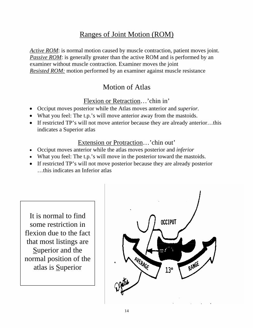
Flexion or Retraction…’chin in’
• Occiput moves posterior while the Atlas moves anterior and superior. • What you feel: The t.p.’s will move anterior away from the mastoids. • If restricted TP’s will not move anterior because they are already anterior…this
indicates a Superior atlas
Extension or Protraction…’chin out’ • Occiput moves anterior while the atlas moves posterior and inferior • What you feel: The t.p.’s will move in the posterior toward the mastoids. • If restricted TP’s will not move posterior because they are already posterior
…this indicates an Inferior atlas
It is normal to find some restriction in
flexion due to the fact that most listings are
Superior and the normal position of the
atlas is Superior
15
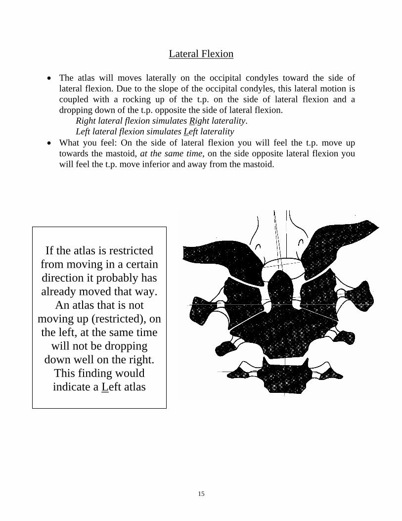
Lateral Flexion
• The atlas will moves laterally on the occipital condyles toward the side of lateral flexion. Due to the slope of the occipital condyles, this lateral motion is coupled with a rocking up of the t.p. on the side of lateral flexion and a dropping down of the t.p. opposite the side of lateral flexion.
Right lateral flexion simulates Right laterality. Left lateral flexion simulates Left laterality
• What you feel: On the side of lateral flexion you will feel the t.p. move up towards the mastoid, at the same time, on the side opposite lateral flexion you will feel the t.p. move inferior and away from the mastoid.
If the atlas is restricted
from moving in a certain direction it probably has already moved that way.
An atlas that is not moving up (restricted), on the left, at the same time
will not be dropping down well on the right.
This finding would indicate a Left atlas
16
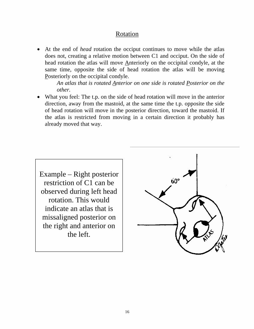
Rotation
• At the end of head rotation the occiput continues to move while the atlas does not, creating a relative motion between C1 and occiput. On the side of head rotation the atlas will move Anteriorly on the occipital condyle, at the same time, opposite the side of head rotation the atlas will be moving Posteriorly on the occipital condyle.
An atlas that is rotated Anterior on one side is rotated Posterior on the other.
• What you feel: The t.p. on the side of head rotation will move in the anterior direction, away from the mastoid, at the same time the t.p. opposite the side of head rotation will move in the posterior direction, toward the mastoid. If the atlas is restricted from moving in a certain direction it probably has already moved that way.
Example – Right posterior
restriction of C1 can be observed during left head
rotation. This would indicate an atlas that is
missaligned posterior on the right and anterior on
the left.
17
Lower Cervicals C2 – C7
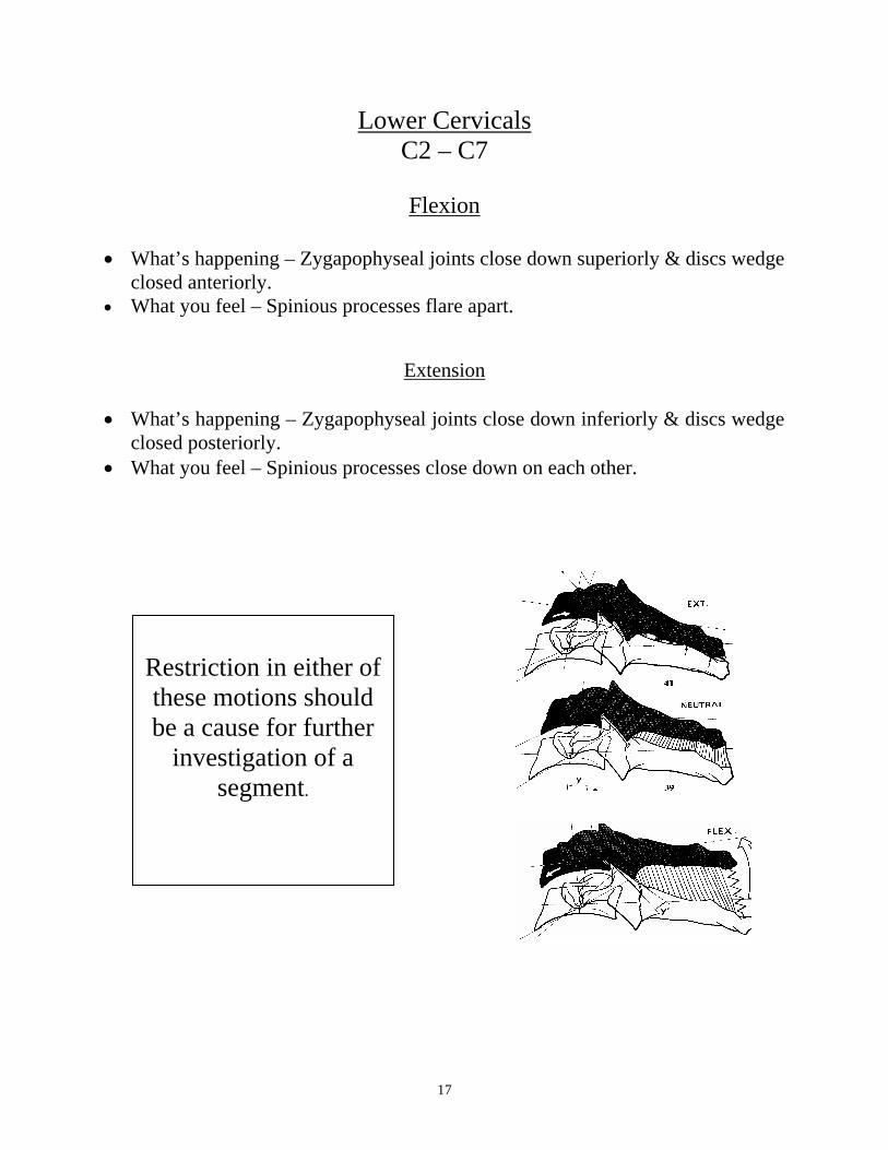
Flexion
• What’s happening – Zygapophyseal joints close down superiorly & discs wedge
closed anteriorly. • What you feel – Spinious processes flare apart.
Extension
• What’s happening – Zygapophyseal joints close down inferiorly & discs wedge closed posteriorly.
• What you feel – Spinious processes close down on each other.
Restriction in either of these motions should be a cause for further
investigation of a segment.
18
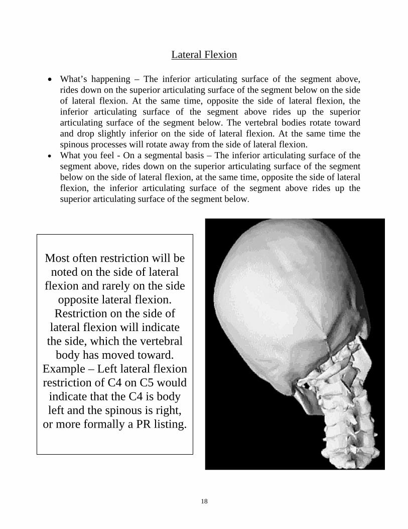
Lateral Flexion
• What’s happening – The inferior articulating surface of the segment above, rides down on the superior articulating surface of the segment below on the side of lateral flexion. At the same time, opposite the side of lateral flexion, the inferior articulating surface of the segment above rides up the superior articulating surface of the segment below. The vertebral bodies rotate toward and drop slightly inferior on the side of lateral flexion. At the same time the spinous processes will rotate away from the side of lateral flexion.
• What you feel - On a segmental basis – The inferior articulating surface of the segment above, rides down on the superior articulating surface of the segment below on the side of lateral flexion, at the same time, opposite the side of lateral flexion, the inferior articulating surface of the segment above rides up the superior articulating surface of the segment below.
Most often restriction will be
noted on the side of lateral flexion and rarely on the side
opposite lateral flexion. Restriction on the side of
lateral flexion will indicate the side, which the vertebral
body has moved toward. Example – Left lateral flexion restriction of C4 on C5 would
indicate that the C4 is body left and the spinous is right,
or more formally a PR listing.
19
Rotation • What’s happening – Each cervical segment’s body rotates toward the side of
rotation, with the greatest degree of rotation taking place at C2 and diminishing as you move down to C7 with the least. This motion will cause the spinous processes to rotate opposite the side of head rotation.
• What you feel – The s.p. will rotate opposite the side of head rotation.
Thoracic Motion All motion analysis is performed preferably from the side of the patient with the patient in the seated position. Patient’s arms are crossed over their chest and patient’s head rests on their forearms. The elbows and shoulders can be used as a lever arm to create motion throughout the thoracic spine.
Flexion • What’s happening – Thoracic IVD’s close down to anterior and posterior
articulations close down superiorly. • What you feel – Spinous processes will flare apart.
Extension
• What’s happening – Thoracic IVD’s close down to posterior and posterior articulations close down inferiorly.
• What you feel – Spinous processes will close down on each other. They may veer off to left or right when sliding inferiorly on spinous process below.
Lateral Flexion
• What’s happening – Thoracic IVD’s close down laterally and vertebral bodies rotate toward the side of lateral flexion (into the cave), at the same time the spinous process will rotate away from the side of lateral flexion (out of the cave).
• What you feel – Spinous processes will rotate and flare away from each other, opposite the side of lateral flexion.
The side, which the spinous shows restriction towards, is the side it will be listed towards. Example – Spinous restricted to the left
during right head rotation would indicate a PL listing.
20
Rotation
• What’s happening – The vertebral bodies rotate toward the side of rotation at the same time, the spinous processes rotate away from the side of rotation.
• What you feel – Spinous processes will rotate away from the side of rotation. It is difficult to gather accurate information from this motion exam due to the fact that thoracic rotation is primarily restricted buy the ribs articulating with the sternum.
Lumbar Motion
Flexion • What’s happening – Lumbar IVD’s close down anteriorly and the posterior
joints open up inferiorly, allowing the spinouses to flare apart. • What you feel – Spinous processes will flare away from each other.
Extension
• What’s happening – Lumbar IVD’s close down posteriorly and the posterior joints open up superiorly, causing the spinouses to close down on each other.
• What you feel – Spinous processes will close down each other.
Lateral Flexion • What’s happening – Lumbar IVD’s close down on the side of lateral flexion
and the shape of the posterior articulations cause the spinouses to rotate slightly toward the side of lateral flexion (into the cave). At the same time the lumbar vertebral bodies will rotate away from the side of lateral flexion.
• What you feel – You should be capable of palpating the rotational motion of the spinous processes in this region during lateral flexion. Also, you can palpate the mammillary process, which will rotate toward the side of lateral flexion at the same time they will close down on each other. Both of these motions can be assessed.
Rotation
• What’s happening – The vertebral bodies rotate toward the side of rotation, at the same time the spinous processes rotate away from the side of rotation.
• What you feel – The spinous processes will move away from the side of rotation
21
Sacrum
Lateral Flexion
• What’s happening – The base of the sacrum will tilt inferior on the side of
lateral flexion • What you feel – With all six palpation fingers laid across the base of sacrum
you will feel a slight anterior/inferior motion take place on the side of lateral flexion.
SI joints/Ilium
IN/EX Rotation
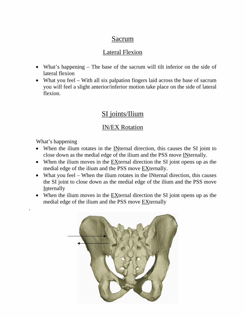
What’s happening • When the ilium rotates in the INternal direction, this causes the SI joint to
close down as the medial edge of the ilium and the PSS move INternally. • When the ilium moves in the EXternal direction the SI joint opens up as the
medial edge of the ilium and the PSS move EXternally. • What you feel – When the ilium rotates in the INternal direction, this causes
the SI joint to close down as the medial edge of the ilium and the PSS move Internally
• When the ilium moves in the EXternal direction the SI joint opens up as the medial edge of the ilium and the PSS move EXternally
.

22
PI Motion
• What’s happening – In the standing position, the patient is instructed to raise
one knee at a time as high as they can. When the knee is raised to 90 degrees and beyond, this causes the ipsilateral ilium to rotate on the surface of the sacral articular surface in the counter-clockwise direction. This counterclockwise motion causes the PSS to move in a Posterior and Inferior direction relative to the sacrum.
• What you feel – The PSS and the sacro-iliac joint on the ipsilateral side is palpated and the amount of Posterior/Inferior motion is noted. This is compared to the range of motion on the opposite side.