west nile disease in tunisia: an overview of 60 years · questo studio offre una sintesi...
TRANSCRIPT
225
Parole chiaveEcologia,Epidemiologia,Equini,Esame,Esseri umani,Tunisia,Virus del Nilo Occidentale.
RiassuntoIl virus della West Nile (WNV) è un virus trasmesso da artropodi importante per la Salute Pubblica. Appartiene al complesso antigenico del virus dell’encefalite giapponese insieme al quale è incluso nel genere Flavivirus, famiglia Flaviviridae. Il virus è in continua diffusione in Tunisia, soprattutto nella zona costiera e meridionale del paese. Dalla prima epidemia umana di WNV, nel 1997, sono aumentate costantemente il numero e l'estensione degli effetti di spillover. Ciononostante, la conoscenza dei fattori scatenanti è ancora piuttosto carente. L'ultima epidemia umana di meningite e meningoencefalite causata da WNV è registrata nel 2012 e, con 86 casi e 6 decessi, ha confermato il fallimento dell'attuale piano predittivo. Questo studio offre una sintesi bibliografica dei documenti pubblicati tra il 1970 e il 2015 e sottolinea le conoscenze esistenti e le lacune che devono essere affrontate nell'ambito della ricerca.
Il virus del Nilo Occidentale in Tunisia:una rassegna sui passati 60 anni di ricerca
KeywordsEpidemiology,Ecology,Equines,Human,Review,Tunisia,West Nile disease.
SummaryWest Nile virus (WNV) is an arthropod borne virus of public health importance. The virus is a member of the genus Flavivirus and belongs to the Japanese encephalitis virus (JEV) antigenic complex under the Flaviviridae family. The WNV is continuously spreading across Tunisia especially in the coastal and Southern area of the country. The first human West Nile disease (WND) epidemic in Tunisia occurred in 1997, since then, the quantity and the extension of spillover effects increased constantly. However, the existing knowledge of factors triggering such events continues to be rather poor. The last epidemic WNV human meningitis and meningoencephalitis recorded in 2012, with 86 cases and 6 deaths, confirmed the failure of the current system in predicting new cases. This review, based on analysis of scientific papers published between 1970 and 2015, summarises the state of knowledge on WNV in Tunisia and highlights the existing knowledge and research gaps that need to be addressed.
Veterinaria Italiana 2017, 53 (3), 225‑234. doi: 10.12834/VetIt.1181.6565.2Accepted: 13.12.2016 | Available on line: 29.09.2017
° Both authors contributed equally to this manuscript.1 Ecole Nationale de Médecine Vétérinaire de Sidi Thabet, Tunisie.
2 Istituto Zooprofilattico Sperimentale dell'Abruzzo e del Molise, Teramo, Italy.3 Institut de la RechercheVétérinaire de Tunisie (IRVT).
*Corresponding author at: Ecole Nationale de Médecine Vétérinaire de Sidi Thabet, Tunisie.Tel.: 00216 20715392, e-mail: [email protected].
Salah Hammami1°, Thameur Ben Hassine1°*, Annamaria Conte2, Jihène Amdouni3,Fabrizio De Massis2, Soufiène Sghaier3 and Sonia Ben Hassen3
West Nile disease in Tunisia: an overview of 60 years
bird and mosquito species (Gubler 2007). In the last 15 years, WNV has dramatically expanded its geographic range and is now considered the most widespread member of the Japanese encephalitis virus (JEV) complex (Bakonyi et al. 2006) as well as the most widespread arbovirus in the world (Brault 2009, Gould et al. 2003, Lanciotti et al. 2006). Given the rising awareness towards WND, notifications of human and equine cases have
IntroductionWest Nile Disease (WND) is the consequence of an arboviral zoonotic infection, transmitted by mosquitoes, which is not restricted by international borders and involves all continents, causing illnesses and deaths in both humans and horses. The causative agent, the West Nile Virus (WNV), belongs to the Flaviviridae family, genus Flavivirus, and is maintained in a transmission cycle involving
226 Veterinaria Italiana 2017, 53 (3), 225‑234. doi: 10.12834/VetIt.1181.6565.2
West Nile Disease in Tunisia Hammami et al.
et al. 2005, Triki et al. 2001, Hachfi et al. 2010). West Nile Virus lineage I was isolated in humans and found to be genetically close related to the strain isolated in Israel in 1998 and in New York in 1999 (Bahri et al. 2010). Between August and October 2003, a second occurrence of WNV infection was observed in the same governorates affected by the first epidemic, although this second epidemic had a larger geographical extension in the Southern area of the country. The disease was confirmed by serum‑neutralization in 36 out of 64 patients and no deaths were recorded (Hachfi et al. 2010, Riabi et al. 2010).
During these 2 epidemics, no clinical cases were reported in horses or in birds. In 2010 and 2011, human WND sporadic cases were recorded in the North Western (Jendouba governorate) and in the Southern (Tataouine and Kebili governorates) areas of the country (ONMNE 2013). A new outbreak was reported in 2012. Between July and December 2012, further 98 WND human cases were confirmed,
increased over the last decades in Europe and in the Mediterranean Basin (Calistri et al. 2010). Humans and horses are considered incidental hosts ‑ given the low level of viraemia that they may develop (Bowen and Nemeth 2007). The fact that infection is mild or unapparent has nothing to do with virus transmission. Nonetheless, it has been shown that the infection can lead to severe diseases or even deaths (ECDC 2011). West Nile Virus is maintained in nature by a primary transmission cycle between mosquitoes and several bird species, which act as amplifying hosts (Hamer et al. 2008, Work et al. 1955). The transmission, epidemiology, and geographic distribution of WND are a complex function of biotic (e.g., host abundance and diversity, bird migration, mosquito distribution) and abiotic (e.g., environmental and climatic aspects) factors (Patz et al. 1996). Both biological factors (e.g., vector diversity, survival of pathogens within the vector populations) and environmental determinants (e.g., climatic conditions, availability of water bodies) may affect the transmission cycles (Owen et al. 2006, Paz 2006).
Tunisia has experienced 3 major epidemics that have affected particularly humans in 1997, 2003, and 2012. Since 2010, sporadic human meningoencephalitis cases have been reported in different regions of Tunisia almost every year. In 2015, an equine case was reported for the first time (OIE 2015). However, the drivers of WNV infection transmission in Tunisia are still poorly understood. The aim of the present review is to report on the emergence of WND and its evolution in Tunisia over the last 60 years and to identify areas on which further investigations should be developed in order to fill existing gaps in current knowledge about WND epidemiology and control in Tunisia.
Epidemiology of West Nile disease in Tunisia
Human WND epidemics and serological investigations in TunisiaIn 1968, serological tests (Inhibition of Haemagglutination; IHA) revealed circulation of WNV in human in different regions of Tunisia (2%, n = 705) (Hannoun 1971). Two years later (1970), seropositivity was confirmed in human by serum‑neutralization in Djerba (3.8%), Tunis (7.8%), and Gabes (7%) (Nabli et al.1970). In autumn 1997, human meningoencephalitis cases due to WNV lineage I (Triki et al. 2001) were observed, for the first time, in the Eastern coastal districts of Tunisia (Governorates of Sfax, Mahdia, and Monastir). A total of 173 cases were recorded with 8 deaths (Feki
0 - 5
5 - 20
20 - 100
100 - 200
Governorates Tunisia
0 100 20050 Km
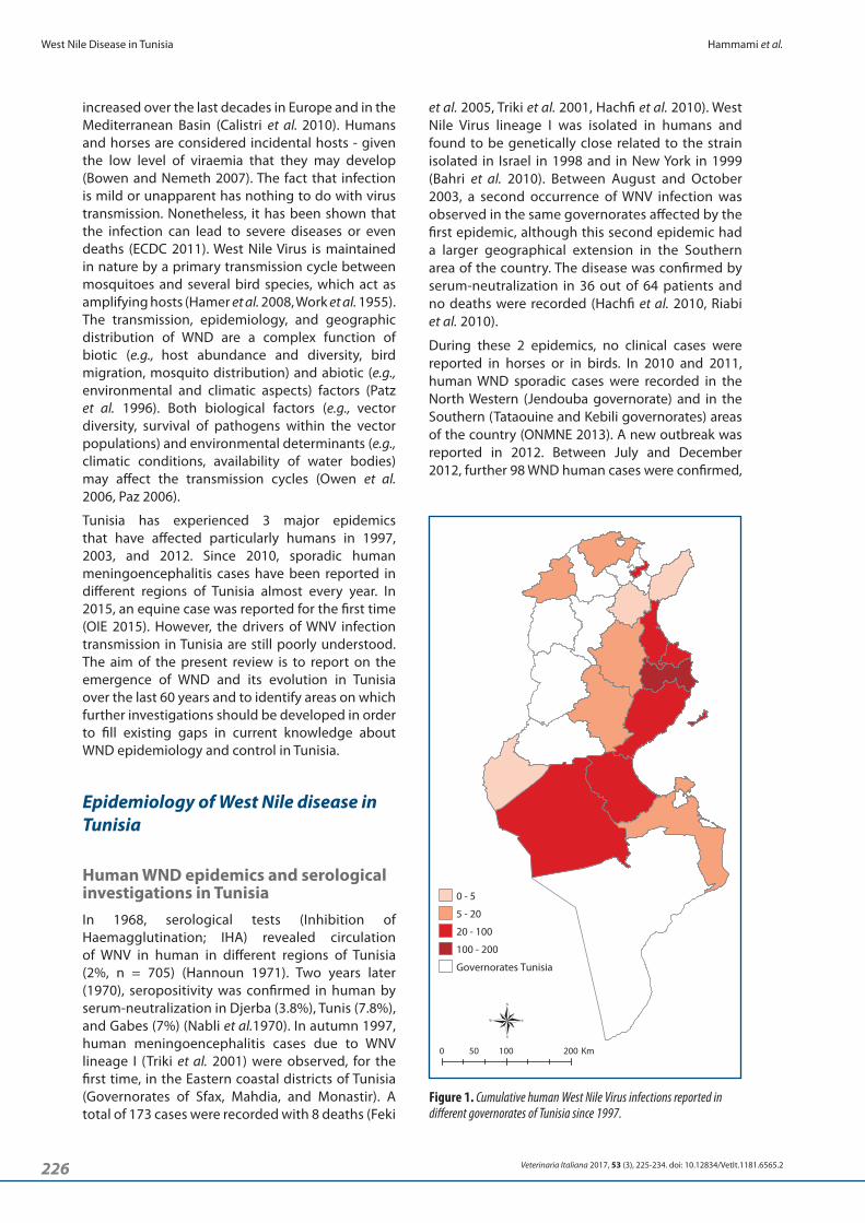
Figure 1. Cumulative human West Nile Virus infections reported in different governorates of Tunisia since 1997.

227Veterinaria Italiana 2017, 53 (3), 225‑234. doi: 10.12834/VetIt.1181.6565.2
Hammami et al. West Nile Disease in Tunisia
Table I. Results of human West Nile virus seropositivity investigations conducted in different governorates of Tunisia from 1970 to 2014.
Governorate Number of patients Period Method Percent of
seropositivity References
Bizerte 335
1968 Inhibition of Haemoagglutination (IHA)
0%
Hannoun 1971
Beja 43 3%
Tunis 126 2%
Nabeul 194 4%
Le Kef 290 2%
Jendouba 5 1%
Sousse 4 0%
Medenine 3 1%
Kairouan 2 0%
Sfax 1 0%
Gafsa 1 0%
Gabes 1 0%
Djerba 1,094
1970 IHA and serumneutralization
3.8%
Nabli et al. 1970Tunis 205 7.8%
Gabes 85 7%
Others 22 9%
Sfax, Mahdia 129 September - December 1997 Elisa IgM 86 % Bahri et al. 2010
Sfax 57 Autumn 1997 IgM in serum and Cerebrospinal Fluid (CSF) 23.4 % Feki et al. 2005
Sousse 34 August - November 2003 Elisa IgG 61% Hachfi et al. 2010
Kairouan
1854 January - December 2007 Elisa IgG
27.7%
Bahri et al. 2010Sfax 7.5%
Bizerte 0.7%Monastir, Kébili, Jendouba, Bizerte,
Tozeur, Kairouan, Nabeul, Sousse, Gabes, Mahdia, Sfax, Sidi Bouzid, Médenine
364 July - December 2012 Elisa IgG 26.92% ONMNE 2013
Monastir 113 August - October 2003 Elisa IgG 33.6% Riabi et al. 2010
WNV investigations in small mammals and equines in TunisiaSince 1970, when serological investigations showed the circulation of WNV in humans in Tunisia (Hannoun 1971, Nabli et al. 1970), several studies have been conducted to check the role of small mammal species and equines in the epidemiological cycle of WND. Early studies on this topic revealed high seropositivity of rodents (Chastel et al. 1977, Chastel et al. 1983).
The first serological studies on horses have been conducted by Haddad in 1980 (Haddad 1980) as a part of a study focusing on a large part of the country (n = 556). The study did not report clinical signs in animals; only 2 animals were positive by Inhibition of Haemaagglutination (IHA) (Haddad 1980). These findings suggested a low viral circulation during this
86 of which were neuroinvasive (ONMNE 2013). In October 2013, 6 human cases were detected in 5 governorates: Gabes, Mahdia, Monastir, Nabeul, and Sousse (ECDC 2013). The reported cases indicate that the most affected areas were primarily coastal areas of the central of Tunisia. However, important number of cases was also registered in Southern areas around oases.
The most favourable period for viral circulation is the late summer and early fall (ONMNE 2013) with the maximum of human cases recorded in this period. Figure 1 shows the cumulative distribution of WND human cases reported in Tunisia since 1997.
Since the onset of the first WND outbreak in Tunisia, several surveys and serological investigations were carried out in humans, especially in the affected areas with the aim to estimate the range of the infection (Table I).
228 Veterinaria Italiana 2017, 53 (3), 225‑234. doi: 10.12834/VetIt.1181.6565.2
West Nile Disease in Tunisia Hammami et al.
in 1980 (Haddad 1980). After this period, the first outbreak was then reported in 1997, the second in 2003. Monitoring human cases of meningitis and meningoencephalitis (MME) due to WNV began in Tunisia only in 2004 (ONMNE 2013). This monitoring, based on passive surveillance, allowed for detecting sporadic cases of WNV meningitis in 2007 in the governorate of Kairouan.
It is worth stressing that, after the establishment of an early warning system in 2010 with the reporting of all cases of MME from April to November, managed by the National Observatory of New and Emerging Diseases (ONMNE), the sensitivity of the reporting system significantly improved, and since then neurological human cases of WND have been reported almost every year. Serological surveys in horses are often conducted with some delay and they are generally carried out after the onset of human epidemics. These surveys generally show a consistency in the identification of areas at risk of WNV circulation.
A third outbreak was recorded in 2012 and it confirmed the lack of the sensitivity of this system in predicting new cases.
Ecology of West Nile virus in Tunisia
Introduction, transmission and spreadMigratory birds have long been suspected as the principal introductory hosts of WNV into new regions of Tunisia (Triki et al. 2001) for several reasons: in temperate areas, outbreaks generally occur during late Summer or early Fall, coinciding with the arrival of large concentrations of migratory
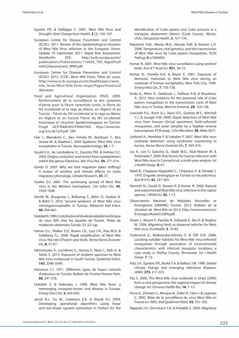
period. Serological surveys in horses have become more frequent after 2005 (Table II). At first, these surveys were conducted in the regions where WND human epidemics had been reported. Seropositivity rates for Immunoglobulin G (IgG), around 30%, were reported in horses (FAO 2009). These findings suggested a significant infection rate in this animal species in the same territories where human cases were reported.
Another survey conducted in 2008 in the North West of Tunisia (Ben Hassine et al. 2011) – an area not affected by any previously known epidemics – also identified a previous active circulation of WNV in this region, with IgG seropositivity around 23% in horses (n = 133). Seroconversion was recorded in October 2008, strictly aligned with periods of maximum human cases recorded.
The most important serological survey in equines has been conducted in 2009 (1,189 tested sera) and confirmed previous results recorded in the areas most affected (Bargaoui et al. 2015). The peculiar environmental conditions of the Southern part of Tunisia, which has a very dry climate with the presence of oases (humid areas), prompted some authors to investigate the characteristics allowing the circulation of WNV in this region (Ben Hassine et al. 2014). The overall seropositivity rate was very high, being it in the order of 42% of tested horses. The serological studies conducted in animals in Tunisia since 1977 are shown in chronological order in Table II.
Virus persistence, silent circulation and spilloverFirst identified in 1968 in Tunisia (Nabli et al. 1970, Hannoun 1971), WNV circulation showed a decline
Table II. Results of animal West Nile virus serological investigations conducted in Tunisia from 1977 to 2011.
Year Animals Parts of Tunisia Seroprevalence Test References
1977 Rodents Nabeul, Tunis, Beja, Gafsa, Médenine, Tataouine 19.8% (n=196) Inhibition of
Haemoagglutination (IHA) Chastel et al. 1977
1980 Rodents and insectivores microbats Several regions of Tunisia 0% (n=103) IHA Chastel et al. 1983
1980 Equines (and the boar) Several regions of Tunisia 2 positives (n=556) IHA Haddad 1980
2006 EquinesCoastal governorates of Tunisia (Gabes, Sfax, Mahdia, Monastir,
Bizerte and Nabeul)32% (n= 269) Competitive ELISA (C-ELISA) Bargaoui et al. 2015
2008 Equines North West of Tunisia (Jendouba, El Kef, Bizerte) 23% (n=133) C-ELISA Ben Hassine et al. 2011
2008 Equines Monastir and Sfax governorates 30% (n= 127)ELISA IgG and IgM
(serumneutralization for confirmation)
Bargaoui 2013
2009 Equines All parts of Tunisia 28% (n=1189) C-ELISA (serumneutralization for confirmation) Bargaoui et al. 2015
2011 Equines Kébili 42.3% (n=284) C-ELISA (serumneutralization for confirmation) Ben Hassine et al. 2014

229Veterinaria Italiana 2017, 53 (3), 225‑234. doi: 10.12834/VetIt.1181.6565.2
Hammami et al. West Nile Disease in Tunisia
(Wasfi et al. 2016). It is generally recognised that the most implicated mosquito species in the transmission of WNV in Tunisia is Culex pipiens, which is the most widely spread mosquito species (Wasfi et al. 2016). The highest densities of C. pipiens are found in stagnant waters rich in organic matter located in the outbreak areas (FAO 2009). Experimentally, C. pipiens populations collected in Tunisia have been shown to be highly susceptible to WNV infection and readily to transmit the virus. When exposed to an infectious blood meal containing WNV, all mosquito strains collected proved to be able to disseminate and transmit the virus (Amraoui et al. 2012). Both forms of C. pipiens and their hybrids coexist in the several sites in Tunisia. This supports the hypothesis that C. pipiens is a bridge vector for the WND in Tunisia (Krida et al. 2015).
Landscape epidemiology associated with West Nile Virus circulation in TunisiaThe aim of the limited number of studies conducted in Tunisia was to investigate the potential association between biotic/abiotic factors and human/horse WND cases or seroprevalence rates, using different statistical methods (risk factor analysis, landscape epidemiology, and transmission dynamic modelling) (Table III). Although not directly applicable to infer geographical variations of the risk of WNV transmission, the results of such studies help identify the main involved factors. Thus, risk factors of geographical variation can be integrated in predictive spatial and/or dynamic models.
Studies focusing on specific risk factor have been carried out in Tunisia based on serological findings in horses as a covariate (Ben Hassine et al. 2011, Ben Hassine et al. 2014). Logistic regression is used to identify risk factors likely to be associated with seropositivity. For landscape epidemiology, 2 large‑scale studies involving the entire country have been conducted to develop prediction maps of areas at risk adopting 2 statistical approaches. The first study was based on seroprevalence in equines (Bargaoui et al, 2015). A logistic model was used to analyse the relation between seropositivity and the covariates. Performance of the model as a classifier was assessed using a Receiver Operating Characteristic (ROC) analysis and computing the area under the ROC curve (AUC). The second study was based on human meningoencephalitis cases recorded in Tunisia in 2012 (Ben Hassine et al. 2015) and had the goal to identify areas at risk for WNV circulation using Mahalanobis distance statistic. Dynamic transmission mdelling has been conducted in 2 recent studies that included Tunisia and many countries of the Mediterranean basin as well as Europe aiming to identify space‑time area at risk for
birds (Ben Hassine et al. 2015); WND outbreaks often occur among humans living in or near wetlands where high concentrations of birds come into contact with large numbers of ornithophilic mosquitoes (Bargaoui et al. 2015). Several studies indicate that the closeness to marshe ecosystems, where migratory bird populations are located, is an important risk factor for WNV circulation in Tunisia (Bargaoui et al. 2015, Ben Hassine et al. 2014, Ben Hassine et al. 2015). Suitable areas for WNV circulation have been identified along all the major roads of migratory birds crossing Tunisia (El Hili 2015) (e.g.: North‑West, Cap Bon in North‑East, and the East‑Coast of the area between Sousse and Sfax).
Serological studies conducted at different points in time show that previously free areas became infected (Haddad 1980, Bargaoui 2013) indicating possible regular introduction of WNV probably from migratory birds. However, the persistent occurrence of WND in Tunisia cannot be caused only by virus reintroduction. Endemic cycles are probably established in some areas of the country, although it is not clear which bird species might play a role in the local persistence of the infection.
Resident birds seem to be particularly suitable for this role, given their density and the ability for some species to cover relatively long distances, independently of seasonal migratory pattern. Migratory birds may introduce WNV into the sylvatic cycle. Then, the virus could be introduced in a peridomestic cycle, involving peridomestic birds and opportunistic mosquitoes. At the same time, it is possible that the virus could be intruded occasionally into an urban cycle, where its amplification can cause human and equine epidemics (Tsai et al. 1998).
The few serological studies conducted in Tunisia on migratory and resident birds showed that Anser anser and Sturnus vulgaris are potential targets for WNV surveillance in wildlife in Tunisia (Bargaoui et al. 2015). Seroprevalence studies conducted in Passer domesticus (House sparrows) – amplifying hosts of WNV in many regions in the world (Nemeth et al. 2006) – in the South of Tunisia (1%) indicate that this species has been heavily involved in maintaining virus circulation in this area (Hammouda et al. 2015). However, the role of migratory and resident birds in the epidemiological cycle of WNV in Tunisia needs further investigations to be fully understood.
The respective roles of mosquito species in WNV transmissionIn 2015 and for the first time, WNV has been isolated and detected in Culex pipiens mosquitoes in Tunisia (Wasfi et al. 2016). Phylogenetic analysis showed that WNV strains belong to lineage 1 and are closely related to the Human Tunisian strain 1997 (PAH 001)

230 Veterinaria Italiana 2017, 53 (3), 225‑234. doi: 10.12834/VetIt.1181.6565.2
West Nile Disease in Tunisia Hammami et al.
fall there are more favourable conditions for WNV transmission than in drier seasons (Bargaoui et al. 2015). High values of NDVI are related to higher photosynthetic activity and improved ecological condition (Gordo 2007). This suitable condition increases the main food supply, such as arthropods, for most migratory birds. Several studies have been based on NDVI for assessment of WNV infection foci and its vector habitat. Seasonal differences in NDVI were among the best predictive factors for determining mosquito distribution abundance in different models (Brownstein et al. 2008, Jacob et al. 2009). The preliminary analysis of the type of vegetation existing in Tunisian provinces with and without an history of WNV human cases seems to indicate the presence of sparsely and bare vegetation as a characteristic of the areas with WNV occurrence; whereas croplands are more present in non‑infected areas (Calistri et al. 2012). Normalized Differentiated Vegetation Index is a highly dynamic factor and more attentive analyses should be conducted to understand the impact of this factor on WNV persistence and spread (Ben Hassine et al. 2015).
While temperature absolute value doesn't seem to influence the Tunisian WNV epidemiological situation (Calistri et al. 2012), lower night time temperature and anomalies of temperature in July
WNV circulation (Conte et al. 2015, Tran et al. 2014).
One of the most explanatory variables in these studies is the presence of humid areas in the North and Central parts of Tunisia. In the South, seropositivity in equines was significantly higher in areas closer to the oasis compared with those surrounding arid areas. The oasis effect encourages birds to congregate around shrinking water sites and to nest in areas in which horses may be closer, thus favouring viral circulation between hosts and mosquitoes. The warmer conditions of these humid areas, with the presence of stagnant water rich in organic materials attract mosquitoes and birds. In addition, this increases the interaction and circulation of WNV (Epstein and Defilippo 2001). In epidemic areas, WNV is thought to be introduced by migrating birds and maintained during the periods of mosquito abundance (Komar 2001, Rappole et al. 2000). As epizootics often occur in areas close to wetlands (Hubálek and Halouzka 1999), it is plausible that the virus would first be amplified in wet areas and later spread to dry surrounding areas.
For Normalized Differentiated Vegetation Index (NDVI), a commonly used measure of vegetation density (Ozdenerol et al. 2008), higher values of the biannual phase of NDVI were associated with higher values of seroprevalence rate in horses and human infections. This means that in humid late spring and
Table III. Overview of climatic and environmental variables associated with West Nile virus circulation in Tunisia.
Infection marker Scale Explicative variables Validation Prediction ReferencesHorse seroprevalence North West of Tunisia Humid area and Ramsar sites No No Ben Hassine et al. 2011
Horse seroprevalence South Tunisia Distance from oasis No No Ben Hassine et al. 2014
Human cases (1997-2011) Country Localization of Humid areas and Ramsar
sites No No Calistri et al. 2012NDVI
Horse seroprevalence Country
Night-time land surface temperature,Yes
(external*) yes Bargaoui et al. 2015biannual phase of NDVI
Distance to the nearest RAMSAR sites
Human meningoencephalitis
(2012)Country
Night-time land surfaceYes
(internal*) yes Ben Hassine et al. 2015Temperature,
Biannual phase of NDVI
Human WNF Outbreaks (2002-2013)
Europe and neighbouring
countries (Tunisia included)
Anomalies of temperature in July
Yes (internal and external) Yes Tran et al. 2014
Anomalies of MNDWI in early June
Presence of wetlands
Location under migratory routesOccurrence of a WNF outbreak the previous
yearHuman and equine clinical
cases notified in Italy, Greece, Portugal, Morocco,
and Tunisia, between 2008 and 2012
Mediterranean basin and Central Europe (Tunisia included)
Altitude, slope, night time Land Surface
Yes (internal) Yes Conte et al. 2015Temperature, Normalized Difference Vegetation Index, Enhanced Vegetation
Index, and daily temperature range*Internal: performance on population underlying the sample; *External: performance on related but slightly different population.

231Veterinaria Italiana 2017, 53 (3), 225‑234. doi: 10.12834/VetIt.1181.6565.2
Hammami et al. West Nile Disease in Tunisia
spreading is well known, but many important questions still remain unanswered, e.g. the geographic origin of each introduction of WNV, the relative importance of the introduction of infected vectors rather than infected birds, and the detailed transmission mechanisms involved in virus establishment into new areas.
In Tunisia, spillover events are increasing in numbers and expanding geographically. However, the knowledge on the factors triggering such events continues to be rather poor. In most cases, the key amplifying bird host and the bridge vector species involved are unknown. Further studies are needed to better understand the eco‑epidemiology of WNV in Tunisia. The reservoir hosts and vectors are crucial for WNV circulation and should be the focus of future studies. With this level of eco‑epidemiological uncertainty, building‑up accurate predictive models of WND outbreak risk is a complicated undertaking, although progresses have been made in this direction.
Entomological surveillance can be improved to:
• identify the species of mosquitoes responsible of transmitting the virus to birds, horses and humans;
• evaluate the seasonal population dynamics of the potential vectors during the year;
• isolate the WNV;
• detect others mosquitos‑borne diseases already present or at risk of introduction in Tunisia.
Entomological surveillance is also crucial for developing an early warning system. However, it would work only if all necessary factors are implemented. Indeed, this surveillance would mobilize staff, techniques, and financial resources for results that sometimes may be inconclusive (Perra et al. 2002). Characterization of circulating strains may help in explaining the absence of clinical cases in infected equines and in testing the hypothesis of the circulation of an attenuated strain, as it has already been done in Texas and in Mexico (Davis et al. 2005). Viral isolation could be obtained by investigation on sentinel domestic birds. This type of monitoring is part of an active surveillance. It is based on seroconversion surveillance on sentinel poultry in several sites. The monitoring of sentinel birds is widely used to detect the transmission of WNV, even at low rate. In the Mediterranean countries, serological surveys of sentinel chicken have given good results in Egypt (Soliman et al. 2010) and Greece (Chaskopoulou et al. 2013). A disadvantage of such active monitoring system is that the costs may become greater, as well as harder to justify, when the target disease is rare (Komar et al. 2001, Leblond et al. 2007). Domestic
have been identified as risk factors (Bargaoui et al. 2015). Summer temperature is an important factor for predicting WNV activity (Kilpatrick et al. 2008, Paz 2006, Savage et al. 1999, Turell et al. 2001). The effect of high temperature on mosquito abundance and vector competency has also been experimentally proven (Dohm et al. 2002).
In Tunisia, space‑time model indicates that most suitable areas for WNV circulation are located in Southern (May), Central (June‑July), Northen (September), and coastal (October‑November) areas. August and December may be considered not suitable periods for West Nile virus circulation. From January to April, no areas suitable for WND were predicted by specific simulation models (Conte et al. 2015).
Consequences for WNV surveillance in Tunisia: knowledge gaps and research prioritiesCurrently, surveillance in Tunisia is based on 3 components:
i. monitoring of human meningitis and meningo‑encephalitis cases (ONMNE 2013);
ii. monitoring of clinical equine encephalitis cases based on the passive reporting of any suspicion of encephalitis within the framework of the integrated network monitoring of rabies (however, in this network the search of WNV is not systematic);
iii. passive surveillance of the circulation of WNV in birds based on the monitoring of avian mortality integrated into the avian influenza surveillance network.
For the entomological component, no continuous monitoring is performed in the risk areas, with the exception of some investigations carried out after human outbreaks. In general, the current monitoring system in place to identify WNV circulation does not meet the requirements of effective early warning system. The goal of such a system should be to provide an early alert to health authorities in order to adopt timely and effective measures to prevent the spread of the infection among animals and humans. It should allow for the isolation and the characterization of the circulating strains. This network should involve both human and animal health authorities at national, regional, and international levels. The monitoring system in Tunisia can significantly be improved by considering the studies on areas and periods at risk.
However, the knowledge of how, when, and from where WNV is introduced into Tunisia remains still very limited. The role of migrating birds on WNV

232 Veterinaria Italiana 2017, 53 (3), 225‑234. doi: 10.12834/VetIt.1181.6565.2
West Nile Disease in Tunisia Hammami et al.
necessary to understand the “oasis effect” on the epidemiology of WND.
The analysis of the relevant literature proposed in this review shows that several routes could be pursued for future research. Efforts are needed to identify vectors and vertebrate hosts and to assess the impact of environment. At the same time, substantial efforts have to be devoted to understand the molecular epidemiology of WNV and the impact of other co‑circulating flaviviruses, like Usutu virus recently detected in the south of Tunisia (Hassine et al. 2014), in order to provide a more detailed understanding and more reliable data for effective risk assessment modelling.
dogs, which are susceptible to WNV infection, could be used as a sentinel species for monitoring WNV circulation in urban areas where horses are few (Resnick et al. 2008). A multi‑year analysis study in Connecticut, USA, indicated that considering both environmental variables and animal sentinel data could be useful for predicting human WNV infection (Liu et al. 2009).
Tunisia has diverse ecological conditions, resulting in biodiversity and, consequently, differential epidemiological transmission patterns. The specific ecological and climatic parameters of each region should be studied and identified. In Southern areas, for example, further research is
Amraoui F., Krida G., Bouattour A., Rhim A., Daaboub J. & Harrat Z. 2012. Culex pipiens, an experimental efficient vector of West Nile and Rift Valley fever viruses in the Maghreb region. PloS One, 7, e36757.
Bahri O., Dhifallah I., Alaya‑Bouafif N.B., Fekih H., Gargouri J.& Triki H. 2010. Étude séroépidémiologique de la circulation du virus West Nile chez l’Homme en Tunisie. Bull Société Pathol Exot,104, 272‑276.
Bakonyi T., Ivanics É., Erdélyi K., Ursu K., Ferenczi E. & Weissenböck H. 2006. Lineage 1 and 2 strains of encephalitic West Nile virus, Central Europe. Emerg Infect Dis,12, 618‑623.
Bargaoui R., Lecollinet S. & Lancelot R. 2015. Mapping the serological prevalence rate of West Nile fever in equids, Tunisia. Transbound Emerg Dis, 62, 55‑66.
Ben Hassine T., Hammami S., Elghoul H. & Ghram A. 2011. Detection of circulation of West Nile virus in equine in the north‑west of Tunisia. Bull Soc Pathol Exot, 104, 266‑271.
Ben Hassine T., Conte A., Calistri P., Candeloro L., Ippoliti C., De Massis F. & Hammamo S. 2017. Identification of suitable areas for West Nile cirus circulation in Tunisia. Transbound Emerg Dis, 64, 449‑458.
Ben Hassine T., De Massis F., Calistri P., Savini G., BelHaj Mohamed B., Ranen A., Hammamo S. 2014. First detection of co‑circulation of West Nile and Usutu viruses in equids in the south‑west of Tunisia. Transbound Emerg Dis, 61, 385‑389.
Bowen R.A. & Nemeth N.M. 2007. Experimental infections with West Nile virus. Curr Opin Infect Dis, 20, 293‑297.
Brault A.C. 2009. Changing patterns of West Nile virus transmission: altered vector competence and host susceptibility. Vet Res, 40, 43.
Brownstein J.S., Rosen H., Purdy D., Miller J.R., Merlino M. & Mostashari F. 2002. Spatial analysis of West Nile virus: rapid risk assessment of an introduced vector‑borne zoonosis. Vector Borne Zoonotic Dis, 2, 157‑164.
References
Calistri P., Giovannini A., Hubalek Z., Ionescu A., Monaco F., Savini G. & Lelli R. 2010. Epidemiology of West Nile in Europe and in the Mediterranean Basin. Open Virol J, 4, 29‑37.
Calistri P., Ben Hassine T., Ippoliti C., Conte A., Danzetta M.L. & Bruno R. 2012. Preliminary analysis of biotic and abiotic factors influencing the occurrence of west nile virus infection in Tunisia. EPIZONE 6th Annual Meeting ‑ Brighton UK June 12th to 14th, 2012.
Chaskopoulou A., Dovas C.I., Chaintoutis S.C., Kashefi J., Koehler P. & Papanastassopoulou M. 2013. Detection and early warning of West Nile virus circulation in Central Macedonia, Greece, using sentinel chickens and mosquitoes. Vector-Borne Zoonotic Dis,13, 723‑732.
Chastel C., Rogues G. & Beaucournu‑Saguez F. 1977. Enquête séroépidémique mixte arbovirus‑arénavirus chez les petits mammifères de Tunisie. Bull Soc Pathol Exot Filiales, 70 (5), 471‑479.
Chastel C., Bach‑Hamba D., Launay H., Le Lay G., Hellal H. & Beaucournu J.C. 1983. Arbovirus infections in Tunisia: new serological survey of small wild mammals. Bull Société Pathol Exot, 76, 21‑33.
Conte A., Candeloro L., Ippoliti C., Monaco F., De Massis F. & Bruno R. 2015. Spatio‑temporal identification of areas suitable for West Nile disease in the Mediterranean Basin and Central Europe. PLoS ONE, 10 (12): e0146024.
Davis C.T., Ebel G.D., Lanciotti R.S., Brault A.C., Guzman H. & Siirin M. 2005. Phylogenetic analysis of North American West Nile virus isolates, 2001‑2004: evidence for the emergence of a dominant genotype. Virology, 342, 252‑265.
Dohm D.J., O’Guinn M.L. & Turell M.J. 2002. Effect of environmental temperature on the ability of Culex pipiens (Diptera: Culicidae) to transmit West Nile virus. J Med Entomol, 39, 221‑225.
El Hili A. 2015. Les oiseaux migrateurs et la grippe aviaire en Tunisie. Newsletter Influenza Aviaire DGSV Tunisie. Rapport technique No.3 Décembre 2005.
233Veterinaria Italiana 2017, 53 (3), 225‑234. doi: 10.12834/VetIt.1181.6565.2
Hammami et al. West Nile Disease in Tunisia
identification of Culex pipiens and Culex restuans in a mosquito abatement district (Cook County, Illinois, USA). Geospatial Health, 3, 157‑176.
Kilpatrick A.M., Meola M.A., Moudy R.M. & Kramer L.D. 2008. Temperature, viral genetics, and the transmission of West Nile virus by Culex pipiens mosquitoes. PLOS Pathog, 4, e1000092.
Komar N. 2001. West Nile virus surveillance using sentinel birds. Ann N Y Acad Sci, 951, 58‑73.
Komar N., Panella N.A. & Boyce E. 2001. Exposure of domestic mammals to West Nile virus during an outbreak of human encephalitis, New York City, 1999. Emerg Infect Dis, 7, 736‑738.
Krida G., Rhim A., Daaboub J., Failloux A‑B. & Bouattour A. 2015. New evidence for the potential role of Culex pipiens mosquitoes in the transmission cycle of West Nile virus in Tunisia. Med Vet Entomol, 29, 124‑128.
Lanciotti R.S., Kerst A.J., Nasci R.S., Godsey M.S., Mitchell C.J. & Savage H.M. 2000. Rapid detection of West Nile virus from human clinical specimens, field‑collected mosquitoes, and avian samples by a TaqMan reverse transcriptase‑PCR assay. J Clin Microbiol, 38, 4066‑4071.
Leblond A., Hendrikx P. & Sabatier P. 2007. West Nile virus outbreak detection using syndromic monitoring in horses. Vector-Borne Zoonotic Dis, 7, 403‑410.
Liu A., Lee V., Galusha D., Slade M.D., Diuk‑Wasser M. & Andreadis T. 2009. Risk factors for human infection with West Nile virus in Connecticut: a multi‑year analysis. Int J Health Geogr, 8, 67.
Nabli B., Chippaux‑Hyppolite C., Chippaux A. & Tamalet J. 1970. Enquête sérologique en Tunisie sur les arbovirus. Bull W H O, 42, 297‑303.
Nemeth N., Gould D., Bowen R. & Komar N. 2006. Natural and experimental West Nile virus infection in five raptor species. J Wildl Dis, 42, 1‑13.
Observatoire National de Maladies Nouvelles et Emergentes (ONMNE) Tunisie. 2013. Bulletin de la situation de West Nile en 2012. http://www.onmne.tn/fr/images/Bulletin2WN.pdf.
Owen J., Moore F., Panella N., Edwards E., Bru R. & Hughes M. 2006. Migrating birds as dispersal vehicles for West Nile virus. EcoHealth, 3, 79‑85.
Ozdenerol E., Bialkowska‑Jelinska E. & Taff G.N. 2008. Locating suitable habitats for West Nile virus‑infected mosquitoes through association of environmental characteristics with infected mosquito locations: a case study in Shelby County, Tennessee. Int J Health Geogr, 7, 12.
Patz J.A., Epstein P.R., Burke T.A. & Balbus J.M. 1996. Global climate change and emerging infectious diseases. JAMA, 275, 217‑223.
Paz S. 2006. The West Nile virus outbreak in Israel (2000) from a new perspective: the regional impact of climate change. Int J Environ Health Res, 16, 1‑13.
Perra A., Zientara S., Murgue B., Zeller H., Hars J. & Lagneau C. 2002. Bilan de la surveillance du virus West Nile en France en 2002. Bull Epidémiol Hebd, 33, 161‑163.
Rappole J.H., Derrickson S.R. & Hubálek Z. 2000. Migratory
Epstein P.R. & Defilippo C. 2001. West Nile Virus and Drought. Glob Change Hum Health, 2 (2), 105‑107.
European Centre for Disease Prevention and Control (ECDC). 2011. Review of the epidemiological situation of West Nile Virus infection in the European Union. Update 19 September 2011. Rapid Risk Assessment: Stockholm: EDC. http://ecdc.europa.eu/en/publications/Publications/110920_TER_Rapid%20risk%20assessment_WNF.pdf.
European Centre for Disease Prevention and Control (ECDC) 2013. ECDC, West Nile Fever, Table on cases. http://www.ecdc.europa.eu/en/healthtopics/west_nile_fever/West‑Nile‑fever‑maps/Pages/historical data.aspx.
Food and Agricultural Organization (FAO). 2009. Renforcement de la surveillance et des systèmes d’alerte pour la fièvre catarrhale ovine, la fièvre du Nil occidental et la rage au Maroc, en Algérie et en Tunisie ‑ Fièvre du Nil occidental et la rage au Maroc en Algérie et en Tunisie Fièvre du Nil occidental: historique et situation épidémiologique en Tunisie. Projet GCP/RAB/002/FRAN.FAO. http://www.fao.org/3/a‑ak152f.pdf. 24P.
Feki I., Marrakchi C., Ben Hmida M., Belahsen F., Ben Jemaa M. & Maaloul I. 2005 Epidemic West Nile virus encephalitis in Tunisia. Neuroepidemiology, 24, 1‑7.
Gould E.A., de Lamballerie X., Zanotto P.M. & Holmes E.C. 2003. Origins, evolution, and vector/host coadaptations within the genus Flavivirus. Adv Virus Res, 59, 277‑314.
Gordo O. 2007. Why are bird migration dates shifting? A review of weather and climate effects on avian migratory phenology. Climate Research, 35, 37.
Gubler D.J. 2007. The continuing spread of West Nile virus in the Western hemisphere. Clin Infect Dis, 45, 1039‑1046.
Hachfi W., Bougmiza I., Bellazreg F., Bahri O., Kaabia N. & Bahri F. 2010. Second epidemic of West Nile virus meningoencephalitis in Tunisia. Médecine Mal Infect, 40, 456‑461.
Haddad N. 1980. Contribution à l’étude séroépidémioclinique du virus WN chez les équidés de Tunisie. Thèse de médecine vétérinaire Tunisie, 53, 221 pp.
Hamer G.L., Walker E.D., Brawn J.D., Loss S.R., Ruiz M.O. & Goldberg T.L. 2008. Rapid amplification of West Nile virus: the role of hatch‑year birds. Vector Borne Zoonotic Dis, 8, 57‑67.
Hammouda A., Lecollinet S., Hamza F., Nasri I., Neb A. & Selmi S. 2015. Exposure of resident sparrows to West Nile virus evidenced in South Tunisia. Epidemiol Infect, 143, 3546‑3549.
Hannoun C.l. 1971. Différents types de foyers naturels d’arbovirus en Tunisie. Bulletin de l’Institut Pasteur Paris, 69, 241‑278.
Hubálek Z. & Halouzka J. 1999. West Nile fever: a reemerging mosquito‑borne viral disease in Europe. Emerg Infect Dis, 5, 643‑650.
Jacob B.J., Gu W., Caamano E.X. & Novak R.J. 2009. Developing operational algorithms using linear and non‑linear squares estimation in Python for the

234 Veterinaria Italiana 2017, 53 (3), 225‑234. doi: 10.12834/VetIt.1181.6565.2
West Nile Disease in Tunisia Hammami et al.
Triki H., Murri S. & Le Guenno B. 2001. Méningoencéphalite à arbovirus West Nile en Tunisie. Med Trop, 61, 487‑490.
Turell M.J., O’Guinn M.L., Dohm D.J. & Jones J.W. 2001. Vector competence of North American mosquitoes (Diptera: Culicidae) for West Nile virus. J Med Entomol, 38, 130‑134.
Tsai T.F., Popovici F., Cernescu C., Campbell G.L. & Nedelcu N.I. 1998. West Nile encephalitis epidemic in southeastern Romania. Lancet, 352, 767‑771.
Work T.H., Hurlbut H.S. & Taylor R.M. 1955. Indigenous wild birds of the Nile Delta as potential West Nile virus circulating reservoirs. Am J Trop Med Hyg, 4, 872‑888.
Wasfi F., Dachraoui K., Cherni S., Bosworth A., Barhoumi W., Dowall S., Chelbi I., Derbali M., Zoghlami Z. & Beier J.C. 2016. West Nile virus in Tunisia, 2014: first isolation from mosquitoes. Acta Tropica, 159, 106‑110.
World Organisation for Animal Health (OIE). 2015.Organisation mondiale de santé animale (OIE), Fièvre de west nile en Tunisie (Available on line at http://www.oie.int/wahis_2/public/wahid.php/Diseaseinformation/ WI/index/newlang/fr, update 2 February 2015).
birds and spread of West Nile virus in the Western Hemisphere. Emerg Infect Dis, 6, 319‑328.
Resnick M.P., Grunenwald P., Blackmar D., Hailey C., Bueno R. & Murray K.O. 2008. Juvenile dogs as potential sentinels for West Nile virus surveillance. Zoonoses Public Health, 55, 443‑447.
Riabi S., Gallian P., Gaaloul I., Simon S., Harrath R. & Hassine M. 2010. Prevalence of IgG antibodies against West Nile virus in blood donors during the 2003 outbreak in Tunisia. Trans R Soc Trop Med Hyg, 104, 507‑509.
Savage H.M., Ceianu C., Nicolescu G., Karabatsos N., Lanciotti R. & Vladimirescu A. 1999. Entomologic and avian investigations of an epidemic of West Nile fever in Romania in 1996, with serologic and molecular characterization of a virus isolate from mosquitoes. Am J Trop Med Hyg, 61, 600‑611.
Soliman A., Mohareb E., Salman D., Saad M., Salama S. & Fayez C. 2010 Studies on West Nile virus infection in Egypt. J Infect Public Health, 3, 54‑59.
Tran A., Sudre B., Paz S., Rossi M., Desbrosse A. & Chevalier V. 2014. Environmental predictors of West Nile fever risk in Europe. Int J Health Geogr, 13, 26.