wg signaling via zw3 and mad restricts ... - the newfeld...
TRANSCRIPT

INVESTIGATION
Wg Signaling via Zw3 and Mad RestrictsSelf-Renewal of Sensory Organ Precursor Cells
in DrosophilaJanine C. Quijano, Michael J. Stinchfield, and Stuart J. Newfeld1
School of Life Sciences, Arizona State University, Tempe, Arizona 85287-4501
ABSTRACT It is well known that the Dpp signal transducer Mad is activated by phosphorylation at its carboxy-terminus. The role ofphosphorylation on other regions of Mad is not as well understood. Here we report that the phosphorylation of Mad in the linkerregion by the Wg antagonist Zw3 (homolog of vertebrate Gsk3-b) regulates the development of sensory organs in the anterior–dorsalquadrant of the wing. Proneural expression of Mad-RNA interference (RNAi) or a Mad transgene with its Zw3/Gsk3-b phosphorylationsites mutated (MGM) generated wings with ectopic sensilla and chemosensory bristle duplications. Studies with pMad-Gsk (anantibody specific to Zw3/Gsk3-b-phosphorylated Mad) in larval wing disks revealed that this phosphorylation event is Wg dependent(via an unconventional mechanism), is restricted to anterior–dorsal sensory organ precursors (SOP) expressing Senseless (Sens), and isalways co-expressed with the mitotic marker phospho-histone3. Quantitative analysis in both Mad-RNAi and MGM larval wing disksrevealed a significant increase in the number of Sens SOP. We conclude that the phosphorylation of Mad by Zw3 functions to preventthe self-renewal of Sens SOP, perhaps facilitating their differentiation via asymmetric division. The conservation of Zw3/Gsk3-bphosphorylation sites in vertebrate homologs of Mad (Smads) suggests that this pathway, the first transforming growth factor b-in-dependent role for any Smad protein, may be widely utilized for regulating mitosis during development.
INTERCELLULAR signaling is essential for proper develop-ment of multicellular organisms. In all animals, highly
conserved proteins belonging to the transforming growthfactor b (TGFb) family perform a multitude of tasks. TGFbproteins can be parsed into the TGFb/Activin or Dpp/BMPsubfamilies. In Drosophila, Dpp signals utilize the type I re-ceptor Thickveins (Tkv), and signal transduction proceedsvia Tkv phosphorylation of carboxy-terminal serines in thesignal transducer Mothers against dpp (Mad). Once Recep-tor phosphorylated, Mad nuclear import occurs, and Madthen forms a complex with Medea. Mad/Medea complexesregulate gene expression together with tissue-specific tran-scription factors (Derynck and Miyazono 2008).
Mad and Medea are members of a highly conserved Smadfamily of TGFb signal transducers. Mad and Smads1/5/8 invertebrates signal for Dpp/BMP subfamily proteins while Me-
dea and Smad4 in vertebrates form complexes with Smadsthat signal for all TGFb proteins (Newfeld and Wisotzkey2006). There are many instances during development wheninteractions between the TGFb pathway and the equally an-cient Wnt-signaling pathway are required. In brief, canonicalWg signal transduction begins with the Frizzled2 Receptorand proceeds via activation of Dishevelled (Dsh). Dsh thenrelays the signal to a ubiquitous cytoplasmic complex thatincludes Zw3 (Gsk3-b in vertebrates), dAPC, dAxin, andArmadillo (Arm; b-catenin in vertebrates). Under nonsignal-ing conditions, Zw3 phosphorylation continuously shunts theubiquitously expressed Arm into the proteasome pathway fordegradation. Upon receiving a Dsh signal, Zw3 is preventedfrom phosphorylating Arm. This leads to Arm nuclear accu-mulation and activation of gene expression in cooperationwith transcription factors such as dTCF (Logan and Nusse2004).
Frequently, the molecular mechanism underlying TGFb–Wnt interactions is binding of Smad proteins to b-cateninand/or TCF. These complexes synergystically activate targetgenes via bipartite enhancer sequences (e.g., Nishita et al.2000). However, a phylogenetic analysis suggested the
Copyright © 2011 by the Genetics Society of Americadoi: 10.1534/genetics.111.133801Manuscript received July 14, 2011; accepted for publication August 13, 2011Supporting information is available online at http://www.genetics.org/content/suppl/2011/08/25/genetics.111.133801.DC1.1Corresponding author: Mail Code 4501, Arizona State University, Tempe, AZ85287-4501. E-mail: [email protected]
Genetics, Vol. 189, 809–824 November 2011 809

existence of another mechanism (Newfeld and Wisotzkey2006). Conserved Zw3/Gsk3-b (serine–threonine kinase)sites were identified in all Mad/Smad1/5/8 subfamily mem-bers. Thus, it was predicted that Mad/Smad1 phosphoryla-tion by Zw3/Gsk3-b represented a cytoplasmic mechanismof Smad–Wnt interaction. This prediction was subsequentlyconfirmed. Fuentealba et al. (2007) demonstrated in verte-brates that Wnt stimulated Gsk3-b phosphorylation ofSmad1, on serine in a central portion of the protein knownas the “linker region”, led to its degradation and the termi-nation of TGFb signaling.
Recently, an analysis in Drosophila employing a Madtransgene with its Zw3/Gsk3-b phosphorylation sites mutated(Mad-Gsk-sites-Mutant; UAS.MGM) and a phospho-specificantibody recognizing Zw3/Gsk3-b-phosphorylated Mad(pMad-Gsk) suggested that Mad is required for Wg signalingin wing development and segment patterning (Eivers et al.2009). In contrast, Zeng et al. (2008) reported an analysis ofMad flip-out clones in wings in combination with biochem-ical studies. These authors concluded that Dpp signaling viaMad antagonizes Wg because Receptor-phosphorylatedMad outcompetes Arm for dTCF binding. Both studies uti-lized expression of the Wg targets Ac and Senseless (Sens)in sensory organ development as their assay.
Among the first steps in sensory organ development is thedirect activation of Ac by Wg. In the wing disk, Ac isexpressed in two rows of proneural cells arrayed along theproximal–distal (P/D) axis in the anterior compartment.These cells bracket the dorsal–ventral (D/V) boundary ofthe disk that expresses Wg, and they will become bristleson the wing margin. The dorsal row of Ac cells becomesa row of widely spaced chemosensory bristles on the dorsalsurface while the ventral row becomes rows of stout mecha-nosensory bristles on the margin and interspersed thinmechanosensory and chemosensory bristles on the ventralsurface (Blair 1992; Couso et al. 1994). Ac is also expressedin proneural cells that become the L1 and L3 sensilla on theanterior–dorsal surface.
Sens is also expressed in two rows of cells along the P/Daxis of the wing disk (in a subset of Ac cells of the anteriorcompartment and extending into the posterior compart-ment) where it plays two roles in sensory organ develop-ment. Sens is a direct target of Wg on the ventral side of theanterior margin within a quadrant that is Apterous andEngrailed negative (Milan et al. 1998). Here Sens functionsas a proneural gene in stout mechanosensory bristle forma-tion and specifies sensory organ precursors (SOP) indepen-dently of Ac and Scute. On the dorsal side of the anteriormargin, within a quadrant that is Apterous positive andEngrailed negative, Sens functions downstream of Ac andScute in chemosensory bristle development. Here Sensspecifies the SOP from within the group of Ac/Scute proneu-ral cells. Along the posterior margin (Engrailed positive)Sens again acts as a proneural gene downstream of Wgand specifies a single row of non-innervated bristles on themargin (Jafer-Nejad et al. 2003, 2006).
The lineage that leads from a single Sens SOP to one typeof adult sensory organ is well known: a mechanosensorynotum bristle (Moore et al. 2004; Miller et al. 2009). Inbrief, notum bristle fate is initiated by Wg signals (Haywardet al. 2008). At stereotypic locations, Wg sets a prepattern byactivating proneural genes such as Ac and Scute in groups ofcells. This begins at 94–96 hr after egg laying (AEL) whenAc expression in the notum first becomes visible. One of thecells in this prepattern group activates Sens to becomea SOP. This activates Delta–Notch-mediated lateral inhibi-tion to prevent other prepattern cells from adopting a SOPfate. The SOP then undergoes three rounds of differentia-tion via asymmetric cell division (each division generatestwo distinct daughter cells, both of which are different fromthe parent). Each division is associated with the asymmetricinheritance of Numb, and the distinct identities of daughtercells are maintained by Notch signaling (Guo et al. 1996).The four terminally differentiated cells (shaft, sheath,socket, and neuron; a cell initially specified as glia under-goes apoptosis (Andrews et al. 2009) are visualized via dif-ferences in gene expression.
To address the paradox presented by Eivers et al. (2009)and Zeng et al. (2008) regarding potential roles for Mad inWg signaling, we obtained the Zw3 phosphorylation-resistantMad transgene (UAS.MGM) and the phospho-specific anti-body (pMad-Gsk). We analyzed them utilizing Ac/Sens andsensory organ development in the wing. We found that Wg-dependent Zw3 phosphorylation of Mad limits self-renewingdivisions in Sens expressing SOP. This restriction is spatiallylimited and occurs only during differentiation of sensoryorgans on the anterior–dorsal quadrant of the wing blade -chemosensory bristles and campaniform sensilla.
Materials and Methods
Drosophila stocks
Mutants are as described: In(2L)dppd5, In(2L)dppd6, dpphr4,dppd-ho, In(2L)dpps4 dppd-ho and In(2L)dpps6 dppd-ho (St.Johnston et al. 1990); dSmad2MB388 (Zheng et al. 2003);gbb1 and gbb4 (Khalsa et al. 1998); Mad11, Mad12, and Df(2L)C28 (Raftery et al. 1995; Sekelsky et al. 1995); Med7
and Med8 (Wisotzkey et al. 1998); sax1 and sax4 (Twomblyet al. 2009); P{lacW}tkvk16713 and tkv7 (Penton et al. 1994;Dworkin and Gibson 2006); and zw3m11 (Siegfried et al.1992). Sca.Gal4 (Nakao and Campos-Ortega 1996) andMS1096.Gal4 (Milan et al. 1998) are as described. UASstrains are as described: CA-Tkv, CA-Sax, and DN-Tkv(Haerry et al. 1998); CA-Zw3 and DN-Zw3 (Bourouis2002); CA-Notch (Fortini et al. 1993) and DN-Notch (Rebayet al. 1993); CA-Baboon (Brummel et al. 1999); dAxinDRGS(Willert et al. 1999); Dpp (Staehling-Hampton and Hoff-mann 1994); Dsh (Axelrod et al. 1996); Gbb (Khalsa et al.1998); lacZ (Brand and Perrimon 1993); Mad (Newfeldet al. 1996); two independently generated lines of MadRNAi(Eivers et al. 2009 and Mike O’Connor, MN); and MGM
810 J. C. Quijano, M. J. Stinchfield, and S. J. Newfeld

(Eivers et al. 2009) and Wg (Hays et al. 1997). Reporters areas described: Ac-LacZ (Van Doren et al. 1992), P{en1}wgen11
(Kassis et al. 1992), and dpp-lacZ-BS3.0 (Blackman et al.1991). General stocks including lacZ and GFP balancers aswell as FLP/FRT stocks for loss-of-function clones are de-scribed in FlyBase (Tweedie et al. 2009). Genetic analyses ofwings and disks followed Takaesu et al. (2005) and Quijanoet al. (2010).
Immunohistochemistry
Third instar larvae were staged utilizing 4-hr egg lays andaging for 116 hr before dissection, yielding a range from 116to 120 hr AEL. Prepupae were staged by placing wanderingthird instar larvae into an empty vial, incubating them for2 hr at 25�, and removing remaining wandering larvae butleaving white prepupae behind. Dissection of prepupae wasperformed 1 hr later, yielding a range in pupal ages of 1–3 hrafter puparium formation (APF). During this stage wingdisks evert into their adult orientation with only a modestincrease in size (Aldaz et al. 2010); pupation proper begins4–6 hr APF with the onset of cuticle secretion.
Larvae and prepupae were inverted and fixed in 4%formaldehyde/PEM, incubated in blocking solution [1%BSA/PBS with 0.1% Triton-X (PBST)], washed once withPBST, and incubated with primary antibodies at 4� overnight.Larvae were washed four times in PBST. Secondary antibod-ies were incubated overnight at 4�. Larvae then were washedfour times in PBST and equilibrated in 70% glycerol/PBST.Wing disks were dissected, mounted in 70% glycerol/PBST,and sealed.
Hybridoma Bank antibodies were the following: mouse22C10, mouse Achaete, mouse Cut (2B10), rat Elav (7E8A10),mouse lacZ (40-1A), mouse Pros (MR1A), mouse Repo(8D12), and mouse Wg (4D4). Additional primary antibodieswere the following: mouse E(Spl)M8 (Jennings et al. 1994),mouse-cleaved Caspase-3 (Leinco), mouse pH3 (AbCam), rab-bit lacZ (Organon Teknika), rabbit pMad-Gsk (Eivers et al.2009), guinea pig pMad-SSVS (Persson et al. 1998; this isthe well-known antibody against Receptor-phosphorylatedMad, which we distinguish from pMad-Gsk), guinea pig Sens(Nolo et al. 2000), rat Su(H) (Gho et al. 1996), guinea pigSox15 and dPax2 (Miller et al. 2009), rat Hairy (Kosman et al.1998), and rat Serrate (Papayannopoulos et al. 1998). Secon-dary antibodies were goat anti-mouse, rabbit, rat, and guineapig Alexa Fluor 488, -546, and -633 (Molecular Probes) andbiotinylated goat anti-rabbit (Vector Labs).
Images were taken capturing a 0.2-mm section every2.0 mm. Intensity-averaged stacks were collected, and indi-vidual slices are shown for prepupal disks while stacks areshown for third instar larval disks. Larval and pupal wingdisks were also analyzed with Vectastain Elite (Vector Labs),which detects biotinylated antibodies. Quantitative analysisof pMad-Gsk-, pH3-, and Sens-expressing cells employedimage stacks for larval and prepupal disks. For pH3 and Sensstudies in Sca.Gal4;Mad, Sca.Gal4;MadRNAi, and Sca.Gal4;MGM disks, we employed unpaired two-tailed t-tests to
determine if the difference between two genotypes, in theaverage number of expressing cells, was statisticallysignificant.
Results
Expression of UAS.MGM induces ectopic sensory organsindependently of Dpp signaling
We first compared phenotypes generated by UAS.MGM tothose of UAS.Mad and UAS.MadRNAi when expressed withan insertion in scabrous (Sca.Gal4). Sca is expressed duringembryonic neurogenesis and again during larval and prepu-pal development. Sca is already visible in the epithelialprecursors of proneural cells and their descendants in wingdisks from the youngest third instar larvae (72–76 hr AEL;Powell et al. 2001). During late larval and prepupal wingdevelopment (Figure 1A, insets), Sca is expressed in a stripeadjacent to the anterior–posterior (A/P) compartmentboundary on the anterior side (primordia of the L3 vein withthree distal sensilla on its dorsal side) and two stripes brack-eting the entire presumptive margin. The anterior marginprimorida contain the precursors of the L1 vein and itstwo proximal sensilla on the dorsal side. The adult ante-rior margin contains three rows of bristles: widely spacedchemosensory bristles on the dorsal side, stout mechanosen-sory bristles atop the D/V boundary, and interspersed thinmechanosensory and chemosensory bristles on the ventralside. Atop the posterior margin there is a single row of non-innervated bristles.
Perhaps because Mad is normally ubiquitously expressed(Sekelsky et al. 1995), Sca.Gal4;UAS.Mad (Sca-Mad) gener-ated wild-type wings 34% of the time (Figure 1A; Support-ing Information, Table S1A) with the remainder displayinga variety of mild ectopic vein phenotypes. Alternatively,Sca.Gal4;UAS.MadRNAi (Sca-MadRNAi) always producedan abnormal phenotype, even in the presence of UAS.Mad.All Sca-MadRNAi wings were smaller than wild type andmissing vein tissue. In addition, many displayed a secondphenotype: ectopic sensilla on the Anterior Cross Vein(ACV), L1, and L3 veins and dorsal chemosensory bristleduplications (Figure 1B, Table S1B). The vein phenotypeis consistent with prior studies of Mad. For example, studiesof wing clones containingMad12 (a deletion of the Receptor-phosphorylated serines; Sekelsky et al. 1995) or Mad10
(a missense mutation that abolishes Receptor phosphoryla-tion; Hoodless et al. 1996) identified numerous defects invein formation (e.g., Marquez et al. 2001; Zeng et al. 2008).
In evaluating similarities and differences between theSca-MadRNAi and Mad mutant clone phenotypes, it shouldbe noted that Eivers et al. (2009) showed in Drosophila S2cells that Mad10 and Mad12 produce full-length (or nearly soin the case of Mad12) proteins amenable to phosphorylationby Zw3. Thus, at this time there are no genomic Mad alleleslacking the Zw3 phosphorylation sites; these were deleted inMadD14 (Chen et al. 1998), but that allele has been lost. In
Zw3-Mad Restricts Sens Self-Renewal 811

addition, the smallest precisely defined deletion of Mad(Df(2L)C28; Wisotzkey et al. 2003) removes at least sevengenes. These facts, taken together, suggest the possibilitythat the true null phenotype for Mad has not yet beenidentified. Thus the tissue-specific depletion of Mad tran-scripts with MadRNAi may be the best method availablefor approximating the Mad null phenotype and could re-veal if Mad has non-Receptor phosphorylation-dependentfunctions.
Sca.Gal4;UAS.MGM (Sca-MGM) resulted in a mixture ofwing phenotypes: wild type; ectopic veins; ectopic sensillaon the ACV, L1 and L3 veins; and dorsal chemosensorybristle duplications (Figure 1C, Table S1C). The fact thatSca-MGM ectopic vein phenotypes are more severe thanSca-Mad (e.g., ectopic vein tissue extending from L2 is seenonly with MGM) suggests that preventing Zw3 phosphory-lation of Mad creates a gain-of-function allele capable ofmimicking ectopic Dpp signaling. Thus one could concludethat Zw3 phosphorylation normally inhibits Mad’s Receptor-dependent functions. This was noted by Eivers et al. (2009)on the basis of the ectopic expression of Dpp target genes inMGM wing disks.
Alternatively, the similarity of Sca-MGM and Sca-MadRNAiectopic sensory organ phenotypes suggests that preventingZw3 phosphorylation of Mad creates a loss-of-function allelefor a previously unsuspected activity of Mad. In theseexperiments, MGM is expressed in otherwise wild-type wingsand therefore must act as a dominant negative with regard toZw3-dependent Mad functions. It seems likely that a Madgenomic null allele (eliminating Zw3 as well as Receptorphosphorylation) would generate a more robust sensoryorgan phenotype.
With the exception of sensilla to bristle transformationgenerated by Smad4 mutant alleles (Takaesu et al. 2005)and Eivers et al. (2009) studies of MGM, sensory organphenotypes are not associated with Dpp in wing develop-ment. This led us to our first hypothesis: this newly uncov-ered activity of Mad in sensory organ development isindependent of Dpp. Supernumerary sense organs (sensillaand bristles) are a hallmark of ectopic Wg signaling, as seenin wings with clones of the Wg antagonist zw3 (zw3M11;Figure 1D, Table S1D; e.g., Blair 1992). The similarity ofphenotypes generated by Sca-MGM (Zw3 phosphorylation-resistant Mad) and zw3M11 clones (loss of zw3 function)
Figure 1 Ectopic sensory organs are generated by proneural expression of MGM. (A, A9, and A99) Sca-Mad wing appears wild type with three sensilla onthe dorsal side of vein L3 (red and black arrowheads) and a thin chemosensory bristle on the anterior–dorsal margin (blue and black arrowheads). Sca-lacZ is visible in the precursors of proneural cells and their descendants in larval (left inset) and pupal (right inset) wing disks (insets are not to scale withthe primary image). (B, B9, and B99) Sca-Mad,MadRNAi wing is missing portions of many veins including L3, but the L3 sensilla appear normal. An ectopicsensillum is present on the wing blade and a dorsal chemosensory bristle duplication (black arrowhead) is visible. Two independently generatedMadRNAi lines produced the same phenotype. (C, C9, and C99) Sca-MGM wing has no vein defects, but there are two ectopic sensilla on dorsal L3(only four fit in the high-magnification view) and a dorsal chemosensory bristle duplication, a phenotype also seen with Sca-Mad, MGM. (D, D9, and D99)Wing with numerous unmarked clones of the null allele zw3M11 displays bunches of ectopic bristles on the wing blade and the margin. Ectopic sensillaare present on dorsal L3 (only four fit in the high-magnification view) and dorsal chemosensory bristle duplications are visible. See Table S1, A–D, forquantification of this phenotypic data.
812 J. C. Quijano, M. J. Stinchfield, and S. J. Newfeld

suggests a second hypothesis: Zw3 phosphorylation of Madis associated with Wg signaling. Furthermore, the disparitybetween the widespread presence of ectopic sensilla and alltypes of margin bristles in zw3M11 clone wings and the mod-est number of ectopic sensilla and dorsal chemosensory bris-tle duplications on the Sca-MGM wing suggests a thirdhypothesis: Zw3 phosphorylation of Mad is part of a spatiallylocalized round of Wg signaling that influences only sensoryorgan development in the wing.
To evaluate the first hypothesis—Dpp independence ofZw3 phosphorylation of Mad—we conducted a comprehen-sive set of loss-of-function studies in wings employing mu-tant clones and viable hypomorphic genotypes for numerousDpp pathway components. If the hypothesis is true, then itpredicts that neither ectopic sensilla nor dorsal chemosen-sory bristle duplications will be present in any genotype. Weanalyzed dpp, Mad, Medea, tkv, gbb, and sax mutants. Gbb isa Dpp/BMP subfamily member that signals through the typeI Receptor Saxophone (Sax) and then Mad during wing-veinformation (Bangi and Wharton 2006). We also analyzeddSmad2MB388 clones. dSmad2 contributes to Activin signal-ing and was initially thought to regulate wing size but notpatterning (Brummel et al. 1999). However, dSmad2 wasrecently proposed to antagonize Mad in wing veins on thebasis of RNAi studies (Sander et al. 2010).
All 13 of these Dpp/Gbb pathway genotypes producedvein defects of varying severity, but none displayed ectopicsensilla or chemosensory bristle duplications (Figure S1,A–I; Table S2, A–M). In many of these genotypes, the ACVwas missing or truncated due to reduced Dpp/Gbb signal-ing. However, in every instance the ACV sensillum, normallylocated atop the dorsal surface of the ACV, was present in itswild-type location or relocated slightly anterior to a positionatop the dorsal surface of L3; gbb1/gbb4 exemplifies theformer (Figure S1G) while dpps4/dppd6 exemplifies the latter(Figure S1A). In addition, like the ACV, the Sensilla of theDorsal Radius appeared in its normal location on proximalL3 even when the L3 vein was completely missing (out ofthe field of view to the left in Figure S1, right column).These results are consist with Mullor et al. (1997) whoreported that altering dpp expression does not impact thesensilla of the dorsal radius, the ACV, or L3. dSmad2MB388
clones did not generate any phenotypes.To rule out the possibility that the sensory organ
phenotype seen with Sca-MGM was associated with over-activation of Dpp or Gbb signaling specifically in Sca-expressing cells, we conducted additional experiments.First, employing Sca.Gal4 we analyzed UAS.Dpp, UAS.CA-Tkv, UAS.CA-Sax (Haerry et al. 1998), and UAS.CA-Baboon(Baboon is an Activin type I receptor upstream of dSmad2;Brummel et al. 1999) and UAS.Gbb. In this assay, Dpp andCA-Baboon resulted in absolute lethality, most likely due toSca.Gal4 embryonic expression. CA-Tkv and CA-Sax (TableS2, N and O) generated ectopic veins but did not displayectopic sensilla or dorsal chemosensory bristle duplications.Gbb wings were largely wild type with a fraction displaying
ectopic tissue between L1 and L2 (Table S2P) due to the roleof Gbb in augmenting Dpp signaling at the wing periphery(Ray and Wharton 2001).
Second, we analyzed Mad, MadRNAi, and MGM withMS1096.Gal4 [expressed throughout the wing blade (Milanet al. 1998; Marquez et al. 2001)] to exclude an artifact dueto the P-element insertion in sca that created Sca.Gal4.MS1096-Mad flattened the Dpp gradient that patterns allveins. The L3 vein that normally responds to maximumDpp signaling was overgrown, but all other veins were re-duced or absent. In contrast, the ACV sensillum and the L3sensilla were present in their normal locations. These wingsdid not contain ectopic sensilla or dorsal chemosensory bris-tle duplications (Figure S1J, Table S1Q). MS1096-MadRNAialso flattened the Dpp gradient and generated wings withmissing veins. Although L3 was truncated after 20% of itsnormal length ectopic sensilla were visible on dorsal L3(Figure S1K, Table S1R). MS1096-MGM leads to ectopicveins with ectopic sensilla on dorsal L3 and dorsal chemo-sensory margin bristle duplications (Figure S1L, Table S1S).These results exclude the insertion in sca as the source of theSca-MGM phenotype. Overall, the wing data support ourfirst hypothesis that Zw3 phosphorylation of Mad is inde-pendent of Dpp signaling.
Zw3 phosphorylation of Mad in larval wing disks isdependent on Wg signal transducers
To test our second and third hypotheses—that Zw3 phos-phorylation of Mad is dependent upon Wg and that Zw3-phosphorylated Mad functions in anterior–dorsal sensoryorgan development—we examined pMad-Gsk expressionin third instar larval wing disks (116–120 hr AEL). We be-gan by examining pMad-Gsk in disks expressing Sca-Mad,Sca-MadRNAi, and Sca-MGM genotypes, and we triple-labeled the disks with pMad-Gsk (green), Ac (red), andSens (blue). Sens in the nucleus of SOP cells is shown inthe blue channel due to its exceptional reliability.
Prior to our analysis we characterized Sca.Gal4 temporaland spatial expression to aid in interpreting the resultingphenotypes. Temporally, Sca.Gal4 expression is visible inearly third instar larval wing disks while Ac expression onthe presumptive wing margin is not visible until mid-thirdinstar (Figure S2A and Van Doren et al. 1992). Spatially,Sca.Gal4 is expressed in many SOP that express Ac and/orSens in both compartments (Figure S2, B and C). The D/Vstripe of Sca expression that functions in L3 sensilla forma-tion overlaps the anterior portion of the parallel stripe of dppexpression that lies just anterior to the A/P compartmentboundary (Masucci et al. 1990). The P/D rows of Ac anteriormargin expression end within the region of overlap betweenthe Sca and dpp stripes. The Sca stripe coincides with thehighest levels of Receptor-phosphorylated Mad in the ante-rior compartment (Figure S2, D and E).
We noted that both Ac and Sens are present in the L1 andL3 sensilla precursor region where Ac is widespread andincludes Sens-expressing cells (Figure S2B). However, we
Zw3-Mad Restricts Sens Self-Renewal 813

could find no reports describing the respective roles of Sensand Ac (proneural vs. SOP specification) in L1 and L3 sen-silla formation. Their respective expression patterns suggestthat the relationship between these genes in sensilla is thesame as in chemosensory bristles. Thus, we conclude that Acacts as a proneural gene and that Sens specifies the SOP foronly two types of sensory organ in the anterior–dorsal quad-rant of the wing.
In our initial experiments, Sca-Mad expression had noobvious effect on Ac or Sens (Figure 2A). In Sca-Mad disks,a subset of Ac and/or Sens cells along the margin and in theL1 and L3 sensilla regions co-expressed pMad-Gsk (Figure 2,B and C). Alternatively, pMad-Gsk expression was occasion-ally seen without Ac or Sens co-expression—perhapsexplained by co-expression with other proneural proteinssuch as Asense or Scute (Cubas et al. 1991; Brand et al.1993). In a subset of co-expressing cells, uniform (cytoplas-mic and nuclear) pMad-Gsk is juxtaposed upon nuclear Acand/or Sens (Figure 2B, inset). In other co-expressing cells,pMad-Gsk is strictly cytoplasmic (Figure 2C, inset). In larvaldisks, pMad-Gsk was present only in the anterior–dorsalcompartment: along the margin pMad-Gsk was visible inthe dorsal row of proneural cells with occasional expressionin the posterior-most cells in the ventral row and in the L1and L3 sensilla regions. pMad-Gsk expression was quite vari-able but visible in every disk.
In Sca-MadRNAi disks (n = 17; Figure 2D), even whenco-expressing UAS.Mad, Ac and Sens expression were nor-mal but pMad-Gsk was absent. In Sca-MGM disks (n = 10;Figure 2E), the same results were obtained. These studiesdemonstrate the requirement for Mad and its two Zw3 phos-phorylation sites for pMad-Gsk expression. Since we wereunable to detect endogenous pMad-Gsk expression withoutUAS.Mad overexpression (with the notable exception of Wgoverexpression, described below), we always co-expressedUAS.Mad. Given this experimental regime, we employed thephenotypically wild-type Sca-Mad genotype (see Figure 1A)as our reference rather than wild type.
Next we examined pMad-Gsk in genotypes with modifiedWg pathways. First, we analyzed Sca-CA-Zw3 [S9A affectingan inhibiting phospho-serine (Bourouis 2002); n = 9], agenotype with modestly reduced canonical Wg signaling.Second, we studied Sca-Dsh (n = 19), a genotype withsignificant overactivation of canonical Wg signaling. Not-withstanding their disparate effects on canonical Wg signal-ing, both genotypes lead to expanded pMad-Gsk within theanterior–dorsal quadrant (Figure 2, F and G). Each of theseassays display the highest level of pMad-Gsk expression/function: in wild type, no expression is detected; in Sca-Madgenotypes, expression is detected but without phenotypicconsequence; and in Dsh and CA-Zw3, elevated expressionis detected with phenotypic consequences. Third, we ana-lyzed Sca-DN-Zw3 [A81T affecting an invariant alaninein the kinase domain (Bourouis 2002); n = 6], a genotypewith modest overactivation of canonical Wg signaling.Sca-DN-Zw3-expressing disks had no pMad-Gsk (Figure
2H). These results implicate the Wg pathway componentsDsh and Zw3 in Mad phosphorylation.
Taken together, the Dsh, CA-Zw3, and DN-Zw3 resultssuggest an unconventional mode of Wg signal transduction:Dsh stimulation of Zw3 to phosphorylate Mad. This cascadecontrasts with canonical Wg signaling in which Dsh preventsZw3 phosphorylation of Arm (Figure S2F). To further ex-plore this unconventional pathway, we examined pMad-Gskin Sca-dAxinDRGS disks [the deletion confers weak consti-tutive activity and modestly reduces canonical Wg signaling(Willert et al. 1999)]. Expression of Sca-dAxinDRGS (n = 9;Figure 2I) had no substantive effect on pMad-Gsk. This re-sult supports the suggestion that an unconventional Wgpathway stimulates Zw3 phosphorylation of Mad becausein the canonical pathway Axin cooperates with Zw3 in thephosphorylation of Arm (Figure S2F).
To confirm our prior studies suggesting that sensoryorgan phenotypes in Sca-MadRNAi and Sca-MGM genotypesare unrelated to Dpp, we examined pMad-Gsk in Sca-DN-Tkv disks. DN-Tkv is an effective means of blocking Dppsignaling (Haerry et al. 1998), and Sca-DN-Tkv wings dis-play crossvein defects (Figure S1I, Table S2J). We foundthat Sca-DN-Tkv had no substantive effect on pMad-Gskexpression (n = 9; Figure 2J).
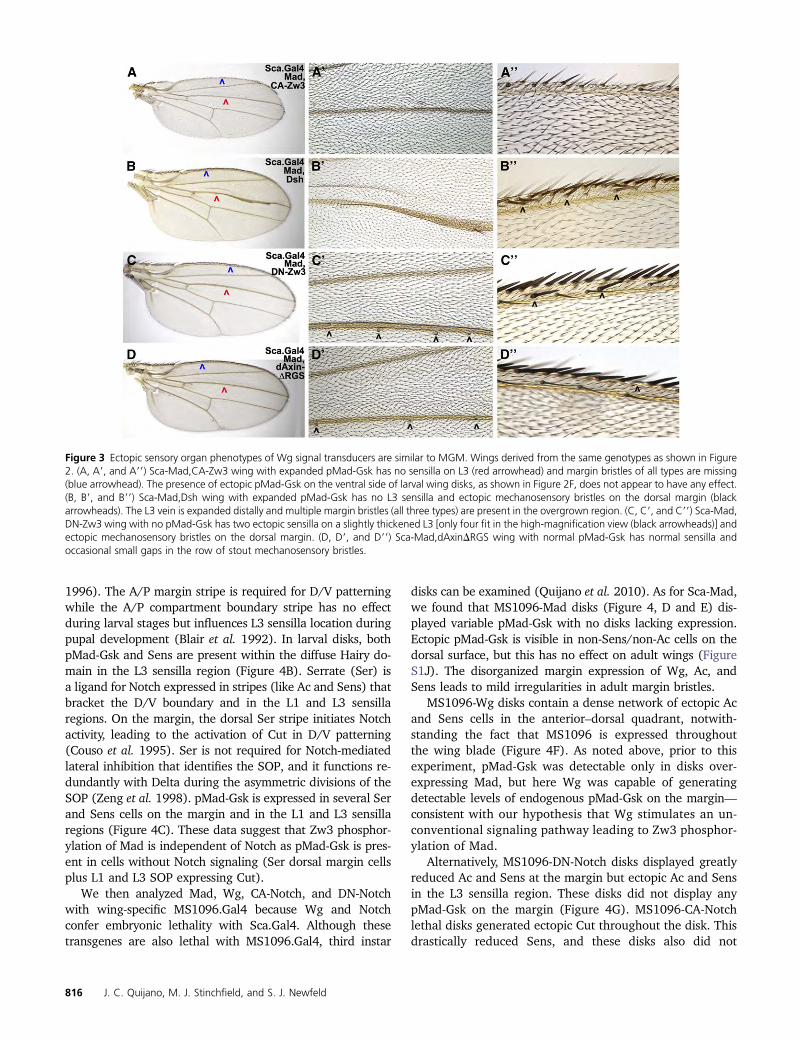
Wings derived from disks with altered Wg-signaling (e.g.,Sca-CA-Zw3 or Sca-Dsh) displayed phenotypes consistentwith the ectopic sensory organ phenotypes of Sca-MGMand are best explained by Wg activation of both its canonicalpathway and the proposed unconventional Dsh-Zw3-Madpathway. Sca-CA-Zw3 wings with significantly reduced Wgsignaling but expanded pMad-Gsk display the opposite phe-notype of Sca-MGM (no pMad-Gsk): Sca-CA-Zw3 wings haveno L1 or L3 sensilla, are missing margin bristles of all types,and display occasional vein truncations (Figure 3A, TableS3A). We attribute the vein and sensilla defects in Sca-CA-Zw3 wings to an overabundance of Zw3-phosphorylated Madand the margin defects to reduced canonical Wg signalingleading to loss of Ac. Sca-Dsh wings with significant ectopicWg signaling and expanded pMad-Gsk also contain the op-posite phenotype of Sca-MGM: no L1 or L3 sensilla and veintruncations that we attribute to an overabundance of Zw3-phosphorylated Mad (Figure 3B, Table S3B). We attribute theectopic mechanosensory bristles on the dorsal margin in Sca-Dsh wings to ectopic canonical Wg signaling.
In addition to sensilla, vein, and margin defects similar tothose of Sca-CA-Zw3, Sca-Dsh wings display distal L3 veinovergrowth with ectopic margin bristles on the overgrownregion. This distal L3 vein phenotype is attributable to theloss of Notch signaling as Dsh also antagonizes the Notchpathway (Axelrod et al. 1996). For example, expression ofa Mind bomb mutant transgene that also antagonizes Notch(Lai et al. 2005) (utilizing Dpp.Gal4 that overlaps the stripeof Sca.Gal4 expression underlying the L3 vein; see also Fig-ure S2D) results in a phenotype with important similaritiesand differences in the Sca-Dsh L3 vein phenotype. The sim-ilarity is that both genotypes resulted in L3 vein distal
814 J. C. Quijano, M. J. Stinchfield, and S. J. Newfeld

widening with multiple ectopic sensory organs (sensilla orbristles), which supports the notion that this aspect ofthe phenotype is due to loss of Notch. The difference is thatthe Mind bomb wing displayed multiple ectopic sensilla onthe L3 vein while the Sca-Dsh wing lacked any sensilla onL3. This difference suggests that the absence of sensilla inthe Sca-Dsh wing is not due to loss of Notch.
Sca-DN-Zw3 wings, with modest ectopic Wg signalingand no pMad-Gsk expression, weakly phenocopied wingswith zw3M11 clones, and the phenotype is again bestexplained by invoking the canonical and proposed uncon-ventional (Dsh-Zw3-Mad) Wg pathways. Loss of the uncon-ventional pathway explains why Sca-DN-Zw3 wings aresimilar to Sca-MGM wings with ectopic vein tissue and ec-topic sensilla (Figure 3C, Table S3C). The presence of ec-topic mechanosensory bristles on the margin is attributed toectopic canonical Wg signaling. Wings with Sca-dAxinDRGSthat have modestly reduced Wg signaling [dAxinDRGS isless potent than CA-Zw3 (Quijano et al. 2010)] and no im-pact on pMad-Gsk expression also did not contain defects insensilla or chemosensory bristles (Figure 3D, Table S3D).We attribute occasional gaps in the stout mechanosensorybristle row to a reduction in canonical Wg signaling.
To better evaluate the hypothesis that expanded pMad-Gsk in Sca-Dsh wings is not associated with Notch, weconducted additional studies in larval wing disks. Cut isa transcription factor activated by Notch in an A/P stripeatop the wing margin (de Celis and Bray 1997) where itfunctions in D/V patterning. Cut is activated independentlyby proneural genes downstream of Wg in SOP where, likeSens, it persists and is required for sensory organ differen-tiation (Jarman and Ahmed 1998). pMad-Gsk is not visiblein the Cut cells atop the margin but is visible in Cut and SensSOP in the L1 and L3 sensilla regions (Figure 4A).
Hairy is a transcription factor downstream of Notch thatis expressed in two intersecting stripes. One runs A/P alongthe margin while the other runs D/V atop the A/Pcompartment boundary and includes diffuse expression inthe L3 sensilla region (Carroll and Whyte 1989, de Celis et al.
Figure 2 pMad-Gsk is expressed in larval wing SOP and responds to Wgsignal transducers. Stacked confocal images of the presumptive wingblade from larvae aged 116–120 hr AEL with anterior to the left (as inFigure 1A, left inset) displaying pMad-Gsk (green), Ac (nuclear; red), andSens (nuclear; blue) except as noted. We maintain this color scheme inFigures 2, 4, 5, 6, and 7. (A) Sca-Mad disk appears wild type with Ac andSens in two rows of SOP (middle arrowhead; Ac anterior margin only). Acand Sens expressing SOP for sensilla (L1: left arrowhead; L3: right arrow-head) are also visible. (B) Sca-Mad disk with several SOP on the dorsalmargin and L1/L3 regions that express nuclear Ac and Sens and uniform(cytoplasmic and nuclear) pMad-Gsk. Boxed area is magnified twofoldand shown as two insets to document the subcellular localization of
pMad-Gsk: three-color (left) and two-color (pMad-Gsk and Ac; right). Theventral-most of the three pMad-Gsk cells to the right of the boxed area isa rare example that does not also express either Ac or Sens. (C) Sca-Maddisk labeled with Wg instead of Ac. Several cells on the margin and L1/L3sensilla regions that express nuclear Sens also express cytoplasmic pMad-Gsk. Overall, pMad-Gsk was seen in 10.26 6.7 cells (n ¼ 108), 4.86 4.5anterior margin cells, 2.8 6 2.8 L1 sensilla region cells, and 2.7 6 2.1 L3sensilla region cells. Insets: three-color (left), two-color [pMad-Gsk andSens (middle)], and pMad-Gsk (right). (D) Sca-Mad,MadRNAi disk with nopMad-Gsk. (E) Sca-MGM disk with no pMad-Gsk, a phenotype also seenwith Sca-Mad, MGM. (F) Sca-Mad,CA-Zw3 disk with expanded pMad-Gsk in the L1/L3 sensilla regions and ectopic pMad-Gsk on the ventralsurface (above the margin rows). (G) Sca-Mad,Dsh disk displays expandedpMad-Gsk in the L1/L3 sensilla regions. (H) Sca-Mad,DN-Zw3 disk withno pMad-Gsk. (I) Sca-Mad,dAxinDRGS disk similar to Sca-Mad (no effecton pMad-Gsk). (J) ScaMad,DN-Tkv disk similar to Sca-Mad (no effect onpMad-Gsk).
Zw3-Mad Restricts Sens Self-Renewal 815
1996). The A/P margin stripe is required for D/V patterningwhile the A/P compartment boundary stripe has no effectduring larval stages but influences L3 sensilla location duringpupal development (Blair et al. 1992). In larval disks, bothpMad-Gsk and Sens are present within the diffuse Hairy do-main in the L3 sensilla region (Figure 4B). Serrate (Ser) isa ligand for Notch expressed in stripes (like Ac and Sens) thatbracket the D/V boundary and in the L1 and L3 sensillaregions. On the margin, the dorsal Ser stripe initiates Notchactivity, leading to the activation of Cut in D/V patterning(Couso et al. 1995). Ser is not required for Notch-mediatedlateral inhibition that identifies the SOP, and it functions re-dundantly with Delta during the asymmetric divisions of theSOP (Zeng et al. 1998). pMad-Gsk is expressed in several Serand Sens cells on the margin and in the L1 and L3 sensillaregions (Figure 4C). These data suggest that Zw3 phosphor-ylation of Mad is independent of Notch as pMad-Gsk is pres-ent in cells without Notch signaling (Ser dorsal margin cellsplus L1 and L3 SOP expressing Cut).
We then analyzed Mad, Wg, CA-Notch, and DN-Notchwith wing-specific MS1096.Gal4 because Wg and Notchconfer embryonic lethality with Sca.Gal4. Although thesetransgenes are also lethal with MS1096.Gal4, third instar
disks can be examined (Quijano et al. 2010). As for Sca-Mad,we found that MS1096-Mad disks (Figure 4, D and E) dis-played variable pMad-Gsk with no disks lacking expression.Ectopic pMad-Gsk is visible in non-Sens/non-Ac cells on thedorsal surface, but this has no effect on adult wings (FigureS1J). The disorganized margin expression of Wg, Ac, andSens leads to mild irregularities in adult margin bristles.
MS1096-Wg disks contain a dense network of ectopic Acand Sens cells in the anterior–dorsal quadrant, notwith-standing the fact that MS1096 is expressed throughoutthe wing blade (Figure 4F). As noted above, prior to thisexperiment, pMad-Gsk was detectable only in disks over-expressing Mad, but here Wg was capable of generatingdetectable levels of endogenous pMad-Gsk on the margin—consistent with our hypothesis that Wg stimulates an un-conventional signaling pathway leading to Zw3 phosphor-ylation of Mad.
Alternatively, MS1096-DN-Notch disks displayed greatlyreduced Ac and Sens at the margin but ectopic Ac and Sensin the L3 sensilla region. These disks did not display anypMad-Gsk on the margin (Figure 4G). MS1096-CA-Notchlethal disks generated ectopic Cut throughout the disk. Thisdrastically reduced Sens, and these disks also did not
Figure 3 Ectopic sensory organ phenotypes of Wg signal transducers are similar to MGM. Wings derived from the same genotypes as shown in Figure2. (A, A9, and A99) Sca-Mad,CA-Zw3 wing with expanded pMad-Gsk has no sensilla on L3 (red arrowhead) and margin bristles of all types are missing(blue arrowhead). The presence of ectopic pMad-Gsk on the ventral side of larval wing disks, as shown in Figure 2F, does not appear to have any effect.(B, B9, and B99) Sca-Mad,Dsh wing with expanded pMad-Gsk has no L3 sensilla and ectopic mechanosensory bristles on the dorsal margin (blackarrowheads). The L3 vein is expanded distally and multiple margin bristles (all three types) are present in the overgrown region. (C, C9, and C99) Sca-Mad,DN-Zw3 wing with no pMad-Gsk has two ectopic sensilla on a slightly thickened L3 [only four fit in the high-magnification view (black arrowheads)] andectopic mechanosensory bristles on the dorsal margin. (D, D9, and D99) Sca-Mad,dAxinDRGS wing with normal pMad-Gsk has normal sensilla andoccasional small gaps in the row of stout mechanosensory bristles.
816 J. C. Quijano, M. J. Stinchfield, and S. J. Newfeld

contain any pMad-Gsk (Figure 4H). Although the DN- andCA-Notch results do not formally exclude the possibility thatNotch influences pMad-Gsk expression, the data clearly re-veal that Wg plays a larger role on the basis of the visibilityof pMad-Gsk without co-expression of Mad when Wg isoverexpressed. Overall, we conclude that the pMad-Gsk lar-val disk studies and the corresponding Sca.Gal4 adult wingphenotypes support our hypothesis that Zw3 phosphoryla-tion of Mad depends upon Wg.
Zw3-phosphorylated Mad is present in sensory organlineage cells in prepupal wings
We then further tested our hypothesis that Zw3-phosphorylatedMad functions specifically in sensilla and dorsal chemo-sensory bristle development in the anterior–dorsal quadrantof the wing. Here we examined pMad-Gsk in Sca-Mad pre-pupal wings (1–3 hr APF) with a set of sensory organ line-age markers. The sensory organ lineage, the cell-type-specific markers employed, and the rationale behind whythese markers were chosen are shown in Figure S3. At 1–3hr APF in prepupal wings, Sens is expressed at high levels inanterior–dorsal chemosensory SOP as these cells begin theirasymmetric divisions. By 8–10 hr APF these divisions arecomplete, and clusters of differentiated cells correspondingto each dorsal chemosensory bristle are present (Jafar-Nejadet al. 2006). Then the mechanosensory SOP cells atop themargin begin to differentiate (Hartenstein and Posakony1989).
To provide a reference for these results, we examinedSac-lacZ and Dpp-lacZ prepupal wings. Here these geneshave the same spatial relationship, to each other and to theSOP markers Sens and Cut, as in larval wings (Ac is notexpressed in prepupal wings; Figure S4). One unexpected
Figure 4 pMad-Gsk expression is dependent on Wg but not on Notchsignaling. Stacked (A–C) and single confocal images (D–H) of the pre-sumptive wing blade as shown in Figure 2 displaying pMad-Gsk (green),Sens (blue), and a cell-type-specific marker (red). Boxed areas are magni-fied twofold and shown in insets to better visualize pMad-Gsk expression.(A) Sca-Mad disk appears wild type with Notch-dependent Cut (nuclear;red) along the margin and Wg-dependent Cut in the L1/L3 sensillaregions. pMad-Gsk is expressed in multiple Cut and Sens cells in the L1(left arrowhead) and L3 (right arrowhead) sensilla regions. (Insets) Three-color (left), two-color [pMad-Gsk and Cut (middle)], and Sens (right). (B)Sca-Mad disk appears wild type with Notch-dependent Hairy (nuclear;red) along the A/P boundary with diffuse expression in the L3 sensillaregion and atop the D/V boundary but not in the L1 sensilla region.pMad-Gsk and Sens cells are visible within the diffuse Hairy domain inthe L3 sensilla region. (C) Sca-Mad disk appears wild type with Ser (red)expression in two rows flanking the intersecting stripes of Hairy on theA/P and D/V boundaries plus diffuse expression in the L1 and L3 sensilla
regions. pMad-Gsk is expressed in several Ser and Sens cells on themargin and in the L3 sensilla region. (D) MS1096-Mad disk withdisorganized Wg-lacZ (red) and Sens on the margin. Sens generallybrackets Wg-lacZ but is occasionally surrounded by Wg-lacZ cells. pMad-Gsk is present in two Sens cells in the dorsal bristle row and in numerousectopic cells throughout the dorsal surface. Overall, pMad-Gsk was visiblein 16.5 6 14.9 cells (n ¼ 6), 13.9 6 12.6 anterior margin cells, 0.8 6 1.3L1 sensilla region cells, and 1.86 1.5 L3 sensilla region cells. (E) MS1096-Mad disk with disorganized Ac (red) and Sens along the margin consis-tent with abnormal Wg-lacZ in this genotype. (Insets) Ac (red) on the leftand pMad-Gsk (green) on the right. There is no pMad-Gsk expression onthe margin but a single pMad-Gsk, Sens, and Cut cell is present in the L3sensilla region (located under the right inset and not visible). (F) MS1096-Wg lethal disk with ectopic Ac (red) and Sens throughout the anterior–dorsal quadrant. (Insets) Ac (red) on the left and pMad-Gsk (green) on theright. pMad-Gsk is present in five cells along the margin (right) that co-express Ac and Sens. Two pMad-Gsk cells are indicated by arrowheads.(G) MS1096-DN-Notch lethal disk with greatly reduced Ac (red) and dis-organized Sens at the margin but numerous ectopic Ac and Sens cells inthe L3 sensilla region. (Insets) Ac (red) on the left and pMad-Gsk (green)on the right. No pMad-Gsk expression is visible. (H) MS1096-CA-Notchlethal disk with ectopic Cut (red) throughout the disk. Several Sens cellsare present at random, and there is no pMad-Gsk. (Insets) Cut (red) onthe left and pMad-Gsk (green) on the right. No pMad-Gsk expression isvisible.
Zw3-Mad Restricts Sens Self-Renewal 817

finding was that the SOP lineage on the wing margin is notidentical to the lineage of the notum: Sox15, a transcriptionfactor marking the socket cell on the notum (Miller et al.2009), is not present in prepupal wings. As a result, weutilized Su(H) to mark the socket cell. In addition, we foundthat the transient glial cell (expressing Repo) is present at1–3 hr APF prior to undergoing apoptosis. An analysis utiliz-ing the apoptosis marker cleaved-Caspase3 confirmed thisfinding, as no cell death was visible in prepupal wings at1–3 hr APF.
We found that, in prepupal wings, pMad-Gsk was co-expressed with every one of our cell-type-specific SOP lineagemarkers (Figure 5). As in larval disks, the number of pMad-Gsk cells was highly variable. Other similarities between pu-pal and larval pMad-Gsk margin expression were: (1) Wg wasalways adjacent to pMad-Gsk (Figure 5H), and no diskslacked pMad-Gsk expressing cells; (2) except for dPax2 andCut (Figure 5, E, I, and J), pMad-Gsk was on the dorsal side;and (3) pMad-Gsk was rarely seen without Sens (Figure 5I).
The subcellular localization of pMad-Gsk varied in pupalwings as it did in larval wings. With all of the markers pMad-Gsk uniform expression (nuclear plus cytoplasm) was themost common (Figure 5). For six of the eight markers, cellswith cytoplasmic pMad-Gsk were present as well as cellsdisplaying uniform expression (dPax2: Figure 5E, Cut: Fig-ure 5J). Alternatively, for Repo and Elav cells (Figure 5, D
Figure 5 pMad-Gsk is present in sensory organ lineage cells in prepupalwing disks. Single confocal slices of the dorsal side of the anterior marginfrom a prepupal wing with a Sca-Mad genotype. Except as noted, wingsare aged 1–3 hr APF and shown with proximal to the left as in Figure 1A,right inset. Wings display pMad-Gsk (green), Sens (blue), and a cell-type-specific marker (red) except as noted. Overall, pMad-Gsk was present in11.4 6 5.6 cells (n = 87), 8.8 6 4.3 anterior margin cells, 1.1 6 1.3 L1sensilla region cells, and 1.66 1.6 L3 sensilla region cells. Boxed areas areenlarged twofold and shown as two insets to better document the sub-cellular localization of pMad-Gsk: three-color (left) and two-color (pMad-Gsk and marker; right). (A) E(spl)M8 is a transcription factor that markscells with active Notch signaling (e.g., shaft and neuron). The two E(spl)
M8 and Sens cells to the left of the box display uniform pMad-Gsk.(Insets) The E(spl)M8 and Sens endocycling cell on the right with twonuclei displays cytoplasmic pMad-Gsk. (B) Su(H) is a transcription factorthat marks the socket cell. (Insets) Three endocycling Su(H) and Sens cellsexpress pMad-Gsk uniformly. (C) Futsch (monoclonal antibody 22C10) isa microtubule-associated protein that marks neurons. (Insets) A 22C10and Sens endocycling cell on the right with uniformly distributed pMad-Gsk. (D) Repo is a transcription factor marking the transient glial cell.(Insets) The Repo and Sens endocycling cell in the center with two nucleiexpresses uniform pMad-Gsk. (E) Cut is a transcription factor and targetof Notch that marks the wing margin in a dense row of cells that do notexpress any sensory organ lineage markers. Cut is also activated in SOP onboth sides of the margin by proneural genes and will mark all cells of thesensory organ lineage. dPax2 is a transcription factor that marks SOP,shaft, and sheath cells. (Insets) The three distal-most cells on the ventralsurface display varying nuclear concentrations of Cut and dPax2 and alsoshow a transition from cytoplasmic to uniform pMad-Gsk (top to bottom).(F) Pros is a transcription factor normally sequestered in the cytoplasm butthat translocates to the nucleus when active in the sheath cell. (Insets) Leftcell has nuclear Pros and Sens with uniformly distributed pMad-Gsk whilethe right cell has cytoplasmic Pros, nuclear Sens, and no pMad-Gsk. (G)Elav is an RNA-binding protein that marks neuronal cells. (Insets) Twoendocycling cells express Elav, Sens, and uniform pMad-Gsk. (H) Confocalstack revealing that Wg on the dorsal margin encompasses pMad-Gsk-and Sens-expressing cells. (Insets) Three Sens cells expressing uniformpMad-Gsk. (I) Lateral view of an everting disk at 0–1 hr APF expressingCut, Sens, and pMad-Gsk. (Insets) The two distal-most Cut and Sens cellsthat straddle the row of Cut-only margin cells express Cut, Sens, anduniform pMad-Gsk. The cell at the right expresses only uniform pMad-Gsk. The circular empty areas in these cells are likely vesicles as the cell onthe left has four of them. (J) At 1–3 hr APF the disk has flattened. (Insets)The Cut and Sens cell on the left displays uniformly distributed pMad-Gsk.The four cells on the right restrict pMad-Gsk to the cytoplasm but only theventral-most expresses Cut and Sens.
818 J. C. Quijano, M. J. Stinchfield, and S. J. Newfeld
and G), pMad-Gsk was only uniformly expressed. Severalexperiments suggest that pMad-Gsk is associated with en-docycles of DNA replication (without mitosis leadingto polyploidy) that occur after the initiation of cell-type-specific gene expression in the neuron, shaft, and socketcells (Hartenstein and Posakony 1989; Audibert et al.2005). For example, E(Spl)M8 (Figure 5A, shaft or neuron),Su(H) (Figure 5B, socket), and 22C10 (Figure 5C, neuron)cells with two nuclei were observed that contain uniformpMad-Gsk. To date, no one has examined endoreplicationin the transient glial cell, but cells with two nuclei express-ing uniform pMad-Gsk and Repo are visible, suggesting thatthis cell type also undergoes endocycling (Figure 5D).
Overall, the prepupal wing data support our thirdhypothesis that Zw3-phosphorylated Mad functions primar-ily in sensilla and dorsal chemosensory bristle development.Furthermore, the association of pMad-Gsk with non-mitoticendocycles of DNA replication in terminally differentiatedsensory organ cells suggests a hypothesis by which MGM (aloss-of-function allele) and Mad-RNAi would lead to ectopicsensory organs. This hypothesis is that the normal functionof Zw3-phosphorylated Mad is to restrict mitosis in thesensory organ lineage.
MGM and Mad-RNAi generate ectopic Sens SOP in theanterior–dorsal quadrant of the wing
To test this hypothesis, we analyzed the co-expression ofpMad-Gsk, Sens, and the chromosome condensation markerphospho-histone3 (pH3) in prepupal wings expressing Sca-Mad,Sca-MadRNAi, or Sca-MGM. pH3 is generally utilized tomark mitotic cells but is also expressed during endocycles ofDNA replication such as those in ovarian follicle cells (Sunet al. 2008). If the role of Zw3-phosphorylated Mad in thesensory organ lineage is to restrict mitosis, then MadRNAiand MGM should display more Sens or pH3 cells than thephenotypically wild-type Sca-Mad. We characterized Sca.Gal4 spatial expression in prepupal wings to aid in interpret-ing these phenotypes. Here the larval relationship betweenthe Sca and Dpp D/V stripes persists: the stripe of Sca over-laps the anterior portion of the parallel stripe of Dpp (Dpppupal enhancer does not activate until 24 hr APF; Affolterand Basler 2007). As a result, in prepupal wings the Sca
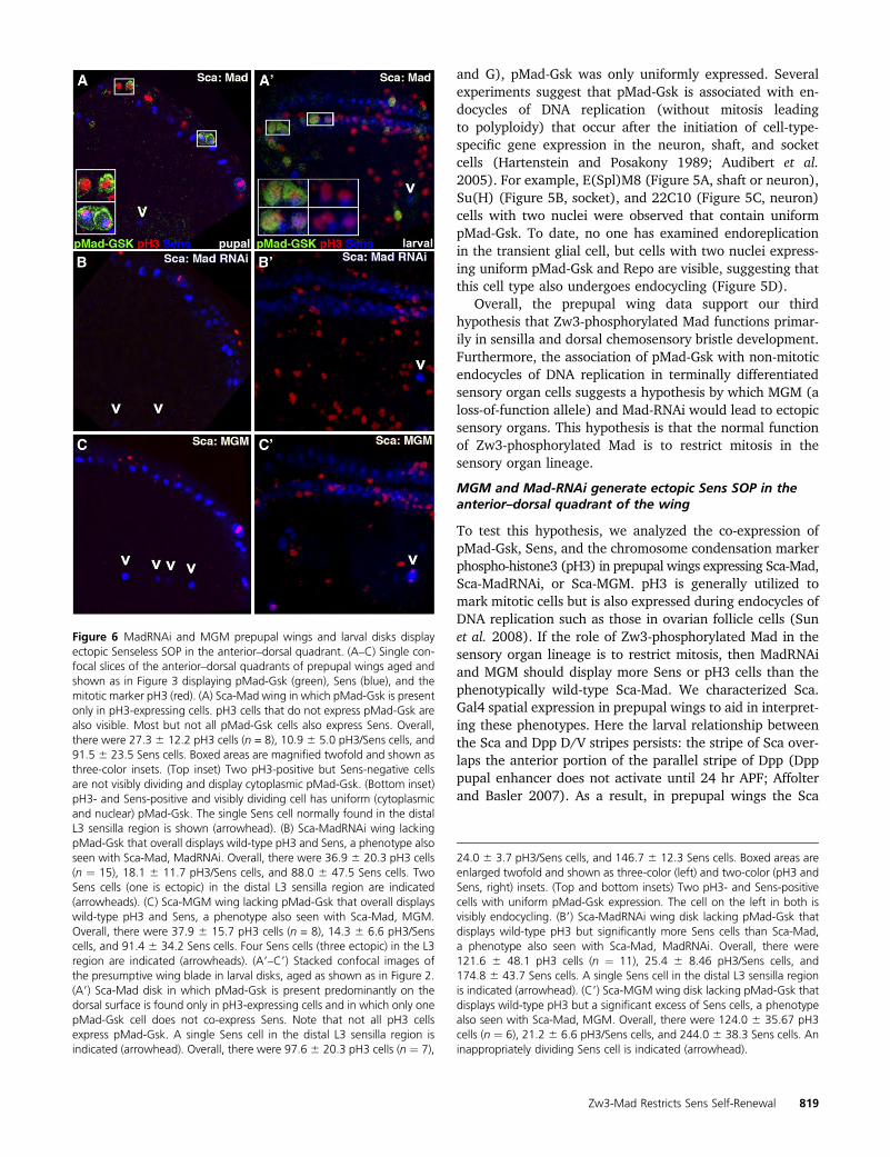
Figure 6 MadRNAi and MGM prepupal wings and larval disks displayectopic Senseless SOP in the anterior–dorsal quadrant. (A–C) Single con-focal slices of the anterior–dorsal quadrants of prepupal wings aged andshown as in Figure 3 displaying pMad-Gsk (green), Sens (blue), and themitotic marker pH3 (red). (A) Sca-Mad wing in which pMad-Gsk is presentonly in pH3-expressing cells. pH3 cells that do not express pMad-Gsk arealso visible. Most but not all pMad-Gsk cells also express Sens. Overall,there were 27.3 6 12.2 pH3 cells (n = 8), 10.9 6 5.0 pH3/Sens cells, and91.5 6 23.5 Sens cells. Boxed areas are magnified twofold and shown asthree-color insets. (Top inset) Two pH3-positive but Sens-negative cellsare not visibly dividing and display cytoplasmic pMad-Gsk. (Bottom inset)pH3- and Sens-positive and visibly dividing cell has uniform (cytoplasmicand nuclear) pMad-Gsk. The single Sens cell normally found in the distalL3 sensilla region is shown (arrowhead). (B) Sca-MadRNAi wing lackingpMad-Gsk that overall displays wild-type pH3 and Sens, a phenotype alsoseen with Sca-Mad, MadRNAi. Overall, there were 36.9 6 20.3 pH3 cells(n ¼ 15), 18.1 6 11.7 pH3/Sens cells, and 88.0 6 47.5 Sens cells. TwoSens cells (one is ectopic) in the distal L3 sensilla region are indicated(arrowheads). (C) Sca-MGM wing lacking pMad-Gsk that overall displayswild-type pH3 and Sens, a phenotype also seen with Sca-Mad, MGM.Overall, there were 37.9 6 15.7 pH3 cells (n = 8), 14.3 6 6.6 pH3/Senscells, and 91.4 6 34.2 Sens cells. Four Sens cells (three ectopic) in the L3region are indicated (arrowheads). (A9–C9) Stacked confocal images ofthe presumptive wing blade in larval disks, aged as shown as in Figure 2.(A9) Sca-Mad disk in which pMad-Gsk is present predominantly on thedorsal surface is found only in pH3-expressing cells and in which only onepMad-Gsk cell does not co-express Sens. Note that not all pH3 cellsexpress pMad-Gsk. A single Sens cell in the distal L3 sensilla region isindicated (arrowhead). Overall, there were 97.6 6 20.3 pH3 cells (n ¼ 7),
24.0 6 3.7 pH3/Sens cells, and 146.7 6 12.3 Sens cells. Boxed areas areenlarged twofold and shown as three-color (left) and two-color (pH3 andSens, right) insets. (Top and bottom insets) Two pH3- and Sens-positivecells with uniform pMad-Gsk expression. The cell on the left in both isvisibly endocycling. (B9) Sca-MadRNAi wing disk lacking pMad-Gsk thatdisplays wild-type pH3 but significantly more Sens cells than Sca-Mad,a phenotype also seen with Sca-Mad, MadRNAi. Overall, there were121.6 6 48.1 pH3 cells (n ¼ 11), 25.4 6 8.46 pH3/Sens cells, and174.8 6 43.7 Sens cells. A single Sens cell in the distal L3 sensilla regionis indicated (arrowhead). (C9) Sca-MGM wing disk lacking pMad-Gsk thatdisplays wild-type pH3 but a significant excess of Sens cells, a phenotypealso seen with Sca-Mad, MGM. Overall, there were 124.0 6 35.67 pH3cells (n ¼ 6), 21.2 6 6.6 pH3/Sens cells, and 244.0 6 38.3 Sens cells. Aninappropriately dividing Sens cell is indicated (arrowhead).
Zw3-Mad Restricts Sens Self-Renewal 819

stripe still coincides with the highest levels of Receptor-phosphorylated Mad in the anterior compartment (FigureS4, C and D).
Our examination of pMad-Gsk and pH3 expression inSca-Mad prepupal wings (which will generally lead tophenotypically normal adult wings; Figure 1A, Table S1A)revealed that pMad-Gsk is present only in pH3-positive cellsand that most, but not all, pMad-Gsk cells also express Sens(Figure 6A). As our hypothesis predicts ectopic pH3 and/orSens expression in the other genotypes, we counted pH3and Sens cells in Sca-Mad disks and found that expressionwas highly variable. Sca-MadRNAi prepupal wings lackingpMad-Gsk (25–35% of adult wings have ectopic sensilla orchemosensory bristle duplications; Figure 1B, Table S1B)
were not significantly different from Sca-Mad wings. How-ever, we noted that there were �22% more Sens cells in theL3 sensilla region of Sca-MadRNAi than Sca-Mad (5.8 vs. 4.6Sens cells in the L3 sensilla region). Careful examinationrevealed that a subset of Sca-MadRNAi wings display a singleectopic Sens cell in this region (Figure 6B). Sca-MGM pre-pupal wings lacking pMad-Gsk (40–60% of adult wings haveectopic sensilla or chemosensory bristle duplications; Figure1C, Table S1C) were also not significantly different from Sca-Mad. Again we noted�25%more Sens cells in the L3 sensillaregion (6.0 vs. 4.6) because a subset of Sca-MGM wings dis-play one or two ectopic Sens cells in this region (Figure 6C).
The presence of ectopic Sens in the L3 sensilla region ofSca-MadRNAi and Sca-MGM prepupal wings suggested thatthe defect due to loss of Zw3-phosphorylated Mad occurredin larvae. Examining Sca-Mad larval disks, we noted thatpMad-Gsk is present only in pH3-expressing cells and thatmost, but not all, pMad-Gsk cells also express Sens. At thisstage, pH3 cells with uniform (cytoplasmic and nuclear)pMad-Gsk expression are actively dividing (Figure 6A9). Inlarval disks, pMad-Gsk expression was visible in more cellsthan in prepupal wings. Sca-MadRNAi larval disks (Figure6B9) contained similar numbers of pH3 cells and pH3/Senscells as Sca-Mad but they had significantly more Sens cells(174.8 6 43.7 vs. 146.7 6 12.3; P , 0.05). Sca-MGM larvaldisks (Figure 6C9) contained similar numbers of pH3 cellsand pH3/Sens cells as Sca-Mad, but they also had signifi-cantly more Sens cells (244.0 6 38.3 vs. 146.7 6 12.3; P ,0.006). Disks with inappropriately dividing Sens cells in theL3 sensilla region are present even in a small sample of sixSca-MGM disks. The excess of Sens cells in Sca-MadRNAiand Sca-MGM larval disks suggests that the mitotic defectdue to loss of Zw3-phosphorylated Mad occurred betweenthe activation of Ac at 95–96 hr AEL and the analysis of Sensat 116–120 hr AEL.
Discussion
Zw3 phosphorylation provokes a distinct response fromreceptor-phosphorylated and nonphosphorylated Mad
Two studies of Zw3/Gsk3-b phosphorylation of Mad/Smad1at the homologous sites in flies and vertebrates (Fuentealbaet al. 2007; Eivers et al. 2009) and one of Gsk3-b phosphor-ylation of Smad3 at a nonhomologous site in mammals (Guoet al. 2008) have shown that phosphorylation leads to Smadpolyubiquitination, degradation, and TGFb signal termina-tion. The data predict that blocking this event via site-directed mutagenesis (as in UAS.MGM) would lead toa gain-of-function allele of Mad—one with hyperactivity thatgenerates phenotypes similar to Mad overexpression. Hyper-activity of MGM was reported by those investigators and cor-roborated by the presence of ectopic veins in our Sca-MGMwing studies. However, detailed analysis of our Sca-MGMwings revealed that hyperactivity is evident only for Mad’sDpp-dependent phenotypes (e.g., ectopic veins). We noteda phenotype in Sca-MGM wings that has not been previously
Figure 7 Model of an unconventional Wg pathway leading to Zw3-phosphorylated Mad activity in non-SOP and SOP cells within the anterior–dorsal quadrant of the wing. (A) Schematic of the anterior–dorsal quad-rant of a late larval wing disk displaying Ac expression. By this time, Wgactivation of Ac has defined the proneural prepattern, and Dpp activationof spalt and omb has specified vein precursor cells. The outlined sectionof the margin and L3 sensilla region is shown as two confocal stacksdepicting Wg-lacZ and Ac (left) and Dpp-lacZ and Ac (right). Two cellsare boxed: a cell expressing high levels of Ac due to feedback from Sensthat will become an L3 sensilla SOP (right) and presumably an L3 veinprecursor (left) that does not express Ac. (B) Schematic of events occur-ring within the L3 vein precursor (left) and the L3 SOP sensilla precursor(right). Dpp does not play a primary role in L3 vein specification, but it isnonetheless required upstream of rhomboid for proper L3 vein formation(Gomez-Skarmeta and Modolell 1996; Biehs et al. 1998). In the L3 veinprecursor (left) that does not express Ac or Sens, Wg-dependent Zw3phosphorylation of Receptor-phosphorylated Mad leads to degradationand the termination of this round of Dpp signaling. Zw3 phosphorylationof nonphosphorylated Mad, if it occurs here, appears to be inconsequen-tial on the basis of the MGM wing phenotype. In the L3 sensilla SOP thatexpresses Ac and Sens, Wg-dependent Zw3 phosphorylation of nonphos-phorylated Mad leads to nuclear accumulation and the restriction of self-renewing mitosis. Zw3 phosphorylation of Receptor-phosphorylatedMad, if it occurs here, may be among the mechanisms used to renderthese cells nonresponsive to Dpp.
820 J. C. Quijano, M. J. Stinchfield, and S. J. Newfeld

associated with Dpp signaling: ectopic sensilla and dorsalchemosensory bristle duplications. The sensory organ pheno-type was also seen with MadRNAi, suggesting that this subsetof the MGM phenotype represents a previously unnoticedloss-of-function phenotype that we have shown is indepen-dent of Dpp and Notch and dependent upon Wg signaling.
One simple explanation for the existence of two distincteffects of Zw3 phosphorylation on Mad activity in wingdevelopment is that Zw3 phosphorylation influences Re-ceptor-phosphorylated Mad distinctly from nonphosphory-lated Mad. We model the unconventional Wg pathway andthis biphasic response in Figure 7. If Mad’s C-terminal SSVSamino acid sequence is already phosphorylated, then Zw3phosphorylation in the linker region leads to ubiquitinationand signal termination. In the larval wing disk, this occurs invein precursor cells and results in the ectopic veins of MGMand DN-Zw3 wings as well as the loss of veins in CA-Zw3 andMadRNAi wings. This aspect of the Zw3–Mad interaction isconsistent with prior studies and is further supported by Gaoet al. (2009). On the basis of studies in mammalian cells, theyidentified Nedd4L and Smurf1 as ubiquitin ligases recogniz-ing doubly phosphorylated (Receptor and Gsk3-b) Smad2/3or Smad1, respectively. Wnt-induced Zw3 phosphorylationof Receptor-phosphorylated Smads likely represents a con-served mechanism for TGFb signal termination.
Our Sca.Gal4 wing assays suggested that nonphosphory-lated Mad can also respond to Zw3 phosphorylation andthat it does so by performing a distinct function. This novelfunction is the prevention of self-renewing mitotic divisionby Sens SOP in the anterior–dorsal quadrant of the wing(a region spatially defined by the presence of Apterous andthe absence of Engrailed). This novel function of Zw3 phos-phorylated but non-Receptor phosphorylated Mad cannotbe achieved when MGM and DN-Zw3 are expressed (fail-ure to restrict self-renewing mitosis in Sens SOP leads toectopic sensory organs) and is hyperactive when CA-Zw3and Dsh are expressed (complete restriction of Sens SOPmitosis leading to loss of sensilla).
Returning to the studies that prompted this analysis,Eivers et al. (2009) reported an increase in Sens expressionalong the anterior margin in larval disks and overgrowth ofdorsal chemosensory bristles in the resulting adult wingwhen expressing MGM with Scalloped.Gal4. Scalloped.Gal4 is expressed widely on both wing surfaces but maxi-mally at the margin where it is required for formation ofbristles (Campbell et al. 1992). Eivers et al. (2009) con-cluded that Wg signals through Mad to repress Sens expres-sion in wing development. Our studies agree with the firstpart of this conclusion and extend their observations by show-ing that MGM-induced Sens increase is limited to a specificcell type (anterior–dorsal SOP) and is perhaps due to the lossof restriction on self-renewal in those cells. Alternatively, Zenget al. (2008) reported that flip-out clones of Mad repressedSens and concluded that Receptor-phosphorylated Mad activ-ity antagonizes Wg signaling. In their study, flip-out cloneswere generated between 72 and 96 hr AEL, before Ac activa-
tion byWg at 94–96 hr AEL and the activation of Sens in SOP.In the earlier period, Dpp and Wg morphogens provide globalpositional information utilized by cells for their initial cell fatedecisions. Thus Dpp antagonism of Wg via Mad during earlythird instar is likely independent of Wg-stimulated Zw3 phos-phorylation of Mad later in wing development.
Recently, an analysis of canonical Wnt signaling in whichGsk3-b moves to the membrane to phosphorylate low densitylipoprotein receptor related protein (LRP) co-receptors forsignal amplification showed that Gsk3-b is then sequesteredinto endosomes. This requires b-catenin and effectively iso-lates Gsk3-b from additional substrates (Taelman et al. 2010).Our data showing that Zw3-phosphorylated Mad is associatedwith an unconventional branch of Wg signaling that does notemploy canonical Wg pathway components in their normalway or any canonical components downstream of Zw3 (e.g.,dAxin) suggest that Zw3/Gsk3-b sequestration may not betriggered by this mechanism or that sequestration is not rapidor complete enough to prevent Zw3 phosphorylation of Mad.
Zw3-phosphorylated Mad is expressed during mitosis inlarval wing disks and functions to prevent Sens SOPself-renewing divisions
The observation that pMad-Gsk is present only in pH3-positive mitotic cells during larval wing development fitswith the work of Fuentealba et al. (2008) who noted pSmad1-Gsk expression only during self-renewing divisions of humanembryonic stem cells. Furthermore, their data revealed thatpSmad1-Gsk is asymmetrically segregated into only one ofthe daughter cells during these divisions. These authors notedthat Receptor-phosphorylated Smad1 was also asymmetric (al-though less so than pSmad1-Gsk). Double phospho-stainingexperiments were not performed, but our data suggest thatthe asymmetrically segregated pSmad1 may be the dual phos-phorylated form (Receptor and Gsk3-b).
Alternatively, our data show that Zw3-phosphorylatedMad that is not Receptor phosphorylated is present in SensSOP that are unaffected by TGFb signaling. These Sens SOPdifferentiate into two non-identical daughter cells via asym-metric division. As a result, in the presence of Zw3-resistantMad (MGM), a larval Sens SOP undergoes a single self-renewing division prior to its typical differentiation divisionto generate an ectopic Sens SOP that becomes either anectopic sensilla or an ectopic dorsal chemosensory bristle.Thus, the activity of Zw3-phosphorylated Mad may be influ-enced by several factors: the presence of Receptor phosphor-ylation, the cell type, and the type of mitosis (self-renewalvs. differentiation).
The nuclear accumulation of pMad-Gsk during mitosis inlarval Sens SOP shares several features with the behavior ofZw3 observed with a GFP-exon trap allele in third instar larvalcentral brain neuroblasts (Wojcik 2008). During differentiation(i.e., an asymmetric division generating a new neuroblastand a distinct ganglion mother cell) it was shown thatZw3 is cytoplasmic during interphase and prophase. At theonset of metaphase, Zw3 nuclear accumulation begins and is
Zw3-Mad Restricts Sens Self-Renewal 821

visible through cytokinesis when Zw3 becomes cytoplasmicagain in each of the two new cells. These similarities suggestthat perhaps Zw3-phosphorylated Mad and/or Zw3 maycontribute to a common function in these cells: facilitationof differentiating vs. self-renewing division in cells capableof both.
New features of Wg, Sens, and Mad activity inwing development
Our data also illuminate new aspects of wing and sensoryorgan formation. From a global patterning perspective, weprovide new insights into how Hedgehog (Hh) and its signaltransducer Cubitus interruptus (Ci) influence L3 sensilladevelopment. Mullor et al. (1997) showed that ectopic Hhthroughout the anterior compartment led to numerousectopic L3 veins that were accompanied by sensilla. In theirview, these cells were fooled into thinking that they were L3cells close to normal Hh at the A/P compartment boundary.Subsequently, Methot and Basler (2001) showed that Ciloss-of-function clones in the posterior compartment cangenerate an ectopic sensilla on L4. Here the interpretationwas that Hh activation of Engrailed is lost in clones, andthus a cell was fooled into thinking that it was in the ante-rior compartment near Hh at the A/P compartment bound-ary. Alternatively, no reports suggest Hh has any role in thedifferentiation of SOP into sensilla. Mullor et al. (1997)explicitly invoke an independent factor X between Hh andL3 sensilla differentiation. Our data suggest that the mostlogical candidate is Wg.
Thus, we propose that Wg continues to influence wingdisk development late in the third instar, beyond its roles inglobal D/V patterning early in the third instar and Acactivation in mid-third instar. With regard to the biochemicalunderpinnings of the unconventional pathway leading toZw3 phosphorylation of Mad during late third instar de-velopment, we note that the mechanism underlying canon-ical Wg pathway interactions between Frizzled and Dsh andbetween Dsh and Zw3 is also currently unknown. It istempting to speculate that an interaction between Zw3 andDark-dependent caspase (Kanuka et al. 2005) that does notinvolve cell death but instead influences the formation ofmechanosensory bristles on the notum may play a role.
We extend the identification of Sens as the primary factorin chemosensory bristle specification to the specification ofsensilla in the anterior–dorsal quadrant. The basis for thisextension may be that all cells in the anterior–dorsal quad-rant express Apterous and none express Engrailed. ThusSens plays the same role in all SOP within this geneticallyand spatially defined quadrant. Alternatively, the stoutmechanosensory bristles on the margin for which Sensserves as a proneural gene, independent of Ac and Scute,derive from the anterior–ventral quadrant that does not ex-press Apterous or Engrailed. Distinct consequences associatedwith Sens function in SOP development in the two quadrantsmay necessitate the restriction of self-renewing mitosis byZw3-phosphorylated Mad to the anterior–dorsal quadrant.
Intriguingly, our data identify the first known non-TGFb-dependent role for any Smad protein in any organism. Whilemany non-Smad-signaling pathways are activated by TGFbreceptors, to date Smads have not been reported to have anyfunctions independent of TGFb signaling. Even the initialstudies of Zw3-phosphorylated Mad indicated that thisevent served to terminate TGFb signaling. We are examin-ing the possibility that Mad has additional TGFb-independentroles by analyzing the effect of EGFR/MapK signaling on Madphosphorylation.
In summary, during wing development the phosphoryla-tion of Mad by Zw3 is not a mechanism of pathway crosstalkbut instead represents a spatially localized round of un-conventional Wg signaling during sensory organ develop-ment. This signal limits the self-renewal of Sens SOP cells,and this limitation may be necessary for SOP cells todifferentiate via asymmetric division leading to adult sen-sory organs. The conservation of Zw3 phosphorylation sitesin Mad’s vertebrate homologs suggests that this mechanismmay be widely utilized for balancing self-renewal and dif-ferentiation during development.
Acknowledgments
We are especially grateful to Eddy DeRobertis for sharingreagents prior to publication. We thank the BloomingtonStock Center, Hugo Bellen, Sarah Bray, the DevelopmentalStudies Hybridoma Bank, Ed Eivers, Ken Irvine, Ed Laufer,Mike O’Connor, Jim Posakony, John Reinitz, and FrancoisSchweisguth for flies, antibodies, and valuable discussions.The authors declare no competing financial interests.
Literature Cited
Affolter, M., and K. Basler, 2007 The Dpp morphogen gradient:from pattern formation to growth regulation. Nat. Rev. Genet. 9:663–674.
Aldaz, S., L. M. Escudero, and M. Freeman, 2010 Live imaging ofDrosophila imaginal disc development. Proc. Natl. Acad. Sci.USA 107: 14217–14222.
Andrews, H., N. Giagtzoglou, S. Yamamoto, K. Schulze, and H.Bellen, 2009 Sequoia regulates cell fate decisions in the sen-sory organs of Drosophila. EMBO Rep. 6: 636–641.
Audibert, A., F. Simon, and M. Gho, 2005 Cell cycle diversityinvolves differential regulation of cyclin E activity in theDrosophila bristle cell lineage. Development 132: 2287–2297.
Axelrod, J., K. Matsuno, S. Artavanis-Tsakonas, and N. Perrimon,1996 Interaction between Wingless and Notch signaling me-diated by dishevelled. Science 271: 1826–1832.
Bangi, E., and K. Wharton, 2006 Dual function of the DrosophilaAlk1/Alk2 ortholog Sax shapes the BMP activity gradient in thewing. Development 133: 3295–3303.
Biehs, B., M. Sturtevant, and E. Bier, 1998 Boundaries in theDrosophila wing imaginal disc organize vein-specific geneticprograms. Development 125: 4245–4257.
Blackman, R., M. Sanicola, L. Raftery, T. Gillevet, and W. Gelbart,1991 An extensive 39 cis-regulatory region directs the imagi-nal disk expression of dpp, a member of the TGFb family inDrosophila. Development 111: 657–665.
822 J. C. Quijano, M. J. Stinchfield, and S. J. Newfeld

Blair, S., 1992 shaggy (zeste-white 3) and the formation of super-numerary bristle precursors in the developing wing blade ofDrosophila. Dev. Biol. 152: 263–278.
Blair, S., A. Giangrande, J. Skeath, and J. Palka, 1992 Developmentof normal and ectopic sensilla in hairy and Hairy wingmutants ofDrosophila. Mech. Dev. 38: 3–16.
Bourouis, M., 2002 Targeted increase in shaggy activity levelsblocks Wingless signaling. Genesis 34: 99–102.
Brand, A., and N. Perrimon, 1993 Targeted gene expression asa means of altering cell fates and generating dominant pheno-types. Development 118: 401–415.
Brand, M., A. Jarman, L. Jan, and Y. Jan, 1993 asense is a Dro-sophila neural precursor gene and is capable of initiating senseorgan formation. Development 119: 1–17.
Brummel, T., S. Abdollah, T. Haerry, M. Shimell, J. Merriam et al.,1999 The Activin receptor Baboon signals through dSmad2and controls cell proliferation but not patterning during larvaldevelopment. Genes Dev. 13: 98–111.
Campbell, S., M. Inamdar, V. Rodrigues, V. Raghavan, M. Palazzoloet al., 1992 The scalloped gene encodes a novel, evolutionarilyconserved transcription factor required for sensory organ differ-entiation in Drosophila. Genes Dev. 6: 367–379.
Carroll, S., and J. Whyte, 1989 The role of the hairy gene duringDrosophila morphogenesis: stripes in imaginal disks. Genes Dev.3: 905–916.
Chen, Y., M. Riese, M. Killinger, and F. Hoffmann, 1998 A geneticscreen for modifiers of Drosophila dpp signaling identifies punt,Mad and 60A. Development 125: 1759–1768.
Couso, J., S. Bishop, and A. Martinez Arias, 1994 The winglesssignaling pathway and the patterning of the wing margin inDrosophila. Development 120: 621–636.
Couso, J., E. Knust, and A. Martinez Arias, 1995 Serrate and wing-less cooperate to induce vestigial gene expression and wing for-mation in Drosophila. Curr. Biol. 5: 1437–1448.
Cubas, P., J. de Celis, S. Campuzano, and J. Modolell, 1991 Proneuralclusters of acheate-scute expression and the generation of sensoryorgans in the Drosophila wing disc. Genes Dev. 5: 996–1008.
de Celis, J., and S. Bray, 1997 Feed-back mechanisms affectingNotch activation at the dorsoventral boundary in the Drosophilawing. Development 124: 3241–3251.
de Celis, J., J. de Celis, P. Ligoxygakis, A. Preiss, C. Delidakis et al.,1996 Functional relationships between Notch, Su(H) and thebHLH genes of the E(spl) complex: the E(spl) genes mediateonly a subset of Notch activities during imaginal development.Development 122: 2719–2728.
Derynck, R., and K. Miyazono, 2008 The TGFb Family Cold SpringHarbor Laboratory Press, Cold Spring Harbor, NY.
Dworkin, I., and G. Gibson, 2006 EGFR and TGF-b signaling con-tributes to variation for wing shape in Drosophila. Genetics 173:1417–1431.
Eivers, E., L. Fuentealba, V. Sander, J. Clemens, L. Hartnett et al.,2009 Mad is required for wg signaling in wing developmentand segment patterning in Drosophila. PLoS ONE 4: e6543.
Fortini, M., I. Rebay, L. Caron, and S. Artavanis-Tsakonas,1993 An activated Notch receptor blocks cell-fate in the de-veloping Drosophila eye. Nature 365: 555–557.
Fuentealba, L., E. Eivers, A. Ikeda, C. Hurtado, H. Kuroda et al.,2007 Integrating patterning signals: Wnt/GSK3 regulates theduration of the BMP/Smad1 signal. Cell 131: 980–993.
Fuentealba, L., E. Eivers, D. Geissert, V. Taelman, and E. DeRobertis,2008 Asymmetric mitosis: unequal segregation of proteinsdestined for degradation. Proc. Natl. Acad. Sci. USA 105: 7732–7737.
Funakoshi, Y., M. Minami, and T. Tabata, 2001 mtv shapes theactivity gradient of the Dpp morphogen through regulation ofthickveins. Development 128: 67–74.
Gao, S., C. Alarcon, G. Sapkota, S. Rahman, P. Chen et al.,2009 Ubiquitin ligase Nedd4L targets activated Smad2/3 tolimit TGFb signaling. Mol. Cell 36: 457–468.
Gho, M., M. Lecourtois, G. Geraud, J. W. Posakony, and F. Schweisguth,1996 Subcellular localization of S(H) in Drosophila sense organcells during Notch signaling. Development 122: 1673–1682.
Gomez-Skarmeta, J., and J. Modolell, 1996 araucan and caupo-lican provide a link between compartment subdivisions andpatterning of sensory organs and veins in the Drosophila wing.Genes Dev. 10: 2935–2945.
Guo, M., L. Jan, and Y. Jan, 1996 Control of daughter cell fatesduring asymmetric division: interaction of Numb and Notch.Neuron 17: 27–41.
Guo, X., A. Ramirez, D. Waddell, Z. Li, X. Liu et al., 2008 Axin andGSK3-b controls Smad3 protein stability and modulate TGFbsignaling. Genes Dev. 22: 106–120.
Haerry, T., O. Khalsa, M. O’Connor, and K. Wharton,1998 Synergistic signaling by two BMP ligands through theSax and Tkv receptors controls wing growth and patterning inDrosophila. Development 125: 3977–3987.
Hartenstein, V., and J. Posakony, 1989 Development of adult sensillaon the wing and notum of Drosophila. Development 107: 389–405.
Hays, R., G. Gibori, and A. Bejsovec, 1997 W signaling generatespattern through two distinct mechanisms. Development 124:3727–3736.
Hayward, P., T. Kalmar, and A. Arias, 2008 Wnt/Notch signalingand information processing during development. Development135: 411–424.
Hoodless, P., T. Haerry, S. Abdollah, M. Stapleton, M. O’Connoret al., 1996 MadR1, a Mad-related protein in BMP2 signaling.Cell 85: 489–500.
Jafar-Nejad, H., M. Acar, R. Nolo, H. Lacin, H. Pan et al.,2003 Senseless acts as a binary switch during sensory organprecursor selection. Genes Dev. 17: 2966–2978.
Jafar-Nejad, H., A. Tien, M. Acar, and H. Bellen, 2006 Senselessand Daughterless confer neuronal identity to epithelial cells inthe wing. Development 133: 1683–1692.
Jarman, A., and I. Ahmed, 1998 The specificity of proneural genesin determining Drosophila sense organ identity. Mech. Dev. 76:117–125.
Jennings, B., A. Preiss, C. Delidakis, and S. Bray, 1994 The Notchpathway is required for E(spl) expression during neurogenesisin Drosophila. Development 120: 3537–3548.
Kanuka, H., E. Kuranaga, K. Takemoto, T. Hiratou, H. Okano et al.,2005 Drosophila caspase transduces Shaggy/GSK-3b kinase ac-tivity in neural precursor development. EMBO J. 21: 3793–3806.
Kassis, J., E. Noll, E. VanSickle, W. Odenwald, and N. Perrimon,1992 Altering the insertional specificity Drosophila transpos-able elements. Proc. Natl. Acad. Sci. USA 89: 1919–1923.
Khalsa, O., J. Yoon, S. Torres-Schumann, and K. Wharton,1998 TGFb/BMP superfamily members, Gbb-60A and Dpp,cooperate to provide pattern information and establish cell iden-tity in the Drosophila wing. Development 125: 2723–2734.
Kosman, D., S. Small, and J. Reinitz, 1998 Rapid preparation ofa panel of polyclonal antibodies to Drosophila segmentationproteins. Dev. Genes Evol. 208: 290–294.
Lai, E., F. Roegiers, X. Qin, Y. Jan, and G. Rubin, 2005 The ubiq-uitin ligase Mind bomb promotes Notch signaling by regulatingthe localization and activity of Serrate and Delta. Development132: 2319–2332.
Logan, C., and R. Nusse, 2004 The Wnt signaling pathway in de-velopment and disease. Annu. Rev. Cell Dev. Biol. 20: 781–810.
Marquez, R., M. Singer, N. Takaesu, W. Waldrip, Y. Kraytsberget al., 2001 Transgenic analysis of the Smad family of TGFbsignal transducers in Drosophila suggests new roles and newinteractions. Genetics 157: 1639–1648.
Zw3-Mad Restricts Sens Self-Renewal 823

Masucci, J., R. Miltenberger, and F. Hoffmann, 1990 Pattern-specificexpression of the Drosophila dpp gene in imaginal disks is regu-lated by 39 cis-regulatory elements. Genes Dev. 4: 2011–2023.
Methot, N., and K. Basler, 2001 An absolute requirement for Ci inHh signaling. Development 128: 733–742.
Milan, M., F. Diaz-Benjumea, and S. Cohen, 1998 Beadex encodesan LMO protein that regulates Apterous LIM-homeodomainactivity in Drosophila wing development: a model for LMO on-cogenes function. Genes Dev. 12: 2912–2920.
Miller, S., T. Avidor-Reiss, A. Polyanovsky, and J. Posakony,2009 Complex interplay of three transcription factors in con-trolling the tormogen differentiation program of Drosophila me-chanoreceptors. Dev. Biol. 329: 386–399.
Moore, A., F. Roegiers, L. Jan, and Y. Jan, 2004 Conversion ofneurons and glia to external-cell fates in the sensory organs ofDrosophila hamlet mutants by a cousin-cousin cell-type re-spec-ification. Genes Dev. 18: 623–628.
Mullor, J., M. Calleja, J. Capdevila, and I. Guerero, 1997 Hedgehogactivity, independent of dpp, participates in wing disc patterning.Development 124: 1227–1237.
Nakao, K., and J. A. Campos-Ortega, 1996 Persistent expressionof genes of the E(spl) complex suppresses neural developmentin Drosophila. Neuron 162: 275–286.
Newfeld, S., and R. Wisotzkey, 2006 Molecular evolution of Smadproteins, pp. 15–35 in Smad Signal Transduction, edited by C.-H.Heldin and P. ten Dijke. Springer-Verlag, Berlin; Heidelberg,Germany; New York.
Newfeld, S., E. Chartoff, J. Graff, D. Melton, and W. Gelbart,1996 Mad encodes a conserved cytoplasmic protein requiredin Dpp/TGFb responsive cells. Development 122: 2099–2108.
Nishita, M., M. Hashimoto, S. Ogata, M. Laurent, N. Ueno et al.,2000 Interaction between Wnt and TGFb signaling pathwaysduring formation of Spemann’s organizer. Nature 403: 781–785.
Nolo, R., L. A. Abbott, and H. Bellen, 2000 Senseless a Zn fingertranscription factor is necessary and sufficient for sensory organdevelopment Drosophila. Cell 102: 349–362.
Papayannopoulos, V., A. Tomlinson, V. Panin, C. Rauskolb, and K.Irvine, 1998 D/V signaling in the Drosophila eye. Science 281:2031–2034.
Penton, A., Y. Chen, K. Staehling-Hampton, J. Wrana, L. Attisanoet al., 1994 Identification of two BMP Type I receptors in Dro-sophila and evidence that Brk25D is a dpp receptor. Cell 78: 239–250.
Persson, U., H. Izumi, S. Souchelnytskyi, S. Itoh, S. Grimsby et al.,1998 The L45 loop in type I receptors for TGFb family mem-bers is a critical determinant in specifying Smad isoform activa-tion. FEBS Lett. 434: 83–87.
Powell, P., C. Wesley, S. Spencer, and R. Cagan, 2001 Scabrouscomplexes with Notch to mediate boundary formation. Nature409: 626–630.
Quijano, J., M. Stinchfield, B. Zerlanko, Y. Gibbens, N. Takaesuet al., 2010 The Sno oncogene antagonizes Wingless signalingin wing disks of Drosophila. PLoS ONE 5: e11619.
Raftery, L., V. Twombly, K. Wharton, and W. Gelbart, 1995 Geneticscreens to identify elements of the dpp pathway in Drosophila.Genetics 139: 241–254.
Ray, R., and K. Wharton, 2001 Context-dependent relationshipbetween the BMPs gbb and dpp during development of the Dro-sophila wing disk. Development 128: 3913–3925.
Rebay, I., R. Fehon, and S. Artavanis-Tsakonas, 1993 Specifictruncations of Drosophila Notch define activated and dominantnegative forms of the receptor. Cell 74: 319–329.
Sander, V., E. Eivers, R. Choi, and E. M. DeRobertis,2010 Drosophila Smad2 opposes Mad signaling during wingvein development. PLoS ONE 5: e10383.
Sekelsky, J., S. Newfeld, L. Raftery, E. Chartoff, and W. Gelbart,1995 Genetic characterization and cloning of Mad, a gene re-quired for dpp function in Drosophila. Genetics 139: 1347–1358.
Siegfried, E., T. Chou, and N. Perrimon, 1992 wg signaling actsthrough zw3, the Drosophila homolog of gsk-3, to regulate enand establish cell fate. Cell 71: 1167–1179.
St. Johnston, D., F. Hoffmann, R. Blackman, D. Segal, R. Grimailaet al., 1990 Molecular organization of the dpp gene in Dro-sophila. Genes Dev. 4: 1114–1127.
Staehling-Hampton, K., and F. Hoffmann, 1994 Ectopic dpp in theDrosophilamidgut alters the expression of homeotic genes, dpp andwg, causing specific morphological defects. Dev. Biol. 164: 502–512.
Sun, J., L. Smith, A. Armento, and W. Deng, 2008 Regulation ofthe endocycle/gene amplification switch by Notch and ecdysonesignaling. J. Cell Biol. 182: 885–896.
Taelman, V., R. Dobrowolski, J. Plouhinec, L. Fuentealba, P. Vorwaldet al., 2010 Wnt signaling requires sequestration of Gsk3 insidemultivesicular endosomes. Cell 143: 1136–1148.
Takaesu, N., E. Herbig, D. Zhitomersky, M. O’Connor, and S. Newfeld,2005 DNA-binding mutations in Smad genes yield dominant-negative proteins or a neomorphic protein that can activate Wgtarget genes in Drosophila. Development 132: 4883–4894.
Tweedie, S., M. Ashburner, K. Falls, P. Leyland, P. McQuilton et al.,2009 FlyBase: enhancing Drosophila gene ontology annota-tions. Nucleic Acids Res. 37: D555–D559.
Twombly, V., E. Bangi, V. Le, B. Malnic, M. Singer et al.,2009 Functional analysis of sax, the Drosophila gene encodingthe BMP type I receptor ortholog of human Alk1 and Alk2.Genetics 183: 563–579.
Van Doren, M., P. Powell, D. Pasternak, A. Singson, and J. Posakony,1992 Spatial regulation of proneural gene activity: auto- andcross-activation of acheate is antagonized by emc. Genes Dev. 6:2592–2605.
Willert, K., C. Logan, A. Arora, M. Fish, and R. Nusse, 1999 ADrosophila Axin homolog, dAxin, inhibits Wnt signaling. Devel-opment 126: 4165–4173.
Wisotzkey, R., A. Mehra, D. Sutherland, L. Dobens, X. Liu et al.,1998 Medea is a Drosophila Smad4 homolog that is differen-tially required to potentiate Dpp responses. Development 125:1433–1445.
Wisotzkey, R., A. Johnson, N. Takaesu, and S. Newfeld, 2003 a/bHydrolase2, a predicated gene adjacent to Mad in Drosophila,belongs to a new global multigene family and is associated withobesity. J. Mol. Evol. 56: 351–361.
Wojcik, E., 2008 A mitotic role for GSK-3b kinase in Drosophila.Cell Cycle 7: 3699–3708.
Zeng, C., S. Younger-Shepherd, L. Jan, and Y. Jan, 1998 Deltaand Serrate are redundant Notch ligands required for asymmet-ric cell divisions within the Drosophila sensory organ lineage.Genes Dev. 12: 1086–1091.
Zeng, Y., M. Rahnama, S. Wang, W. Lee, and E. Verheyen,2008 Inhibition of Drosophila Wg signaling involves competi-tion between Mad and Arm for dTcf. PLoS ONE 3: e3893.
Zheng, X., J. Wang, T. Haerry, A. Wu, J. Martin et al., 2003 TGF-bsignaling activates steroid hormone receptor expression duringneuronal remodeling in the Drosophila brain. Cell 112: 303–315.
Communicating editor: J. A. Birchler
824 J. C. Quijano, M. J. Stinchfield, and S. J. Newfeld

GENETICSSupporting Information
http://www.genetics.org/content/suppl/2011/08/25/genetics.111.133801.DC1
Wg Signaling via Zw3 and Mad RestrictsSelf-Renewal of Sensory Organ Precursor Cells
in DrosophilaJanine C. Quijano, Michael J. Stinchfield, and Stuart J. Newfeld
Copyright © 2011 by the Genetics Society of AmericaDOI: 10.1534/genetics.111.133801


