what‟s new in dicom
TRANSCRIPT

Whatõs New in DICOM
DICOM Workshop @ SPIE MEDICAL IMAGING
February 2011
Bas Revet, Philips Healthcare ð(Chair WG 6)

ÅThe DICOM Standard
ÅImages
ÅBeyond Radiology, Cardiology and Radio Therapy
ÅManipulating with Images
ÅAnalyzing Image Data and Documenting the Results
ÅImaging Workflow
ÅImage Distribution
ÅApplication Distribution
ÅFitting DICOM in the IT world
ÅThe Standard is still evolving
Content
2SPIE Medical Imaging ð Feb 2011

What and Who is DICOM
3SPIE Medical Imaging - Feb 2011
The DICOM Standard
All almost 30 years going strong é

• DICOM is the International Standard for Medical Imaging and related information
Å images,
Åwaveforms,
Å derived measurements and assessments,
Å image presentation controls,
Åworkflow management in the imaging dept.
ÅBeyond Radiology and Cardiology: Pathology, Ophthalmology, Surgery, Dermatology, é
ÅPublished as NEMA PS3 and as ISO 12052
SPIE Medical Imaging - Feb 2011 4
The DICOM Standard

The DICOM Work Process
Å Continuous process for evolution of the standard
ð Itõs just òDICOMó, not DICOM 3.0, 3.1, etc.
ð Conformance is to SOP Classes, not to a ôversionõ of the Standard
Å Change Proposals for minor corrections
ð About 100 / year
ð Anybody can submit
Å Supplements for major changes ðnew object types, services, compression schemes
ð About 10 / year
ð Require Work Item approved by DICOM Standards Committee
Å Consolidated edition published every year
ð Most recently, January 2010 (2009 version)
ð Available free at DICOM web site
SPIE Medical Imaging - Feb 2011 5

The DICOM Working Groups
Produced by 27 Working Groups:
6SPIE Medical Imaging - Feb 2011
1 Cardiac and Vascular Information 15 Digital Mammography and CAD
2 Projection Radiography and Angiography 16 Magnetic Resonance
3 Nuclear Medicine 17 3D
4 Compression 18 Clinical Trials and Education
5 Exchange Media 19 Dermatologic Standards
6 Base Standard (coordination, main editor) 20 Integration of Imaging and
Information Systems7 Radiotherapy
8 Structured Reporting 21 Computed Tomography
9 Ophthalmology 22 Dentistry
10 Strategic Advisory 23 Application Hosting
11 Display Function Standard 24 Surgery
12 Ultrasound 25 Veterinary Medicine
13 Visible Light 26 Pathology
14 Security 27 Web Technology for DICOM

Images
Where it all started é
SPIE Medical Imaging - Feb 2011 7
ôClassicõ Images
First Generation of images:
ÅCR, CT, MR, US, MN
ÅSecondary Capture (for what not fit)
ÅMulti -frame XA and XRF, US, NM, PET
ÅDigital Detector: DX, MG (mammo), IO (inter-
oral)
ÅVisible Light: Microscopy, Photographic, Video
ÅFamily RT data object, including Images
8SPIE Medical Imaging - Feb 2011

ôEnhancedõ Images
9SPIE Medical Imaging - Feb 2011
Extension of classic multi-frame images allowing the
specification of properties of each frame
ÅEnhanced MR, spectroscopy
ÅEnhanced CT
ÅEnhanced XA and XRF
ÅEnhanced PET
ÅEnhanced US (3D)
ÅOphthalmic Tomography
ÅX-Ray 3D Angiographic, Craniofacial, Breast Tomosynthesis
ÅIn development: Enhanced NM
ÅAlso used by: Segmentation, IVUS-OCT, etc.

Å Multiframe objects to support 1000+ slice studies
Å Image header supports functional group attributes changing during acquisition
Å Dimensions allow multiple views of data
Å Common structure used for all new image IODs
Å Sup XXX Enhanced Nuclear Medicine (Proposed)
Å Sup 43 Enhanced US Volume (Approved 2010)
Å Sup 141 Enhanced MR Color (Approved 2009)
Å Sup 43 Enhanced Ultrasound (Approved 2009)
Å Sup 125 Breast Tomosynthesis (Approved 2008)
Å Sup 117 Enhanced PET (Approved 2008)
Å Sup 110 Ophthalmic Coherence Tomography (Approved 2007)
Å Sup 116 3-D X-Ray (Approved 2007)
Å Sup 83 Enhanced XA/XRF Image (Approved 2004)
Å Sup 58 Enhanced CT (Approved 2003)
Å Sup 49 Enhanced MR (Approved 2001)
SPIE Medical Imaging - Feb 2011 10
ôEnhancedõ Images
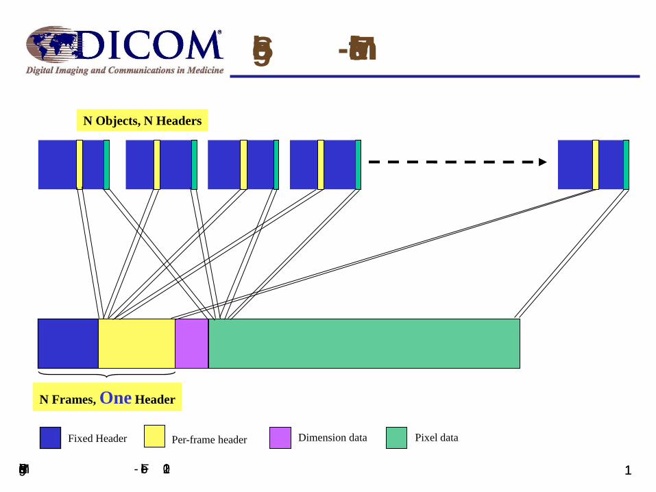
Single-frame to MultiFrame
N Objects, N Headers
N Frames, One Header
Pixel dataDimension dataPer-frame headerFixed Header
SPIE Medical Imaging - Feb 2011 11
New Clinical Domains
SPIE Medical Imaging - Feb 2011 12
Beyond Radiology, Cardiology and
Radio Therapy
Reusing proven concepts é

New Imaging Domains
Ophthalmology
ÅSup 144 Ophthalmic Axial Measurements (Approved 2010)
ÅSup 146 Ophthalmic Visual Field (OPV) (Approved 2010)
Pathology
ÅSup 145 Whole Slide Imaging (Approved 2010)
– Splits (Hugh) image into tiles and uses enhanced multi-
frame framework
– Enable existing viewing pipelines to be used
– Compression Transfer Syntaxes to reduce amount data
SPIE Medical Imaging - Feb 2010 13

Whole Slide Imaging
planes, color planes
Thumbnail Image
Intermediate Image Tiles
Baseline Image Tiles
Multi - frame image (single object)
Multi - frame image (single object) may include multiple Z -
Single frame image
ÅPyramid concept to navigate across matrix of tiles
SPIE Medical Imaging - Feb 2010 14

ÅIntravascular Ultrasound (IVUS) (Approved 2001)
ÅIntravascular OCT (Optical Coherence
Tomography) (in progress)
15SPIE Medical Imaging - Feb 2011
New Technologies Cardiology
Radiotheraphy
RT Second Generation
ÅRadiotherapy Working Group decided to
create a new family of SOP Classes
ÅBased on the new Enhanced Multi-frame
paradigm and other new DICOM
developments
ÅPreparing version for Public Comment (Supplement 147)
SPIE Medical Imaging - Feb 2011 16
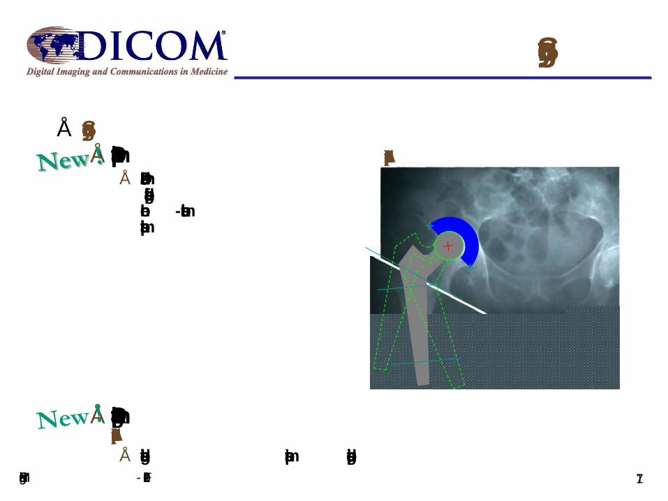
Å Surgery
ÅSup 131 Implant Description (Approved 2010)
Å 2 D and 3D modelsand surgical plans for bone-mountable implants
ÅSup 134 Implantation Plan SR Document Storage (Approved 2010)
Å containing the used impants and positioning
Surgery
17SPIE Medical Imaging - Feb 2011